Dermatology · Medicine
Candidiasis
Also known as Cutaneous candidiasis · Candidal intertrigo · Oral candidiasis (thrush) · Vulvovaginal candidiasis · Candidal balanitis · Candidal paronychia · Chronic mucocutaneous candidiasis
Candidiasis is a yeast infection caused predominantly by Candida albicans and increasingly by non-albicans species (C. glabrata, C. tropicalis, C. parapsilosis) and the multidrug-resistant C. auris. For MBBS final-proficiency, candidates must master the cutaneous forms (intertrigo, napkin/diaper dermatitis, candidal paronychia), the mucosal forms (oral thrush, angular cheilitis, vulvovaginal candidiasis, balanitis), the risk factors (moisture, occlusion, antibiotics, diabetes, immunosuppression, pregnancy), the bedside diagnosis by KOH showing budding yeasts and pseudohyphae, and the stepwise antifungal ladder (topical nystatin/azoles, oral fluconazole/itraconazole, echinocandins for invasive disease). They must also recognise chronic mucocutaneous candidiasis as a marker of immune dysregulation (STAT1 gain-of-function, AIRE/APECED, IL-17 pathway defects) and Candida auris as an emerging nosocomial threat.
On this page & tools
Your progress
Saved locally on this device.
Exam tags
Red flags
Overview and Definition [1]
Candidiasis is a superficial fungal infection caused by dimorphic yeasts of the genus Candida. The human skin and mucous membranes are colonised by these organisms from early infancy, usually without causing disease. Disease develops when the equilibrium between the organism, the local microenvironment and host immunity is disturbed. Candida albicans is responsible for the majority of superficial infections, but the proportion of infections due to non-albicans species has grown. Candida glabrata is frequently associated with vulvovaginal disease and is often less susceptible to fluconazole; C. tropicalis and C. parapsilosis are important in hospital settings and catheter-related infections; C. krusei is intrinsically fluconazole-resistant; and Candida auris is an emerging multidrug-resistant organism that has caused nosocomial outbreaks on every inhabited continent. The dermatology and medicine syllabi focus on recognising the clinical forms, confirming the diagnosis at the bedside with KOH microscopy, and selecting the appropriate level of antifungal therapy. [1][10][12]
For clinical and exam purposes, it is useful to separate superficial candidiasis from invasive disease. Superficial (cutaneous and mucocutaneous) candidiasis affects the stratum corneum, mucosal surfaces and peri-ungual tissues. Invasive candidiasis denotes bloodstream infection (candidemia) or deep-seated infection of organs such as the heart, eyes, liver, spleen or central nervous system. The same patient can move across this spectrum: a premature infant with napkin candidiasis can develop candidemia from a central line; an immunocompromised patient with oral thrush can develop oesophageal candidiasis. The dermatology and MBBS examinations concentrate on the superficial forms, but they also test the red flags that push management from outpatient to inpatient care. A solid understanding of both ends of the spectrum is therefore essential for safe practice and examination success. [1]
Classification
Candidiasis is classified by anatomical site and by the nature of the host immune response. The categories are not mutually exclusive; a patient with diabetes may have intertrigo, oral thrush and balanitis simultaneously. The classification below is the framework an examiner expects a candidate to present in a viva or structured SAQ. [1][2]
Cutaneous versus mucocutaneous versus invasive candidiasis
Cutaneous
Mucosal
Nail and peri-ungual
Chronic mucocutaneous
Invasive
A second axis distinguishes uncomplicated sporadic infection from recurrent or persistent disease. Recurrent vulvovaginal candidiasis is conventionally defined as four or more symptomatic episodes within 12 months. Chronic mucocutaneous candidiasis is defined by chronic, treatment-refractory infection of skin, nails and mucous membranes, often beginning in early childhood. This distinction is clinically important because recurrent or persistent disease obliges the clinician to look for an underlying local or systemic predisposition and to consider long-term suppressive therapy rather than a short course of topical treatment. [4][15]
Candida species and their clinical relevance
Candida albicans is a diploid organism that can switch between yeast, pseudohyphal and hyphal growth forms. It is responsible for roughly 70-80% of vulvovaginal infections and most cases of oral and cutaneous candidiasis in immunocompetent hosts. Candida glabrata is a smaller yeast that does not form hyphae and often causes asymptomatic colonisation or mild disease; it is notable for reduced fluconazole susceptibility. Candida tropicalis is a true hypha-forming species that is more common in tropical countries and in patients with diabetes, leukaemia and indwelling lines. Candida parapsilosis is associated with skin colonisation, catheter-related infections and neonatal disease. Candida krusei is intrinsically resistant to fluconazole and is more often seen in haematology-oncology patients. Candida auris is unique because it can persist on skin and environmental surfaces, is often pan-resistant, and causes outbreaks. [10][11][12]
Epidemiology and Risk Factors
Candida species are normal commensals of the oral cavity, gastrointestinal tract and vaginal vault. Superficial candidiasis develops when the balance between colonisation and host resistance shifts. The exact incidence of cutaneous candidiasis in the community is unknown because many cases are treated empirically without laboratory confirmation, but intertrigo is a frequent reason for dermatology and general practice consultations in hot, humid climates and in settings with high rates of obesity and diabetes. Vulvovaginal candidiasis is one of the commonest vaginal infections globally: around 75% of women report at least one lifetime episode, and 5-8% experience recurrent disease. A systematic review of the global burden estimated that approximately 138 million women experience recurrent vulvovaginal candidiasis each year, with substantial impact on quality of life and healthcare costs. [5][6]
Key risk factors for candidiasis
Local factors are the most common and the most modifiable. Moisture and occlusion in skin folds raise the local pH, macerate the stratum corneum and remove the mechanical barrier to yeast penetration. Obesity increases the number, depth and surface area of skin folds. Incontinence, excessive sweating, tight synthetic clothing and poor hygiene all contribute. Napkin dermatitis in infants is almost always a combination of irritant contact dermatitis and secondary candidal infection, because the warm, moist, occlusive napkin environment is ideal for yeast overgrowth. [13]
Systemic factors are equally important. Diabetes mellitus, whether type 1 or type 2, predisposes to candidiasis through hyperglycaemia, impaired neutrophil chemotaxis and phagocytosis, and glycosuria. HIV infection is a major risk factor, especially when the CD4 count falls below 200 cells per microlitre; oral and oesophageal candidiasis are classic AIDS-defining opportunistic infections. Haematological malignancy, transplant immunosuppression, corticosteroids (topical, inhaled or systemic), and biologics that block IL-17 or TNF-α all impair antifungal immunity. Broad-spectrum antibiotics reduce lactobacilli and allow vaginal or gastrointestinal Candida to overgrow. Pregnancy, oral contraceptives and oestrogen therapy increase vaginal glycogen. Nutritional deficiencies of iron, zinc, folate and vitamin B12 are associated with angular cheilitis and persistent oral candidiasis. Interestingly, Candida is not transmitted primarily by sexual contact in the way that trichomoniasis or gonorrhoea is; rather, VVC usually represents endogenous overgrowth. However, symptomatic balanitis in a male partner may require simultaneous treatment to prevent ping-pong recurrence. [1][2][16]
Pathophysiology
Candida is a dimorphic fungus. In its commensal yeast form it is relatively benign; under permissive conditions it undergoes morphological switching to pseudohyphal and hyphal forms that can invade epithelium and elicit a brisk inflammatory response. The transition from yeast to hypha is driven by environmental cues such as temperature, pH, nutrient availability and contact with epithelial surfaces. Hyphal formation is not merely a morphological change but a virulence trait: hyphae can penetrate tissue, resist phagocytosis and promote biofilm development. [12]
Several classes of virulence factors have been defined. Secreted aspartyl proteinases (Saps) degrade host proteins, facilitate tissue invasion and may interfere with host immune responses. Phospholipases damage cell membranes. Adhesins, particularly the agglutinin-like sequence (Als) family, allow the organism to adhere to epithelial cells, extracellular matrix and medical devices. Biofilm formation is particularly important on mucosal surfaces and prosthetic materials: organisms within biofilms are up to 1000 times more resistant to antifungals and are protected from host immune effectors. C. albicans can also undergo a white-opaque switch, a heritable epigenetic change that affects mating, host adaptation and antifungal tolerance. [12]
Host defence against Candida depends on the integrity of the epithelial barrier, competition from commensal bacteria, neutrophils, macrophages and the T-helper 17 (Th17) lymphocyte axis. The Th17 response, mediated by interleukin-17, IL-22 and IL-23, is critical for mucocutaneous antifungal immunity. Genetic defects that impair this pathway predispose to chronic mucocutaneous candidiasis. The most important single defect is STAT1 gain-of-function: hyperphosphorylation of STAT1 suppresses the IL-17 response and produces a syndrome of recurrent or persistent mucocutaneous candidiasis, often accompanied by type I interferonopathy features and occasionally by other infections. Other described defects include STAT3 loss-of-function (autosomal-dominant hyper-IgE syndrome), AIRE mutations causing autoimmune polyendocrinopathy-candidiasis-ectodermal dystrophy (APECED), and mutations in IL-17 receptor, IL-17F or CARD9. [8][15]
The inflammatory response to candidal invasion explains the clinical findings. In the skin, neutrophils accumulate around invading hyphae and produce the characteristic satellite pustules. In the oral mucosa, epithelial hyperplasia and neutrophilic microabscesses produce the white pseudomembranous plaques. In chronic infection, a delayed-type hypersensitivity reaction may produce the firm white plaques of hyperplastic candidiasis. Understanding these mechanisms allows the clinician to predict which patients are at risk: anyone with barrier disruption, altered flora or defective Th17 immunity will be vulnerable to superficial candidiasis, and the same patients may be prone to recurrence when treatment stops. [1]
Candida also has a remarkable ability to form biofilms on mucosal and prosthetic surfaces. In biofilms, yeast cells are embedded in an extracellular matrix that confers resistance to both antifungals and host immune attack. This explains why device-associated candidiasis (central lines, urinary catheters, dentures) is harder to eradicate and why removing the device is often necessary. [12]
Clinical Presentation
The clinical presentation of candidiasis is dictated by the site of infection and the host immune response. Superficial disease is usually localised, itchy or sore, and recognisable by the combination of erythema, moisture and satellite lesions. Mucosal disease may be mild and self-limiting or severe and recurrent, especially in immunosuppression. Chronic mucocutaneous disease is persistent and may be disfiguring. The following sections describe the common presentations in the order they are most likely to appear in clinical practice and in examinations. [1]
Cutaneous candidiasis
Candidal intertrigo is the prototypical cutaneous form and the one most frequently examined. It appears as a bright-red, moist, shiny, well-demarcated plaque in a skin fold, with a scalloped or collarette edge and characteristic satellite pustules or papules at the periphery. The patient may complain of itching, burning, soreness or malodour. Common sites are the submammary folds, axillae, infrabdominal pannus, inguinal creases, perineum and interdigital toe webs. Interdigital infection (errosio interdigitalis blastomycetica) produces maceration, fissuring and erythema between the fingers, particularly in people whose hands are wet for long periods. Secondary bacterial infection, usually with staphylococci or streptococci, can produce purulent crusting, a foul smell and rapid spread. [1]
Napkin (diaper) candidiasis typically develops on a background of irritant napkin dermatitis. It presents as a beefy-red, confluent eruption that involves the convexities and the skin folds, with small satellite papules and pustules at the margin. This contrasts with irritant napkin dermatitis, which is usually confined to the convex surfaces and spares the folds. The infant may be uncomfortable and may have concomitant oral thrush. A breastfeeding infant may transmit candidiasis to the mother’s nipples, causing painful, erythematous nipples with radiating pain during feeding. [13]
Mucosal candidiasis
Oral candidiasis (thrush) has several recognisable forms. Pseudomembranous thrush is the classic presentation: creamy white plaques that can be scraped off an erythematous base, leaving a bleeding surface. It is common after antibiotics, inhaled corticosteroids and immunosuppression. Erythematous (atrophic) candidiasis presents as a red, burning, sore mucosa without white plaques; it is often associated with dentures and is sometimes called denture stomatitis. Angular cheilitis affects the oral commissures with soreness, fissuring and maceration; it is frequently multifactorial, with candida, staphylococci, moisture pooling from dentures or lip licking, and nutritional deficiency all contributing. Chronic hyperplastic candidiasis appears as a firm, white plaque that cannot be scraped off; it is considered potentially premalignant and may show dysplasia on biopsy. Denture-related stomatitis is another common variant: the mucosa beneath a denture becomes red and inflamed because the denture acts as a reservoir for candida and traps moisture. Patients should be advised to clean dentures thoroughly and to remove them at night. [3][16]
Vulvovaginal candidiasis is characterised by intense vulval itching, soreness, dyspareunia, external dysuria and a non-offensive, creamy or curdy white vaginal discharge. The vulva may be erythematous and oedematous; satellite pustules may be seen on the labia. Symptoms are usually most prominent in the week before menses. Candidal balanitis produces erythematous papules or plaques on the glans and prepuce, sometimes with a white exudate, fissuring and pruritus. It is more common in uncircumcised men, in diabetes, and in men whose partners have VVC. [6][7]
Nail and peri-ungual disease
Chronic candidal paronychia is the characteristic nail involvement. It produces boggy, erythematous swelling of the proximal and lateral nail folds, with loss of the cuticle, transverse ridging and Beau lines of the nail plate. It is usually multifactorial: the primary problem is irritant contact dermatitis from wet work, with secondary candidal colonisation and episodic bacterial flares. The condition is more common in barbers, hairdressers, cleaners, cooks, dental workers, health-care workers and swimmers. It is distinguished from acute bacterial paronychia by its chronic course, multi-finger distribution, absence of severe pain and fluctuance, and the presence of cuticle loss rather than pus lifting the cuticle. [14]
Chronic mucocutaneous candidiasis
CMC presents with persistent, widespread, treatment-resistant candidiasis of skin, nails and mucous membranes from infancy or early childhood. The infection may be disfiguring, with thick, hyperkeratotic plaques on the face, scalp and hands. It may be associated with endocrine autoimmunity (hypoparathyroidism, hypothyroidism, adrenal insufficiency) in APECED, dental enamel hypoplasia, nail dystrophy and alopecia. The key clinical clue is the lifelong, mucocutaneous-restricted pattern of recurrent or persistent candidiasis, which differs from the episodic, infection-related pattern seen in HIV. [8][15]
Differential Diagnosis
The differential diagnosis of candidiasis is broad because red, scaly, pustular or erosive eruptions in skin folds and mucous membranes have many causes. The key is to combine the clinical picture with a simple KOH examination. [1][2]
Candidal intertrigo versus its mimics
Tinea cruris/corporis
Inverse psoriasis
Erythrasma
Bacterial intertrigo
Hailey-Hailey disease
Other important differentials include seborrhoeic dermatitis for facial and scalp involvement, atopic dermatitis and allergic contact dermatitis for napkin dermatitis, irritant contact dermatitis for napkin and lip-licker rashes, lichen planus and oral hairy leukoplakia for oral white plaques, herpes simplex for painful oral erosions, and nail-unit squamous cell carcinoma or melanoma for refractory single-digit chronic paronychia. KOH microscopy, bacterial culture and, when necessary, biopsy, usually resolve the uncertainty. [1][2]
A bedside framework for distinguishing fold rashes
When confronted with a red rash in a skin fold, ask three questions. First, is the lesion scaly and annular with central clearing? If yes, think tinea corporis. Second, is the plaque smooth, well-demarcated and without satellite pustules? If yes, think inverse psoriasis. Third, does the rash fluoresce coral-red under Wood's lamp? If yes, think erythrasma. If the answer to all three is no and the patient has moist, shiny erythema with satellite pustules, candidal intertrigo is the most likely diagnosis. This framework avoids unnecessary investigations and allows empirical antifungal therapy to be started promptly. [1]
Clinical and Bedside Assessment
A focused history should establish the duration, sites, frequency of recurrence and the presence of risk factors. Ask specifically about recent or current antibiotics, inhaled or systemic corticosteroids, diabetes symptoms, pregnancy, sexual history, denture use, wet work, obesity, immunosuppression and HIV risk factors. The physical examination should inspect all skin folds, the oral cavity and commissures, the genitalia, the nails and the napkin area in infants. [1]
The key bedside sign is the satellite pustule: a small pustule or papule sitting just beyond the edge of a red, moist fold plaque. This sign is highly suggestive of candidiasis and is not seen in inverse psoriasis or dermatophyte infection. A Wood's lamp is useful in the differential diagnosis of groin and axillary rashes: erythrasma shows coral-red fluorescence, tinea versicolor shows copper-orange fluorescence, and candidiasis does not fluoresce in this way. [1]
When to suspect an underlying systemic disease is a critical exam concept. Severe or recurrent oral candidiasis in an adult without an obvious local cause should prompt testing for HIV and diabetes mellitus. Recurrent vulvovaginal candidiasis should trigger a fasting glucose or HbA1c and consideration of HIV testing. Chronic paronychia that is persistent or recurrent should lead to screening for diabetes and, if refractory, HIV and iron studies. Candidiasis from infancy with endocrine abnormalities should raise the possibility of chronic mucocutaneous candidiasis or APECED. [3][6]
Focused history and examination checklist
A structured approach ensures no risk factor or differential is missed. History should cover duration, sites, recurrence, recent antibiotics, inhaled or systemic steroids, diabetes symptoms, pregnancy, denture use, wet work, obesity, sexual history, immunosuppression and HIV risk. Examination should inspect all skin folds, oral cavity and commissures, genitalia, nails and napkin area. Look specifically for satellite pustules, cuticle loss, white plaques that scrape off, and signs of secondary bacterial infection. Ask about symptoms of systemic disease such as weight loss, fever, night sweats and dysphagia. Record vital signs and look for sepsis only if invasive candidiasis is suspected. [1]
Investigations
KOH microscopy is the cornerstone of diagnosis for cutaneous candidiasis. A skin scraping, nail-fold swab or mucosal smear is placed on a slide with 10% potassium hydroxide, gently heated and examined under high power. Candida shows ovoid budding yeast cells (blastoconidia) and elongated pseudohyphae that remain attached like links of sausages. True hyphae may also be seen. Dermatophytes, by contrast, show branching, septate hyphae with no yeast forms. The distinction is important because it determines therapy: topical azoles treat both, but tinea cruris may also respond to allylamines such as terbinafine, whereas candidiasis generally does not. [1]
Culture is reserved for atypical, recurrent, treatment-refractory or severe disease, and when species identification is needed. Sabouraud dextrose agar or CHROMagar Candida allows presumptive species identification. Antifungal susceptibility testing is important for Candida auris, non-albicans species such as C. glabrata and C. krusei, and for patients who fail standard therapy. Blood cultures are essential when invasive candidiasis is suspected. Endoscopy is used for oesophageal candidiasis when the diagnosis is uncertain or when symptoms are refractory to antifungal therapy. [7][9]
Screening for predisposing disease is part of the work-up for recurrent, severe or unusual presentations. This includes fasting glucose or HbA1c for diabetes; HIV Ag/Ab testing; serum iron studies, ferritin, folate and vitamin B12 for nutritional deficiency; endocrine panel (calcium, phosphate, parathyroid hormone, thyroid-stimulating hormone, cortisol, ACTH) when CMC or APECED is suspected; and referral for immunological and genetic testing in chronic mucocutaneous candidiasis. [3][8]
When is advanced testing needed?
Most cases of cutaneous or mucosal candidiasis do not require advanced testing. KOH microscopy is sufficient for diagnosis, and empirical topical therapy is appropriate. Culture is indicated when the diagnosis is uncertain, the infection is recurrent or refractory, a non-albicans species is suspected, or the patient is immunocompromised. Antifungal susceptibility testing is essential for Candida auris, C. glabrata and C. krusei, and when first-line therapy has failed. Molecular methods such as MALDI-TOF mass spectrometry or internal transcribed spacer sequencing are used in reference laboratories to identify unusual species. In chronic mucocutaneous candidiasis, genetic testing for STAT1, STAT3, AIRE and IL-17 pathway genes is arranged by an immunologist. [1][7]
[1]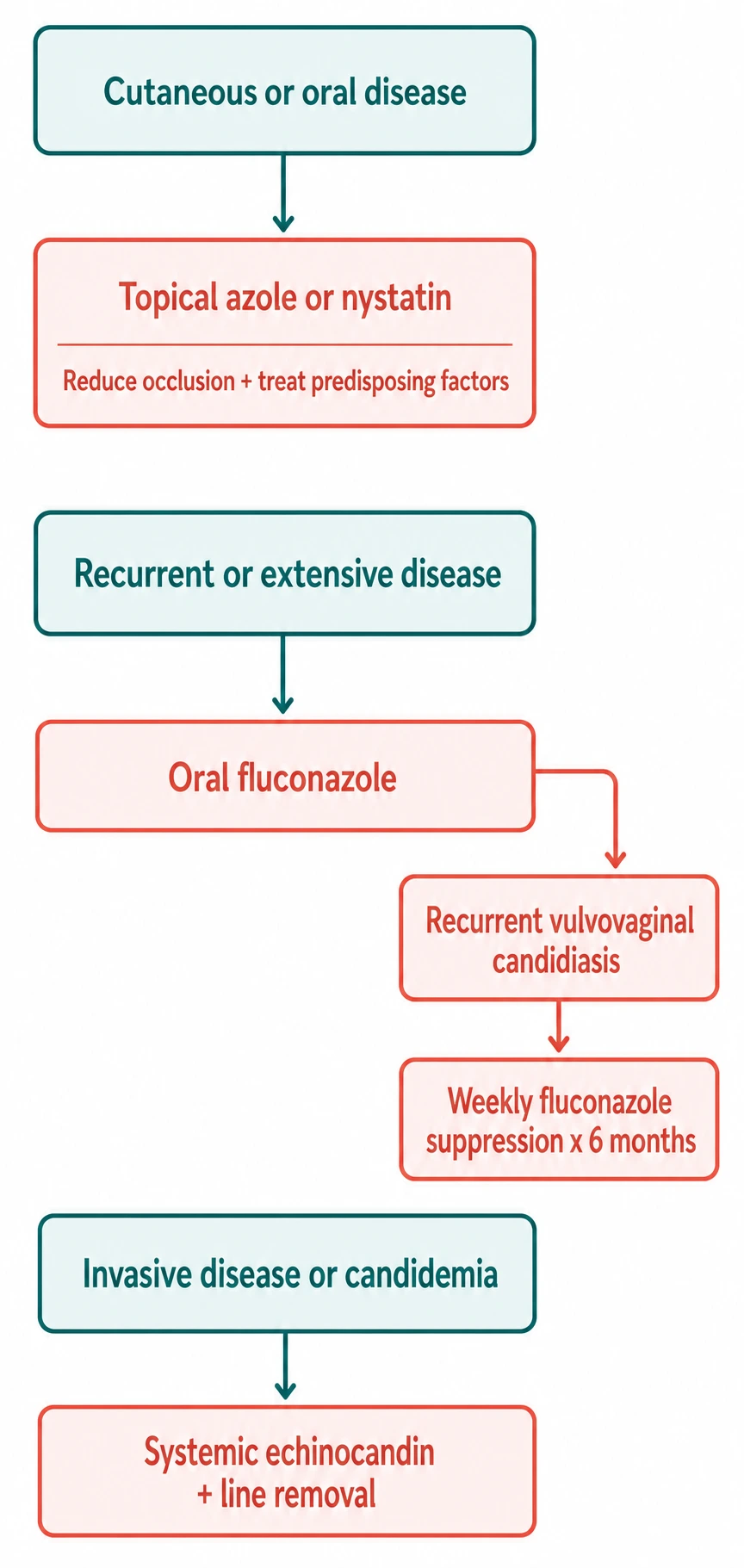
Management — Resuscitation
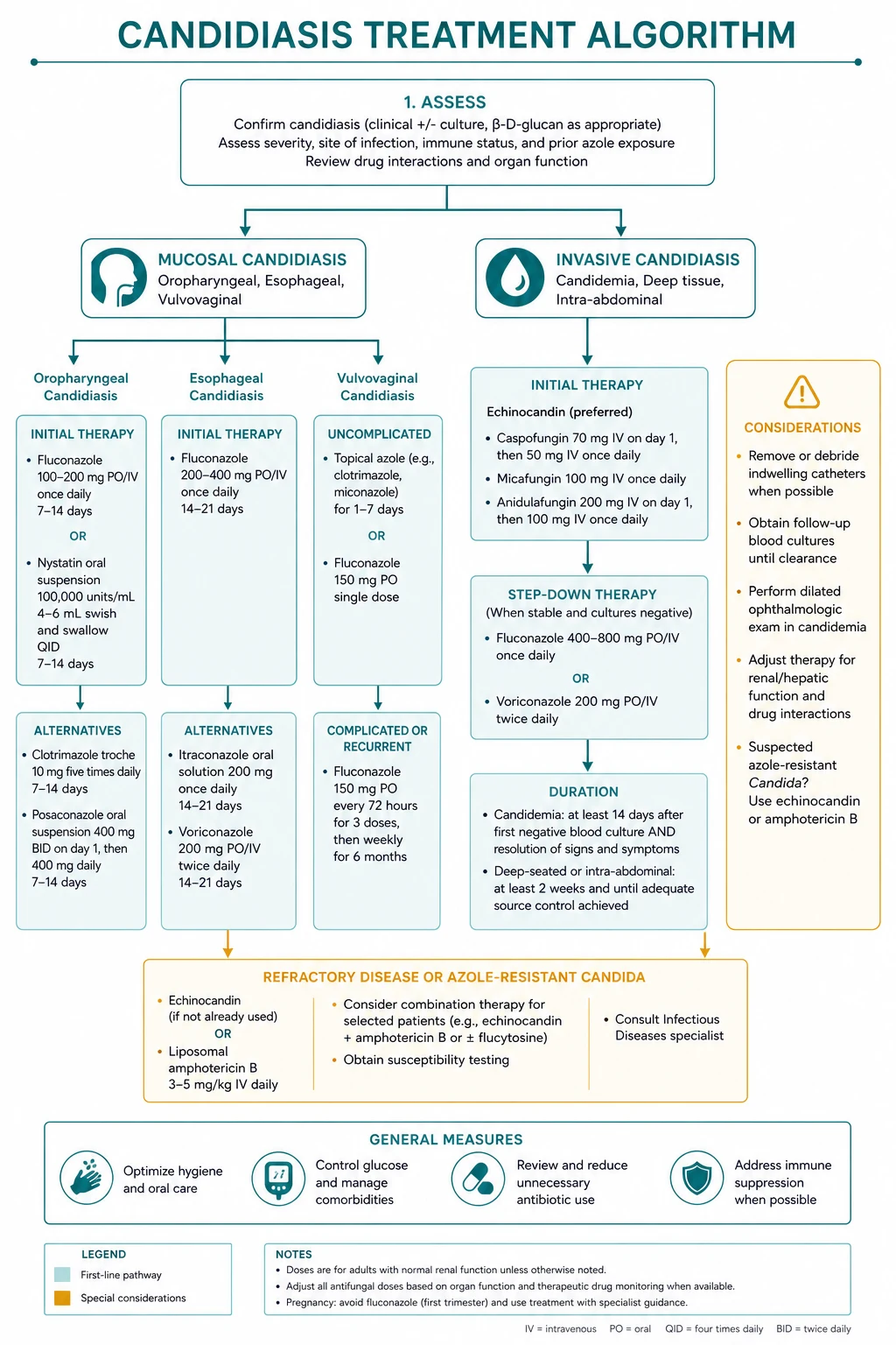
Life-threatening candidiasis is not the usual cutaneous form but candidemia or invasive candidiasis. Triggers are indwelling vascular catheters, broad-spectrum antibiotics, parenteral nutrition, prolonged ICU stay, immunosuppression, gastrointestinal surgery and haematological malignancy. Presenting features include persistent fever, hypotension, sepsis, and positive blood cultures. The IDSA 2016 guideline recommends an echinocandin as first-line therapy for candidemia in non-neutropenic adults. Options include caspofungin 70 mg loading dose then 50 mg intravenously daily, micafungin 100 mg IV daily, or anidulafungin 200 mg loading dose then 100 mg IV daily. [7][9]
In neutropenic patients, the choice is more nuanced. Echinocandins are preferred, but amphotericin B preparations may be used, particularly when there is hepatosplenic involvement or a high risk of resistant organisms. Liposomal amphotericin B 3 mg/kg daily is the usual formulation. Once neutropenia resolves and the patient is stable, step-down to fluconazole is appropriate. Empirical antifungal therapy should be considered in febrile neutropenia that persists despite broad-spectrum antibiotics, especially when there are risk factors for invasive candidiasis. [7]
Equally important is source control. The infected central venous catheter should be removed whenever feasible; persistent candidemia is much more likely if the line remains in place. Any abscess should be drained, and ophthalmology review should be arranged to exclude candidal endophthalmitis, which occurs in a minority of patients with candidemia and can threaten vision. Once the patient is clinically stable, the isolate is known to be susceptible, and follow-up blood cultures are negative, step-down to fluconazole 400-800 mg orally daily is appropriate. [7][9]
Candida auris is managed as a healthcare-associated pathogen: isolate the patient, notify infection control and public-health authorities, identify the species with MALDI-TOF or molecular methods, and treat according to susceptibility. Many C. auris isolates are resistant to multiple antifungal classes; treatment may require combination therapy or agents such as amphotericin B or echinocandins, with expert infectious-diseases input. [10][11]
Management — Definitive and Stepwise
Cutaneous candidiasis (intertrigo, napkin, flexural)
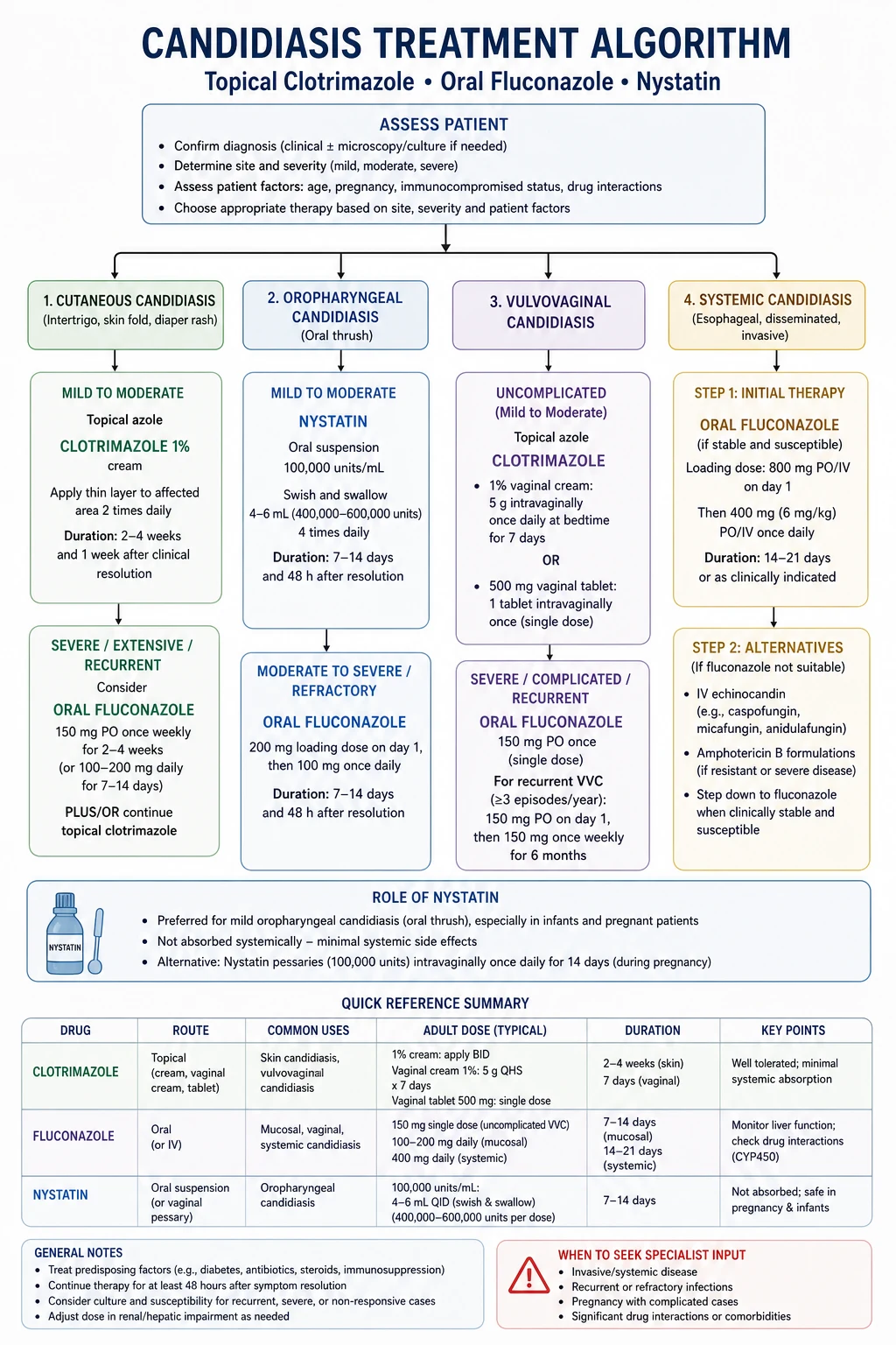
First-line therapy is topical nystatin or a topical azole (clotrimazole 1%, miconazole 2%, econazole 1%, ketoconazole 2%) applied twice daily for 2-4 weeks. The key adjuncts are drying and reducing occlusion: keep folds clean and dry, use a cool hair dryer, apply absorbent or antifungal powder, lose weight if obese, and treat underlying diabetes or incontinence. Napkin dermatitis is treated with frequent nappy changes, airing, barrier creams (zinc oxide or petrolatum) and a topical azole or nystatin if satellite lesions are present. [1][2]
Oral candidiasis and angular cheilitis
Mild oral thrush responds to nystatin suspension (100,000 units per mL, 4-6 mL swished and swallowed four times daily for 7-14 days) or clotrimazole troches 10 mg dissolved in the mouth five times daily. Miconazole oral gel is a useful alternative. For moderate to severe disease, immunosuppression or recurrent infection, oral fluconazole 100-200 mg daily for 7-14 days is preferred. Angular cheilitis is treated with a topical antifungal cream to the commissures plus attention to dentures, nutritional deficiencies and moisture control; a mild topical steroid may be added briefly for inflammation. [3][16]
Vulvovaginal candidiasis and balanitis
Uncomplicated VVC is treated with a single dose of oral fluconazole 150 mg or a topical azole cream or pessary (clotrimazole, miconazole, econazole) for 1-7 days. Pregnancy should be treated with topical azoles only; oral fluconazole is avoided, especially in the first trimester. Severe or recurrent VVC may require fluconazole 150 mg every 72 hours for three doses, then suppressive therapy with fluconazole 100-200 mg weekly for 6 months after confirming the diagnosis by culture and excluding diabetes. Recurrent balanitis should prompt testing for diabetes and HIV; sexual partners with VVC should be treated to prevent ping-pong infection. [6][7]
Chronic candidal paronychia
Management has four pillars. First, protect the hands from wet work by using cotton-lined rubber gloves, applying emollients after washing, and using barrier creams. Second, apply a topical antifungal (clotrimazole, miconazole or ciclopirox) to the nail fold for 4-8 weeks. Third, use a topical anti-inflammatory if dermatitis is prominent. Fourth, for refractory cases, give oral itraconazole (200 mg twice daily for 1 week per month, for 2-3 months) or fluconazole 150 mg weekly. Surgical marsupialisation is reserved for refractory disease after several months of conservative therapy. [14]
Chronic mucocutaneous candidiasis
CMC is managed with long-term systemic azoles. Fluconazole or itraconazole is used first-line; refractory cases or azole-resistant organisms may require posaconazole. The underlying immune defect should be investigated by an immunologist, with genetic testing for STAT1, STAT3, AIRE and IL-17 pathway defects. In STAT1 gain-of-function disease, the JAK1/2 inhibitor ruxolitinib can restore IL-17 responses and produce clinical remission. Endocrine complications such as hypoparathyroidism, adrenal insufficiency and hypothyroidism must be screened for and treated. [8][17][18][19][20]
Drug dosing and monitoring quick reference
Topical nystatin and azoles are safe and effective for most superficial disease. Nystatin works by binding to ergosterol in the fungal cell membrane and creating pores; it is not absorbed systemically and is safe in infants, children and pregnancy. Topical azoles (clotrimazole, miconazole, econazole, ketoconazole) inhibit fungal CYP450-dependent 14α-demethylase, blocking ergosterol synthesis. They are generally applied twice daily for 2-4 weeks. Oral fluconazole is a triazole with excellent bioavailability and tissue penetration. A single 150 mg dose is standard for uncomplicated VVC; more severe or recurrent disease requires 100-200 mg daily for 7-14 days. Itraconazole is given as 200 mg daily for 5-7 days, or as a 200 mg twice-daily pulse for one week per month in chronic paronychia. Posaconazole is reserved for refractory or resistant disease. Echinocandins (caspofungin, micafungin, anidulafungin) inhibit β-1,3-D-glucan synthase and are used intravenously for invasive candidiasis; they are not absorbed orally and are therefore inappropriate for superficial disease. Fluconazole interacts with warfarin, sulfonylureas, statins and calcineurin inhibitors, so drug interactions must be checked. [1][6][7]
[1] [1] [1] [1]The Australian Therapeutic Guidelines emphasise moisture control, barrier creams and topical nystatin or azole for cutaneous and oral candidiasis. Oral fluconazole is reserved for severe, recurrent or extensive disease. Chronic paronychia is treated with wet-work avoidance and topical azole or ciclopirox; oral fluconazole 150 mg weekly pulse is commonly used for refractory cases.
Specific Subtypes and Scenarios
Candidal intertrigo in obesity and diabetes
The patient is often overweight or diabetic. The fold is red, moist, sore and satellite-pustuled. Management combines a topical azole, glycaemic control, weight reduction, absorbent powder and, if bacterial superinfection is suspected, a topical or oral antistaphylococcal agent. Recurrence is common if folds remain moist. Patients should be counselled to dry the folds thoroughly after bathing, to wear loose cotton clothing, and to consider weight loss if obesity is present. The addition of a short course of a mild topical corticosteroid may reduce inflammation, but steroids should never be used alone because they can worsen the infection. If the rash is extensive or recurrent, oral fluconazole 100-200 mg daily for 7-14 days is appropriate. [1]
Napkin candidiasis
A previously improving irritant napkin rash suddenly becomes bright red, confluent and involves the folds, with satellite pustules. Treat the napkin dermatitis with frequent changes, barrier cream and airing, and add a topical azole or nystatin for 7-14 days. Oral thrush in the infant and candidal mastitis in the breastfeeding mother should be considered. The mother may need topical antifungal treatment to the nipples to prevent cross-infection. If the infant has oral thrush, both mother and infant should be treated concurrently. Severe or resistant cases in infants may require oral fluconazole under specialist guidance. [13]
Angular cheilitis
Angular cheilitis is a mixed infection at the wet, occluded oral commissures. Candida is often present, but Staphylococcus aureus or Streptococcus pyogenes may also contribute. Treatment should cover both fungal and bacterial organisms if needed: a topical antifungal cream, and a topical antiseptic or antibiotic if bacterial involvement is suspected. Control dentures and moisture pooling; check iron, folate and vitamin B12; and consider diabetes and HIV testing in recurrent cases. In patients with dentures, the appliance should be cleaned, soaked and removed at night. Nutritional supplementation is appropriate if deficiencies are found. A brief course of a mild topical steroid may reduce inflammation, but only in combination with antifungal therapy. [16]
Oesophageal candidiasis
Oesophageal candidiasis is an AIDS-defining illness and also occurs in other immunosuppressed states. Patients have odynophagia, dysphagia and retrosternal pain. Empirical systemic therapy with fluconazole 200-400 mg daily for 14-21 days is indicated, especially in HIV with CD4 count less than 200 cells per microlitre. Endoscopy is reserved for refractory cases, atypical features or when CMV or HSV co-infection is suspected. Topical therapy alone is ineffective. [7]
Candida auris colonisation and infection
C. auris colonises skin, especially the axilla and groin, and can persist for months. It causes outbreaks in long-term care facilities and ICUs. Infection may present as fever, sepsis, wound infection, otitis or pneumonia. Multidrug resistance is common; treatment is guided by antifungal susceptibility testing. Contact precautions, environmental cleaning and public-health notification are essential. Decolonisation strategies are not well established. [10][11]
Self-test: why does this patient keep getting thrush?
A 42-year-old woman presents with her fifth episode of vulvovaginal candidiasis in 12 months. She is not pregnant, not diabetic on known history, and not sexually active with new partners. What are the three most important next steps? [1]
- Confirm the diagnosis by culture to exclude non-albicans species or alternative pathology.
- Screen for diabetes with fasting glucose or HbA1c, and consider HIV testing if risk factors exist.
- Discuss suppressive therapy: fluconazole 100-200 mg weekly for 6 months, with the caveat that recurrence is common after stopping.
Complications and Pitfalls
Complications of superficial candidiasis are usually local. Secondary bacterial infection can superimpose on intertrigo and produce cellulitis, lymphangitis or sepsis. Nail dystrophy and onycholysis are common in chronic paronychia. Fissuring and bacterial superinfection complicate angular cheilitis. Maternal nipple candidiasis can occur in breastfeeding mothers of infants with napkin or oral candidiasis. [1][14]
Systemic spread is rare in immunocompetent hosts, but in neutropenic or critically ill patients, Candida can translocate across damaged gut mucosa or enter the bloodstream via intravenous catheters, causing candidemia, endophthalmitis, endocarditis and hepatosplenic candidiasis. Candidemia carries a mortality of 20-40% even with treatment, and delayed echinocandin therapy worsens outcomes. Candida auris outbreaks have been linked to ventilator-associated pneumonia, bloodstream infection and wound infection, with crude mortality exceeding 30% in some series. These complications are more common with non-albicans species and multidrug-resistant isolates. [7][10][11]
Pitfalls include missing the underlying cause (diabetes, HIV, immunosuppression, nutritional deficiency), treating with topical steroids alone which can worsen infection, using oral fluconazole in pregnancy, misdiagnosing chronic paronychia as acute bacterial paronychia leading to unnecessary incision, and failing to investigate chronic mucocutaneous candidiasis for associated endocrine autoimmunity or genetic defects. In invasive candidiasis, a common pitfall is failing to remove the infected central line, which perpetuates candidemia. Another pitfall is relying on empirical topical therapy for oesophageal candidiasis: because topical agents do not reach the oesophageal mucosa, systemic fluconazole is required. In immunosuppressed patients, recurrent thrush may herald immunological failure or poor adherence to antiretroviral therapy. [2][14]
Prognosis and Disposition
Uncomplicated cutaneous and mucosal candidiasis has an excellent prognosis when treated with topical or oral antifungals and when predisposing factors are controlled. Most patients can be managed in primary care. Recurrent vulvovaginal candidiasis has a high relapse rate after suppressive therapy is stopped; patients should be warned that recurrence is common. Chronic paronychia recurs in 30-50% of patients if wet work is resumed before the cuticle has regenerated. Chronic mucocutaneous candidiasis requires lifelong management and surveillance for endocrine complications. Invasive candidiasis and Candida auris infection carry significant mortality, especially in ICU patients and those with persistent neutropenia. [5][9]
Patients who are discharged should receive clear instructions on treatment duration, hygiene measures and when to return. Topical therapy should generally continue for 7-14 days, or for one week after clinical clearance, to reduce relapse. Those started on oral fluconazole for severe or recurrent disease need follow-up to confirm response and to review culture results. Anyone with recurrent or atypical disease should be told to return for HIV and diabetes testing if these have not already been performed. Clear communication reduces non-adherence and prevents the development of resistant organisms. [1][6]
Prevention and patient counselling
Prevention focuses on removing local and systemic risk factors. Patients with intertrigo should keep folds dry, wear loose cotton clothing, avoid tight synthetic garments, and lose weight if overweight. Those with napkin dermatitis should change nappies frequently, allow airing, and use barrier creams. People with chronic paronychia must protect their hands from wet work with cotton-lined gloves and apply emollients after washing. Patients on inhaled corticosteroids should rinse their mouth or use a spacer to reduce oral candidiasis. Women with recurrent VVC should avoid unnecessary antibiotics, wear cotton underwear, and consider probiotic or boric acid strategies under medical supervision. Good glycaemic control is essential in diabetes. HIV-positive patients should adhere to antiretroviral therapy. Denture wearers must clean and remove dentures at night. These simple measures often prevent recurrence as effectively as prolonged antifungal courses. [1][2]
Disposition and follow-up
Most patients with uncomplicated cutaneous or mucosal candidiasis can be discharged from primary care with topical therapy and lifestyle advice. Review is needed if symptoms fail to improve within 1-2 weeks, if the disease is recurrent, or if an underlying systemic condition is suspected. Referral to dermatology is appropriate for atypical rashes, diagnostic uncertainty or refractory disease. Referral to infectious diseases is indicated for invasive candidiasis, Candida auris infection or persistent candidemia. Referral to immunology is indicated for suspected chronic mucocutaneous candidiasis. Women with recurrent VVC should have a follow-up appointment after completing suppressive therapy to assess relapse and reinforce preventive measures. [6][9]
Special Populations
Infants and children: Napkin candidiasis is common; treat with barrier creams, frequent nappy changes and topical nystatin or azole. Oral thrush in infants is treated with miconazole gel or nystatin suspension. Oral fluconazole is reserved for severe or refractory disease. Breastfeeding mothers may need simultaneous nipple treatment. [13]
Pregnancy: Vulvovaginal candidiasis is more common in pregnancy because of elevated oestrogen and glycogen deposition. Treat with a topical imidazole such as clotrimazole or miconazole for 7 days. Avoid oral fluconazole in pregnancy, especially in the first trimester, because of teratogenicity concerns. Recurrent episodes should prompt glucose testing. [6]
Elderly: Dentures, xerostomia, incontinence, diabetes and multiple medications increase risk. Pay attention to drug interactions: fluconazole inhibits CYP3A4 and CYP2C9 and can increase levels of warfarin, sulfonylureas, statins, tacrolimus, cyclosporine and some antiretrovirals. Skin fragility and reduced mobility can make oral hygiene and skin-fold care difficult; occupational therapy and nursing support may be needed to prevent recurrence. [3]
Pregnancy: Use topical azoles (clotrimazole, miconazole) for VVC; avoid oral fluconazole, particularly in the first trimester. Treating the male partner is generally not needed unless he has symptomatic balanitis. Pregnant patients with recurrent symptoms should be evaluated for diabetes. [6]
Diabetes: Hyperglycaemia impairs neutrophil chemotaxis and phagocytosis, and glycosuria provides a fertile medium. Optimise glycaemic control as a core part of management. Diabetic patients with intertrigo or balanitis may need more prolonged or systemic therapy. [2]
HIV and immunosuppression: Oral and oesophageal candidiasis are common opportunistic infections. Antifungal therapy is needed, but antiretroviral therapy or reduction of immunosuppression is the definitive long-term strategy. Routine primary antifungal prophylaxis is not recommended; secondary prophylaxis is reserved for frequent or severe recurrences. Patients with advanced HIV should be assessed for other opportunistic infections. [3][7]
Elderly: Dentures, xerostomia, incontinence, diabetes and multiple medications increase risk. Pay attention to drug interactions: fluconazole inhibits CYP3A4 and CYP2C9 and can increase levels of warfarin, sulfonylureas, statins, tacrolimus, cyclosporine and some antiretrovirals. Swallowing problems and reduced mobility can make oral hygiene and skin-fold care difficult; occupational therapy and nursing support may be needed to prevent recurrence. [3]
[1]Evidence, Guidelines and Regional Differences
The IDSA 2016 candidiasis guideline is the landmark reference for invasive disease. It recommends echinocandin first-line for candidemia, removal of infected central lines, ophthalmology review, and fluconazole step-down once the patient is stable and the isolate is susceptible. The AWMF 2021 VVC guideline defines uncomplicated and complicated disease and recommends culture for recurrent cases. For cutaneous disease, evidence supports topical nystatin and azoles as first-line, with oral fluconazole for extensive or recurrent disease. The Taudorf 2019 evidence-based review summarises topical and systemic treatments for cutaneous candidiasis and highlights the importance of treating predisposing factors. [1][6][7]
Regional practice varies in important ways. In the United Kingdom, NICE CKS and BASHH favour topical azoles for uncomplicated VVC, and oral fluconazole only when topical therapy has failed; in pregnancy, only topical imidazoles are used. In India and many low- and middle-income settings, single-dose oral fluconazole 150 mg is widely used for uncomplicated VVC because of cost, convenience and climate. The US and IDSA approach uses either oral or topical agents first-line for VVC, and oral fluconazole for balanitis. For chronic paronychia, Australia and UK guidelines emphasise wet-work avoidance and topical anti-inflammatory or antifungal therapy before oral agents. These regional differences matter for exam questions that quote guideline tables. [6][10]
Regional differences are important in clinical practice. In the United Kingdom, NICE and BASHH favour topical azoles for uncomplicated VVC in pregnancy and recommend oral fluconazole only when topical therapy fails. In the United States, single-dose oral fluconazole 150 mg is widely used for uncomplicated VVC. In Australia and India, weekly fluconazole pulse is commonly used for chronic candidal paronychia and recurrent VVC. Infection-control practices for Candida auris are standardised globally because of its outbreak potential. [6][10]
Landmark studies
The IDSA 2016 update summarised two decades of randomised trials showing that echinocandins produce faster clearance of candidemia and lower mortality than fluconazole in the first 24-48 hours of therapy in unstable patients. The AWMF 2021 vulvovaginal candidosis guideline compiled evidence that short-course topical azoles and single-dose oral fluconazole are equivalent for uncomplicated VVC, but that recurrent disease requires longer suppressive regimens. The global burden systematic review by Denning and colleagues quantified the enormous prevalence and quality-of-life impact of RVVC. Taudorf and colleagues' 2019 review of cutaneous candidiasis established the evidence base for topical nystatin and azoles and for oral fluconazole in extensive or recurrent cutaneous disease. Case series of posaconazole and ruxolitinib in CMC have shown that refractory disease can respond to either newer antifungals or immunomodulation directed at the underlying defect. [1][5][6][7][17][18]
Exam Pearls
CANDIDA
Risk factors for candidiasis: MOIST
Exam application bank (NEET-PG / INICET)
One-line answer
Candidiasis is a yeast infection caused predominantly by Candida albicans and increasingly by non-albicans species (C. glabrata, C. tropicalis, C. parapsilosis) and the multidrug-resistant C. auris. For MBBS final-proficiency, candidates must master the cutaneous forms (intertrigo, napkin/diaper dermatitis, candidal paronychia), the mucosal forms (oral thrush, angular cheilitis, vulvovaginal candidiasis, balanitis), the risk factors (moisture, occlusion, antibiotics, diabetes, immunosuppression, pregnancy), the bedside diagnosis by KOH showing budding yeasts and pseudohyphae, and the stepwise antifungal ladder (topical nystatin/azoles, oral fluconazole/itraconazole, echinocandins for invasive disease). They must also recognise chronic mucocutaneous candidiasis as a marker of immune dysregulation (STAT1 gain-of-function, AIRE/APECED, IL-17 pathway defects) and Candida auris as an emerging
Worked stems (answer without another resource)
Stem 1 — Classic presentation. Map symptoms to mechanism; name the first investigation and first treatment step with dose/route if drug therapy is standard. [1]
Stem 2 — Unstable / complicated. List red flags that force immediate resuscitation, theatre, ICU, antidote, or reperfusion — and what you do in the first 15 minutes. [1]
Stem 3 — Atypical group. Elderly, pregnancy, child, or immunocompromised: how presentation and thresholds change. [1]
Stem 4 — Differential trap. Name the three closest mimics and one discriminator for each. [1]
Stem 5 — Disposition. Who goes home with safety-netting, who is admitted, who needs HDU/ICU/theatre, and what follow-up is mandatory. [1]
Rapid viva checklist
- Definition + classification
- Pathophysiology chain
- Bedside signs / criteria
- Score with exact components (if any)
- Emergency bundle
- Definitive therapy with doses
- Complications of disease and of treatment
- Special populations
- Guideline/trial name if classic
- Three exam traps
Coverage self-check
If you cannot answer any stem above from this page alone, re-read the matching section — the page is intended to be self-sufficient for final-prof and NEET-PG/INICET questions on Candidiasis.
References
- [1]Taudorf EH, Jemec GBE, Hay RJ, et al. Cutaneous candidiasis - an evidence-based review of topical and systemic treatments to inform clinical practice J Eur Acad Dermatol Venereol, 2019.PMID 31287594
- [2]Hay RJ. The management of superficial candidiasis J Am Acad Dermatol, 1999.PMID 10367915
- [3]Millsop JW, Fazel N. Oral candidiasis Clin Dermatol, 2016.PMID 27343964
- [4]Sobel JD. Recurrent vulvovaginal candidiasis Am J Obstet Gynecol, 2016.PMID 26164695
- [5]Denning DW, Kneale M, Sobel JD, et al. Global burden of recurrent vulvovaginal candidiasis: a systematic review Lancet Infect Dis, 2018.PMID 30078662
- [6]Farr A, Effendy I, Frey Tirri B, et al. Guideline: Vulvovaginal candidosis (AWMF 015/072, level S2k) Mycoses, 2021.PMID 33529414
- [7]Pappas PG, Kauffman CA, Andes DR, et al. Clinical Practice Guideline for the Management of Candidiasis: 2016 Update by the Infectious Diseases Society of America Clin Infect Dis, 2016.PMID 26679628
- [8]Okada S, Asano T, Moriya K, et al. Human STAT1 Gain-of-Function Heterozygous Mutations: Chronic Mucocutaneous Candidiasis and Type I Interferonopathy J Clin Immunol, 2020.PMID 32852681
- [9]Pappas PG, Lionakis MS, Arendrup MC, et al. Invasive candidiasis Nat Rev Dis Primers, 2018.PMID 29749387
- [10]Du H, Bing J, Hu T, et al. Candida auris: Epidemiology, biology, antifungal resistance, and virulence PLoS Pathog, 2020.PMID 33091071
- [11]Chowdhary A, Jain K, Chauhan N. Candida auris Genetics and Emergence Annu Rev Microbiol, 2023.PMID 37406342
- [12]Lopes JP, Lionakis MS. Pathogenesis and virulence of Candida albicans Virulence, 2022.PMID 34964702
- [13]Chiriac A, Wollina U. Diaper dermatitis-a narrative review of clinical presentation, subtypes, and treatment Wien Med Wochenschr, 2024.PMID 37861874
- [14]Rocha BP, Verardino G, Leverone A, et al. Histopathological analysis of chronic paronychia Int J Dermatol, 2023.PMID 36631425
- [15]Kirkpatrick CH. Chronic mucocutaneous candidiasis Eur J Clin Microbiol Infect Dis, 1989.PMID 2502409
- [16]Cabras M, Maxia A, Montaldo C, et al. Treatment of angular cheilitis: A narrative review and authors' clinical experience Oral Dis, 2020.PMID 31464357
- [17]Firinu D, Pisanu M, Piras B, et al. Successful treatment of chronic mucocutaneous candidiasis caused by azole-resistant Candida albicans with posaconazole Clin Dev Immunol, 2011.PMID 21197459
- [18]Higgins E, Al Shehri T, McAleer MA, et al. Use of ruxolitinib to successfully treat chronic mucocutaneous candidiasis caused by gain-of-function signal transducer and activator of transcription 1 (STAT1) mutation J Allergy Clin Immunol, 2015.PMID 25662309
- [19]Mössner R, Diering N, Bader O, et al. Ruxolitinib Induces Interleukin 17 and Ameliorates Chronic Mucocutaneous Candidiasis Caused by STAT1 Gain-of-Function Mutation Clin Infect Dis, 2016.PMID 26787170
- [20]Bloomfield M, Dalal H, Broides A, et al. Utility of Ruxolitinib in a Child with Chronic Mucocutaneous Candidiasis Caused by a Novel STAT1 Gain-of-Function Mutation J Clin Immunol, 2018.PMID 29934865