Dermatology · Medicine
Cellulitis and erysipelas
Also known as Cellulitis · Erysipelas · Non-necrotising acute dermo-hypodermal infection · Periorbital cellulitis · Orbital cellulitis
Cellulitis and erysipelas are acute, non-necrotising bacterial infections of the dermis and subcutaneous tissue (cellulitis) or the upper dermis with superficial lymphatics (erysipelas), caused most often by streptococci and Staphylococcus aureus. Fellowship-level assessment demands mastery of the clinical and anatomical distinction between erysipelas and cellulitis, risk stratification and the exclusion of life-threatening mimics (necrotising fasciitis, deep venous thrombosis, severe inflammatory dermatoses), recognition of predisposing factors (tinea pedis, lymphoedema, venous insufficiency, immunocompromise), the role of bacteraemia and its rarity in uncomplicated disease, rational empiric antibiotic therapy (flucloxacillin/cephalexin; clindamycin, vancomycin, or linezolid for MRSA or severe penicillin allergy), the special urgency of periorbital versus orbital cellulitis, and the prevention of recurrence including secondary prophylaxis.
On this page & tools
Your progress
Saved locally on this device.
Exam tags
Red flags
Overview
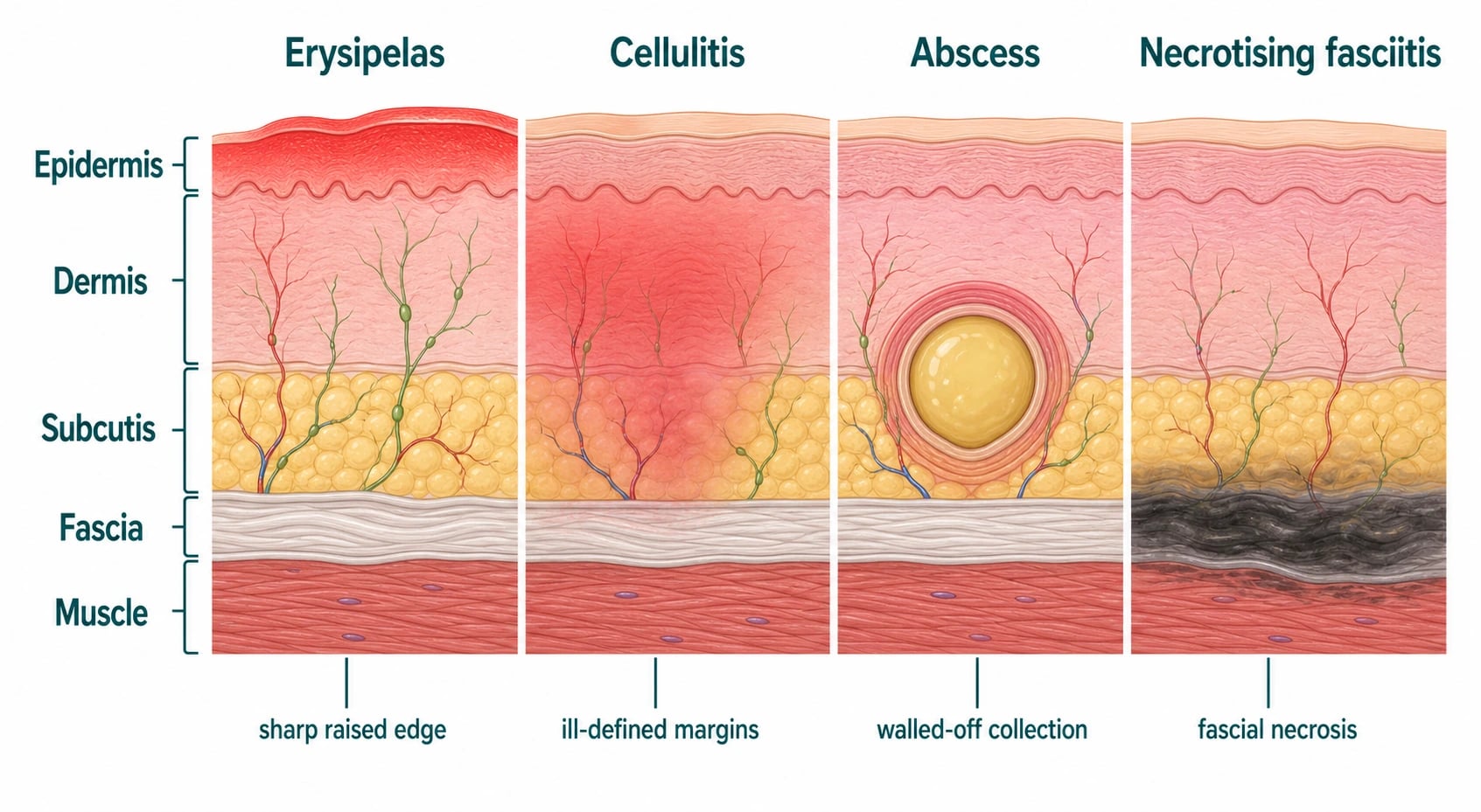
Cellulitis is an acute, non-necrotising, spreading bacterial infection of the dermis and subcutaneous tissue, presenting as a warm, erythematous, oedematous, tender area with ill-defined margins. Erysipelas is a more superficial form involving the upper dermis and superficial lymphatics, with a sharply demarcated, raised advancing edge. Both are caused most often by β-haemolytic streptococci (especially group A, S. pyogenes) and Staphylococcus aureus. Fellowship-level competence requires precise clinical phenotyping, confident exclusion of dangerous mimics (especially necrotising fasciitis and deep venous thrombosis), recognition of predisposing factors, rational empiric antibiotic selection, the special urgency of periorbital/orbital disease, and strategies to prevent the common and disabling problem of recurrence.[1][2][3]
Clinical assessment & bedside tools
A thorough bedside assessment is essential to differentiate true cellulitis from look-alikes, identify the portal of entry, and grade severity before deciding on site-of-care and route of antibiotics: [1]
- Mark the edge of erythema with an indelible ink pen or take a dated photograph; spread beyond the mark in 24-48 h means treatment failure or wrong diagnosis.
- Examine the inter-toe web spaces for maceration, fissuring, and tinea pedis — the single commonest portal in lower-limb cellulitis. KOH microscopy of scrapings is a quick, cheap bedside test.
- Palpate for fluctuance suggesting an abscess (requires incision and drainage, not antibiotics alone).
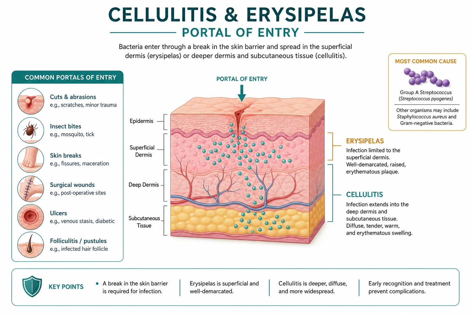
- Look for portal of entry: surgical wounds, ulcers, eczema, cracks in heels, IV cannulation, insect bites, fungal nail infection, and pediculosis in homeless or institutional patients.
- Check for lymphangitis (linear erythema tracking proximally) and tender regional lymphadenopathy — features that argue for streptococcal infection.
- Quantify severity: fever, tachycardia, hypotension, sepsis features, pain out of proportion, bullae, crepitus, and skin necrosis all suggest severe disease (Class III or IV).
- Comorbidity screen: diabetes, immunosuppression, cirrhosis, IVDU, peripheral vascular disease, lymphoedema, chronic kidney disease, sickle cell — all increase risk of severe disease and lower the threshold for IV therapy.
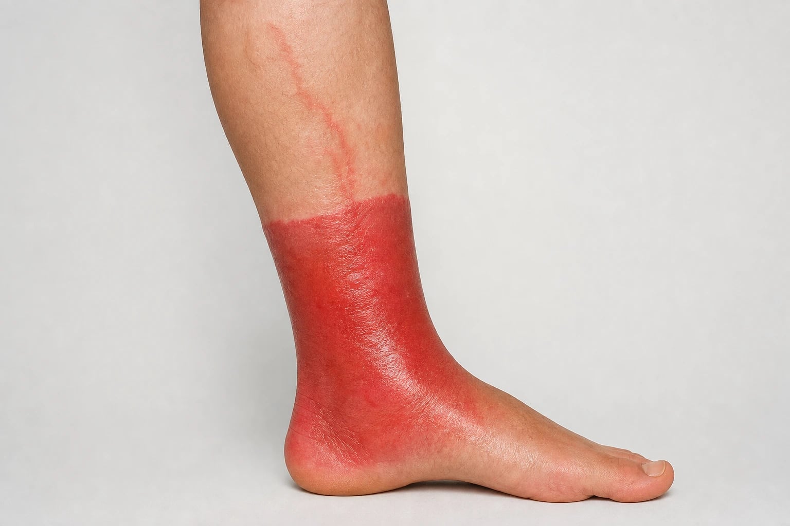
- Erysipelas vs cellulitis border sign: erysipelas has a raised, sharply demarcated leading edge; cellulitis has a flat, indistinct margin. [1]
Quick numbers for the examiner
Differential diagnosis — pseudocellulitis and dangerous mimics
A large fraction of patients referred for "cellulitis" have an alternative diagnosis (pseudocellulitis), and a smaller fraction have a dangerous mimic. The bedside marks the difference. [1]
Necrotising fasciitis — the do not miss
- Cutaneous pain out of proportion to the examination.
- Rapid progression beyond the mark; dusky or violaceous skin; haemorrhagic bullae; skin necrosis.
- Systemic toxicity disproportionate to skin appearance.
- Anaesthesia over the affected area (cutaneous nerve death).
- Crepitus suggests gas-forming organisms.
- Surgical emergency — broad-spectrum IV antibiotics within 1 hour (meropenem or piperacillin-tazobactam plus linezolid/vancomycin with clindamycin added for toxin suppression) and immediate debridement by a senior surgeon. Do not delay surgery for imaging.
- LRINEC score may aid but is not a substitute for clinical suspicion. [1]
Progression of necrotising fasciitis — staging for the bedside
The natural history of necrotising soft-tissue infection evolves through recognisable bedside stages, and naming them improves early recognition when the signs are still subtle: [1]
- Stage 1 (early, 0–2 days) — tender erythema with swelling and warmth disproportionate to examination findings; pain that exceeds clinical signs; fever, tachycardia, and a rising CRP. Misdiagnosis as uncomplicated cellulitis is common; the LRINEC score may still be low.
- Stage 2 (intermediate, 2–4 days) — tense oedema, dusky or violaceous skin discolouration, haemorrhagic bullae, and serosanguinous "dishwater" drainage from any breach; cutaneous anaesthesia appears as the superficial nerves infarct; systemic toxicity mounts (lactate >2 mmol/L, AKI, confusion).
- Stage 3 (late, 4–7+ days) — frank skin necrosis with eschar formation, crepitus from gas-forming organisms (type II polymicrobial or Clostridium), septic shock, and multi-organ failure. Mortality now exceeds 50% even with surgery. [1]
Recognition at Stage 1 is the only route to a good outcome: any cellulitis with pain out of proportion, rapid spread beyond the marked edge at 24–48 h, bullae, anaesthesia, or rising lactate mandates urgent senior surgical review and operative exploration — do not wait for confirmatory imaging.[2][4]
Orbital (postseptal) cellulitis
- Proptosis, painful or limited eye movements (ophthalmoplegia), reduced visual acuity, chemosis with or without systemic toxicity.
- Most commonly from ethmoid or maxillary sinusitis in children.
- Urgent CT of orbits and sinuses with contrast; IV ceftriaxone plus metronidazole plus vancomycin; ENT and ophthalmology input; surgical drainage if subperiosteal or orbital abscess. [1]
Investigations
- Blood cultures: low yield (about 1-2%) in uncomplicated disease; recommended for sepsis, severe disease, immunocompromise, recurrent cellulitis, piercing or tattoo exposure, IVDU, or suspected bacteraemia.
- Pus swab: if there is a wound, ulcer, or purulent exudate. Culture and sensitivity. Add PVL PCR for recurrent or family-cluster abscesses (USA300 / SCCmec dominance where relevant).
- Skin biopsy: rarely used; consider for atypical features (vasculitis, Sweet syndrome, pyoderma gangrenosum, malignancy mimic) when diagnosis is unclear.
- Imaging: ultrasound for suspected abscess; CT or MRI if necrotising fasciitis or orbital cellulitis suspected; plain radiographs if gas or osteomyelitis.
- Severity bloods: FBC, CRP, U&E, glucose, HbA1c, venous and arterial blood gas, lactate for sepsis.
- Immunocompromise screen: HIV, HbA1c, consider hepatitis B/C and immunoglobulins for recurrent disease.
- Blood culture contamination minimisation: clean skin with 2% chlorhexidine, draw 2 sets from different sites; do not draw from an existing cannula. [1]
Special scenarios
- Periorbital (preseptal) cellulitis: in children, usually from sinusitis or local trauma; treated with oral co-amoxiclav or cefalexin for 7 days; if systemic features or red eye are present → admit for IV ceftriaxone plus metronidazole and CT; rule out orbital extension. [1]
Periorbital cellulitis — anatomy, organisms, and management
Periorbital (preseptal) cellulitis is a dermal–subcutaneous infection anterior to the orbital septum — the fibrous sheet that anchors the orbital rim to the tarsal plates and forms the anterior boundary of the orbit. Because the septum is a physical barrier, infection does not communicate with the orbit itself, and vision, ocular motility, and pupil reflexes remain normal; this is the key bedside discriminator from orbital cellulitis. In children under 9 years the commonest antecedent is bacteraemic seeding from ipsilateral ethmoid or maxillary sinusitis (the ethmoid bone in this age group is fenestrated, allowing direct venous communication); in older children and adults the portal is more often local trauma, an insect bite, impetigo, or a hordeolum.[11][15]
Organisms vary with the portal. Sinusitis-related cases are typically S. pneumoniae, S. pyogenes, S. aureus, or (in unimmunised children) Haemophilus influenzae type b. Trauma-related cases are predominantly S. aureus and streptococci, with Pasteurella multocida after cat or dog bites around the lid. Anaerobes are uncommon unless there is a dental or sinus source. [1]
Treatment is stratified by age and severity. Mild childhood periorbital cellulitis without systemic features is treated with oral co-amoxiclav (or cefalexin in penicillin allergy) for 7 days, with review at 48 h. Children under 1 year, those who look systemically unwell, those who cannot tolerate oral therapy, or those in whom orbital (postseptal) extension cannot be excluded clinically are admitted for IV ceftriaxone plus metronidazole, plus urgent contrast-enhanced CT of the orbits and sinuses. Adult periorbital cellulitis follows the same principles but the threshold for IV therapy is lower (co-amoxiclav IV, or ceftriaxone plus metronidazole if there is dental or sinus involvement). Failure to defervesce within 48 h, worsening eyelid oedema, or any new ocular symptom mandates re-examination for orbital extension.[11][15]
- Animal or human bite wounds: Pasteurella multocida and Capnocytophaga; amoxicillin-clavulanate is first-line; consider tetanus and rabies prophylaxis.
- Saltwater or brackish-water exposure: Vibrio vulnificus; doxycycline plus ceftriaxone; patients with cirrhosis are at risk of fulminant septicaemia.
- Freshwater exposure: Aeromonas hydrophila — doxycycline plus ciprofloxacin.
- Fish-tank / aquarium / fish-handling: Mycobacterium marinum — biopsy, prolonged combination antibiotics.
- Lymphoedema-associated recurrent cellulitis: aggressive compression, lymphoedema therapist input, treat tinea pedis; long-term penicillin V 250 mg BD if two or more episodes per year (CREST guidance; UK NICE NG141).
- Dermatologic barrier defects: atopic dermatitis, blistering disorders, ichthyosis — treat the underlying skin disease to prevent recurrent infection.
- Necrotising fasciitis (see above): surgical emergency. [1]
Special populations
- Children: most lower-limb cellulitis is streptococcal; empirical flucloxacillin or cephalexin; facial (periorbital) is usually S. pneumoniae, S. aureus, or S. pyogenes.
- Pregnancy: avoid tetracyclines and fluoroquinolones; penicillin and cephalosporins are safe; lactation-compatible antibiotics: amoxicillin, cephalexin, erythromycin.
- Diabetes / PVD: lower threshold for admission; consider mixed polybacterial infection if a chronic ulcer is the portal; broader cover if systemically unwell.
- Cirrhosis / chronic liver disease: Capnocytophaga (after dog bites) and Vibrio vulnificus (after seawater exposure) cause fulminant sepsis; aggressive empirical cover with ceftriaxone plus doxycycline.
- Elderly: atypical presentation is common (delirium instead of fever); blood cultures are useful; longer antibiotic duration may be needed; review CKD dose adjustments.
- Immunocompromised: HIV, transplant, chemotherapy, biologics — neutropenic sepsis; broader empirical cover (e.g., piperacillin-tazobactam plus vancomycin). [1]
Regional deltas
[1]The MARKED bedside framework for cellulitis
Complications and prognosis
- Bacteraemia and sepsis — uncommon in mild-moderate disease but rising in immunocompromised hosts.
- Abscess formation — requires incision and drainage; antibiotics alone fail.
- Necrotising fasciitis — the do not miss; mortality 25% overall, higher with delayed surgery.
- Thrombophlebitis and DVT — from inflammation and immobility.
- Lymphangitis → lymphoedema — recurrent episodes damage lymphatic channels; chronic lymphoedema further predisposes to further cellulitis (the vicious cycle).
- Streptococcal toxic shock — invasive GAS with hypotension and multi-organ failure — admit to ICU; clindamycin suppresses exotoxin production.
- Permanent pigmentary or scarring sequelae, especially after bullous cellulitis or abscess.
- Post-streptococcal glomerulonephritis — rare in adults but a recognised complication following streptococcal cellulitis.
- Recurrence is the single most common complication: 30% of patients have another episode within 3 years, often from the same portal of entry. [1]
Special considerations for adults
- Marking the edge of erythema prevents diagnostic drift at 48 h.
- Tinea pedis is the commonest portal in lower-limb disease — examine inter-toe web spaces; skin scrapings for KOH microscopy; oral terbinafine 250 mg OD for 4 weeks for typical dermatophyte infection.
- Compression therapy (after the acute phase) accelerates healing, reduces lymphoedema, and halves recurrence in venous insufficiency.
- Long-term penicillin V prophylaxis (250 mg BD) is supported by the ORIENT trial (2013) and Cochrane review (2024) for patients with frequent recurrence despite optimisation of underlying risk factors.
- Patient advice at discharge: foot care, tinea prevention, moisturise ankles, prompt attention to fissures, and education on when to return (rapid spread, fever, blister). [1]
Pathophysiology and microbiology
- Group A β-haemolytic streptococci (S. pyogenes) are the commonest cause of erysipelas and a leading cause of cellulitis; groups B, C, and G streptococci also contribute (especially in diabetes and older adults).
- Staphylococcus aureus is the other principal pathogen in cellulitis; MRSA is an important cause in endemic regions, in recurrent or severe disease, and in specific exposures (athletes, incarcerated populations, IVDU).
- Infection typically enters through a skin barrier breach: tinea pedis (the single commonest predisposing factor for leg cellulitis), fissures, ulcers, eczema, trauma, insect bites, IV cannulation, or surgery.
- Bacteraemia is uncommon in uncomplicated cellulitis and erysipelas (approximately 1-2% in typical cases), so routine blood cultures are low-yield in immunocompetent adults with mild-moderate disease, but are warranted in severe disease, sepsis, immunocompromise, and specific exposures.[6][8][11]
Clinical features
Erysipelas
A bright red, shiny, warm, tender, indurated plaque with a sharply demarcated, raised advancing edge, typically on the face or lower leg, often with proximal lymphangitis and lymphadenopathy, fever, chills, and malaise. "Butterfly" facial involvement is classic. The sharp demarcation reflects the superficial lymphatic involvement.[3][7]
Cellulitis
A warm, erythematous, oedematous, tender area with poorly defined margins (because the infection is deeper, in dermis/subcutis), most often on a lower limb. There may be vesicles, bullae, petechiae, or necrotic patches in severe cases; regional lymphadenopathy and fever are common. The unilateral distribution is a key discriminator from stasis dermatitis and other bilateral inflammatory conditions.[1][2]
Special sites
- Periorbital (preseptal) cellulitis — infection anterior to the orbital septum; eyelid erythema/oedema without proptosis, ophthalmoplegia, or visual change; usually managed with oral antibiotics but monitored closely for posterior extension.
- Orbital (postseptal) cellulitis — infection posterior to the orbital septum, usually from adjacent sinusitis; presents with proptosis, painful or limited eye movement (ophthalmoplegia), reduced vision, and systemic toxicity, and is an ophthalmic/ENT emergency requiring urgent imaging (CT), IV antibiotics, and often surgical drainage of an abscess or sinus.[11][15]
Diagnosis
The diagnosis is clinical. Useful adjuncts: [1]
- Skin swabs/microbiology are useful only when there is an associated wound, ulcer, or purulent drainage (culture the portal of entry).
- Blood cultures for severe disease, sepsis, immunocompromise, or specific exposures (low yield otherwise).[6]
- Blood tests (FBC, CRP, U&E, glucose) for severity assessment and comorbidity.
- Imaging (ultrasound to distinguish abscess from cellulitis; CT/MRI if necrotising infection or orbital involvement suspected). Plain radiographs if gas or osteomyelitis suspected.
- Procalcitonin is not routinely required but may support a bacterial cause in difficult cases.
Differential diagnosis and "pseudocellulitis"
A large fraction of patients referred for "cellulitis" have an alternative diagnosis (pseudocellulitis), and misdiagnosis drives unnecessary admission and antibiotics. Important mimics include:[4]
- Stasis (venous) dermatitis and lipodermatosclerosis — bilateral, chronic, dependent; not cellulitis though may be secondarily infected.
- Contact dermatitis, atopic dermatitis — pruritic, bilateral or geometric.
- Deep venous thrombosis — unilateral leg swelling; exclude with ultrasound if uncertain.
- Necrotising fasciitis — severe pain out of proportion, rapid spread, systemic toxicity, bullae, creptius, skin necrosis; surgical emergency (use a validated scoring tool such as LRINEC cautiously; do not delay surgery).
- Acute gout, septic arthritis, osteomyelitis.
- Inflammatory dermatoses: erythema nodosum, Sweet syndrome, pyoderma gangrenosum, fixed drug eruption.
- Erythema migrans (Lyme), herpes zoster early, insect envenomation.
- Carcinoma erysipeloides (inflammatory breast/lymphatic metastasis). [1]
The unilateral, warm, tender, spreading pattern with systemic upset distinguishes true cellulitis from most bilateral, chronic, pruritic mimics.[2][4]
Risk stratification and severity
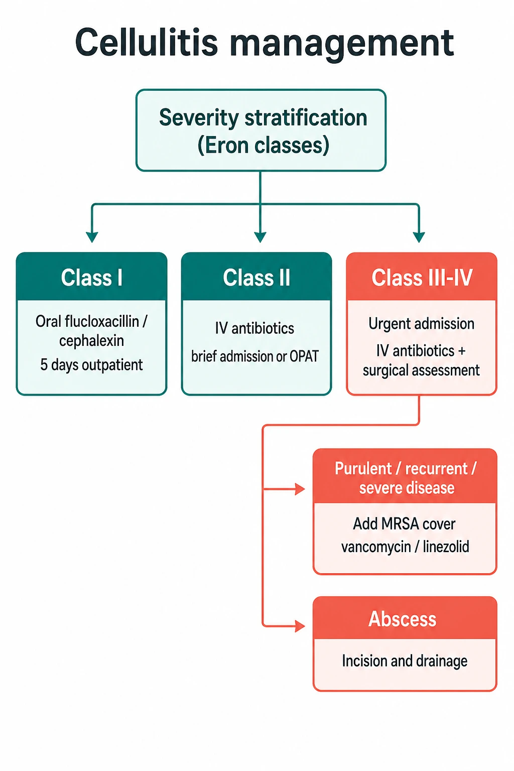
Use a severity framework (e.g., Eron/Lewis classes or similar) to guide the site of care and route of antibiotics:[2][5]
- Class I — no signs of systemic toxicity; oral antibiotics, outpatient.
- Class II — systemic toxicity or comorbidity; IV antibiotics, brief admission or outpatient parenteral therapy.
- Class III–IV — systemic toxicity, unstable comorbidity, or a limb/life-threatening infection (gas, necrosis, immunocompromise); urgent admission, IV antibiotics, and surgical assessment. [1]
Management
Antibiotic therapy
Empiric therapy targets streptococci and methicillin-susceptible S. aureus; broaden for MRSA in the appropriate setting.[4][9][12]
- Typical (non-purulent) cellulitis/erysipelas, oral: flucloxacillin (or cephalexin) for 5 days, extending if not improved; macrolides (clarithromycin) for penicillin allergy. A 5-6 day course is sufficient for most uncomplicated cases.[13][14]
- Severe or IV therapy: IV flucloxacillin (or cefazolin); clindamycin for penicillin allergy or toxin suppression; vancomycin or linezolid for suspected MRSA or severe penicillin allergy.
- Suspected MRSA: add vancomycin, linezolid, daptomycin, or clindamycin (if susceptible) — particularly for purulent infection, recurrent disease, prior MRSA, IVDU, severe sepsis, or endemic regions.
- Purulent infection (abscess): incision and drainage is primary; antibiotics adjunctive depending on size, systemic signs, and immune status.
- Specific exposures: animal bites (amoxicillin-clavulanate for Pasteurella); water exposure (Vibrio/Aeromonas — doxycycline plus a third-generation cephalosporin).
Treatment doses — first-line and alternatives
Specific doses for the commonest regimens. Adjust for renal and hepatic function, body weight in children, and local resistance patterns. [1]
Oral therapy for uncomplicated cellulitis / erysipelas (Class I): [1]
- Flucloxacillin 500 mg QDS (four times daily) for 5 days — the standard first-line dose in the UK and Australia for non-severe cellulitis and erysipelas.
- Cefalexin 500 mg TDS (three times daily) for 5 days — alternative with better streptococcal cover; preferred in penicillin-allergic patients able to take cephalosporins.
- Clarithromycin 500 mg BD (twice daily) for 5 days — for true penicillin allergy.
- Amoxicillin 500 mg TDS — when streptococcal infection predominates (e.g. classic erysipelas) and staphylococcal cover is not required. [1]
IV therapy for moderate–severe cellulitis (Class II–III): [1]
- Flucloxacillin 1–2 g IV 6-hourly (QDS) — switch to oral once afebrile and improving.
- Cefazolin 1–2 g IV 8-hourly — alternative in penicillin allergy with tolerable cephalosporin cross-reactivity.
- Benzylpenicillin (penicillin G) 1.2–2.4 g IV 6-hourly PLUS flucloxacillin — when both S. pyogenes and S. aureus must be covered and the patient is hospitalised.
- Clindamycin 300–450 mg IV 6-hourly — for penicillin-allergic patients, for toxin suppression in invasive group A streptococcal infection (suppresses exotoxin production), or as an adjunct in necrotising fasciitis alongside broad-spectrum cover.
- Vancomycin 25–30 mg/kg loading then 15–20 mg/kg 12-hourly (trough 15–20 mg/L) — for suspected MRSA, severe sepsis, or treatment failure.
- Linezolid 600 mg IV/PO 12-hourly — alternative to vancomycin; excellent oral bioavailability; reserve for MRSA or vancomycin intolerance. [1]
Secondary prophylaxis after recurrent cellulitis (≥2 episodes/year): [1]
- Penicillin V 250 mg BD orally — long-term, supported by the ORIENT trial and Cochrane review (2017; updated 2024).
- Erythromycin 250 mg BD — alternative in penicillin allergy. [1]
Paediatric doses (per kg body weight, approximate): [1]
- Flucloxacillin 12.5–25 mg/kg QDS oral (max 500 mg/dose); 25–50 mg/kg QDS IV (max 2 g/dose).
- Cefalexin 12.5–25 mg/kg TDS oral (max 1 g/dose); cefazolin 25 mg/kg 8-hourly IV.
- Co-amoxiclav (for periorbital cellulitis) — 0.5 mL/kg of 125/31 suspension TDS, or 25/5 mg/kg TDS. [1]
Antibiotic doses at a glance
Supportive and adjunctive measures
- Elevate the affected limb; mark the edge of erythema to monitor response.
- Treat the portal of entry: tinea pedis (antifungal), leg ulcers, interdigital fissures, eczema.
- Analgesia, hydration, and management of comorbidities (diabetes, oedema).
- Treat predisposing factors to prevent recurrence: lymphoedema (compression), venous insufficiency, obesity, tinea pedis.[9][10]
Prevention of recurrence
Recurrent cellulitis is common (up to one-third within 3 years) and disabling. Prevention requires eliminating the portal of entry (aggressive treatment of tinea pedis and interdigital maceration), compression for lymphoedema/venous insufficiency, and — for those with frequent recurrences despite optimisation — long-term secondary antibiotic prophylaxis (e.g., low-dose oral penicillin V or erythromycin), supported by Cochrane evidence for reduced recurrence.[9][10]
Complications
Bacteraemia and sepsis (especially in immunocompromised hosts), abscess formation, necrotising fasciitis, venous thromboembolism from immobility, lymphatic damage with recurrent episodes ("lymphangitis → lymphoedema → further cellulitis" vicious cycle), and permanent limb swelling.[6][9]
Clinical pearl
[1]Red flags
Exam application bank (NEET-PG / INICET)
One-line answer
Cellulitis and erysipelas are acute, non-necrotising bacterial infections of the dermis and subcutaneous tissue (cellulitis) or the upper dermis with superficial lymphatics (erysipelas), caused most often by streptococci and Staphylococcus aureus. Fellowship-level assessment demands mastery of the clinical and anatomical distinction between erysipelas and cellulitis, risk stratification and the exclusion of life-threatening mimics (necrotising fasciitis, deep venous thrombosis, severe inflammatory dermatoses), recognition of predisposing factors (tinea pedis, lymphoedema, venous insufficiency, immunocompromise), the role of bacteraemia and its rarity in uncomplicated disease, rational empiric antibiotic therapy (flucloxacillin/cephalexin; clindamycin, vancomycin, or linezolid for MRSA or severe penicillin allergy), the special urgency of periorbital versus orbital cellulitis, and the pre
Worked stems (answer without another resource)
Stem 1 — Classic presentation. Map symptoms to mechanism; name the first investigation and first treatment step with dose/route if drug therapy is standard. [1]
Stem 2 — Unstable / complicated. List red flags that force immediate resuscitation, theatre, ICU, antidote, or reperfusion — and what you do in the first 15 minutes. [1]
Stem 3 — Atypical group. Elderly, pregnancy, child, or immunocompromised: how presentation and thresholds change. [1]
Stem 4 — Differential trap. Name the three closest mimics and one discriminator for each. [1]
Stem 5 — Disposition. Who goes home with safety-netting, who is admitted, who needs HDU/ICU/theatre, and what follow-up is mandatory. [1]
Rapid viva checklist
- Definition + classification
- Pathophysiology chain
- Bedside signs / criteria
- Score with exact components (if any)
- Emergency bundle
- Definitive therapy with doses
- Complications of disease and of treatment
- Special populations
- Guideline/trial name if classic
- Three exam traps
Coverage self-check
If you cannot answer any stem above from this page alone, re-read the matching section — the page is intended to be self-sufficient for final-prof and NEET-PG/INICET questions on Cellulitis and erysipelas.
Expanded exam teaching (depth pass)
Clinical reasoning
For Cellulitis and erysipelas, examiners test whether you can prioritise life threats, choose the right first test, and give specific therapy (agent, dose, route, timing). Generic phrases without numbers score poorly.
Mechanism → feature map
Build a short chain: cause → pathophysiologic intermediate → clinical feature → complication. Every major symptom in the classic vignette should sit on that chain.
Investigation strategy
- Bedside/first-line tests that change immediate management
- Confirmatory or staging tests
- What a normal result does not exclude
- When not to delay treatment for imaging (unstable patient)
Management ladder
- Resuscitation / ABC / sepsis or haemorrhage bundle as relevant
- Specific antidote / procedure / antimicrobial / reperfusion / surgery
- Supportive care and monitoring targets
- Definitive long-term therapy and secondary prevention
- Disposition and safety-net advice
Special populations
Always prepare one line each for children, pregnancy, elderly, renal/hepatic impairment, and immunocompromised patients when the topic allows.
Pitfalls that fail candidates
- Treating the number not the patient
- Missing pregnancy status when relevant
- Imaging before stabilisation
- Wrong empiric cover or wrong antidote timing
- Incomplete counselling on recurrence, adherence, or red-flag return
Cellulitis and erysipelas are acute, non-necrotising bacterial infections of the dermis and subcutaneous tissue (cellulitis) or the upper dermis with superficial lymphatics (erysipelas), caused most often by streptococci and Staphylococcus aureus. Fellowship-level assessment demands mastery of the clinical and anatomical distinction between erysipelas and cellulitis, risk stratification and the exclusion of life-threatening mimics (necrotising fasciitis, deep venous thrombosis, severe inflammato [1]
Structured revision sheet
Must-know numbers and names
List every score, size threshold, dose, and time window from this topic on a blank page from memory, then check against the sections above.
Three classic MCQ angles
- Most likely diagnosis given a vignette
- Next best step in management
- Most appropriate investigation
Three classic SAQ angles
- Pathophysiology in five steps
- Management algorithm with doses
- Complications and prevention
Clinical station flow
Greet → focused history → targeted exam → investigations → explain diagnosis → emergency care → definitive plan → safety-net / follow-up → answer examiner questions on mechanism and pitfalls.
[1]References
- [1]Raff AB, Kroshinsky D. Cellulitis: A Review JAMA, 2016.PMID 27434444
- [2]Rrapi R, Chand S, Kroshinsky D. Cellulitis: A Review of Pathogenesis, Diagnosis, and Management Med Clin North Am, 2021.PMID 34059247
- [3]Bystritsky RJ. Cellulitis Infect Dis Clin North Am, 2021.PMID 33494874
- [4]Boettler MA, Kaffenberger BH, Chung CG. Cellulitis: A Review of Current Practice Guidelines and Differentiation from Pseudocellulitis Am J Clin Dermatol, 2022.PMID 34902109
- [5]Long B, Gottlieb M. Diagnosis and Management of Cellulitis and Abscess in the Emergency Department Setting: An Evidence-Based Review J Emerg Med, 2022.PMID 34657784
- [6]Gunderson CG, Martinello RA. A systematic review of bacteremias in cellulitis and erysipelas J Infect, 2012.PMID 22101078
- [7]Rodrigues MA, Caetano M, Amorim I, et al. [Non-Necrotizing Acute Dermo-Hypodermal Infections: Erysipela and Infectious Cellulitis] Acta Med Port, 2021.PMID 33971117
- [8]Stevens DL, Bryant AE. Streptococcus pyogenes Impetigo, Erysipelas, and Cellulitis 2022.PMID 36479753
- [9]Peghin M, Graziano E, Rovelli C, et al. Prevention and treatment of recurrent cellulitis Curr Opin Infect Dis, 2023.PMID 36853755
- [10]Dalal A, Eskin-Schwartz M, Mimouni D, et al. Interventions for the prevention of recurrent erysipelas and cellulitis Cochrane Database Syst Rev, 2017.PMID 28631307
- [11]Yadalla D, Jayagayathri R, Padmanaban K, et al. Bacterial orbital cellulitis - A review Indian J Ophthalmol, 2023.PMID 37417106
- [12]Shu Z, Cao J, Li H, et al. Efficacy and safety of first- and second-line antibiotics for cellulitis and erysipelas: a network meta-analysis of randomized controlled trials Arch Dermatol Res, 2024.PMID 39240378
- [13]Mo Y, Tan WC, Cooper BS. Antibiotic duration for common bacterial infections-a systematic review JAC Antimicrob Resist, 2025.PMID 39881797
- [14]Chambliss ML MD, MSPH, Rumball A DO, Brown CM MD. Skin and Soft Tissue Infections Am Fam Physician, 2026.PMID 42202366
- [15]Anosike BI, Ganapathy V, Nakamura MM. Epidemiology and Management of Orbital Cellulitis in Children J Pediatric Infect Dis Soc, 2022.PMID 35438766
- [16]Clebak KT, Malone MA. Skin Infections Prim Care, 2018.PMID 30115333