Dermatology · Medicine
Cicatricial alopecia
Also known as Cicatricial alopecia · Scarring alopecia · Lichen planopilaris (LPP) · Frontal fibrosing alopecia (FFA) · Central centrifugal cicatricial alopecia (CCCA) · Folliculitis decalvans · Dissecting cellulitis of the scalp
Cicatricial (scarring) alopecia is a group of disorders in which hair follicles are destroyed and replaced by fibrous tissue, producing permanent, irreversible hair loss. The hallmark clinical sign is loss of follicular ostia. The North American Hair Research Society (NAHRS) classifies primary cases by inflammatory infiltrate into lymphocytic (lichen planopilaris, frontal fibrosing alopecia, discoid lupus, CCCA, pseudopelade of Brocq), neutrophilic (folliculitis decalvans, dissecting cellulitis) and mixed (acne keloidalis nuchae) forms. Early diagnosis by trichoscopy and biopsy of the active margin is essential because destroyed follicles cannot regrow. Management targets the inflammatory mechanism: lymphocytic disease uses corticosteroids, hydroxychloroquine, mycophenolate and JAK inhibitors; neutrophilic disease uses antibiotics, isotretinoin and TNF inhibitors. Hair transplantation is reserved for burnt-out, quiescent disease.
On this page & tools
Your progress
Saved locally on this device.
Exam tags
Red flags
Overview & Definition
Cicatricial alopecia, also called scarring alopecia, is not a single diagnosis but a group of disorders that share a common final pathway: destruction of the hair follicle and its replacement by scar (fibrous) tissue. The result is permanent hair loss because the stem cells required to regenerate the follicle are lost.[2][5]
The clinical hallmark that separates cicatricial alopecia from non-cicatricial (non-scarring) alopecia is the absence of follicular ostia — the tiny openings of hair follicles on the skin surface are no longer visible. In non-scarring alopecias such as androgenetic alopecia, alopecia areata or telogen effluvium, the follicle remains structurally intact, so hair regrowth is at least theoretically possible. In cicatricial alopecia, once the follicular opening is lost, the follicle cannot be restored and the hair loss is irreversible.[2]
Cicatricial alopecia can be primary (the inflammatory process targets the follicle itself) or secondary (the follicle is destroyed by an external process). Primary cicatricial alopecias are rare, with a combined prevalence estimated in the range of 0.02 to 0.11 percent depending on the population studied, but their impact on quality of life is high because the cosmetic loss is permanent.[5][8]
[1]Classification
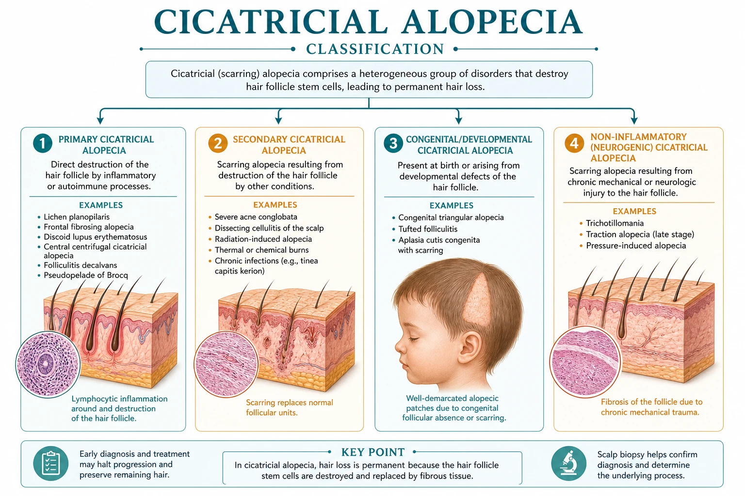
The most widely used classification is the consensus statement of the North American Hair Research Society (NAHRS). It divides primary cicatricial alopecia into three groups based on the predominant inflammatory infiltrate seen on scalp biopsy: lymphocytic, neutrophilic and mixed.[2][6]
Lymphocytic group
In this group a band-like or perifollicular lymphocytic infiltrate damages the upper (permanent) portion of the hair follicle, particularly the isthmus and bulge region where follicular stem cells reside. Diseases include: [1]
- Lichen planopilaris (LPP) — the prototypical lymphocytic cicatricial alopecia.
- Frontal fibrosing alopecia (FFA) — now considered a clinical variant of LPP with preferential frontal-temporal involvement.
- Discoid lupus erythematosus (DLE) — the scalp manifestation of chronic cutaneous lupus.
- Central centrifugal cicatricial alopecia (CCCA) — the most common scarring alopecia in women of African ancestry.
- Pseudopelade of Brocq — historically described as a separate entity; now usually regarded as an end-stage, burnt-out appearance of LPP or DLE. [1]
Neutrophilic group
Neutrophilic infiltration around and within the follicle produces pustules, folliculitis, abscesses and sinus tracts. The two main diseases are: [1]
- Folliculitis decalvans — recurrent pustules and tufted hairs, with Staphylococcus aureus commonly cultured.
- Dissecting cellulitis of the scalp (perifolliculitis capitis abscedens et suffodiens) — deep, boggy, interconnected nodules and sinus tracts. [1]
Mixed group
A mixed infiltrate is seen in: [1]
- Acne keloidalis nuchae — fibrosing folliculitis at the nape of the neck, most common in Black men.
- Acne necrotica — superficial pustular lesions that heal with varioliform scarring.
- Erosive pustular dermatosis of the scalp — pustular, erosive process that follows trauma or surgery in sun-damaged scalp. [1]
Secondary cicatricial alopecia
Secondary forms are not classified by NAHRS because the follicle is destroyed by an external process rather than a primary folliculocentric disease. Examples include physical burns, ionising radiation, severe infections (kerion, cutaneous tuberculosis, tertiary syphilis), cutaneous malignancies infiltrating the scalp, and iatrogenic injury from surgery or chronic trauma. In resource-limited settings, tinea capitis kerion and destructive pyogenic infections remain important preventable causes of scarring hair loss in children.[3][6]
Epidemiology & Risk Factors
Primary cicatricial alopecias are uncommon but the epidemiology varies markedly by subtype. LPP shows a female predominance of roughly 3:1 and most commonly affects Caucasian women in the fourth to sixth decades. FFA is overwhelmingly a disease of postmenopausal women, with a typical age of onset between 50 and 70 years, although it is increasingly reported in premenopausal women and in men.[1][6]
CCCA is the most common form of cicatricial alopecia in women of African ancestry. It usually begins in the third to fifth decade and progresses slowly over years. Traction from tight braids, weaves, locs, chemical relaxers and heat styling are recognised risk factors, although genetic susceptibility and grooming practices interact. The name "central centrifugal" describes the vertex starting point and outward expansion pattern.[9]
Folliculitis decalvans affects men more often than women, with onset usually in early to mid-adult life. Staphylococcus aureus is frequently isolated, and chronic nasal carriage may be present. Dissecting cellulitis is most common in young Black men and is one of the four components of the follicular occlusion tetrad, together with acne conglobata, hidradenitis suppurativa and pilonidal sinus disease. Acne keloidalis nuchae is almost confined to Black men and is strongly linked to close shaving or clipping of curly nape hair and friction from collars or helmets.[3][7]
Recent studies have also highlighted systemic associations. Patients with primary cicatricial alopecia, particularly lymphocytic subtypes, appear to have higher rates of metabolic comorbidity (dyslipidaemia, diabetes, hypertension) and possibly cardiovascular disease. The relationship is likely multifactorial, involving shared inflammatory pathways, obesity-related hormonal changes and reduced physical activity because of psychosocial impact. In discoid lupus, ultraviolet exposure and smoking are important environmental risk factors for disease activity and scarring.[5][8]
Epidemiological anchors
Pathophysiology
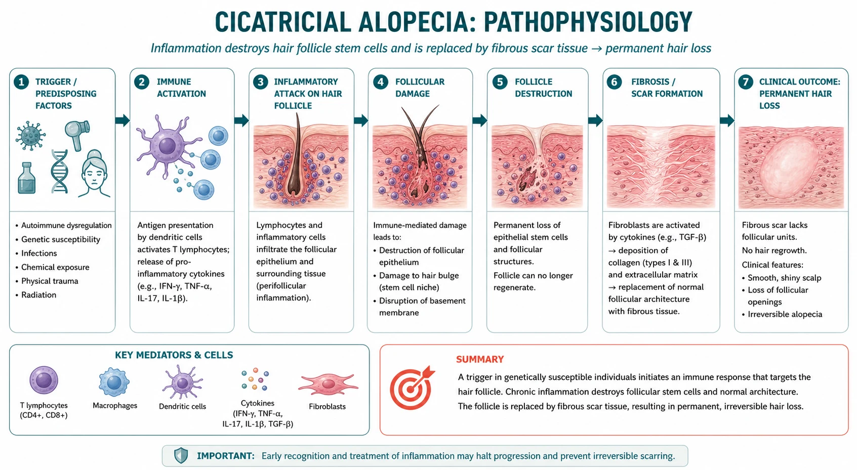
The key event in all cicatricial alopecias is the loss of the follicular stem-cell niche. The permanent portion of the follicle — the isthmus and bulge — contains epithelial stem cells that normally regenerate the lower follicle during each hair cycle. When this region is destroyed, the follicle cannot repopulate and the follicular tract is replaced by dense collagenous scar tissue.[5][8]
In lymphocytic disease, autoreactive or aberrantly activated T cells infiltrate around the follicular infundibulum and isthmus. The sebaceous gland is often an early target, and sebaceous loss is a consistent histological finding. Cytokines such as interferon-γ, TNF-α and interleukin-17 drive a lichenoid or interface inflammatory reaction, while transforming growth factor-β (TGF-β) promotes fibroblast activation and collagen deposition. Epithelial cells may undergo epithelial-mesenchymal transition (EMT), losing their epithelial phenotype and acquiring a fibroblast-like phenotype that contributes to dermal fibrosis.[5][8]
In neutrophilic disease, neutrophils accumulate within and around the follicle, often in response to bacterial colonisation or a primary follicular occlusion defect. The intense neutrophilic inflammation destroys the follicular wall, allows adjacent follicles to merge (creating the tufted hairs of folliculitis decalvans), and produces abscesses, sinus tracts and granulation tissue. Chronic inflammation drives hypertrophic scarring and keloid formation in acne keloidalis nuchae.[3][7]
The concept of follicular immune privilege collapse has been proposed as a unifying mechanism. The anagen hair bulb normally maintains immune privilege by downregulating MHC class I and producing immunosuppressive molecules. In alopecia areata this immune privilege is lost but the follicle is preserved. In cicatricial alopecia, the inflammatory attack is more destructive and the immune-privileged stem-cell niche is permanently lost, leading to scar.[5]
Clinical Presentation
Patients usually present with one or more patches of hair loss that are slowly progressive. The first symptom may be itching (pruritus), burning, pain or tenderness (trichodynia) at the site of active inflammation. In dark-skinned individuals, post-inflammatory hyperpigmentation or hypopigmentation may be prominent around the margins. [1]
The common physical signs across all cicatricial alopecias are: [1]
- Patches of hair loss with an irregular or smooth border.
- Loss of follicular ostia in the bald skin, giving a smooth, shiny, atrophic appearance.
- Erythema and scale at the active inflammatory margin.
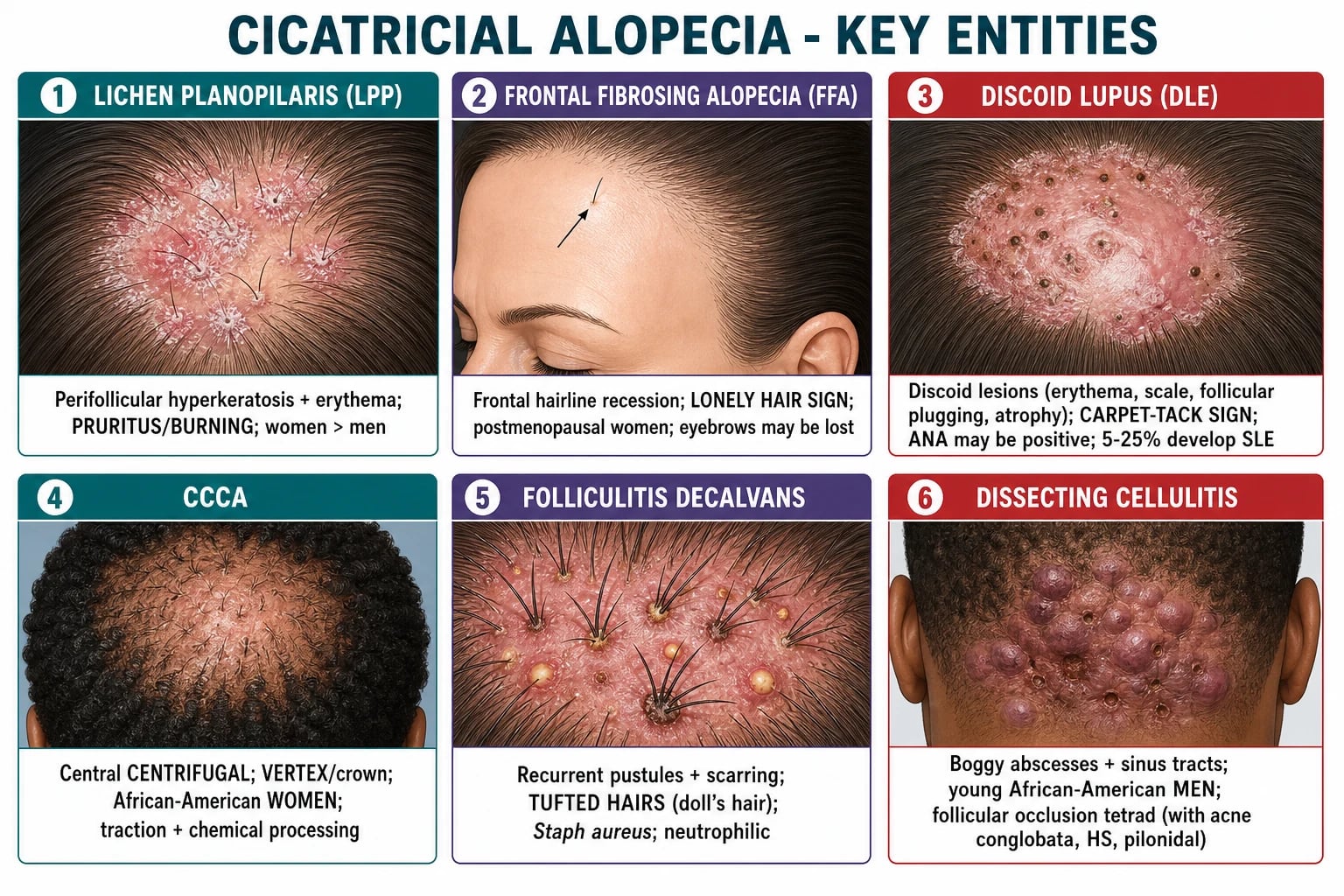
- Perifollicular hyperkeratosis — white, scaly collarettes around remaining hairs, characteristic of LPP/FFA.
- Tufted hairs — multiple hair shafts emerging from a single dilated follicular opening, pathognomonic for folliculitis decalvans.
- Pustules, crusting, nodules, sinus tracts or keloid formation in neutrophilic or mixed disease. [1]
A crucial concept is the active inflammatory margin. In active disease, the border of the bald patch is red, scaly, pustular or indurated. The central scarred area is pale, smooth and has no follicular openings. Treatment should be directed at the active margin; the scarred centre is irreversible. [1]
Atypical presentations are frequently tested. FFA may involve the eyebrows, eyelashes or body hair, and may be accompanied by facial papules. CCCA may present with mild scaling or burning before visible hair loss. Pseudopelade of Brocq presents with multiple small, white, atrophic patches resembling "footprints in the snow" and no active inflammation. Dissecting cellulitis can be mistaken for infection because of large abscesses. In children, scarring alopecia is uncommon and should prompt exclusion of tinea capitis (especially kerion), trichotillomania with chronic traction, and inflammatory skin diseases.[2][4][6]
Differential Diagnosis
The first branching point in any alopecia is scarring versus non-scarring. Non-scarring alopecias are far more common and must be distinguished before committing to a diagnosis of cicatricial alopecia. [1]
Alopecia areata produces smooth, patchy hair loss with normal follicular openings and often exclamation-mark hairs at the margin. The scalp is not scarred. A pull test may be positive. In contrast, cicatricial alopecia has absent follicular ostia and a smooth, shiny surface. [1]
Androgenetic alopecia causes patterned hair loss (Hamilton-Norwood in men, Ludwig in women) with miniaturised hairs and preserved follicular openings. The frontal hairline in women is usually maintained. FFA is distinguished by frontal recession, perifollicular hyperkeratosis and loss of follicular ostia. [1]
Traction alopecia is initially reversible if the traction is removed. It affects the sites of traction (frontal, temporal, marginal). Chronic traction can become scarring (late-stage traction alopecia), particularly in women who wear tight braids or weaves for many years. Early disease shows preserved follicular openings; late disease shows loss. [1]
Tinea capitis is a dermatophyte infection that produces scaling, erythema, broken hairs, occipital lymphadenopathy and sometimes kerion. It is more common in children. Wood's lamp examination, KOH microscopy and fungal culture distinguish it. Kerion can cause scarring if untreated. [1]
Trichotillomania produces bizarre, irregular patches with hairs of varying lengths because the patient pulls and breaks hairs. The scalp is not scarred unless there is chronic, severe traction. A history of hair-pulling or concurrent anxiety disorders is supportive. [1]
Secondary causes of scarring must be considered in the appropriate context: a history of burns, radiotherapy, scalp surgery, severe infection, or a slowly enlarging plaque that may represent cutaneous squamous cell carcinoma, basal cell carcinoma, or lymphoma. In patients with long-standing DLE or dissecting cellulitis, chronic non-healing ulceration should raise concern for Marjolin squamous cell carcinoma.[2][4][6]
Clinical & Bedside Assessment
The examination begins with a systematic inspection of the scalp under good light, preferably with a dermatoscope. The examiner should map the pattern of hair loss, note the presence or absence of follicular openings, identify active inflammation, and look for pustules, crusting, scale, keloid or sinus tracts. [1]
A focused examination includes: [1]
- Inspection of the whole scalp — note the pattern (vertex, frontal, patchy, diffuse), symmetry, and any scarring.
- Palpation — scarred skin is often smooth, shiny, atrophic, firm or sclerotic. Active inflammation feels indurated, boggy or tender.
- Follicular ostia check — use a dermatoscope to confirm whether follicular openings are present. This is the single most important discriminator.
- Hair-pull test — grasp about 50 hairs and gently pull. In active non-scarring alopecia more than 5 to 6 hairs may come out; in burnt-out scarring alopecia the pull test is usually negative because there are no hairs to extract. At the active margin, it may be positive if inflammation is ongoing.
- Trichoscopy — use a handheld dermatoscope to identify specific patterns (see below).
- Scalp biopsy — a 4 mm punch biopsy taken from the active inflammatory margin (not the centre) provides the definitive histological classification.
- Photographs — serial standardised photographs are essential for monitoring progression or response. [1]
Named bedside signs include: [1]
- Lonely hair sign — a single terminal hair at the advancing frontal hairline in FFA, representing the last surviving follicle in the area of recession.
- Carpet-tack sign — when the adherent scale of DLE is lifted, follicular plugs are seen on the underside; this is a sign of discoid lupus.
- Tufted hairs — multiple hairs emerging from a single ostium in folliculitis decalvans; sometimes called "doll's hair" or "paintbrush" hair.
- Footprints in the snow — the appearance of multiple small white atrophic patches in pseudopelade of Brocq.[2][4]
Investigations
Investigations are chosen to confirm the diagnosis, classify the disease, and exclude mimics. [1]
Trichoscopy
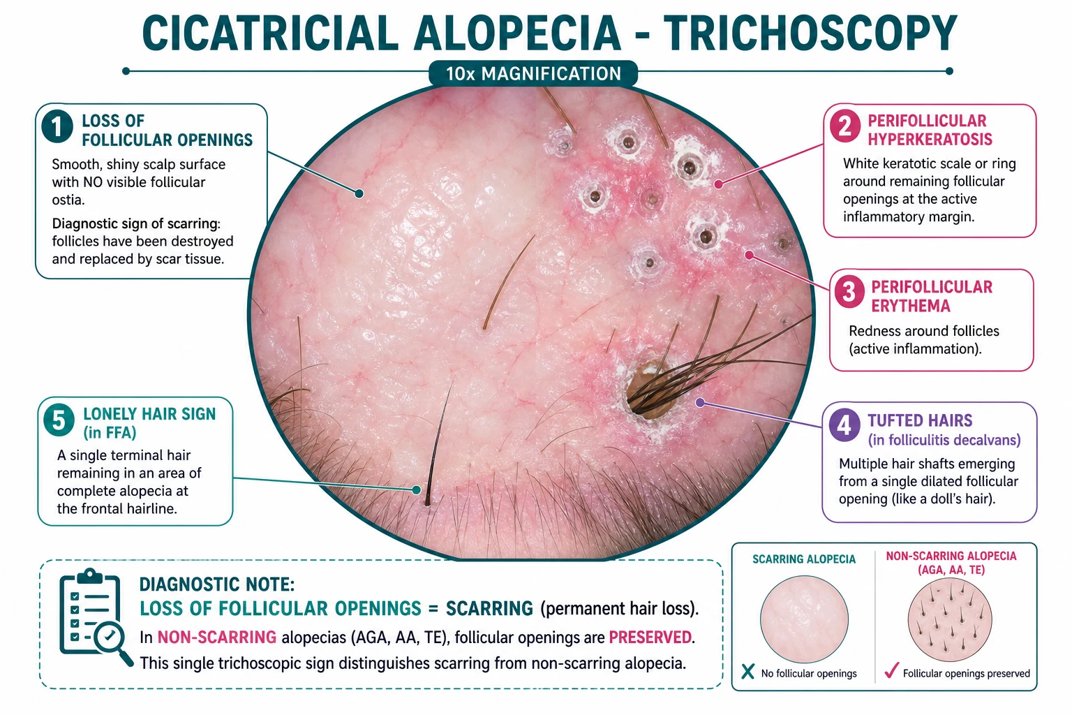
Trichoscopy is the first-line investigation. It is non-invasive and often diagnostic. The hallmark feature of scarring alopecia is the absence of follicular openings. Additional findings vary by subtype: [1]
- Lymphocytic disease: perifollicular scale, perifollicular erythema, white dots (scarred follicular openings), tubular hair casts, and areas of complete loss of follicular openings.
- FFA: lonely hair sign, perifollicular erythema/scale at the frontal margin, and loss of follicular openings in the frontal band.
- DLE: thick arborizing vessels, white structureless areas, keratotic plugs, and prominent scaling.
- CCCA: reduced hair density at the vertex, peripilar white halos, broken hairs, and loss of follicular openings in the central scarred area.
- Folliculitis decalvans: tufted hairs, pustules, yellowish follicular openings, haemorrhagic crusts, and reduced follicular density.
- Dissecting cellulitis: yellow structureless areas, blood vessels, and empty hair follicles with minimal scaling. [1]
Scalp biopsy
A 4 mm punch biopsy should be taken from the active inflammatory margin — the junction of normal hair-bearing skin and scarred skin. The centre of the scar contains only dense fibrous tissue and is non-diagnostic. Biopsy should be deep enough to include the subcutaneous fat, oriented along the direction of hair growth, and the specimen should be handled gently to avoid crush artefact. [1]
Histological findings by group: [1]
- Lymphocytic: lichenoid or perifollicular lymphocytic infiltrate at the follicular isthmus, loss of sebaceous glands, and replacement of follicles by fibrous tract remnants. Direct immunofluorescence may show cytoid bodies at the dermo-epidermal junction in LPP; in DLE, lupus bands (linear IgG, IgM, C3) are seen at the basement membrane.
- Neutrophilic: intrafollicular and perifollicular neutrophils, pustules, abscesses, granulation tissue, and later fibrous tracts. In folliculitis decalvans, the merging of adjacent follicles produces the tufted hair appearance.
- End-stage/burnt-out: dense fibrous tracts replace hair follicles, absent sebaceous glands, minimal inflammation, and sometimes residual scarring. The original subtype may no longer be identifiable. [1]
Microbiology
Pustular or crusted lesions should be swabbed for bacterial culture, including Staphylococcus aureus. In neutrophilic disease, culture guides antibiotic choice. If tinea capitis is suspected, take skin scrapings and hairs for KOH microscopy and fungal culture or PCR. In chronic sinus tracts, consider anaerobic culture. [1]
Autoimmune and systemic screening
In patients with DLE or any clinical suggestion of lupus, test: [1]
- ANA by immunofluorescence.
- Anti-dsDNA, anti-Smith, anti-Ro/La, complement C3/C4 if systemic features are present.
- Full blood count, renal function, urinalysis to screen for systemic lupus involvement. [1]
For other subtypes, consider thyroid function tests, fasting lipids, glucose/HbA1c, and iron studies because metabolic comorbidities are common and may influence management or patient counselling. [1]
Imaging and photography
Serial clinical photographs are the simplest and most valuable monitoring tool. High-frequency ultrasonography and reflectance confocal microscopy are research tools and not required for routine care. In dissecting cellulitis with extensive sinus tracts, computed tomography or magnetic resonance imaging may be needed to define the extent of disease before surgery.[4][6]
Management — Resuscitation
Although cicatricial alopecia is not immediately life-threatening, the first consultation is time-critical in the sense that every day of active inflammation destroys more follicles. The "resuscitation" phase focuses on preventing further irreversible loss. [1]
Immediate priorities: [1]
- Confirm that the alopecia is scarring by trichoscopy (loss of follicular ostia) and, if the diagnosis is uncertain, biopsy from the active margin.
- Stop trauma and trigger factors — advise the patient to avoid tight hairstyles, extensions, chemical relaxers, heat, and harsh scalp treatments. For FFA, avoid high-forehead traction hairstyles. For AKN, stop close clipping.
- Start anti-inflammatory therapy promptly — do not wait for biopsy results if the clinical picture is clear. Topical corticosteroids and/or intralesional corticosteroids can be started at the first visit.
- Treat active infection if pustules are present; empiric antistaphylococcal antibiotics are reasonable while awaiting culture.
- Counsel the patient that treatment aims to stop inflammation and preserve remaining hair; regrowth in already scarred areas is not expected.
- Arrange appropriate baseline investigations and specialist follow-up. [1]
The most important counselling point is that destroyed follicles cannot regrow hair. Early treatment can halt progression, but it cannot reverse established scarring. This realistic expectation helps prevent disappointment and improves adherence.[3][6]
Management — Definitive & Stepwise
Treatment is directed by the NAHRS inflammatory group. The goal is not to regrow hair but to stop inflammation and preserve remaining follicles. Management is therefore a long-term, stepwise process. [1]
Lymphocytic cicatricial alopecia
First-line — topical and intralesional corticosteroids [1]
- Potent topical corticosteroids such as clobetasol propionate 0.05% foam, solution or shampoo applied to the active margin once or twice daily for 4 to 8 weeks, then tapered to a maintenance schedule. Foam preparations are cosmetically acceptable and may improve penetration. Use intermittent "pulse" therapy to minimise skin atrophy.
- Intralesional triamcinolone acetonide 2.5 to 10 mg per mL injected into the active inflammatory margin every 4 to 6 weeks. Use lower concentrations (2.5 mg/mL) for the face and eyebrows to avoid atrophy. Intralesional steroids are particularly useful for localised LPP and FFA. [1]
First-line systemic — hydroxychloroquine [1]
- Hydroxychloroquine 200 to 400 mg orally daily is the most commonly used systemic agent for lymphocytic cicatricial alopecia, including LPP, FFA and DLE. Dose is weight-based (ideally less than 5 mg/kg/day of real body weight to reduce retinopathy risk). Onset is slow; assess response at 3 to 6 months. Baseline and annual ophthalmological review is mandatory because of retinal toxicity. [1]
Adjunctive and second-line systemic options [1]
- Tetracyclines (doxycycline 100 mg orally twice daily or minocycline 100 mg daily) have anti-inflammatory and anti-matrix-metalloproteinase effects and are often used as steroid-sparing agents.
- Mycophenolate mofetil 1 to 2 g orally daily is used for refractory or extensive lymphocytic disease. Monitoring includes full blood count and liver function tests.
- Systemic corticosteroids (e.g., prednisolone 0.5 to 1 mg/kg/day orally) can be used for short-term control of active flares, but long-term use is limited by toxicity. Taper over weeks to months while introducing a steroid-sparing agent.
- Pioglitazone 15 to 30 mg orally daily, a PPAR-γ agonist, has been used as an adjunct in LPP, with variable evidence.
- JAK inhibitors (tofacitinib, baricitinib, ritlecitinib) are emerging off-label options for refractory disease, particularly in patients with extensive inflammation or concomitant alopecia areata. They require infection screening, lipid monitoring and counselling about thromboembolic and malignancy risks.
- 5-alpha-reductase inhibitors (finasteride 1 to 5 mg orally daily or dutasteride 0.5 mg orally daily) are often used in FFA, especially when there is clinical or histological overlap with androgenetic alopecia. [1]
Neutrophilic cicatricial alopecia
Folliculitis decalvans [1]
- Antistaphylococcal therapy is the cornerstone. The classic combination is oral rifampicin 300 mg twice daily plus clindamycin 300 mg twice daily for 10 to 12 weeks. The rationale is that rifampicin penetrates biofilms and clindamycin suppresses staphylococcal toxins and inflammation; resistance is reduced by combination therapy.
- Long-term maintenance with doxycycline 100 mg twice daily or erythromycin/clarithromycin is often required because relapse is common.
- Antiseptic shampoos containing chlorhexidine, zinc pyrithione or ketoconazole reduce colonisation. Topical clindamycin and mupirocin to the nares are used for nasal S. aureus carriage.
- For refractory disease, oral isotretinoin 0.5 to 1 mg/kg/day, dapsone or TNF inhibitors (adalimumab, infliximab) may be considered. [1]
Dissecting cellulitis of the scalp [1]
- Oral isotretinoin is the mainstay of treatment. Dose is 0.5 to 1 mg/kg/day for 4 to 6 months, with the aim of disease remission. It acts by reducing sebum production, normalising follicular keratinisation and modulating inflammation. It is highly teratogenic; strict pregnancy prevention is essential.
- Antibiotics such as doxycycline, clindamycin or trimethoprim-sulfamethoxazole are used for their anti-inflammatory properties and for secondary infection.
- TNF inhibitors (adalimumab, infliximab) are effective for refractory dissecting cellulitis.
- Intralesional corticosteroids and surgical drainage or excision may be needed for large abscesses or sinus tracts. Surgery should be performed only in quiescent disease because active disease flares after manipulation. [1]
Mixed group — acne keloidalis nuchae
- Trigger avoidance: stop close clipping, use a clipper guard of grade 2 (6 mm) or longer, and reduce friction from collars or helmets.
- Topical therapies: clindamycin or benzoyl peroxide for pustules; topical corticosteroids or retinoids for papules.
- Intralesional triamcinolone 10 to 40 mg/mL for fibrotic plaques and keloid formation.
- Systemic agents: doxycycline, clindamycin-rifampicin combination, or isotretinoin for extensive disease.
- Surgery: excision of large keloidal plaques or laser hair epilation for persistent disease. [1]
Adjunctive and supportive care
- Topical minoxidil 5% solution or foam does not reverse scarring but may help preserve or thicken remaining terminal hairs. It is commonly used as an adjunct in CCCA and FFA.
- Camouflage: wigs, hairpieces, scalp micropigmentation and careful hairstyling are important for psychosocial support.
- Platelet-rich plasma (PRP) and low-level laser therapy are emerging adjuncts with limited evidence; they should not replace anti-inflammatory therapy.
- Psychosocial support: hair loss has a profound impact on self-esteem, particularly in women and in cultures where hair is central to identity. Screen for anxiety and depression and offer support or referral. [1]
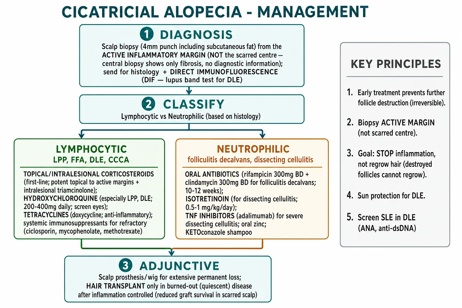
Management ladder — LADDER
LADDER
Local corticosteroids + hydroxychloroquine (HCQ) first-line.
Neutrophilic disease — rifampicin + clindamycin, doxycycline, isotretinoin.
Early treatment prevents permanent follicle loss.
Biopsy active margin to classify and exclude tinea, lupus, SCC.
Regrowth is impossible in scarred areas; preserve what remains.
Hair transplantation only in quiescent disease.
Specific Subtypes & Scenarios
Lichen planopilaris (LPP)
LPP presents with one or more patches of scarring hair loss, often on the vertex or crown. The active margin shows perifollicular erythema and perifollicular hyperkeratosis (white scale around follicles). Patients often complain of itching or burning. Histology shows a lichenoid infiltrate at the follicular isthmus, loss of sebaceous glands, and fibrous tract remnants. Management is topical/intralesional corticosteroids plus hydroxychloroquine; second-line agents include mycophenolate, systemic corticosteroids and pioglitazone. [1]
Frontal fibrosing alopecia (FFA)
FFA is a lymphocytic scarring alopecia that causes progressive recession of the frontal and temporal hairline in postmenopausal women. The lonely hair sign — a single terminal hair at the advancing hairline — is highly characteristic. Eyebrow and eyelash loss are common, and facial papules may be present. It is considered a clinical variant of LPP. Treatment includes 5-alpha-reductase inhibitors (finasteride or dutasteride), hydroxychloroquine, topical/intralesional corticosteroids, and sometimes mycophenolate or JAK inhibitors. Sun protection is important because photo-exposure may worsen disease at the hairline.[1]
Discoid lupus erythematosus (DLE)
DLE of the scalp presents as well-demarcated erythematous plaques with adherent scale, follicular plugging, dyspigmentation and scarring. The carpet-tack sign is seen when scale is lifted and follicular plugs are visible on its underside. DLE is the most common form of chronic cutaneous lupus. Approximately 5 to 25 percent of patients with DLE may develop systemic lupus erythematosus, so systemic screening is indicated. First-line therapy is potent topical or intralesional corticosteroids plus hydroxychloroquine; refractory disease may require thalidomide, methotrexate, mycophenolate or retinoids. Strict photoprotection and smoking cessation are essential.[3]
Central centrifugal cicatricial alopecia (CCCA)
CCCA begins at the vertex or crown and expands centrifugally. It is most common in women of African ancestry. Traction, chemical relaxers and heat are contributing factors. The active margin may show perifollicular hyperkeratosis, while the centre is scarred. Treatment is centred on stopping all traumatic grooming practices, topical corticosteroids, intralesional triamcinolone, and hydroxychloroquine. Minoxidil is used as an adjunct to preserve remaining hair. Early diagnosis is critical because the central vertex is cosmetically and psychologically important.[9]
Pseudopelade of Brocq
Pseudopelade is the end-stage, burnt-out appearance of multiple small, white, atrophic patches with no active inflammation. It is often described as "footprints in the snow". Because there is no active inflammation, immunosuppressive therapy is not indicated. The clinician should search for an active underlying process (LPP, DLE) producing new lesions; if none are present, management is cosmetic camouflage and monitoring. [1]
Folliculitis decalvans
This neutrophilic disease presents with recurrent pustules, crusting and tufted hairs. Staphylococcus aureus is frequently cultured. The standard treatment is the rifampicin-clindamycin combination for 10 to 12 weeks, with maintenance antibiotics and decolonisation measures. Chronic relapsing disease may require isotretinoin, dapsone or TNF inhibitors. Nasal mupirocin and antiseptic washes reduce carriage and relapse rates. [1]
Dissecting cellulitis of the scalp
Dissecting cellulitis presents with deep, boggy, tender nodules and abscesses on the scalp that communicate through sinus tracts. It is part of the follicular occlusion tetrad. It disproportionately affects young Black men. Treatment is long-term, often with isotretinoin as the mainstay. Refractory disease responds to TNF inhibitors, and surgery is reserved for quiescent, localised disease. Chronic disease carries a risk of squamous cell carcinoma, so persistent non-healing areas need biopsy.[7]
Acne keloidalis nuchae (AKN)
AKN affects the nape of the neck in Black men, beginning as perifollicular papules and pustules and progressing to keloidal plaques, tufted hairs and sinus tracts. It is classified as a mixed cicatricial alopecia. Management focuses on stopping close shaving, topical antibiotics/retinoids, intralesional corticosteroids, and systemic agents for extensive disease. Surgery or laser hair removal is used for refractory plaques. [1]
Complications & Pitfalls
The most important complication is permanent, irreversible hair loss. Even with optimal treatment, hair cannot be restored in areas where follicles have been replaced by scar. Other complications include: [1]
- Psychosocial morbidity — hair loss causes significant anxiety, depression, reduced quality of life and social isolation. The impact is often underestimated.
- Secondary infection — pustules and sinus tracts can become chronically colonised with S. aureus.
- Keloid formation — particularly in AKN, dissecting cellulitis and in individuals with keloid tendency.
- Squamous cell carcinoma — long-standing DLE and dissecting cellulitis can develop SCC, sometimes called Marjolin ulcer. Any non-healing ulcer, nodule or change in a chronic scar should be biopsied.
- Iatrogenic skin atrophy — prolonged potent topical corticosteroids or repeated high-concentration intralesional steroids can cause atrophy, telangiectasia and striae.
- Ocular involvement — DLE can affect eyelids and conjunctiva; FFA can cause eyelash loss. [1]
Common pitfalls include: [1]
- Biopsying the centre of the scar rather than the active margin. The centre shows only end-stage fibrosis and will not give a diagnosis.
- Treating burnt-out scar with minoxidil alone and expecting regrowth. Minoxidil cannot regenerate destroyed follicles.
- Missing tinea capitis in children and treating with immunosuppressants instead of oral antifungals.
- Failing to screen for systemic lupus in scalp DLE.
- Delaying treatment while waiting for biopsy results, allowing further follicle destruction.
- Performing hair transplantation during active disease, leading to poor graft survival and disease flare. [1]
Prognosis & Disposition
The prognosis of cicatricial alopecia depends on how early treatment is started and how quickly inflammation is controlled. Established scar is permanent, but progression can almost always be halted if active disease is treated promptly. The most realistic outcome is stabilisation with preservation of remaining hair. [1]
Indicators of good prognosis include: [1]
- Early diagnosis with limited involvement.
- Rapid response to first-line therapy (reduction of erythema, scale, pustules, and halting of expansion).
- Good adherence to treatment and trigger avoidance.
- Absence of keloid tendency or extensive scarring. [1]
Indicators of poor prognosis include: [1]
- Late presentation with extensive central scarring.
- Rapidly progressive disease despite first-line therapy.
- Keloid formation or extensive sinus tract disease.
- Continued traction, chemical processing or smoking.
- Non-adherence to treatment. [1]
Most patients are managed in the outpatient dermatology clinic. The follow-up interval depends on disease activity: monthly injections for active LPP/FFA, every 6 to 12 weeks for stable patients on hydroxychloroquine, and urgent review if new pustules, nodules, rapid expansion or non-healing ulceration develop. Patients should be taught to photograph the affected areas so that progression can be tracked between visits. [1]
Special Populations
Pregnancy and breastfeeding
Management must be modified during pregnancy. Avoid isotretinoin, finasteride, dutasteride, methotrexate, mycophenolate, thalidomide and TNF inhibitors. Hydroxychloroquine is generally considered safe and is continued in lupus patients during pregnancy because the risk of disease flare outweighs drug risk. Topical corticosteroids and intralesional triamcinolone can be used with caution; limit the amount and avoid potent steroids over large areas. Doxycycline and other tetracyclines should be avoided after the first trimester because of effects on fetal bone and teeth. Rifampicin is usually avoided in pregnancy unless essential. Breastfeeding considerations are similar; discuss case-by-case. [1]
Children
Cicatricial alopecia is uncommon in children. The most frequent cause of childhood scarring alopecia is tinea capitis, particularly kerion, which must be treated with oral antifungal therapy (griseofulvin or terbinafine) rather than immunosuppressants. Trichotillomania and traction alopecia are common but usually reversible. Juvenile LPP, FFA and DLE are rare but recognised; management mirrors adult protocols, with particular caution regarding steroid atrophy and growth effects of systemic agents. [1]
African ancestry and CCCA
Women of African ancestry with vertex hair loss should be assumed to have CCCA until proven otherwise. A sensitive history of hair-care practices is essential; patients may not recognise tight braids, weaves or chemical relaxers as harmful. Counselling on protective styling (low tension, no chemicals, no heat) is as important as medication. AKN in Black men similarly requires culturally sensitive advice about clipping and friction. [1]
Elderly patients
Postmenopausal women with frontal hairline recession are the classic FFA demographic. However, elderly patients may have scalp atrophy, actinic damage and multiple comorbidities that increase the risk of topical steroid side effects and systemic immunosuppression. Hydroxychloroquine retinopathy risk increases with age and duration; ophthalmological screening is especially important. [1]
Immunocompromised patients
In patients with HIV, transplant or other immunosuppression, consider infectious and neoplastic causes of scarring. Disseminated tinea capitis, deep fungal infections, cutaneous tuberculosis and lymphoma can all present with scarring alopecia. Biopsy and culture are essential before starting immunosuppressive therapy. [1]
Evidence, Guidelines & Regional Differences
The NAHRS classification, first published in 2001 and refined in subsequent consensus meetings, remains the international framework for defining and classifying primary cicatricial alopecia. It is purely descriptive, dividing disease by inflammatory infiltrate, but it provides a rational basis for treatment.[6]
High-quality randomised controlled trials are scarce because the diseases are rare and heterogeneous. Evidence for treatment is therefore largely based on case series, expert consensus and retrospective cohorts. Key evidence summaries include: [1]
- Hydroxychloroquine is the most widely used systemic agent for lymphocytic disease, supported by retrospective series and consensus guidelines. Response is slow; a 6-month trial is usually needed before declaring failure.
- Rifampicin-clindamycin combination is the most accepted regimen for folliculitis decalvans, supported by small cohorts and the rationale for biofilm eradication.
- Isotretinoin is the mainstay for dissecting cellulitis, supported by case series and expert recommendations.
- TNF inhibitors (adalimumab, infliximab) have emerging evidence in refractory neutrophilic and mixed disease, including dissecting cellulitis and AKN.
- JAK inhibitors are increasingly reported for refractory LPP/FFA, but long-term safety data are limited and their use is off-label. [1]
Regional differences matter. In India and many low-resource settings, hydroxychloroquine is affordable and widely available, but isotretinoin requires strict pregnancy prevention programmes and rifampicin-clindamycin may be limited by cost or access. In the United Kingdom, NICE and British Association of Dermatologists guidance emphasise photoprotection in DLE and shared-care prescribing for biologics. In the United States, American Academy of Dermatology guidelines support mechanism-based therapy and early referral for scalp biopsy. In Europe, EADV consensus documents stress the psychosocial impact and the need for quality-of-life assessment. Clinicians should follow local formularies and pregnancy-prevention regulations.[3]
[1] [1] [1]Exam Pearls
[1]Exam application bank (NEET-PG / INICET)
One-line answer
Cicatricial (scarring) alopecia is a group of disorders in which hair follicles are destroyed and replaced by fibrous tissue, producing permanent, irreversible hair loss. The hallmark clinical sign is loss of follicular ostia. The North American Hair Research Society (NAHRS) classifies primary cases by inflammatory infiltrate into lymphocytic (lichen planopilaris, frontal fibrosing alopecia, discoid lupus, CCCA, pseudopelade of Brocq), neutrophilic (folliculitis decalvans, dissecting cellulitis) and mixed (acne keloidalis nuchae) forms. Early diagnosis by trichoscopy and biopsy of the active margin is essential because destroyed follicles cannot regrow. Management targets the inflammatory mechanism: lymphocytic disease uses corticosteroids, hydroxychloroquine, mycophenolate and JAK inhibitors; neutrophilic disease uses antibiotics, isotretinoin and TNF inhibitors. Hair transplantation is
Worked stems (answer without another resource)
Stem 1 — Classic presentation. Map symptoms to mechanism; name the first investigation and first treatment step with dose/route if drug therapy is standard. [1]
Stem 2 — Unstable / complicated. List red flags that force immediate resuscitation, theatre, ICU, antidote, or reperfusion — and what you do in the first 15 minutes. [1]
Stem 3 — Atypical group. Elderly, pregnancy, child, or immunocompromised: how presentation and thresholds change. [1]
Stem 4 — Differential trap. Name the three closest mimics and one discriminator for each. [1]
Stem 5 — Disposition. Who goes home with safety-netting, who is admitted, who needs HDU/ICU/theatre, and what follow-up is mandatory. [1]
Rapid viva checklist
- Definition + classification
- Pathophysiology chain
- Bedside signs / criteria
- Score with exact components (if any)
- Emergency bundle
- Definitive therapy with doses
- Complications of disease and of treatment
- Special populations
- Guideline/trial name if classic
- Three exam traps
Coverage self-check
If you cannot answer any stem above from this page alone, re-read the matching section — the page is intended to be self-sufficient for final-prof and NEET-PG/INICET questions on Cicatricial alopecia.
[1]References
- [1]Griggs J, Trüeb RM, Gavazzoni Dias MFR, et al. Fibrosing alopecia in a pattern distribution J Am Acad Dermatol, 2021.PMID 31926219
- [2]Pirmez R. The dermatoscope in the hair clinic: Trichoscopy of scarring and nonscarring alopecia J Am Acad Dermatol, 2023.PMID 37591567
- [3]Ezemma O, Khetarpal S, Sperling L, et al. Treatment modalities for lymphocytic and neutrophilic scarring alopecia J Am Acad Dermatol, 2023.PMID 37591564
- [4]Mathur M, Tosti A, Shepherd J, et al. Trichoscopy of primary cicatricial alopecias: an updated review J Eur Acad Dermatol Venereol, 2020.PMID 31566830
- [5]Harries MJ, Meyer K, Chaudhry I, et al. Fibrosis and stem cell epithelial-mesenchymal transition in primary cicatricial alopecias J Am Acad Dermatol, 2019.PMID 30639879
- [6]Uchiyama M, Nagai Y, Takahashi K, et al. Primary cicatricial alopecia: Recent advances in evaluation and diagnosis based on trichoscopic and histopathological observation, including overlapping and specific features J Dermatol, 2022.PMID 34866229
- [7]Sood S, Mesinkovska N. Biologic and Small Molecule Treatments for Primary Neutrophilic Cicatricial Alopecias: An Evidence-Based Review J Cutan Med Surg, 2023.PMID 37489902
- [8]Wang EHC, Monga P, Glassman SJ, et al. Primary cicatricial alopecias are characterized by dysregulation of shared gene expression pathways PNAS Nexus, 2022.PMID 35899069
- [9]Heymann WR. Central centrifugal cicatricial alopecia: Beyond the hot comb J Am Acad Dermatol, 2023.PMID 37775048
- [10]Jimenez F, Alam M, Vogel JE, et al. Hair transplantation: Basic overview J Am Acad Dermatol, 2021.PMID 33905785