Dermatology · Medicine
Congenital melanocytic naevus
Also known as Congenital melanocytic naevus (CMN) · Giant congenital melanocytic naevus · Garment naevus · Bathing trunk naevus · Congenital naevus · CMN syndrome · Neurocutaneous melanocytosis (NCM)
A congenital melanocytic naevus (CMN) is a melanocytic naevus present at birth or within the first weeks of life, caused by a postzygotic mosaic activating mutation in the RAS-MAPK pathway (NRAS Q61 in ~80-90% of giant CMN; BRAF V600E in some smaller CMN; mosaic BRAF fusions). Classified by projected adult size into small (under 1.5 cm), medium (1.5-19.9 cm) and large/giant (≥20 cm or ≥5% BSA). Giant CMN carries a 5-15% lifetime melanoma risk (often pre-pubertal, arising in the deep dermis) and 5-10% risk of neurocutaneous melanocytosis (NCM) when on the posterior axis with ≥20 satellite naevi. Management is multidisciplinary; prophylactic excision is controversial; MEK inhibitors are an emerging targeted option for NRAS-mutant disease.
On this page & tools
Your progress
Saved locally on this device.
Exam tags
Red flags
Definition, Anatomy and Classification
A congenital melanocytic naevus (CMN) is a melanocytic naevus present at birth, or — in the "tardive" or "congenital-type" form — appearing within the first 4-6 weeks of life. It is composed of naevomelanocytes (naevus cells) that failed to complete their migration from the neural crest to the epidermis during the first trimester, arresting in the dermis, around adnexal structures, around nerves and in the subcutis.[5] This arrest distinguishes CMN histologically from an acquired naevus, in which naevus cells are confined to the epidermis (junctional) and papillary dermis.
CMN anatomy — the BONES mnemonic
BONES
Naevus cells splay apart dermal collagen in a 'single-file' or sheet-like pattern — the histological hallmark of CMN.
Naevus cells encircle pilosebaceous units, eccrine coils and ducts, and apocrine glands — never seen in acquired naevi.
Naevus cells cuff small dermal nerves; a classic diagnostic clue in difficult cases.
Giant CMN cells can track along septa, between adipocytes, into fascia and even skeletal muscle — explains why superficial excision never removes all naevus cells.
Clinical correlate: surface becomes verrucous, cerebriform or cobblestone, with coarse terminal hairs, especially in giant CMN.
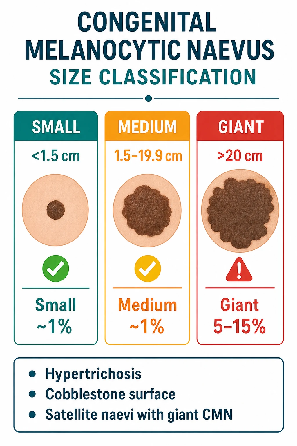
CMN are classified by projected adult size (the size the lesion is expected to reach in adulthood, derived by multiplying the largest diameter in infancy by a growth factor — historically 1.7, now 2 in some consensus schemes):[2][5]
CMN size classification — the bedside numbers
The 2020 international CMN consensus (Ott et al., supported by the ERN-Skin network) refined the classification by adding (1) number of satellite naevi (none, fewer than 20, 20-50, more than 50), (2) additional morphological features (colour heterogeneity, surface rugosity, subcutaneous nodules, proliferative nodules) and (3) the "CMN syndrome" designation when neurological, skeletal or other organ involvement is present. This is the classification examiners now expect at fellowship level. [1]
Small CMN (under 1.5 cm)
- Most common (~1 in 100 births)
- Cosmetic concern; melanoma risk ≈1% (≈ background)
- Surface often smooth; may become slightly raised and pigmented over time
- Maternal anxiety is the main indication for any intervention
- Biopsy only for changing / suspicious lesions
Medium CMN (1.5-19.9 cm)
- Intermediate frequency
- Melanoma risk ≈1%; NCM risk low unless posterior axis / many satellites
- Often on trunk; can be cosmetically disfiguring if visible
- Selective excision for cosmesis / repeated trauma acceptable
- Baseline photography + annual review is reasonable
Large / giant CMN (>=20 cm or >=5% BSA)
- Rare (~1 in 20 000 births)
- 5-15% lifetime melanoma risk (often pre-pubertal, deep dermal)
- 5-10% risk of NCM; risk highest on posterior axis with 20 or more satellites
- Multidisciplinary team from birth: dermatology, paediatrics, neurology, plastic surgery, psychology
- MRI brain + whole spine in the first 6 months; lifelong surveillance
Eponymous giant CMN patterns
Giant CMN are described by their cutaneous distribution:[2]
- Bathing-trunk naevus — circumferential involvement of the lower trunk, buttocks and thighs ("swimsuit distribution").
- Cape / shoulder naevus — over the upper back, shoulders and arms; highest NCM risk (posterior axial location).
- Stocking / glove naevus — entire limb, often with distal sparing.
- Dermal melanocytosis ("Mongolian-spot-like") — slate-grey, deep dermal lesions with indistinct borders; coexist with classic giant CMN in ~1-2% of cases. [1]
Epidemiology and Risk Factors
CMN epidemiology — the key numbers
CMN is the most common tumour of infancy by histological definition. Small CMN occur in approximately 1% of live births, medium CMN in approximately 1 in 1 000, and giant CMN in approximately 1 in 20 000. There is no clear sex, ethnic or geographic gradient for the giant form, although some series report a slight female predominance and a higher prevalence in Caucasian and Asian populations than in Black African populations.[2]
Risk factors are largely those of melanoblast proliferation in utero: [1]
- Maternal parity and age — multiparity and older maternal age have been weakly associated in case-control studies.
- In-utero drug exposures — occasional case reports link CMN to maternal use of methimazole, misoprostol, and ergotamine, but causation is unproven.
- Gestational events — first-trimester bleeding, infections and maternal stress have been implicated anecdotally.
- Fertility treatment and IVF — a weak association, likely reflecting multiple early cell divisions.
- Family history — overwhelmingly negative; CMN is a postzygotic mosaic event (see pathophysiology), so there is no significant recurrence risk in siblings, offspring or future pregnancies. [1]
The most important "host" risk factor is the distribution and size of the CMN itself: posterior-axial location, >20 satellite naevi, and head/neck involvement drive NCM risk, while the projected adult size drives melanoma risk. [1]
Pathophysiology and Molecular Genetics
Embryological origin
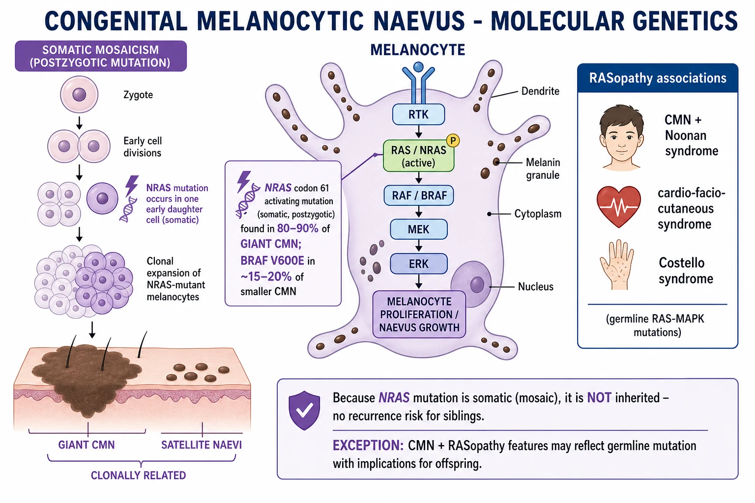
Melanocytes are derived from the neural crest. A subset of melanoblasts migrates dorsolaterally between the ectoderm and somites around weeks 6-8 of gestation, populating the epidermis, hair follicles, uveal tract, leptomeninges, mucosae and striae vascularis. CMN are thought to arise when an activating RAS-MAPK pathway mutation occurs in a single neural-crest-derived melanoblast at this stage; the daughter cells then carry the mutation wherever they migrate, producing a patchy, segmental, sometimes enormous pigmented lesion.[5][6]
Driver mutations
NRAS Q61 (Q61K / Q61R)
- 80-90% of giant CMN; 50-70% of medium CMN
- Somatic, postzygotic — not inherited
- Activates both RAF-MEK-ERK and PI3K-AKT pathways
- Drives proliferation + senescence escape in deep dermal naevomelanocytes
- Druggable: MEK inhibitors (binimetinib, trametinib) are emerging targeted options
BRAF V600E
- ~15-20% of CMN, mostly smaller lesions
- Same mutation as in acquired naevi and ~50% of cutaneous melanomas
- Activates RAF-MEK-ERK pathway only
- Most common in small / medium CMN; less common in giant CMN
- Targetable: BRAF inhibitors (dabrafenib, vemurafenib) plus MEK inhibitors
Mosaic BRAF fusions
- Recurrent cause of CMN — Kao et al. 2024
- Druggable by MAPK pathway inhibition
- Distinct from BRAF V600E point mutations
- Some at risk of malignant transformation
- Increasingly recognised with NGS
In the remaining 10-20% of CMN, BRAF V600E or, increasingly recognised, mosaic BRAF fusions are the driver. Mosaic BRAF fusions (e.g. with AGK, TRIM24, MKL2) were characterised in 2024 as a recurrent cause of CMN, targetable by MAPK pathway inhibition.[9] A small minority of CMN are driven by HRAS, KRAS or other rare alterations.
Mosaicism — why the lesion is large and the children are unaffected
Because the activating mutation occurs after fertilisation (postzygotic), only a fraction of the body's cells carry it. If the mutation occurs early in the melanoblast lineage (before extensive migration), a large anatomical territory is populated with mutant cells → giant CMN with clonally related satellite naevi. If late, the lesion is small.[3][7] This is the classic "type 1 segmental mosaicism" pattern; the older literature described it as "paradominant inheritance" — a germline-silent mutation that is not transmissible because the germline is unaffected.
The key practical consequence is that CMN is NOT a hereditary condition — there is no significant recurrence risk in siblings, offspring or subsequent pregnancies. Families can be reassured. [1]
Oncogene-induced senescence (OIS)
Paradoxically, the NRAS Q61 mutation that drives CMN also triggers oncogene-induced senescence via p16/CDKN2A and p53 pathways — the molecular reason that most naevomelanocytes in a CMN stop proliferating. The rare cells that escape senescence ("second hit") are thought to be the ones that give rise to melanoma in giant CMN.[4][5]
RASopathy associations
CMN may coexist with germline RAS-MAPK pathway disorders — the so-called RASopathies: [1]
- Noonan syndrome (most common; PTPN11, SOS1, RAF1, KRAS, NRAS). Multiple CMN can be a cutaneous marker.
- Cardio-facio-cutaneous (CFC) syndrome (BRAF, MAP2K1, MAP2K2, KRAS).
- Costello syndrome (HRAS).
- Noonan syndrome with multiple lentigines (formerly LEOPARD syndrome; PTPN11, RAF1) — multiple lentigines, ECG conduction abnormalities, hypertrophic cardiomyopathy. [1]
In these syndromes, a germline RAS pathway mutation + a somatic "second hit" in the melanoblast lineage produces the CMN. The index case is a candidate for clinical genetics referral.[3]
Clinical Presentation and Natural History
At birth
A CMN is typically a brown to black pigmented macule, patch or plaque present at birth. The colour may be uniform or heterogeneous (areas of light brown, dark brown, black, occasionally blue-grey). The surface is often smooth in the neonate and may be hairless or covered in fine vellus hair. The borders are usually well-defined in small CMN and irregular but reasonably sharp in giant CMN. [1]
Infancy and childhood
The lesion grows proportionally with the child (the "growth factor" approach to projected adult size relies on this). It often: [1]
- Becomes raised and more verrucous ("cobblestone" or "cerebriform" surface).
- Develops hypertrichosis — coarse, dark terminal hairs growing from the lesion, especially over the midline back and scalp.
- Develops darkening at puberty under hormonal influence.
- May lighten in early childhood (in some series 30-50% of medium CMN become less pigmented). [1]
Adult
Small/medium CMN tend to plateau, darken slightly with UV exposure, and persist lifelong. Giant CMN often continue to develop proliferative nodules — small, raised, often more deeply pigmented nodules that are histologically and clinically distinct from melanoma (see differential diagnosis below). [1]
Satellite naevi
Satellite naevi are smaller congenital-type pigmented macules or papules scattered around the body, distant from the main lesion. They are: [1]
- Clonally related to the main CMN (carry the same NRAS / BRAF mutation).
- Often present at birth but may continue to appear through childhood.
- The single most powerful bedside predictor of NCM: >20 satellite naevi + posterior axis is the classic high-risk phenotype.[3]
Proliferative nodules vs melanoma
A proliferative nodule is a benign nodular proliferation of naevomelanocytes within a giant CMN, present in up to 10% of giant CMN. Clinically it is a sudden, rapidly growing, often pink or pigmented nodule. Histologically it lacks the cytological atypia, mitoses and necrosis of melanoma. The distinction can be difficult even for expert dermatopathologists; aneuploidy by FISH or CGH, p16 / p21 immunohistochemistry and comparative genomic hybridisation are used. [1]
[1]Melanoma Risk in CMN
Melanoma risk by CMN size — the bedside numbers
Risk by size
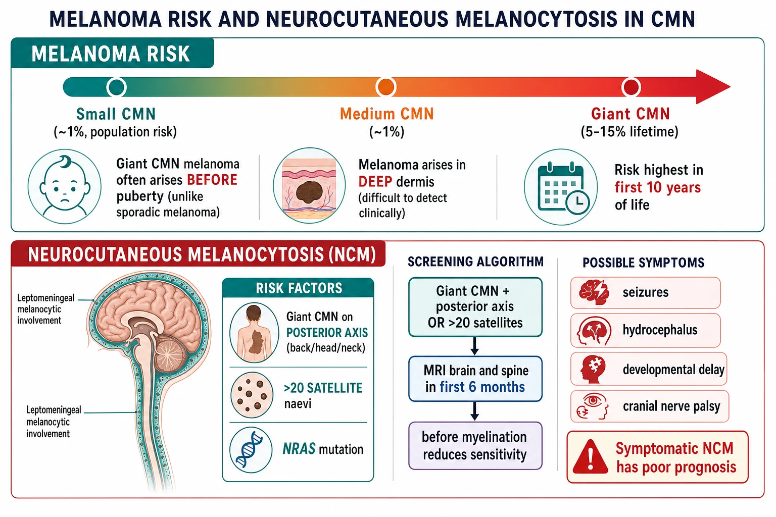
| Size | Lifetime melanoma risk | Characteristics |
|---|---|---|
| Small (under 1.5 cm) | ~1% (≈ population risk) | Melanoma rare; usually junctional (clinically detectable) |
| Medium (1.5-19.9 cm) | ~1% | Similar to small; selective excision reasonable for change |
| Giant (≥20 cm or ≥5% BSA) | 5-15% | Melanoma often arises before puberty; arises in the deep dermis or CNS (difficult to detect clinically) |
Key features of melanoma in CMN
- Giant CMN melanoma differs from sporadic melanoma:[4]
- Onset: often before age 10 (unlike sporadic melanoma which peaks in adults).
- Site of origin: frequently the deep dermis (naevus cells in the deep dermal / subcutaneous tissue) rather than the epidermis — making early clinical detection difficult.
- Histology: may be nodular, spindle cell, or small round blue cell (neuroblastoma-like) melanoma.
- Clinical signs of malignant change: new nodule (rapidly growing), colour change (darkening, blue-black, depigmentation), ulceration or bleeding, pain, or satellite lesions becoming elevated.
Neurocutaneous Melanocytosis (NCM)
- Definition: proliferation of melanocytes in the leptomeninges (pia and arachnoid) and/or brain parenchyma.[3]
- Risk: ~5-10% of patients with giant CMN develop symptomatic NCM. Risk is highest when:[2][3]
- Giant CMN is on the posterior axis (back, head, neck).
- 20 or more satellite naevi are present.
- NRAS mutation present (virtually all giant CMN).
- Symptoms: seizures, hydrocephalus (obstructive — due to melanin / reticulo-macrophage infiltration at the foramen of Magendie and Luschka), developmental delay, cranial nerve palsies, spinal cord compression (melanocytosis in the spinal leptomeninges).
- Screening: MRI brain and whole spine in the first 6 months of life (before myelination reduces MRI sensitivity) for all patients with giant CMN + posterior axial location or 20 or more satellites.[3][8]
- Prognosis: symptomatic NCM has poor prognosis (often fatal; median survival under 3 years from symptom onset) due to obstructive hydrocephalus, seizures, or primary CNS melanoma.
- Asymptomatic NCM: some patients have radiological NCM without symptoms — managed with neurological surveillance.
Differential Diagnosis of a Pigmented Congenital Lesion
Congenital melanocytic naevus (CMN)
- Present at birth or first weeks of life
- Brown-black, well-defined patch, plaque or nodule
- Histology: deep dermal naevus cells, periadnexal / perineural extension
- Somatic NRAS / BRAF / BRAF-fusion mutation
- Management by size + location + NCM risk
Becker's naevus (organoid hamartoma)
- Usually appears in adolescence (not at birth)
- Sharply demarcated tan-brown patch with hypertrichosis
- Shoulder, scapula, chest most often; unilateral
- Increased androgen receptor density
- Cosmetic; laser / electrolysis for hair
Cafe-au-lait macule (CALM)
- Uniform light-brown macule, well-defined edge
- May be present at birth or appear in infancy
- No surface change, no hypertrichosis
- 6 or more lesions / axillary freckling = NF1
- Wood's lamp accentuates in skin of colour
Mongolian spot / dermal melanocytosis
- Slate-blue / grey, poorly-defined patch
- Lumbosacral, buttock, back
- Regresses by age 5 in most cases
- Histology: dendritic melanocytes in deep dermis
- Extensive / persistent = consider nevus of Ota / Ito / Hori
Nevus sebaceous (of Jadassohn)
- Yellow-orange, waxy, hairless plaque at birth
- Flattens in childhood, becomes verrucous at puberty
- Risk of secondary BCC / SCC / trichoblastoma (~5-15% lifetime)
- Wnt pathway / HRAS / KRAS mutations
- Prophylactic excision often considered
Plexiform neurofibroma
- Soft, bag-like, often overlying CALM (NF1)
- Hypertrichosis possible
- Can be deeply invasive (head and neck 'dumbo-ear')
- NF1 / NF2 family
- MRI + NF1 multidisciplinary follow-up
Other differentials include: congenital smooth-muscle hamartoma (often hypertrichotic but pale-fawn), congenital pigmented dermatofibroma, congenital juvenile xanthogranuloma (yellow-orange, transient), congenital mastocytoma (Darier's sign positive), and the PHACE / Sturge-Weber spectrum when a facial vascular component coexists with a CMN. [1]
Clinical Examination and Bedside Assessment
A full assessment of a child with a CMN includes: [1]
- Total-skin examination — document every CMN, every satellite and its size (largest diameter), distribution, colour, surface and presence of hypertrichosis.
- Neurological examination — level of consciousness, fontanelle, head circumference, cranial nerves, primitive reflexes (in infants), tone, gait, skin for neurocutaneous stigmata, seizure history.
- Ophthalmology assessment — fundoscopy, intraocular pressure, slit-lamp; mandatory in palpebral CMN, head/neck CMN, and any giant CMN.
- Baseline total-body photography — standardised, age- and surface-area-corrected images at 6, 12, 24 months and then yearly; this is the single most useful surveillance tool.
- Dermoscopy — useful for the smaller CMN and for monitoring the dermoscopic signature of an individual lesion; giant CMN are too large for standard dermoscopy.
- Palpation — depth (dermal vs subcutaneous), nodularity, warmth, tenderness. [1]
ABCDE (asymmetry, border, colour, diameter, evolution) is the standard melanoma screen but is much less reliable in giant CMN because of the heterogeneous baseline colour, irregular border and constant growth. In this context, the most important bedside sign is evolution — a focal change in colour, surface, nodularity or ulceration that is out of step with the rest of the lesion. [1]
Investigations
MRI brain and whole spine
MRI findings of NCM: [1]
- T1 hyperintense leptomeningeal signal (paramagnetic effect of melanin).
- T1 hyperintense parenchymal melanocytosis in the temporal lobes, cerebellum, brainstem, thalami.
- Leptomeningeal enhancement post-contrast.
- Hydrocephalus (obstructive — melanin deposition at the foramina of Magendie and Luschka, or within the ventricular system).
- Mass lesions (parenchymal melanoma, leptomeningeal melanoma). [1]
Biopsy
- Excisional biopsy (1-3 mm clinical margin, full-thickness to subcutis, single piece) — for small/medium CMN that change or require histology.
- Incisional / punch biopsy of a suspicious nodule in a giant CMN — must reach the deep dermis to capture the deep naevomelanocytes where melanoma arises.
- Shave biopsy is contraindicated — it cannot sample the deep dermal component and may transect a melanoma, causing difficulty with depth measurement (Breslow thickness). [1]
Histopathology
CMN histology — the five deep features
Histology does not reliably predict melanoma risk; a banal-appearing CMN can give rise to melanoma, and an atypical-appearing CMN may remain quiescent for life.[5]
Molecular testing
- Targeted NGS panel for NRAS, BRAF, BRAF fusions, KRAS, HRAS — increasingly routine, especially when systemic therapy is being considered.
- FISH / CGH for difficult proliferative nodule vs melanoma.
- Germline RASopathy panel when a clinical geneticist suspects Noonan, CFC or Costello syndrome. [1]
Reflectance confocal microscopy and high-frequency ultrasound
Used in specialist centres to assess the depth and architecture of suspicious areas within a giant CMN; they do not replace biopsy. [1]
Management
Small and medium CMN
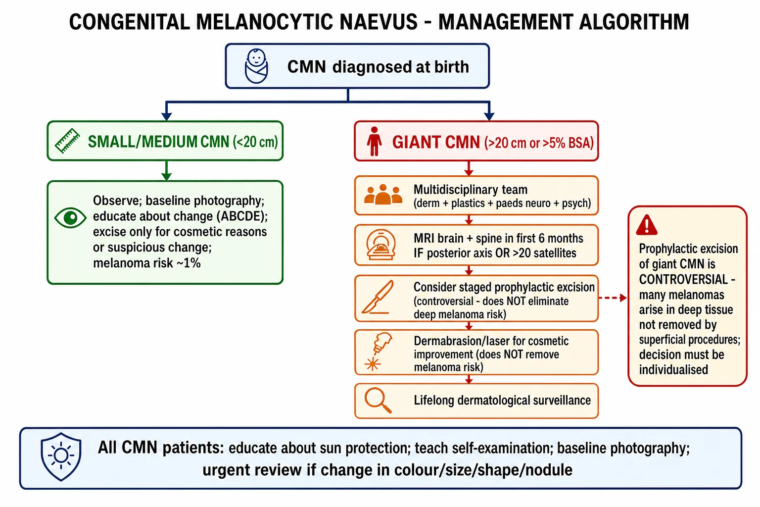
- Observe with baseline photography; re-photograph at 6 months, 12 months and then yearly.
- Educate parents and (age-appropriately) the child on the ABCDE criteria and the signs of malignant change (new nodule, ulceration, bleeding, rapid growth, pain, itch).
- Excision is not mandatory; consider it for cosmetic reasons, repeated trauma, functional impairment, or any change suspicious for melanoma.
- Lasers (Q-switched ruby, alexandrite) may be used cosmetically; risk of repigmentation, scarring, and depigmentation.
- Dermatology review every 1-3 years depending on the lesion. [1]
Giant CMN — multidisciplinary approach
Giant CMN requires a multidisciplinary team from birth:[2][3]
- Paediatric dermatology — primary medical lead; baseline photography; serial examination; biopsy of any change.
- Paediatric neurology — clinical surveillance; MRI brain and whole spine in the first 6 months if posterior-axis / >20 satellites; repeat MRI every 1-2 years if NCM present.
- Paediatric neurosurgery — symptomatic NCM (hydrocephalus, mass effect).
- Plastic / paediatric surgery — staged excision, tissue expansion, skin grafts, cultured epithelial autografts.
- Ophthalmology — palpebral / head and neck CMN; baseline assessment, intraocular pressure, fundoscopy.
- Clinical genetics — RASopathy features, sibling counselling, prenatal counselling for future pregnancies.
- Psychology / psychiatry — body-image, peer interactions, school integration.
- Pain service / wound care — for ulcerated, eroded or pruritic lesions. [1]
Prophylactic excision — controversial
Arguments for prophylactic excision
- Reduces the cosmetic burden and the social / psychological impact of a giant CMN
- Removes the visible, clinically monitorable component of the lesion
- Some series report reduced (but not eliminated) melanoma risk from the excised component
- Modern tissue expansion, serial excision and skin-substitute techniques have improved outcomes
Arguments against prophylactic excision
- Many giant-CMN melanomas arise in the DEEP dermis / subcutis — not removed by superficial excision
- Procedure carries significant morbidity: scarring, alopecia, contracture, infection, graft failure
- No high-quality evidence that excision reduces melanoma-specific mortality
- Satellite naevi and NCM risk are not altered by excision of the main lesion
- Psychological impact of repeated major surgery in childhood must be weighed
The modern consensus is that prophylactic complete excision of a giant CMN is not a melanoma-preventing procedure; it may be offered for cosmetic, functional or psychological reasons after a careful, family-centred, multidisciplinary discussion. Partial / staged excision of cosmetically or functionally significant areas (e.g. face, scalp, hands) is reasonable; complete excision is rarely achievable and is not the goal. [1]
Curettage, dermabrasion and laser
- Curettage / dermabrasion performed in early infancy (first 4-6 weeks, before the naevomelanocytes mature and anchor to the deeper dermis) can lighten the lesion and improve texture. It does not remove deep naevus cells and does not eliminate melanoma risk.[2]
- Ablative laser (CO2, erbium:YAG) — flattens the verrucous surface; does not eliminate risk.
- Pigment-selective laser (Q-switched ruby, Q-switched alexandrite, Nd:YAG 1064 nm) — lightens pigment; significant repigmentation rate; risk of depigmentation in skin of colour.
Bottom line: cosmetic procedures are valuable for quality of life, but the family must understand that none of them removes the deep naevomelanocytes or eliminates the melanoma risk. [1]
MEK inhibitors — emerging targeted therapy
The discovery that giant CMN are driven by NRAS Q61 has opened the door to MEK inhibitor therapy (binimetinib, trametinib, selumetinib). Case reports and early phase 2 trials in infants and children with giant CMN ± symptomatic NCM have shown: [1]
- Marked lightening of the cutaneous lesion (visible within 4-12 weeks).
- Stabilisation or improvement of NCM-related seizures and neurological symptoms.
- Tolerable toxicity profile (rash, CK elevation, ocular events, growth plate effects in young children). [1]
MEK inhibitors are not yet standard of care; they are used in specialist centres under trial protocols or compassionate access, and are reserved for symptomatic NCM, bulky unresectable disease, or where other measures have failed.[9]
BRAF-fusion CMN may respond to MEK / ERK inhibitors in a similar rationale, given that BRAF-fusion signals through the same MAPK pathway.[9]
Management of neurocutaneous melanosis
- Asymptomatic NCM (radiological only) — neurology surveillance, repeat MRI every 1-2 years, antiepileptics if seizures develop.
- Symptomatic NCM — antiepileptics, consideration of MEK inhibitor under trial, CSF diversion (VP shunt, ETV) for hydrocephalus, neurosurgical resection of symptomatic mass lesions.
- Prognosis of symptomatic NCM is poor: median survival under 3 years from symptom onset; main causes of death are hydrocephalus, status epilepticus, and primary CNS melanoma.[8]
Palpebral CMN
CMN involving the eyelid is associated with ocular melanocytosis (melanocytes in the uveal tract, conjunctiva, sclera), which carries a risk of glaucoma and uveal melanoma. Management includes mandatory ophthalmology assessment, serial intraocular pressure and fundoscopy, and staged lid-sparing excision when cosmesis or function warrant it.[1]
Complications and Pitfalls
Disease complications
- Melanoma (5-15% lifetime risk in giant CMN, often pre-pubertal, deep dermal)
- Neurocutaneous melanosis (5-10% of giant CMN, often fatal when symptomatic)
- Pruritus, xerosis, eczema within the lesion
- Ulceration, fissure, infection (intertriginous sites)
- Psychosocial / body-image morbidity (cosmetic, peer, school)
- Functional impairment (palpebral, periocular, periarticular)
Procedure complications
- Bleeding, haematoma, infection, dehiscence (excision)
- Scarring, keloid, hypertrophic scar, alopecia (scalp)
- Contracture (especially across joints)
- Graft failure / donor-site morbidity
- Repigmentation, post-inflammatory hyper-/hypo-pigmentation (laser)
- Partial response (no procedure is curative)
Classic management errors
- Reassuring a parent about a changing nodule — always biopsy
- Ordering a shave biopsy that misses the deep dermal component
- Delaying MRI in a child with neurological symptoms
- Missing ocular involvement in a palpebral CMN
- Over-promising 'cure' with laser / dermabrasion
- Ignoring the family's psychological burden
- Failing to offer clinical genetics referral when RASopathy features are present
Prognosis and Disposition
CMN prognosis — what the examiner needs
Follow-up schedule for a giant CMN: [1]
- Dermatology — every 3-6 months for the first 5 years, then 6-12 monthly.
- Neurology — baseline MRI in the first 6 months, repeat at 12-24 months if NCM present, then every 1-2 years.
- Ophthalmology — baseline, then yearly if palpebral / head and neck CMN.
- Plastic surgery — at diagnosis and as needed for staged excision.
- Psychology — regular review, school support, peer support groups.
- Self / parent surveillance — monthly total-body examination using baseline photographs; any change → same-day dermatology contact. [1]
Disposition — most small/medium CMN can be discharged to community dermatology with annual review and self-surveillance. Giant CMN should remain in a specialist multidisciplinary service for life. [1]
Special Populations
Pregnancy and prenatal counselling
A CMN in a pregnant woman may darken or become more verrucous under hormonal influence. Excision is rarely required during pregnancy. Pre-conception or prenatal counselling for parents of a child with a giant CMN should include: (1) reassurance about the postzygotic mosaic nature and the very low recurrence risk; (2) discussion of MRI safety in the first 6 months of life (no contrast if possible); (3) information about emerging in-utero and neonatal MEK inhibitor experience; (4) family psychological support. [1]
Paediatric CMN
Melanoma in children is rare; Spitz naevi and atypical proliferative nodules are the commonest differential within a giant CMN. Threshold for biopsy differs from adults — a single, rapidly growing, ulcerated or bleeding nodule should be excised, but stable heterogeneity and hypertrichosis are not, on their own, indications. [1]
RASopathies
CMN may be the cutaneous marker of an underlying germline RASopathy. Index cases with a giant CMN and any of: short stature, broad neck, congenital heart disease (pulmonary stenosis, HCM), developmental delay, characteristic facies, sparse hair, or a family history, should be referred to clinical genetics for germline testing (PTPN11, SOS1, RAF1, BRAF, MAP2K1/2, KRAS, HRAS, NRAS). [1]
Skin of colour (Fitzpatrick V-VI)
CMN in darkly pigmented skin is darker, more verrucous, and more hypertrichotic; it is also more difficult to detect early melanomatous change because of the baseline pigment. Dermoscopy in skin of colour is a specialist skill; clinicopathological correlation is essential. Acral lentiginous melanoma and mucosal melanoma are proportionally more common than in lighter skin types and must remain in the differential. [1]
Immunosuppression
There is no large evidence base for an increased CMN-melanoma risk in transplant or HIV patients, but general melanoma surveillance is more vigilant in this group; giant CMN should continue to be followed in a specialist service. [1]
Evidence Base, Guidelines and Regional Differences
The most important evidence underpinning modern CMN management: [1]
- NRAS Q61 driver in giant CMN — Kinsler lab, 2013 and subsequent. The 2022 Exp Dermatol paper (PMID 35020224) and the 2020 Br J Dermatol genotype-phenotype study (Polubothu & Kinsler, PMID 31111470) underpin the modern classification and the rationale for MEK inhibition.[6][7]
- Mosaic BRAF fusions — Kao et al., J Invest Dermatol 2024 (PMID 37716647).[9]
- Neurocutaneous melanosis — Ramaswamy et al. Epilepsy Behav 2020 (PMID 32272368) systematic review; Ruth Semin Pediatr Neurol 2024 (PMID 39389659) on CMN syndrome.[3][8]
- 2020 international CMN consensus (Ott et al., J Am Acad Dermatol and ERN-Skin) — the current size + satellite + additional-feature classification; underpins modern size categorisation.
- Management guidelines — BAD (UK), AAD / ASPS (US) and EDF (EU) guidelines are broadly concordant on the need for multidisciplinary care, MRI screening in high-risk phenotypes, and the controversial nature of prophylactic excision.
- MEK inhibitor trials — phase 2 trials of binimetinib, trametinib and selumetinib in NRAS-mutant giant CMN (NCT identifier trials; published case series); endpoints include lesion lightening, NCM stabilisation, quality of life and safety.
Regional differences are mostly logistical: in high-resource settings, MRI screening at 4-6 months is routine; in resource-limited settings, the same imaging is available only on clinical indication, and clinical surveillance with baseline photography is the cornerstone. [1]
Exam Pearls and High-Yield Minutiae
[1]Red Flags
Exam application bank (NEET-PG / INICET)
One-line answer
A congenital melanocytic naevus (CMN) is a melanocytic naevus present at birth or within the first weeks of life, caused by a postzygotic mosaic activating mutation in the RAS-MAPK pathway (NRAS Q61 in ~80-90% of giant CMN; BRAF V600E in some smaller CMN; mosaic BRAF fusions). Classified by projected adult size into small (under 1.5 cm), medium (1.5-19.9 cm) and large/giant (≥20 cm or ≥5% BSA). Giant CMN carries a 5-15% lifetime melanoma risk (often pre-pubertal, arising in the deep dermis) and 5-10% risk of neurocutaneous melanocytosis (NCM) when on the posterior axis with ≥20 satellite naevi. Management is multidisciplinary; prophylactic excision is controversial; MEK inhibitors are an emerging targeted option for NRAS-mutant disease.
Worked stems (answer without another resource)
Stem 1 — Classic presentation. Map symptoms to mechanism; name the first investigation and first treatment step with dose/route if drug therapy is standard. [1]
Stem 2 — Unstable / complicated. List red flags that force immediate resuscitation, theatre, ICU, antidote, or reperfusion — and what you do in the first 15 minutes. [1]
Stem 3 — Atypical group. Elderly, pregnancy, child, or immunocompromised: how presentation and thresholds change. [1]
Stem 4 — Differential trap. Name the three closest mimics and one discriminator for each. [1]
Stem 5 — Disposition. Who goes home with safety-netting, who is admitted, who needs HDU/ICU/theatre, and what follow-up is mandatory. [1]
Rapid viva checklist
- Definition + classification
- Pathophysiology chain
- Bedside signs / criteria
- Score with exact components (if any)
- Emergency bundle
- Definitive therapy with doses
- Complications of disease and of treatment
- Special populations
- Guideline/trial name if classic
- Three exam traps
Coverage self-check
If you cannot answer any stem above from this page alone, re-read the matching section — the page is intended to be self-sufficient for final-prof and NEET-PG/INICET questions on Congenital melanocytic naevus.
[1]References
- [1]Camargo CP, Saliba M, Saad EA, et al. Treatments of palpebral congenital melanocytic nevus: a systematic review Acta Cir Bras, 2023.PMID 38055392
- [2]Viana AC, Gontijo B, Bittencourt FV. Giant congenital melanocytic nevus An Bras Dermatol, 2013.PMID 24474093
- [3]Ruth J. Congenital melanocytic nevus syndrome: An association between congenital melanocytic nevi and neurological abnormalities Semin Pediatr Neurol, 2024.PMID 39389659
- [4]Alos L, Carrasco A, Teixido C, et al. Melanoma on congenital melanocytic nevi Pathol Res Pract, 2024.PMID 38518732
- [5]Zayour M, Lazova R. Congenital melanocytic nevi Clin Lab Med, 2011.PMID 21549240
- [6]Kinsler VA, O'Hare P, Jacques T, et al. Spatiotemporal expression of NRAS and occurrence of giant congenital melanocytic nevi Exp Dermatol, 2022.PMID 35020224
- [7]Polubothu S, Kinsler VA. Does the gene matter? Genotype-phenotype and genotype-outcome associations in congenital melanocytic naevi Br J Dermatol, 2020.PMID 31111470
- [8]Ramaswamy V, Delaney H, Haque S, et al. Epilepsy in isolated parenchymal neurocutaneous melanosis: A systematic review Epilepsy Behav, 2020.PMID 32272368
- [9]Kao JC, Kao JH, Alomari AK, et al. Mosaic BRAF Fusions Are a Recurrent Cause of Congenital Melanocytic Nevi Targetable by MAPK Pathway Inhibition J Invest Dermatol, 2024.PMID 37716647