Dermatology · Medicine
Cryoglobulinaemia
Also known as Cryoglobulinaemia · Cryoglobulinemic vasculitis · Mixed cryoglobulinaemia
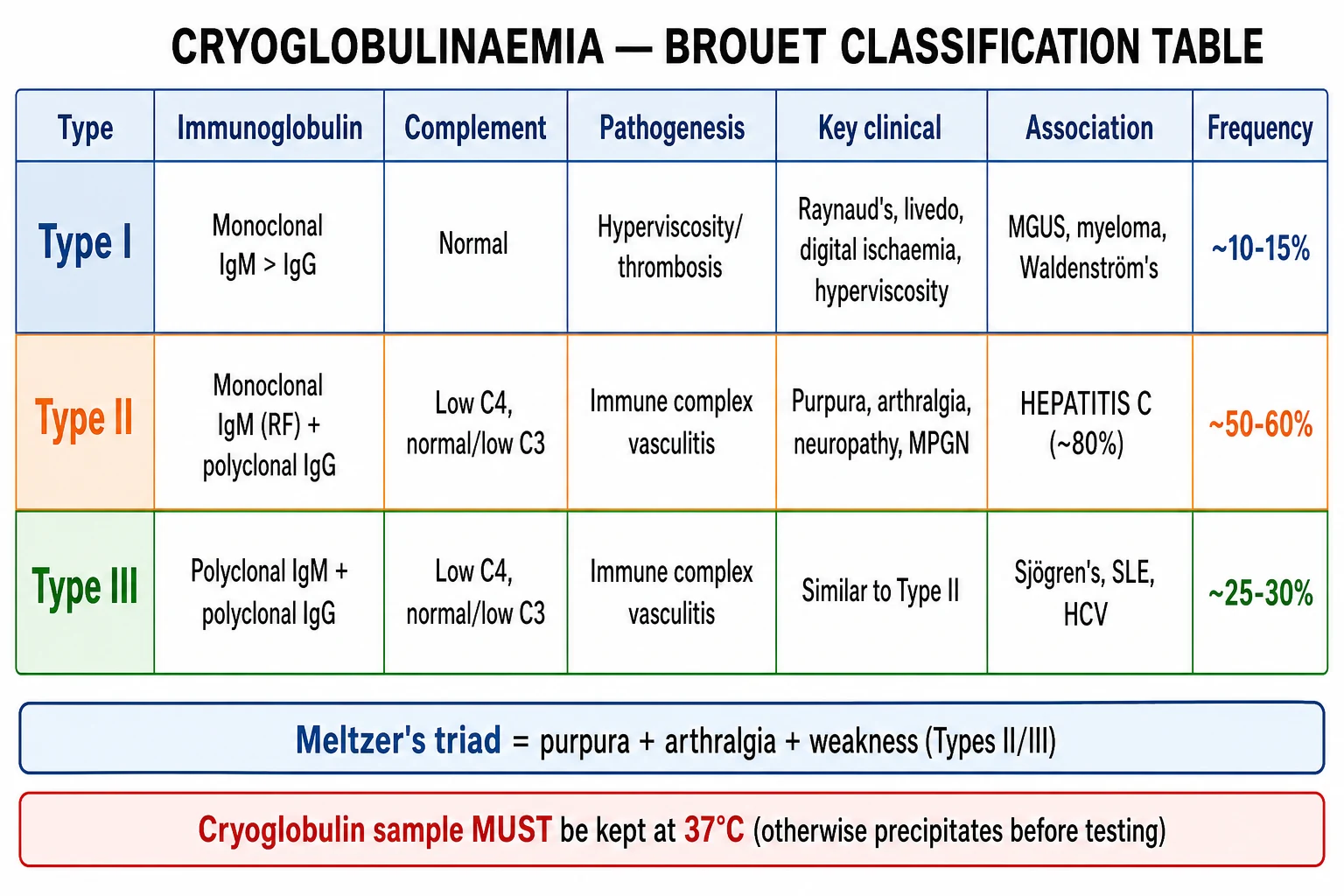
Cryoglobulinaemia is the presence of circulating immunoglobulins that reversibly precipitate below 37°C and dissolve on rewarming. Classified by the Brouet system: Type I (monoclonal IgM/IgG; hyperviscosity/thrombosis; MGUS, myeloma, Waldenström's), Type II (mixed monoclonal IgM with rheumatoid factor activity + polyclonal IgG; immune complex vasculitis; hepatitis C in ~80%), and Type III (polyclonal IgG + IgM; immune complex vasculitis; connective tissue disease, infection). Mixed types (II/III) present with Meltzer's triad (purpura + arthralgia + weakness) and cause leukocytoclastic vasculitis, peripheral neuropathy, and MPGN-like glomerulonephritis. The complement pattern of low C4 with normal or low C3 is the classic laboratory hallmark. Management centres on treating the underlying cause: direct-acting antivirals (DAAs) cure HCV and cryoglobulinaemia in most patients; rituximab for refractory or severe mixed cryoglobulinaemia; plasma exchange for hyperviscosity or rapidly progressive disease. NEET-PG/INICET assessment demands mastery of the Brouet classification, Meltzer's triad, the HCV association, the low-C4 complement pattern, the warm-sample handling requirement, and the DAA/rituximab treatment ladder.
On this page & tools
Your progress
Saved locally on this device.
Exam tags
Red flags
Overview and Definition
Cryoglobulinaemia literally means "cold antibodies in the blood." It is defined by the presence of circulating immunoglobulins that reversibly precipitate at temperatures below 37°C and redissolve on rewarming. The precipitate is composed of immunoglobulins (and sometimes complement), and its physical behaviour is the diagnostic property exploited in the laboratory test. Importantly, the circulating cryoglobulins themselves are not necessarily pathogenic at body temperature; tissue injury occurs when the cryoglobulins precipitate in the cooler peripheral capillaries, or when they form immune complexes that deposit in vessel walls and activate complement.[1][3]
The term encompasses a spectrum of disorders. Some patients have circulating cryoglobulins but no symptoms (essential cryoglobulinaemia), while others develop organ-threatening vasculitis, hyperviscosity or glomerulonephritis. The clinical expression depends on the amount, temperature solubility, immunoglobulin class and clonality of the cryoglobulins, as well as the presence of an underlying infection, autoimmune disease or malignancy.[1]
The Brouet classification is the universally accepted framework. It divides cryoglobulins into three types based on the immunoglobulin composition and clonality. Type I is a single monoclonal immunoglobulin. Types II and III are mixed cryoglobulins composed of two or more immunoglobulin classes, with the key distinction that Type II contains a monoclonal IgM with rheumatoid factor activity, whereas Type III is polyclonal. The clinical and laboratory implications of this distinction are profound: Type I behaves as a hyperviscosity and occlusion syndrome, while mixed types behave as immune-complex-mediated small-vessel vasculitis. The term essential cryoglobulinaemia is used when no underlying infection, autoimmune disease or malignancy can be identified; this label is becoming less common as more precise associations are recognised.[1][3]
Brouet classification at a glance
Type I
Type II (mixed)
Type III (mixed)
Epidemiology and Risk Factors
Mixed cryoglobulinaemia (Types II and III combined) accounts for approximately 80-90% of all cryoglobulinaemia in adults, with Type II being the most common individual type and Type III often representing a less specific, reactive state. Type I is rare, making up roughly 10-15% of cases, and is usually discovered in the context of an established lymphoproliferative disorder.[1]
The dominant risk factor for mixed cryoglobulinaemia is chronic hepatitis C virus (HCV) infection. The widely quoted figure is that roughly 80% of mixed cryoglobulinaemia, and especially Type II disease, is HCV-associated. However, this proportion is falling in regions with high uptake of HCV treatment. In populations where HCV prevalence has declined, the relative contribution of autoimmune disease (particularly Sjögren's syndrome) and idiopathic (essential) cryoglobulinaemia has risen.[1][5]
Other important associations are stratified by Brouet type: [1]
- Type I: monoclonal gammopathy of undetermined significance (MGUS), multiple myeloma, Waldenström's macroglobulinaemia, chronic lymphocytic leukaemia, lymphoma. These conditions produce a monoclonal immunoglobulin that can precipitate in the cold.
- Type II: chronic HCV infection (dominant), hepatitis B virus (HBV), HIV, and occasionally connective tissue disease.
- Type III: autoimmune disease, particularly Sjögren's syndrome (the most common autoimmune cause), systemic lupus erythematosus, rheumatoid arthritis; also chronic infections such as HCV, HBV, HIV, endocarditis, leishmaniasis and Epstein-Barr virus.[2][10]
Key epidemiological facts
Environmental and host factors include cold exposure, which can precipitate symptoms in patients with circulating cryoglobulins; the female predominance in autoimmune-associated disease; and age, with Type I occurring in older adults with lymphoproliferative disease. Notably, the prevalence of HCV varies by region, so the epidemiology of HCV-associated cryoglobulinaemia is strongly regional. In countries with high HCV prevalence and limited access to direct-acting antivirals, mixed cryoglobulinaemia remains common. In countries that have achieved HCV elimination targets, cryoglobulinaemia is increasingly seen in the context of autoimmune disease or is labelled essential.[5]
Pathophysiology
The pathophysiology of cryoglobulinaemia is best understood by separating Type I from mixed Types II and III, because the mechanisms differ fundamentally. [1]
Type I cryoglobulinaemia: hyperviscosity and vascular occlusion
Type I cryoglobulinaemia is composed of a monoclonal immunoglobulin, most often IgM (IgM is the most efficient at cold precipitation) but sometimes IgG or, rarely, IgA. Because the protein is monoclonal, it is produced in large quantity and has a uniform structure. In the cooler parts of the circulation (ears, nose, fingers, toes), the immunoglobulin precipitates directly from the plasma. This creates several problems.[1]
First, the high concentration of circulating paraprotein increases serum viscosity. Hyperviscosity reduces blood flow through the microcirculation, impairs oxygen delivery, and causes endothelial injury. Second, the precipitated cryoglobulin physically occludes small vessels and promotes thrombosis. The result is a non-inflammatory vasculopathy: the vessels are blocked, but there is no immune-complex deposition, no complement activation, and no leukocytoclastic vasculitis. This is why Type I does not cause palpable purpura, arthralgia or glomerulonephritis in the classic sense. The clinical picture is one of hyperviscosity (visual disturbance, headache, mucosal bleeding) and cold-sensitive digital ischaemia (Raynaud's, livedo, acrocyanosis, ulcers).[1]
Mixed cryoglobulinaemia: immune-complex vasculitis
Mixed cryoglobulins (Types II and III) are composed of two or more immunoglobulin classes. The defining feature is the presence of an IgM with rheumatoid factor (RF) activity: it binds to the Fc portion of IgG. In Type II, this IgM is monoclonal; in Type III, it is polyclonal. The IgM-IgG immune complexes form in the circulation and deposit in the walls of small vessels (post-capillary venules) in the skin, joints, peripheral nerves, kidneys and gastrointestinal tract.[1][3]
Once deposited, the immune complexes activate the classical complement pathway. C1q binds to the Fc region of the IgG within the immune complex, triggering the C4-C2-C3 cascade. Because C4 is consumed early in the classical pathway, the hallmark laboratory finding is disproportionately low C4 with normal or mildly low C3. The terminal complement components and anaphylatoxins (C3a, C5a) recruit neutrophils. Neutrophils release lysosomal enzymes and reactive oxygen species, fragmenting the vessel wall and producing leukocytoclastic vasculitis (nuclear dust from disintegrated neutrophils).[1]
In HCV-associated Type II cryoglobulinaemia, chronic HCV infection drives the clonal expansion of B cells, particularly marginal-zone-like B cells. The HCV envelope protein E2 binds to CD81 on B cells and lowers the activation threshold. This leads to the expansion of a monoclonal or oligoclonal B-cell population that produces the IgM-RF cryoglobulin. Eradication of HCV removes this drive, and in most patients the cryoglobulinaemia resolves. This elegant pathophysiological link explains why antiviral therapy is the cornerstone of management.[5]
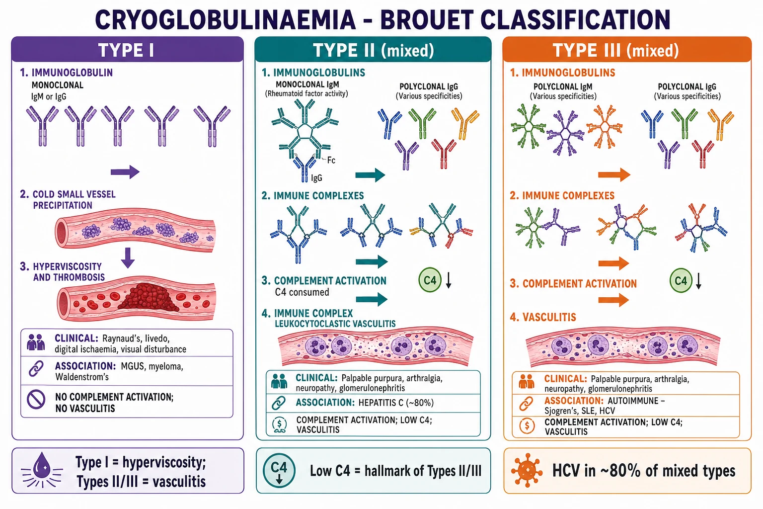
Why low C4 and normal or low C3?
The complement pattern is a favourite exam point. The classical pathway is initiated by immune complexes and consumes C4 and C2 before C3. Because the classical pathway is the dominant driver, C4 is disproportionately low. C3 may be normal or mildly reduced because the alternative pathway can also be activated by damaged endothelium, but the classical pathway dominance produces the characteristic low C4 with normal or low C3. This pattern is a strong clue to mixed cryoglobulinaemia and contrasts with alternative-pathway diseases such as C3 glomerulopathy, where C3 is low but C4 is normal.[1]
Clinical Presentation
Mixed cryoglobulinaemia: Meltzer's triad and beyond
The classic presentation of mixed cryoglobulinaemia is Meltzer's triad: palpable purpura, arthralgia and weakness. This triad is not invariable, but when present it is highly suggestive. The purpura is typically palpable, recurrent and located on the lower legs and other dependent areas, reflecting leukocytoclastic vasculitis of the post-capillary venules. It may be preceded by a burning sensation, and crops of lesions can appear after cold exposure or prolonged standing.[1][3]
Arthralgia is usually symmetrical, affects small and medium joints, and is non-erosive. It differs from rheumatoid arthritis because it is episodic and does not cause joint deformity. Weakness is often described as a proximal myopathy-like fatigue, but careful examination frequently reveals an underlying peripheral neuropathy. Neuropathy is a major source of morbidity in mixed cryoglobulinaemia and can present as a distal symmetrical sensory or sensorimotor polyneuropathy, or as mononeuritis multiplex.[1]
Other organ involvement includes: [1]
- Skin: palpable purpura, livedo reticularis, Raynaud's phenomenon, digital ulcers, urticaria, skin necrosis.
- Kidneys: membranoproliferative glomerulonephritis (MPGN-like), presenting with proteinuria, haematuria, casts, nephrotic syndrome, nephritic syndrome or acute kidney injury. Renal disease is a major determinant of prognosis.
- Peripheral nerves: sensory neuropathy, sensorimotor neuropathy, mononeuritis multiplex.
- Gastrointestinal tract: mesenteric vasculitis causing abdominal pain, intestinal ischaemia, bleeding or perforation.
- Liver: chronic HCV infection, chronic hepatitis, cirrhosis, hepatocellular carcinoma.
- Central nervous system: stroke is uncommon but described, particularly in hyperviscosity states.[1][5]
Type I cryoglobulinaemia: hyperviscosity and digital ischaemia
Type I cryoglobulinaemia presents with symptoms of hyperviscosity and vascular occlusion, not vasculitis. The patient may report visual blurring or amaurosis fugax, headache, dizziness, vertigo, tinnitus, mucosal bleeding, and fatigue. On examination, fundoscopy may show retinal venous engorgement, haemorrhages or exudates. The hands and feet may show acrocyanosis, Raynaud's phenomenon, livedo reticularis, digital ischaemia or ulceration. The digits are often cold-sensitive and may develop non-healing ulcers or gangrene. There is no palpable purpura, no glomerulonephritis and no classical Meltzer's triad, unless the patient has coincident mixed cryoglobulins.[1]
Atypical presentations
Atypical presentations are important for exams because they test whether the candidate understands the underlying mechanism rather than just the classic triad. A patient may present with isolated peripheral neuropathy, isolated glomerulonephritis, or severe abdominal pain before skin lesions appear. Some patients have only fatigue and arthralgia without purpura. In the elderly, Type I cryoglobulinaemia may be mistaken for peripheral arterial disease or cholesterol emboli. In pregnant patients, the hyperviscosity of Type I can worsen, and immune-complex disease may flare because of physiological changes in complement and circulating immune complexes. In immunosuppressed patients, the presentation may be muted or the underlying infection may be occult.[1]
[1]Differential Diagnosis
The differential diagnosis of cryoglobulinaemia depends on whether the patient presents with palpable purpura and systemic vasculitis (mixed types) or with hyperviscosity and digital ischaemia (Type I). The key is to distinguish cryoglobulinaemic vasculitis from other small-vessel vasculitides, and Type I cryoglobulinaemia from other causes of hyperviscosity and occlusive vasculopathy.[1]
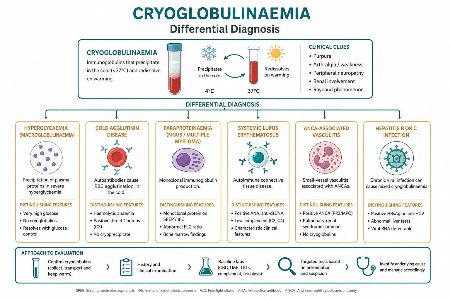
| Mimic | Distinguishing features |
|---|---|
| IgA vasculitis (Henoch-Schönlein purpura) | Palpable purpura, arthritis, abdominal pain, renal involvement; more common in children; biopsy shows IgA-dominant deposition on direct immunofluorescence; normal or low C4 less characteristic; no cryoglobulins or HCV association. |
| ANCA-associated vasculitis (microscopic polyangiitis, granulomatosis with polyangiitis, eosinophilic granulomatosis with polyangiitis) | ANCA positive in most cases; pauci-immune glomerulonephritis on biopsy; no cryoglobulins; may have pulmonary haemorrhage or upper airway disease. |
| Antiphospholipid syndrome | Thrombosis, recurrent miscarriage, livedo reticularis; anticardiolipin or anti-beta-2-glycoprotein-I antibodies, lupus anticoagulant; no palpable purpura or immune-complex vasculitis; complement usually normal. |
| Hyperviscosity syndrome (Waldenström's macroglobulinaemia, multiple myeloma) | Very high IgM or IgG paraprotein, high serum viscosity, retinal changes, mucosal bleeding; may coexist with Type I cryoglobulinaemia; no complement consumption or leukocytoclastic vasculitis. |
| Cholesterol emboli | Livedo reticularis, blue toe syndrome, renal failure after vascular intervention or aortic manipulation; eosinophilia; no cryoglobulins, no purpura. |
| Calciphylaxis | Painful necrotic skin lesions in patients with renal failure and hyperphosphataemia; high calcium-phosphate product; skin biopsy shows vascular calcification; no cryoglobulins. |
| Septic emboli / infective endocarditis | Fever, positive blood cultures, vegetation on echocardiography, Osler nodes, Janeway lesions; splinter haemorrhages; no cryoglobulins. |
| Thrombotic vasculopathy | Coagulopathy, thrombocytosis, cryofibrinogenaemia; no monoclonal immunoglobulin or immune-complex deposition; biopsy shows fibrin thrombi without vasculitis. |
Cryoglobulinaemia versus key mimics
IgA vasculitis
ANCA vasculitis
Antiphospholipid syndrome
Clinical and Bedside Assessment
A focused clinical assessment in suspected cryoglobulinaemia should answer three questions: is this mixed cryoglobulinaemia (vasculitis) or Type I (hyperviscosity/occlusion)? What organs are involved? What is the underlying cause? [1]
History
Key history points include: [1]
- Cold sensitivity: do symptoms worsen with cold exposure? Raynaud's, livedo, acrocyanosis and cold-induced purpura are clues.
- Infection risk: prior blood transfusion, intravenous drug use, tattoos, needlestick injury, or residence in a region with high HCV prevalence. Hepatitis B and HIV should also be considered.
- Autoimmune symptoms: dry eyes, dry mouth (Sjögren's), arthralgia, rash, photosensitivity, oral ulcers (SLE), arthritis (rheumatoid arthritis).
- Malignancy symptoms: bone pain, fatigue, weight loss, night sweats, recurrent infections (myeloma, lymphoma, Waldenström's).
- Neurological symptoms: numbness, tingling, burning pain, weakness, mononeuritis multiplex pattern.
- Renal symptoms: foamy urine (proteinuria), dark urine (haematuria), oedema, reduced urine output.
- Gastrointestinal symptoms: abdominal pain, rectal bleeding, mesenteric ischaemia symptoms. [1]
Examination
The bedside examination should also assess the severity and tempo of disease. Rapidly progressive purpura, new ulceration, or evolving neurological deficit suggest urgent intervention. Measure blood pressure because hypertension may indicate renal involvement or be a complication of renal disease. Examine the abdomen for tenderness, peritonism or melena, and ask about stool colour. In the patient with suspected Type I disease, fundoscopy is essential to look for retinal venous engorgement, haemorrhages, cotton-wool spots or papilloedema. Auscultate the heart for murmurs and check peripheral pulses to differentiate vasculitis from infective endocarditis or peripheral arterial disease. [1]
- Skin: palpable purpura on the lower legs, livedo reticularis, Raynaud's, digital ulcers, acrocyanosis, skin necrosis, urticaria.
- Joints: symmetrical non-erosive arthralgia, often small joints of the hands and feet.
- Neurological: distal sensory loss, reduced ankle reflexes, mononeuritis multiplex (wrist drop, foot drop).
- Fundoscopy: retinal venous engorgement, haemorrhages, exudates, papilloedema in hyperviscosity.
- Cardiovascular: signs of endocarditis if septic emboli are a concern; blood pressure for renal involvement or hypertensive emergency.
- Abdomen: tenderness, guarding, rectal bleeding in mesenteric vasculitis; hepatomegaly or stigmata of chronic liver disease in HCV/HBV.
- Lymph nodes and spleen: lymphadenopathy or splenomegaly may suggest lymphoproliferative disease. [1]
Investigations
The investigation of suspected cryoglobulinaemia is directed at confirming the diagnosis, identifying the Brouet type, defining organ involvement, and finding the underlying cause. [1]
Cryoglobulin detection and the 37°C rule
The cryoglobulin screen is the defining test. The crucial practical point is that the sample must be handled at 37°C from collection to centrifugation. Blood should be drawn into a pre-warmed tube, transported at 37°C (for example in an incubator or water bath), and centrifuged at 37°C. The serum is then cooled to 4°C for up to 7 days. A precipitate that forms on cooling and redissolves on rewarming is a cryoglobulin. If the sample is allowed to cool to room temperature before centrifugation, the cryoglobulins precipitate in the clot and are discarded, producing a false-negative result. This is one of the most commonly tested practical points in examinations.[1][3]
Characterisation of cryoglobulins
Once a cryoglobulin is detected, the precipitate is washed and redissolved, and immunofixation or immunoelectrophoresis is used to identify the immunoglobulin classes. This determines whether the cryoglobulin is Type I, II or III. Quantification of the cryoglobulin level is sometimes performed, although the clinical correlation is imperfect. [1]
Complement and serology
- Complement: low C4 with normal or low C3 is the classic pattern in mixed cryoglobulinaemia. C4 is disproportionately low because of classical pathway activation.
- Rheumatoid factor: positive in mixed cryoglobulinaemia because the cryoglobulin IgM has RF activity. A strongly positive RF in the presence of HCV or purpura is a clue.
- Viral screen: HCV antibody and HCV RNA are mandatory in all mixed cryoglobulinaemia. Hepatitis B serology (HBsAg, anti-HBc) and HIV should also be checked.
- Autoimmune screen: ANA, anti-SSA/Ro, anti-SSB/La (Sjögren's), anti-dsDNA, anti-Smith, complement C3/C4, ESR, CRP.
- Serum protein electrophoresis and immunofixation: identifies monoclonal paraprotein in Type I or Type II disease. Free light chains should be checked if myeloma is suspected.
- Full blood count, renal function, liver function, urinalysis: baseline assessment and screening for organ involvement. [1]
Biopsy
- Skin biopsy: of a fresh purpuric lesion shows leukocytoclastic vasculitis (fragmented neutrophils, fibrinoid necrosis of vessel walls, extravasated red cells). Direct immunofluorescence may show immunoglobulin and complement deposition, but IgA-dominant deposition would point to IgA vasculitis rather than cryoglobulinaemia.[1]
- Renal biopsy: if proteinuria, haematuria or renal impairment is present. The typical pattern is membranoproliferative glomerulonephritis (MPGN-like) with mesangial and subendothelial deposits, often described as "tram-track" double contours on light microscopy. Immunofluorescence shows IgM and complement deposition.[1]
Other investigations
- Nerve conduction studies: to characterise peripheral neuropathy.
- Imaging: abdominal ultrasound or CT for liver disease; mesenteric imaging if GI ischaemia is suspected; echocardiography if endocarditis is a differential.
- Bone marrow biopsy: if lymphoproliferative disease is suspected. [1]
CRYO
Management — Resuscitation
Emergency presentations of cryoglobulinaemia are uncommon but potentially life-threatening. They fall into three categories: hyperviscosity crisis (Type I), rapidly progressive glomerulonephritis, and life-threatening vasculitis or mesenteric ischaemia. The initial management is supportive while definitive therapy is arranged.[1][6]
Hyperviscosity crisis in Type I cryoglobulinaemia
The patient may present with visual disturbance, altered mental status, headache, seizures, or mucosal bleeding. Immediate management includes: [1]
- Plasma exchange to rapidly reduce the paraprotein concentration and serum viscosity. This is the most effective emergency treatment.
- Intravenous fluids to maintain intravascular volume and improve perfusion.
- Treat the underlying lymphoproliferative disorder (for example, rituximab-based chemotherapy for Waldenström's macroglobulinaemia or multiple myeloma).
- Warm the patient and avoid cold exposure.
- Ophthalmology review if visual symptoms are present. [1]
Rapidly progressive glomerulonephritis or severe vasculitis
This is usually seen in mixed cryoglobulinaemia. The combination of plasma exchange, rituximab and high-dose corticosteroids is the standard emergency approach. Plasma exchange removes circulating cryoglobulins and immune complexes; rituximab depletes the B cells producing the pathogenic immunoglobulins; and corticosteroids suppress the acute inflammatory response. Cyclophosphamide may be added for severe renal disease.[6]
Mesenteric ischaemia or GI bleeding
Urgent surgical and gastroenterology input is required. CT angiography may identify mesenteric vasculitis. Medical management with plasma exchange and immunosuppression is combined with surgical exploration if bowel perforation or infarction is suspected. [1]
[1]Management — Definitive and Stepwise
The definitive management of cryoglobulinaemia is treatment of the underlying cause. Symptomatic and immunosuppressive therapies are added according to severity and organ involvement. [1]
Step 1: Treat the underlying cause
- HCV-associated mixed cryoglobulinaemia: Direct-acting antivirals (DAAs) are the cornerstone. Modern DAA regimens cure HCV in more than 95% of patients and achieve sustained virological response (SVR). In most patients, cryoglobulinaemia resolves after SVR. The choice of DAA regimen depends on HCV genotype, prior treatment history, cirrhosis status and renal function. Examples include sofosbuvir/velpatasvir for 12 weeks, or glecaprevir/pibrentasvir for 8-12 weeks. Ribavirin may be added in selected patients with advanced fibrosis or prior treatment failure, but its use is diminishing in the modern DAA era.[5][7][8]
- HBV-associated disease: antiviral therapy with nucleos(t)ide analogues such as tenofovir or entecavir.
- HIV-associated disease: antiretroviral therapy.
- Lymphoproliferative disease (Type I): treat the underlying malignancy. Rituximab is commonly used for B-cell disorders. For Waldenström's macroglobulinaemia, regimens include bendamustine-rituximab or bortezomib-based therapy. For multiple myeloma, use myeloma-directed therapy.
- Autoimmune disease (Sjögren's, SLE): manage the underlying disease with corticosteroids, hydroxychloroquine, and immunosuppressants as indicated.[10]
Step 2: Non-pharmacological measures
- Cold avoidance: keep extremities warm, avoid cold environments, use gloves and warm socks, avoid cold drinks if pharyngeal symptoms are present.
- Elevate dependent areas to reduce purpura and oedema.
- Analgesia for arthralgia and neuropathy; neuropathic pain agents such as gabapentin or amitriptyline may be needed.
- Skin care for ulcers and necrotic lesions.
- Nephrotoxin avoidance if renal involvement is present (NSAIDs, aminoglycosides, contrast agents where possible).
- Vaccination against hepatitis A and B if not immune; pneumococcal and influenza vaccination if immunosuppression is planned. [1]
Step 3: Mild to moderate mixed cryoglobulinaemia
For patients with skin-limited or mild systemic disease that does not respond to treatment of the underlying cause: [1]
- Corticosteroids: oral prednisolone 0.5-1 mg/kg/day, tapering over weeks to months. Steroids suppress the acute vasculitis but do not remove the cryoglobulin source.
- Colchicine or low-dose corticosteroids may be used for recurrent purpura.
- Hydroxychloroquine may be used in autoimmune-associated disease, particularly Sjögren's. [1]
Step 4: Severe, refractory or organ-threatening disease
Rituximab is typically given as 375 mg/m² intravenously once weekly for 4 weeks. Infusion reactions, including cytokine-release symptoms, are most common with the first dose and can be reduced with premedication such as paracetamol and an antihistamine. B-cell depletion usually lasts 6-12 months. Hypogammaglobulinaemia and delayed-onset neutropenia can occur, and reactivation of hepatitis B is a risk; therefore, HBV status must be known before starting therapy. The GISC consensus supports rituximab as first-line immunosuppression for severe mixed cryoglobulinaemia, particularly when the underlying cause cannot be immediately treated or when organ involvement is present. [1]
- Rituximab (anti-CD20 monoclonal antibody) is the preferred immunosuppressive agent for mixed cryoglobulinaemia. It depletes B cells, including those producing the pathogenic cryoglobulins. The GISC consensus recommends rituximab for severe mixed cryoglobulinaemia, particularly non-HCV disease or HCV disease that persists despite SVR. Standard dosing is 375 mg/m² weekly for 4 weeks, sometimes followed by maintenance or a "4+2" protocol (four weekly doses plus two monthly doses) for refractory disease. Rituximab should be used with caution in untreated HCV infection because B-cell depletion can transiently increase HCV viraemia; it is usually combined with or given after DAA therapy.[6]
- Corticosteroids: pulse intravenous methylprednisolone 0.5-1 g daily for 3 days, followed by oral prednisolone, for severe organ involvement.
- Cyclophosphamide: oral or intravenous pulses for severe glomerulonephritis or refractory vasculitis, particularly in SLE-associated disease. It is usually reserved for the most severe cases because of toxicity.
- Azathioprine or mycophenolate mofetil may be used as steroid-sparing agents in relapsing or maintenance therapy.
- Plasma exchange: used for hyperviscosity, rapidly progressive glomerulonephritis, or life-threatening vasculitis. It is a bridge to definitive therapy (DAAs, rituximab, malignancy treatment) rather than a standalone cure.[1][6]
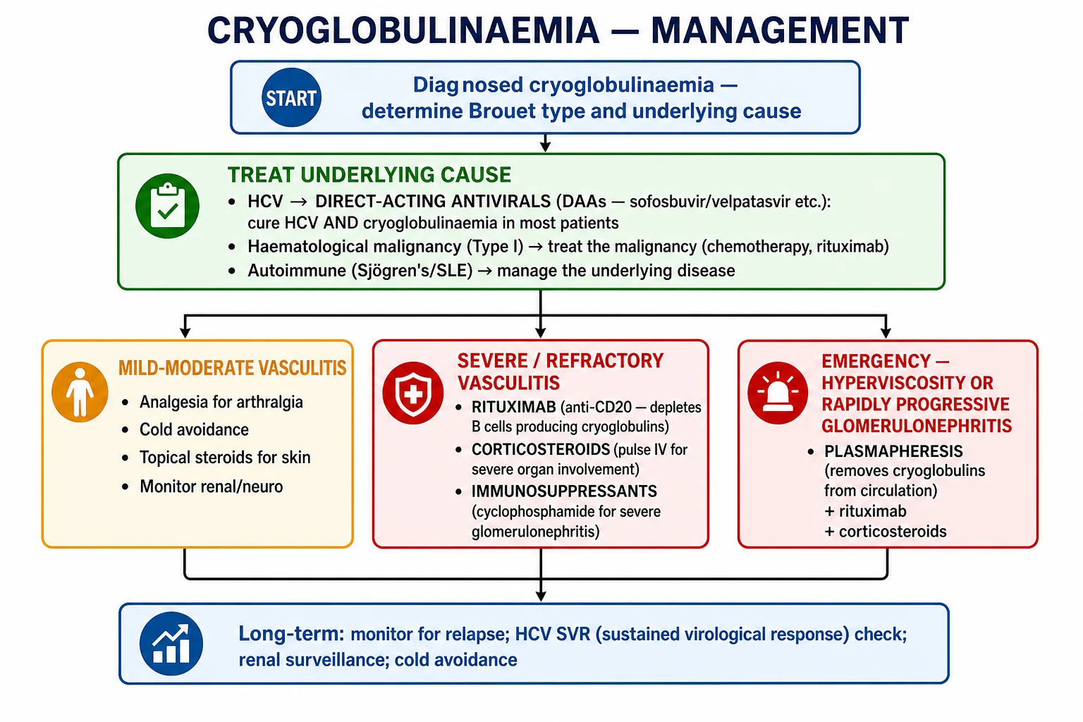
Stepwise management of mixed cryoglobulinaemia
Identify and treat the cause: HCV DAAs, HBV therapy, autoimmune disease therapy, or lymphoma/myeloma treatment.
Cold avoidance, elevation, analgesia and skin care for all patients.
Mild systemic disease: oral corticosteroids, colchicine or hydroxychloroquine depending on association.
Severe or refractory disease: rituximab + corticosteroids; add cyclophosphamide for severe renal disease.
Emergency: plasma exchange for hyperviscosity, rapidly progressive glomerulonephritis or life-threatening vasculitis.
Specific Subtypes and Scenarios
Type I cryoglobulinaemia
Type I is associated with a monoclonal gammopathy. The management priority is to treat the underlying lymphoproliferative disorder and reduce the paraprotein burden. Rituximab is often the first-line treatment for B-cell-associated Type I. Plasma exchange is used for symptomatic hyperviscosity. Because there is no vasculitis, corticosteroids and cyclophosphamide are less effective unless there is an associated lymphoproliferative disease that requires them.[1]
Type II cryoglobulinaemia (HCV-associated)
This is the most common scenario. Modern DAA therapy has transformed the prognosis. Most patients achieve SVR and remission of cryoglobulinaemia. However, some patients, particularly those with long-standing disease or established glomerulonephritis, have persistent cryoglobulins or organ damage despite HCV eradication. In these patients, rituximab is indicated. The VASCUVALDIC trial showed that sofosbuvir plus ribavirin for 24 weeks produced sustained virological response in most patients with HCV-associated cryoglobulinaemic vasculitis and was associated with clinical improvement in vasculitis. Modern interferon-free DAA regimens are better tolerated and have largely replaced interferon-based therapy. Real-world cohorts report that cryoglobulins disappear in a substantial proportion of patients after SVR, although some patients, particularly those with long-standing disease, have persistent low-level cryoglobulins or residual organ damage.[7]
Type II/III cryoglobulinaemia with autoimmune disease
Sjögren's syndrome is the most common autoimmune association. The renal involvement is typically MPGN-like. Management combines treatment of the underlying autoimmune disease with rituximab and/or corticosteroids. Hydroxychloroquine is often used for Sjögren's. Cyclophosphamide may be needed for severe lupus-associated disease.[10]
Essential (idiopathic) mixed cryoglobulinaemia
When no infection, autoimmune disease or malignancy is found, the disease is labelled essential. Rituximab is often the treatment of choice for symptomatic essential mixed cryoglobulinaemia. Relapses are common, and maintenance therapy may be needed. [1]
Cryoglobulinaemia with peripheral neuropathy
Peripheral neuropathy can be the dominant manifestation. It is often a sensory or sensorimotor polyneuropathy, or mononeuritis multiplex. Nerve conduction studies help define the pattern. Treatment of the underlying cause and rituximab are the mainstays; plasma exchange may be used for rapidly progressive neuropathy. [1]
Cryoglobulinaemic glomerulonephritis
Renal disease is a major complication and prognostic factor. The typical biopsy shows MPGN-like changes. Management includes treatment of the underlying cause, corticosteroids, rituximab, and cyclophosphamide for severe disease. Plasma exchange is considered for rapidly progressive disease. Long-term outcomes depend on the severity of renal damage at presentation and the response to therapy.[1]
Complications and Pitfalls
Disease complications
- Chronic kidney disease from cryoglobulinaemic glomerulonephritis.
- End-stage kidney disease requiring dialysis or transplantation in severe untreated renal disease.
- Permanent peripheral neuropathy with sensory loss, motor deficits and disability.
- Digital ulceration, gangrene and amputation in Type I or severe mixed disease.
- Gastrointestinal ischaemia, bleeding or perforation from mesenteric vasculitis.
- Stroke or central nervous system vasculitis (rare).
- Progressive liver disease, cirrhosis and hepatocellular carcinoma in HCV-associated disease.
- Bacterial infections of skin ulcers and immunosuppression-related infections. [1]
Diagnostic pitfalls
- False-negative cryoglobulin test due to improper sample handling (not kept at 37°C). This is the single most important pitfall.
- Misinterpreting the complement pattern as lupus nephritis when the low C4 is actually due to classical pathway activation by cryoglobulin immune complexes.
- Missing HCV infection because the patient has no risk factors or obvious liver disease.
- Assuming all palpable purpura is IgA vasculitis in children or ANCA vasculitis in adults, without considering cryoglobulinaemia.
- Overlooking Type I cryoglobulinaemia in a patient with a monoclonal gammopathy and digital ischaemia, because the clinician is looking for vasculitis. [1]
Management pitfalls
- Using rituximab alone in untreated HCV-associated disease without antiviral therapy. DAAs should be the first-line treatment; rituximab is reserved for refractory or severe disease.
- Using corticosteroids as definitive therapy for HCV-associated cryoglobulinaemia; steroids suppress symptoms but do not eradicate HCV.
- Delaying plasma exchange in hyperviscosity crisis or rapidly progressive glomerulonephritis.
- Failing to monitor renal function and urinary protein after starting treatment.
- Insufficient cold avoidance, leading to recurrent symptoms.
- Missing the underlying malignancy in Type I cryoglobulinaemia; a full haematological work-up is essential. [1]
Prognosis and Disposition
The prognosis of cryoglobulinaemia depends on the type, the underlying cause, the severity of organ involvement, and the response to treatment. [1]
HCV-associated mixed cryoglobulinaemia
Modern DAA therapy has transformed the outlook. Most patients achieve SVR, and the majority experience remission of cryoglobulinaemia and improvement in skin, joint and neurological symptoms. However, the response is not universal. Some patients have persistent cryoglobulins or residual organ damage despite HCV eradication. Factors associated with incomplete response include long disease duration, advanced renal disease, severe neuropathy, and established glomerulosclerosis. Patients with persistent symptoms after SVR should be considered for rituximab.[5][9]
Type I cryoglobulinaemia
Prognosis is determined by the underlying lymphoproliferative disorder. Effective treatment of the malignancy (for example, Waldenström's macroglobulinaemia or multiple myeloma) usually resolves the cryoglobulinaemia. Plasma exchange provides rapid relief of hyperviscosity symptoms. Long-term outcomes depend on the response to malignancy-directed therapy. [1]
Autoimmune-associated mixed cryoglobulinaemia
Prognosis is linked to control of the underlying autoimmune disease. Sjögren's-associated cryoglobulinaemia has a higher risk of lymphoma and should be monitored. Renal involvement is associated with a worse prognosis and requires long-term nephrology follow-up. [1]
Disposition and follow-up
Patients with mild disease can be managed as outpatients with treatment of the underlying cause and cold avoidance. Patients with renal involvement, severe neuropathy, gastrointestinal involvement, or hyperviscosity require inpatient management or rapid specialist referral. Follow-up should include regular monitoring of renal function, urinalysis, complement levels, cryoglobulin levels, RF, and neurological status. Even after successful treatment of HCV, patients should be monitored for relapse, particularly if cryoglobulins persist.[1]
Prognostic factors
Special Populations
Pregnancy
Cryoglobulinaemia is uncommon in pregnancy, but management requires special consideration. Type I hyperviscosity can worsen because of increased plasma volume and haemodilution. Plasma exchange is generally safe in pregnancy if hyperviscosity is symptomatic. Mixed cryoglobulinaemia may flare because of physiological changes in immune function. Corticosteroids and rituximab require careful risk-benefit discussion; rituximab crosses the placenta in the second and third trimesters and can cause neonatal B-cell depletion. Cyclophosphamide is contraindicated in pregnancy. DAAs are generally avoided in pregnancy unless clearly indicated. Cold avoidance and supportive care remain important. [1]
Elderly patients
Elderly patients are more likely to have Type I cryoglobulinaemia associated with MGUS or lymphoproliferative disease. They may present atypically with digital ischaemia or falls due to neuropathy. Polypharmacy and renal impairment increase the risk of drug toxicity. Corticosteroids should be used cautiously because of infection, diabetes, osteoporosis and cognitive effects. The threshold for malignancy work-up should be low. [1]
Immunocompromised patients
Patients with HIV, transplant recipients, or those on immunosuppressive therapy may have muted inflammatory responses, making the diagnosis more difficult. Opportunistic infections can mimic vasculitis. Immunosuppression increases the risk of complications from corticosteroids and rituximab. HBV reactivation is a risk with rituximab, so prophylactic antiviral therapy is required if HBsAg or anti-HBc is positive. [1]
Anticoagulated patients
Anticoagulation does not cause cryoglobulinaemia, but it can worsen purpura and bleeding from vasculitic lesions. The decision to continue or stop anticoagulation should be individualised based on the indication for anticoagulation and the severity of bleeding. [1]
Paediatric patients
Cryoglobulinaemia is rare in children. When it occurs, it is usually associated with infection or autoimmune disease. HCV-associated cryoglobulinaemia is less common in children in endemic regions, although vertical transmission occurs. Management principles are similar to adults, with dose adjustments for weight. [1]
In regions with high HCV prevalence and limited access to DAA therapy, mixed cryoglobulinaemia remains a major cause of immune-complex glomerulonephritis and systemic vasculitis. In high-income countries with HCV elimination programmes, the proportion of cryoglobulinaemia attributable to autoimmune disease is rising. Access to rituximab and plasma exchange also varies by region, affecting the management of severe disease.
Evidence, Guidelines and Regional Differences
The evidence base for cryoglobulinaemia has changed dramatically with the advent of DAA therapy. Before DAAs, interferon-alpha plus ribavirin was the standard HCV treatment, but response rates were lower and relapse was common after stopping therapy. The modern DAA era has produced high SVR rates and durable remission of cryoglobulinaemia in most patients. [1]
Landmark studies
- VASCUVALDIC study (Saadoun et al., 2016): a randomised controlled trial of sofosbuvir plus ribavirin for HCV-associated cryoglobulinaemic vasculitis. The study demonstrated high rates of virological response and clinical improvement in vasculitis. This trial helped establish the primacy of antiviral therapy in HCV-associated disease.[7]
- Real-world DAA studies (Passerini et al., 2018): observational data showing that modern DAA regimens are safe and effective in HCV-cryoglobulinaemia, with disappearance of cryoglobulins in a majority of patients after SVR.[8]
- Dissociated responses (Pozzato et al., 2020): a prospective study showing that while DAAs achieve virological response in most patients, some have persistent haematological or immunological abnormalities (cryoglobulins, RF positivity) after SVR. This supports the use of rituximab in refractory cases.[9]
- GISC rituximab consensus (Quartuccio et al., 2023): evidence and consensus-based recommendations from the Italian Study Group of Cryoglobulinemia for the use of rituximab in mixed cryoglobulinaemia. The document supports rituximab as first-line immunosuppressive therapy for severe or refractory mixed cryoglobulinaemia, particularly in non-HCV or HCV-persistent disease.[6]
Guidelines
There is no single international guideline dedicated solely to cryoglobulinaemia. Management is guided by the underlying disease guidelines: EASL/AASLD guidelines for HCV, ACR/EULAR recommendations for Sjögren's and SLE, and haematology guidelines for lymphoproliferative disorders. The Italian GISC consensus provides the most specific guidance for rituximab use in mixed cryoglobulinaemia.[6]
Regional differences
- HCV prevalence: regions with high HCV prevalence (parts of Eastern Europe, North Africa, South Asia) still see a large burden of HCV-associated cryoglobulinaemia. In contrast, countries with universal HCV treatment access are seeing fewer new cases.
- DAA access: DAA availability and cost have historically varied, though generic production has improved access in many low- and middle-income countries.
- Rituximab and plasma exchange access: advanced immunosuppressive therapy and apheresis may be limited in resource-constrained settings, where corticosteroids and antiviral therapy are the mainstay.
- Autoimmune disease burden: in regions with high Sjögren's or SLE prevalence, autoimmune-associated cryoglobulinaemia may dominate. [1]
Try this: what is the first-line treatment for HCV-associated mixed cryoglobulinaemia?
Direct-acting antivirals (DAAs). HCV eradication with modern DAA regimens cures the infection in more than 95% of patients and usually leads to remission of the cryoglobulinaemia. Rituximab is reserved for severe, refractory or non-HCV disease.
Exam Pearls and High-Yield Minutiae
[1]Exam application bank (NEET-PG / INICET)
One-line answer
Cryoglobulinaemia is the presence of circulating immunoglobulins that reversibly precipitate below 37°C and dissolve on rewarming. Classified by the Brouet system: Type I (monoclonal IgM/IgG; hyperviscosity/thrombosis; MGUS, myeloma, Waldenström's), Type II (mixed monoclonal IgM with rheumatoid factor activity + polyclonal IgG; immune complex vasculitis; hepatitis C in ~80%), and Type III (polyclonal IgG + IgM; immune complex vasculitis; connective tissue disease, infection). Mixed types (II/III) present with Meltzer's triad (purpura + arthralgia + weakness) and cause leukocytoclastic vasculitis, peripheral neuropathy, and MPGN-like glomerulonephritis. The complement pattern of low C4 with normal or low C3 is the classic laboratory hallmark. Management centres on treating the underlying cause: direct-acting antivirals (DAAs) cure HCV and cryoglobulinaemia in most patients; rituximab for
Worked stems (answer without another resource)
Stem 1 — Classic presentation. Map symptoms to mechanism; name the first investigation and first treatment step with dose/route if drug therapy is standard. [1]
Stem 2 — Unstable / complicated. List red flags that force immediate resuscitation, theatre, ICU, antidote, or reperfusion — and what you do in the first 15 minutes. [1]
Stem 3 — Atypical group. Elderly, pregnancy, child, or immunocompromised: how presentation and thresholds change. [1]
Stem 4 — Differential trap. Name the three closest mimics and one discriminator for each. [1]
Stem 5 — Disposition. Who goes home with safety-netting, who is admitted, who needs HDU/ICU/theatre, and what follow-up is mandatory. [1]
Rapid viva checklist
- Definition + classification
- Pathophysiology chain
- Bedside signs / criteria
- Score with exact components (if any)
- Emergency bundle
- Definitive therapy with doses
- Complications of disease and of treatment
- Special populations
- Guideline/trial name if classic
- Three exam traps
Coverage self-check
If you cannot answer any stem above from this page alone, re-read the matching section — the page is intended to be self-sufficient for final-prof and NEET-PG/INICET questions on Cryoglobulinaemia.
[1]References
- [1]Roccatello D, Saadoun D, Ramos-Casals M, et al. Cryoglobulinaemia Nat Rev Dis Primers, 2018.PMID 30072738
- [2]Retamozo S, Quartuccio L, Ramos-Casals M. Cryoglobulinemia Med Clin (Barc), 2022.PMID 35216803
- [3]Ferri C, Zignego AL, Pileri SA. Cryoglobulins J Clin Pathol, 2002.PMID 11825916
- [4]Roccatello D, Saadoun D, Ramos-Casals M, et al. Cryoglobulinaemia Nat Rev Dis Primers, 2018.PMID 30072792
- [5]Desbois AC, Cacoub P, Saadoun D. Cryoglobulinemia: An update in 2019 Joint Bone Spine, 2019.PMID 30731128
- [6]Quartuccio L, Baldini C, Bartoloni E, et al. Management of mixed cryoglobulinemia with rituximab: evidence and consensus-based recommendations from the Italian Study Group of Cryoglobulinemia (GISC) Clin Rheumatol, 2023.PMID 36169798
- [7]Saadoun D, Thibault V, Si Ahmed SN, et al. Sofosbuvir plus ribavirin for hepatitis C virus-associated cryoglobulinaemia vasculitis: VASCUVALDIC study Ann Rheum Dis, 2016.PMID 26567178
- [8]Passerini M, Merli M, La Mura V, et al. Are direct-acting antivirals safe and effective in hepatitis C virus-cryoglobulinemia? virological, immunological, and clinical data from a real-life experience Eur J Gastroenterol Hepatol, 2018.PMID 30138160
- [9]Pozzato G, Mazzaro C, Gerosa M, et al. Direct-acting antiviral agents for hepatitis C virus-mixed cryoglobulinaemia: dissociated virological and haematological responses Br J Haematol, 2020.PMID 32790920
- [10]François H, Mariette X. Renal involvement in primary Sjögren syndrome Nat Rev Nephrol, 2016.PMID 26568188