Dermatology · Medicine
Cryotherapy
Also known as Cryotherapy · Cryosurgery · Liquid nitrogen therapy · Cryopeeling
Cryotherapy (cryosurgery) uses liquid nitrogen at -196 C to destroy cutaneous lesions by controlled freezing through repeated freeze-thaw cycles. Cell death results from four mechanisms — extracellular ice formation (cellular dehydration), intracellular ice formation (membrane disruption), vascular stasis (thrombosis and ischaemia), and apoptosis — and the lethal target temperature is approximately -20 to -30 C. The ice ball must extend a 2-3 mm halo beyond the visible lesion margin; 1-2 freeze-thaw cycles per session for benign lesions and two cycles for premalignant/malignant lesions. The commonest indications are viral warts, actinic keratosis, molluscum contagiosum, seborrhoeic keratosis, solar lentigo, Bowen disease, and carefully selected superficial BCC. Expected sequence: pain/erythema at freezing, blister at 24-48 h, crust at 7-14 days, then a healed macule. The most important long-term complication is permanent hypopigmentation (melanocytes are more cold-sensitive than keratinocytes), worst in skin of colour (Fitzpatrick IV-VI). NEVER use cryotherapy for suspected melanoma or any lesion requiring histology.
On this page & tools
Your progress
Saved locally on this device.
Exam tags
Red flags
Overview and Definition
Cryotherapy, also called cryosurgery, is the controlled, localised destruction of cutaneous tissue by freezing with liquid nitrogen, the coldest practical cryogen available (boiling point -196 C). The technique exploits the fact that living cells tolerate only a narrow temperature range: when tissue is cooled rapidly to the lethal isotherm of approximately -20 to -30 C, a predictable cascade of physical and biological injuries kills the target cells while the surrounding tissue is relatively spared. The destroyed tissue is left in situ to slough over the following days to weeks, healing by secondary intention beneath a crust.[1]
Although liquid nitrogen is by far the dominant agent in modern dermatology, it is worth knowing the alternatives. Nitrous oxide (boiling point -89 C) and carbon dioxide snow (-78.5 C) are warmer cryogens that were historically used for "cold cautery" and are still employed in some dental and minor-procedure settings, but they cannot achieve the destructive depth or the rapid cooling rate of liquid nitrogen and are unsuitable for treating malignancy. Argon-helium systems are used experimentally in solid-organ cryoablation but are not part of cutaneous practice. The clinical relevance is that when an examiner or a guideline says "cryotherapy" in a dermatological context, they mean liquid nitrogen delivered by open spray, cotton-tipped applicator, or contact probe, and the dosimetry quoted below refers specifically to that agent.[1]
The procedure is one of the commonest destructive interventions in office dermatology worldwide because it is quick (a treatment session lasts seconds to a couple of minutes), requires no anaesthetic in most cases, leaves no suture marks, is portable, and is inexpensive. Its principal limitations are equally characteristic: it destroys tissue rather than excising it, so no specimen is available for histology; it produces predictable but unavoidable hypopigmentation that is permanent and disfiguring in darker phototypes; and it carries a non-trivial recurrence rate for warts and a measurable one even for superficial basal cell carcinoma. A fellowship-level candidate must hold both faces of the technique in mind — its place as a first-line destructive modality, and the disciplined restraint required never to deploy it where a diagnosis is uncertain or where histology is essential.[1]
Classification (Delivery Techniques)
There are three principal ways to deliver liquid nitrogen to a lesion, and the choice is dictated by lesion size, thickness, site, patient tolerance (especially in children), and the precision required. The three techniques differ in how well they control the lateral spread of cold and the depth of the ice ball. [1]
Open spray
- Most common office technique
- LN2 sprayed from a canister or unit 1-2 cm from lesion
- Best for warts, AK, multiple discrete lesions
- Rapid cooling, deep ice ball
- Conical/focused, spray-diameter cones for precision
- Risk of over-freezing if uncontrolled
Cotton-tipped applicator
- Cotton swab dipped in LN2, touched onto lesion
- Gentle, well tolerated in children
- Best for molluscum contagiosum and small lesions
- Limited depth and precision
- Lower risk of lateral over-freeze
- Cotton may stick to frozen tissue
Forceps / copper or bronze disc
- Cold forceps grasp pedunculated lesions (e.g. skin tags, filiform warts)
- Copper/bronze disc dipped in LN2 for contact therapy
- Highly controlled, limited lateral spread
- Best for pedunculated or precisely-defined lesions
- Less destructive than open spray
- Requires repeated dipping between lesions
Cryopeeling is a fourth, field-based variant in which a fine LN2 mist is sprayed over a whole cosmetic unit (typically the face) to treat widespread actinic damage or, in selected centres, inflammatory acne. It is performed by experienced operators and is rarely examined in detail at MBBS level beyond recognising the name.[1]
Epidemiology and Risk Factors
Cryotherapy is a treatment rather than a disease, so its "epidemiology" is really a description of which patients present for it and which patients are at risk from it. The procedure is performed in primary care, dermatology out-patient clinics, and minor-operations lists across nearly every health system. The commonest indication worldwide is viral warts, particularly common hand and foot warts in children, adolescents, and young adults — populations in whom human papillomavirus infection of the skin is most prevalent. Actinic keratosis drives large volumes of cryotherapy in older fair-skinned adults with cumulative sun exposure, especially outdoor workers and those with skin types I-II. Molluscum contagiosum and seborrhoeic keratosis generate a steady stream of paediatric and elderly presentations respectively.[6]
The "risk" axis matters as much as the indication axis. Four patient groups warrant particular caution before a cryogun is picked up. Patients with darker phototypes (Fitzpatrick IV-VI) carry a high baseline risk of permanent hypopigmentation, which can be cosmetically disabling on the face and neck. Patients with cold-intolerance disorders — Raynaud phenomenon, cryoglobulinaemia, cryofibrinogenaemia, cold urticaria, and cold haemoglobinuria — can develop serious systemic or ischaemic consequences. Patients on anticoagulants or with thrombocytopenia bleed and blister more readily. Immunosuppressed patients heal poorly, develop secondary infection more often, and frequently have refractory or recurrent lesions that cryotherapy alone cannot control.[3]
Pathophysiology — The Four Mechanisms of Cell Death
The destructive effect of liquid nitrogen on tissue is not a single process but the convergence of four mechanisms that act on different time scales and across different temperature gradients. A candidate who can articulate the cascade — and explain why each step matters — answers most of what an examiner probes at viva. [1]
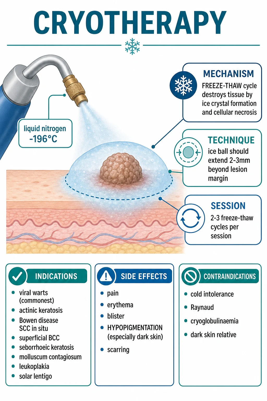
Mechanism 1 — Extracellular ice and the solute-effect. As tissue cools below freezing, ice forms first in the extracellular space because the extracellular fluid is dilute relative to the cytoplasm. The ice excludes solutes, so the remaining unfrozen extracellular fluid becomes progressively hyperosmolar. Water is drawn out of the cells along the osmotic gradient, producing severe cellular dehydration, denaturation of proteins, and a toxic intracellular concentration of salts (the solute-effect). The cells shrink, their membranes are stressed, and many die of this biochemical injury alone before any ice ever forms inside them.[1]
Mechanism 2 — Intracellular ice and membrane disruption. If cooling is rapid enough, the intracellular fluid supercools and ice crystals nucleate inside the cell. Intracellular ice is the single most lethal mechanism in cryosurgery: it physically disrupts the plasma membrane and the membranes of organelles, ruptures the cytoskeleton, and produces irreversible structural damage. This is why the rate of cooling matters and why the open-spray technique (which cools rapidly) is more destructive than the slower cotton-applicator method for the same apparent freeze time.[1]
Mechanism 3 — Vascular stasis and ischaemia. Cold injures the microvasculature of the dermis. Endothelial cells are damaged, capillaries become leaky and then thrombose, and the target tissue is secondarily infarcted over the hours following treatment. This vascular mechanism extends the zone of destruction beyond the cells that were directly frozen and is particularly important for thicker or vascular lesions such as pyogenic granuloma.[1]
Mechanism 4 — Apoptosis. At the periphery of the ice ball, where temperatures are sub-lethal but still cold, cells are not killed outright. Instead they undergo programmed cell death over the following hours, extending the margin of destruction. Apoptosis explains why the histological zone of necrosis is larger than the visible ice ball suggested at the time of treatment.[1]
The thaw phase is as important as the freeze phase, a fact examiners reward. During a slow thaw, the small ice crystals that formed rapidly coalesce and grow into larger crystals (recrystallisation), magnifying membrane injury and allowing the solute-effect to persist longer. A slow, passive thaw is therefore deliberately sought — the operator waits for the tissue to return fully to body temperature before re-freezing, and never applies heat or pressure to accelerate thawing. Each freeze-thaw cycle widens the zone of complete necrosis: a second cycle ensures that cells that survived the first (particularly at the deep margin) are killed, which is why malignant and premalignant lesions always receive two cycles.[2]
A final mechanistic point that explains the signature complication: melanocytes are markedly more cold-sensitive than keratinocytes. Melanocytes are destroyed at warmer temperatures than keratinocytes, so any freeze intense enough to treat a lesion is also intense enough to ablate the melanocyte population at the site. The result is permanent hypopigmentation — the single most important long-term adverse effect of cryotherapy and the dominant consideration when treating skin of colour.[9]
Clinical Presentation — Lesions Selected for Cryotherapy
The "presentation" of a cryotherapy topic is really the presentation of the lesions it treats. Candidates must be able to recognise the discrete, well-demarcated, predominantly superficial lesions for which cryotherapy is appropriate, and to distinguish them from lesions that demand excision and histology. [1]
Viral warts (common, plantar, plane, filiform, periungual) are firm, hyperkeratotic papules or plaques with disruption of the normal skin lines and, often, thrombosed capillary loops visible as black dots. They present in children and young adults, on the hands, feet, and periungually, and are frequently painful when on the sole. Actinic keratoses are rough, gritty, erythematous or skin-coloured patches on chronically sun-exposed skin of older fair-skinned adults; they are characteristically "felt better than seen". Molluscum contagiosum presents as small, firm, umbilicated pearly papules in children, often in clusters and often with surrounding eczematous "molluscum dermatitis". Seborrhoeic keratoses are the "stuck-on" waxy tan-brown plaques of older adults. Solar lentigines are flat brown macules on sun-exposed skin. Bowen disease (squamous cell carcinoma in situ) presents as a persistent, slowly enlarging, scaly erythematous patch or plaque. Superficial basal cell carcinoma presents as a slow-growing, often subtly scaly, faintly rolled erythematous patch on the trunk or limbs.[6]
The patient history before cryotherapy must capture more than the lesion itself. The operator should ask specifically about cold intolerance (Raynaud, cold urticaria, cryoglobulinaemia), document the phototype, ask about anticoagulants and bleeding disorders, pregnancy, previous keloid or hypertrophic scarring, and any implanted electronic device if spray is to be used close to a pacemaker. These are not bureaucratic boxes: each one changes whether cryotherapy is the right choice and how aggressively it can be delivered.[3]
Differential Diagnosis — When Cryotherapy Is and Is Not the Right Tool
Because cryotherapy destroys rather than excises, the decision to use it is in large part a decision about what does not need histology. The table below frames the decision the way an examiner frames it: which lesions are first-line for cryotherapy, which are acceptable only in carefully selected circumstances, and which must never be frozen. [1]
| Category | Lesions | Rationale |
|---|---|---|
| Cryotherapy FIRST-LINE | Viral warts, actinic keratosis, molluscum contagiosum, seborrhoeic keratosis, solar lentigo, small Bowen disease | Benign or premalignant; clinical diagnosis secure; destructive treatment adequate; no histology required |
| Cryotherapy ACCEPTABLE (selected) | Low-risk superficial BCC (trunk/limb, under 1 cm); hypertrophic lichen planus; prurigo nodularis; keloids (with intralesional steroid); cutaneous leishmaniasis (selected regions) | Surgery not suitable or patient preference; lower-risk lesion; counseled on follow-up; cryotherapy as one option among several |
| Excision PREFERRED (not cryotherapy) | Nodular or aggressive BCC; invasive SCC; lesions over 2 cm; lesions on the face (H-zone) where recurrence matters | Histology of margins essential; deep extension; high-stakes site; cryotherapy cannot guarantee clearance |
| Cryotherapy NEVER | Suspected melanoma; any undiagnosed pigmented lesion; lesions where histology is needed | Cryotherapy forfeits the specimen and delays diagnosis of melanoma, which is potentially fatal |
The single most dangerous pitfall in cryotherapy is the casual freezing of a pigmented lesion that turns out to be melanoma. The discipline that prevents it is simple: if there is any diagnostic doubt, dermoscopy now and biopsy before any destructive treatment. A "wart" or "seborrhoeic keratosis" that is irregular, changing, darkly and variably pigmented, or bleeding is melanoma until proven otherwise, and melanoma is never a cryotherapy indication.[1]
Clinical and Bedside Assessment
The pre-treatment assessment is brief but decisive. The operator confirms the diagnosis clinically and, where appropriate, dermoscopically; measures the lesion and notes its thickness; identifies the site and its proximity to sensitive structures (eye, nail unit, nerve, cartilage); records the phototype; and screens for cold-intolerance. Dermoscopy is mandatory for any pigmented lesion before destructive treatment — the cost of dermoscopy is seconds; the cost of destroying a melanoma is a life. A baseline clinical photograph is good practice, especially for BCC and Bowen disease, to document the pre-treatment appearance and to support follow-up.[1]
Response is assessed session to session. For warts, the lesion should blacken and reduce in thickness and area over successive treatments; for actinic keratosis, the rough scale resolves and a faint erythematous or hypopigmented macule remains; for superficial BCC, the treated area should heal with atrophy and hypopigmentation and is reviewed at three to six months with a low threshold for biopsy if any residual lesion persists. Patients are given a written safety-net: the expected blister, the timeframe for crusting, and the signs that should trigger reattendance (spreading erythema, increasing pain, pus, recurrence, darkening).[3]
Investigations
For most indications no investigation is required — cryotherapy is built on a confident clinical diagnosis. The only pre-treatment test the operator must consider is whether a biopsy is needed first, and the answer is yes whenever the diagnosis is in doubt, whenever the lesion is pigmented, whenever a malignant or premalignant diagnosis (BCC, Bowen) has not been histologically confirmed, and whenever a treated lesion recurs. In selected patients a cold-intolerance workup is relevant: cryoglobulins, cold agglutinin titre, full blood count, and complement when cryoglobulinaemia or cold agglutinin disease is suspected. For superficial BCC treated by cryotherapy, baseline dermoscopy and clinical photography, with a planned review at three to six months and a low threshold for biopsy, are the minimum standard of follow-up.[8]
Good documentation — the lesion, site, freeze time per cycle, number of cycles, and the size of the ice-ball halo — protects both patient and clinician. This is especially important for BCC and Bowen disease, where the follow-up decision depends on knowing exactly what was treated and how aggressively.[8]
Management — The Procedural Technique (Immediate)
The standard open-spray technique proceeds in defined steps. The lesion and surrounding skin are cleaned. The spray nozzle is positioned approximately 1 to 2 cm from the lesion and the liquid nitrogen is released in a continuous or pulsed spray, painting the freeze across the lesion until a uniform ice ball extends 2 to 3 mm beyond the visible margin. The spray is stopped and the tissue is allowed to thaw completely, returning to body temperature and to normal colour. This constitutes one freeze-thaw cycle. For benign lesions one or two cycles suffice; for premalignant and malignant lesions two cycles are mandatory. Sessions are repeated at intervals of 2 to 4 weeks, most commonly every three to four weeks for warts.[2]
The cotton-tipped applicator technique uses a cotton swab dipped in liquid nitrogen and touched onto the lesion. It is gentler, well tolerated in children, and suited to molluscum contagiosum and small facial lesions, but it delivers a shallower and less precisely controlled freeze than the spray. Care must be taken that the frozen cotton does not adhere to the skin and tear it on withdrawal. The forceps or copper-disc technique applies cold through a metal contact: a cold pair of forceps can grasp a pedunculated skin tag or filiform wart, while a copper or bronze disc dipped in liquid nitrogen delivers contact therapy with limited lateral spread and excellent control. These contact methods are less destructive than the open spray and are reserved for the lesions they suit.[2]
Analgesia is usually unnecessary for short benign freezes. For sensitive sites (digits, genitalia, periungual) or for the long freezes required for superficial BCC, a topical anaesthetic under occlusion or an injected local anaesthetic improves tolerance. Children may benefit from distraction and, where available, vapocoolant spray. The operator and any assistant must wear eye protection and avoid spraying towards the face; the patient should be warned of the brief sharp stinging and the throbbing that follows.[3]
Equipment and operator safety
Liquid nitrogen is stored in a vacuum-insulated dewar and delivered through a hand-held spray unit with a trigger and a selection of interchangeable nozzles or aperture cones (a narrow cone concentrates the spray for small lesions, a wide cone for field treatment). The unit must be kept upright, in a ventilated room (liquid nitrogen expands almost 700-fold as it vaporises and can displace oxygen in a confined space), and away from the eyes. Operator safety measures are non-negotiable: eye protection for the operator and any assistant, because splashing liquid nitrogen or ricocheting spray can freeze the cornea; gloves when handling the dewar; and avoidance of spraying towards the patient's face. The patient is positioned comfortably — seated or supine depending on the site — and the surrounding skin is protected with a cone or a petrolatum barrier when treating close to the eye or other sensitive structures. Cotton wool and a discard bin should be to hand, and the trigger should always be released and the unit vented before refilling or storing.[2][3]
The dosimetry of a freeze is judged by two visible endpoints: the size of the ice ball (which must extend the required halo beyond the lesion) and the freeze time (timed from the moment the ice ball reaches the lesion edge, not from the moment the trigger is pressed). With practice the operator reads the ice ball by its colour and contour: a uniform, glistening white disc with a sharp margin indicates adequate freeze, while a mottled or incomplete freeze under-treats the lesion. For malignant lesions, where depth matters, some operators also use a thermocouple needle placed beneath the lesion to confirm that the tissue has reached the lethal isotherm; this is routine in specialist cryosurgery units but uncommon in primary care.[1]
Management — Definitive and Stepwise Ladder by Indication
The freeze time and the number of cycles are not arbitrary: they are calibrated to the lesion's thickness, biological behaviour, and risk of recurrence. The table consolidates the protocol by indication; the prose that follows expands the ladder for the commonest indications. [1]
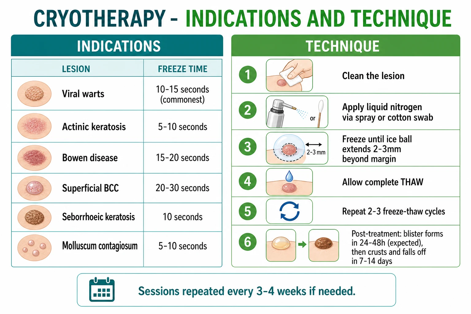
| Indication | Freeze time per cycle | Cycles | Halo | Session interval | Notes |
|---|---|---|---|---|---|
| Viral warts (commonest) | 10-15 s | 2 | 2-3 mm | q3-4 wk | Pare hyperkeratosis first; recurrence 20-30% |
| Actinic keratosis | 5-8 s | 1 | 2 mm | as needed | Lesion-directed; field therapy for multiple AK |
| Bowen disease (small) | 15-20 s | 2 | 3-5 mm | review | Excision/PDT/5-FU for larger lesions |
| Superficial BCC (selected) | 30 s | 2 | 5 mm | review 3-6 mo | Low-risk trunk/limb only; recurrence 5-15% |
| Molluscum contagiosum | 5-10 s | 1 | 1-2 mm | as needed | Brief; children; may leave hypopigmentation |
| Seborrhoeic keratosis | 5-10 s | 1-2 | 2 mm | as needed | Cosmetic; consider curettage for histology |
| Solar lentigo | 5 s | 1 | 1-2 mm | as needed | Cosmetic; sun-protection to prevent recurrence |
The wart ladder
The wart treatment ladder begins with patient-applied salicylic acid paint or plaster (typically 12-26% salicylic acid, applied daily after soaking and filing for up to 12 weeks), which is first-line for most common and plantar warts and is at least as effective as cryotherapy in head-to-head trials. Cryotherapy is the standard office-based option, applied every three to four weeks for up to three or four sessions. For resistant or thick plantar warts, paring the callus before freezing and combining with salicylic acid between sessions improves clearance. Needling (repeated puncture of a plantar wart under local anaesthetic to provoke an immune response) is an option for refractory lesions. Caustics (cantharidin), podophyllotoxin (for genital warts), bleomycin intralesional, and pulsed-dye or CO2 laser are reserved for recalcitrant cases. Referral is indicated for extensive warts, warts in immunosuppression, and warts resistant to several months of treatment. The recent VRAIE multicentre randomised trial (Chanal and colleagues, 2025) compared salicylic acid, liquid nitrogen, 5-fluorouracil and imiquimod in previously-treated plantar warts and found no single agent clearly superior, reinforcing that plantar warts are stubborn and that a stepped, patient-tailored approach is reasonable.[6][7]
The actinic keratosis ladder
For discrete actinic keratoses, lesion-directed cryotherapy is first-line: a single 5-8 second freeze with a 2 mm halo is usually adequate, and lesions are reviewed and re-treated if they persist. For multiple lesions or field cancerisation, lesion-by-lesion cryotherapy is inefficient and field-directed therapy is preferred — topical 5-fluorouracil, imiquimod, photodynamic therapy, or chemical peels — with cryotherapy reserved for the few thick or hyperkeratotic lesions that do not respond to field treatment. The systematic review by Worley and colleagues (2023) confirms that cryotherapy, field therapies, and photodynamic therapy all have comparable moderate efficacy for individual AK lesions, and that field therapy is favoured when lesion count is high.[4][5]
The superficial BCC ladder
Excision is the first-line treatment for BCC of any subtype, with histological margin control. Cryotherapy has a defined but narrow role: it is acceptable for carefully selected, low-risk, superficial BCC on the trunk or limbs, typically under 1 cm, where the patient is unsuitable for or declines surgery. The protocol is aggressive — 30 seconds per cycle, two cycles, with a 5 mm halo — and follow-up at three to six months with a low threshold for biopsy is mandatory. European consensus guidelines (Peris and colleagues, 2019) place cryotherapy as one of several nonsurgical options for low-risk superficial BCC alongside imiquimod, 5-FU, and photodynamic therapy, but emphasise that surgery is first-line and that cryotherapy is not appropriate for nodular or aggressive BCC.[8]
Escalation triggers across all indications are: failure to clear after three to four cryotherapy sessions, recurrence after apparent clearance, any clinical suspicion of invasion (induration, rapid growth, ulceration in a treated BCC/Bowen), or any change in a treated lesion that raises the possibility of misdiagnosis. The response is always biopsy and excision with histology, not more cryotherapy.[1]
Specific Subtypes and Scenarios
Plantar warts deserve special attention because they are painful, deeply set, and refractory. The hyperkeratotic callus should be pared before freezing so that the lethal isotherm reaches the deeper wart tissue; the freeze is held longer than for a common wart, and combination with salicylic acid between sessions improves outcomes. Plantar warts have the highest recurrence rate of any wart site.[6]
Periungual warts are technically difficult because of the nail apparatus. Over-aggressive freezing risks permanent nail dystrophy, and cryotherapy is relatively contraindicated in patients with Raynaud phenomenon or digital ischaemia because of the risk of digit-threatening vasospasm. Where treatment is necessary, careful, limited freezing with protection of the nail fold is required, and alternatives (topical, intralesional bleomycin, laser) should be considered.[3]
Genital warts are generally not treated with liquid nitrogen spray in primary care; first-line is topical podophyllotoxin or imiquimod, with cryotherapy performed by a specialist where indicated because of the sensitivity of the tissue and the risk of scarring in a sexually significant site. Molluscum contagiosum in children is treated with a brief 5-10 second freeze; it is quick and effective but the operator must counsel parents about possible hypopigmentation, and given that molluscum is self-limiting, watchful waiting is often the better choice for uncomplicated cases.[10]
Superficial BCC managed by cryotherapy is discussed above. Keloids and hypertrophic scars can be treated with a light freeze combined with intralesional corticosteroid (triamcinolone acetonide), but recurrence is common. Cutaneous leishmaniasis is treated with cryotherapy in some endemic regions on the rationale that freezing kills the parasite; the evidence base is limited and lesion selection matters.[1]
Complications and Pitfalls
The complications of cryotherapy fall into immediate, early, and late groups, and a candidate should be able to enumerate each group with the timeframe. [1]
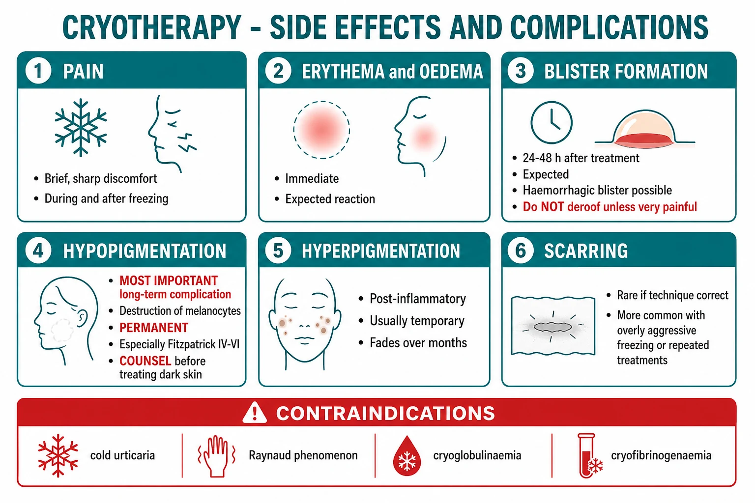
Immediate complications include the expected pain (brief, sharp, stinging during freezing, throbbing for minutes after thaw), erythema and oedema at the treatment site, occasional headache from the cold, and periauricular tinnitus from nitrogen gas venting through the Eustachian tube when treating lesions near the ear. Early complications within 48 hours include blister formation, which is expected and indeed indicates that the freeze reached adequate depth — the blister may be haemorrhagic and should not be deroofed unless very painful, in which case it is aspirated with a sterile needle leaving the roof intact as a biological dressing. Secondary infection and bleeding are the other early events, the latter more common in anticoagulated patients.[3]
Late complications are dominated by hypopigmentation, which is the most important long-term complication of cryotherapy and is permanent. Because melanocytes are destroyed at warmer temperatures than keratinocytes, any freeze intense enough to treat the lesion also ablates the melanocyte population at the site. The result is a persistently lighter macule that is cosmetically obvious and particularly disfiguring on the face and neck of patients with skin of colour (Fitzpatrick IV-VI). Hyperpigmentation is post-inflammatory and usually transient, fading over months. Scarring and atrophy occur with overly aggressive freezing or repeated treatments at the same site. Nail dystrophy follows over-aggressive treatment of periungual warts. Nerve damage and digital ischaemia are rare but serious risks at peripheral sites, particularly in patients with Raynaud phenomenon. Periocular injury (ectropion, corneal exposure, eye injury) is a risk when treating lesions near the eye.[9]
Recurrence rates are part of the complication picture: viral warts recur in 20-30% of cases after a course of cryotherapy, and superficial BCC recurs in 5-15% of adequately treated low-risk lesions — hence the mandatory follow-up for BCC.[8]
The classic pitfalls are three. First, treating a melanoma or undiagnosed pigmented lesion with cryotherapy, which forfeits the specimen and delays the diagnosis of a potentially fatal disease. Second, deploying cryotherapy on dark skin without explicit counselling about permanent hypopigmentation. Third, undertreating a malignant lesion (a single short freeze) and falsely reassuring the patient that it has been "dealt with", when in fact the deep margin has not been killed and the lesion will recur, possibly with invasion. Each pitfall is preventable by the disciplined sequence: confirm diagnosis, select the right indication, dose the freeze correctly, and follow up appropriately.[1]
Contraindications
Absolute contraindications are few but absolute: any lesion for which histology is required (suspected melanoma, undiagnosed pigmented lesion, lesion of uncertain nature), and any patient with a serious cold-intolerance disorder in whom systemic consequences are possible. Relative contraindications include darker phototypes (Fitzpatrick IV-VI), where the risk-benefit balance must be explicitly discussed; peripheral and digital sites in patients with Raynaud phenomenon or compromised circulation; thin, atrophic skin in the elderly; immunosuppression (poor healing, infection, refractory lesions); and lesions over cartilage (ear, nose) where perichondritis is a risk. Pregnancy is not a contraindication because there is no systemic absorption of significance.[3]
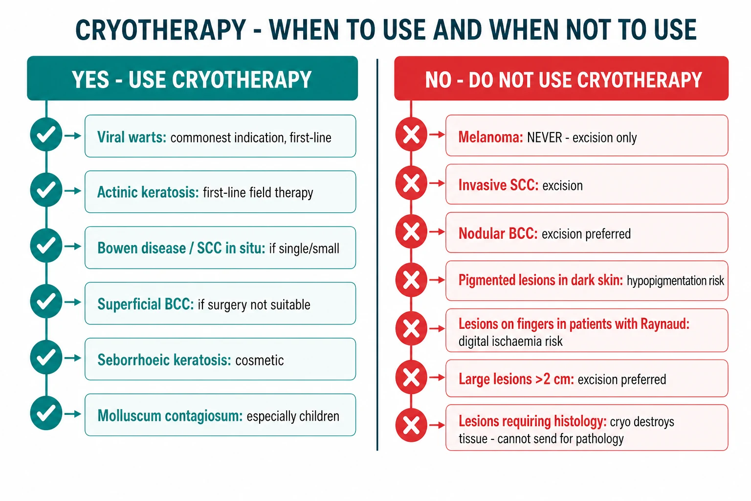
When NOT to Use Cryotherapy
The clearest way to internalise the contraindications is the explicit list of lesions that must not be frozen. Suspected melanoma is the cardinal "never": cryotherapy destroys the tissue, forfeits the histology, prevents staging, and can be fatal by delaying diagnosis. Invasive squamous cell carcinoma requires excision with margin control. Nodular, morpheaform, infiltrative, or otherwise aggressive BCC requires excision (often Mohs) because cryotherapy cannot guarantee clearance of the deep and lateral margins. Lesions requiring histology of any kind — diagnostic uncertainty, recurrent lesion, lesion that has failed to respond to expected treatment — must be biopsied before any destructive treatment. Pigmented lesions on the face of dark-skinned patients carry an unacceptable cosmetic risk from hypopigmentation. Large lesions (over 2 cm) are better managed by excision for cosmesis and margin control.[1]
Prognosis and Disposition
The natural history of a cryotherapy-treated lesion is predictable and worth quoting to the patient: pain and erythema immediately, blister within 24-48 hours (often haemorrhagic, may be large), crust at 7-14 days that then sloughs to reveal a healing pink then hypopigmented macule, and a settled hypopigmented scar by four to six weeks. Clearance rates depend on the indication. Viral warts clear in approximately 50-70% of patients after three or four sessions, with a recurrence rate of 20-30%. Actinic keratoses clear in the majority after a single adequate freeze, though new lesions continue to appear in a field-cancerised area. Superficial BCC cleared by cryotherapy recurs in 5-15% of cases, which is why structured follow-up at three to six months with a low threshold for biopsy is the standard.[8]
Disposition depends on the indication. Warts, molluscum, seborrhoeic keratosis, and solar lentigo are managed in primary care with cryotherapy or topical therapy and discharged on clearance. Actinic keratosis is managed with a combination of lesion-directed and field therapy, with periodic dermatology review for high-burden field cancerisation. BCC and Bowen disease treated by cryotherapy must be followed up by the treating dermatology service with a defined review and biopsy-if-persistent pathway, because recurrence is silent and missed recurrence of a BCC can progress to invasion. The safety-net advice to every patient is to return if the treated lesion recurs, darkens, ulcerates, or enlarges — features that demand re-evaluation for malignancy.[1]
Aftercare
Clear aftercare advice is part of competent practice and is examinable. The patient is told to wash the area normally and to apply a simple emollient such as white soft paraffin (Vaseline) or an antibiotic ointment if crusted. The blister that forms at 24-48 hours is expected and should be left intact; if it is large and painful it may be aspirated with a sterile needle, leaving the roof in place as a biological dressing. The crust falls off over one to three weeks and should not be picked. Sun protection of the treated site during and after healing reduces post-inflammatory hyperpigmentation, though it cannot prevent the hypopigmentation that is intrinsic to the technique. Patients are warned that the final result is a lighter macule and, in darker skin, that this is likely to be permanent. Sessions for warts are repeated at intervals of three to four weeks until clearance is achieved.[3]
Special Populations
Children are commonly treated for warts and molluscum. Pain tolerance is lower, so brief freezes, distraction, and topical or vapocoolant anaesthesia help. Parents must be counselled about hypopigmentation, particularly in dark-skinned children, and the self-limiting nature of molluscum means that watchful waiting is often the better choice. Patients with darker phototypes (Fitzpatrick IV-VI) constitute a relative contraindication for cryotherapy at cosmetically significant sites because of permanent hypopigmentation; alternatives such as salicylic acid for warts, electrocautery, shave, or laser should be considered and the risk discussed explicitly. Older adults often have thin atrophic skin and a heavy burden of actinic damage; lighter freezes, attention to anticoagulation, and a low threshold for field therapy rather than lesion-by-lesion cryotherapy are appropriate. Immunocompromised patients (solid-organ transplant recipients, haematological malignancy, HIV) frequently have refractory warts and aggressive actinic field cancerisation; cryotherapy is one tool among several, but any atypical or refractory lesion must be biopsied to exclude squamous cell carcinoma. Pregnant women can be treated safely as there is no significant systemic absorption, though molluscum and genital warts in pregnancy may merit deferral where the condition is self-limiting. Patients on anticoagulants are at greater risk of bleeding and haemorrhagic blistering and should be warned and treated with shorter freezes and pressure as needed.[3]
Advantages and Disadvantages
CRYO
The advantages of cryotherapy explain its enduring popularity: it is quick (a treatment session lasts 10-30 seconds), requires no anaesthetic in most cases, leaves no sutures, is portable (a hand-held liquid nitrogen canister can be used in any clinic), and is cost-effective compared with excision. The disadvantages are equally clear and define the boundaries of its use: it provides no histology, so any diagnostic doubt must be resolved before treatment; it produces permanent hypopigmentation that is cosmetically significant in skin of colour; it is painful; it carries a recurrence rate that demands follow-up; and over-aggressive use produces scarring and nerve or nail damage. A candidate who can state both lists — and who volunteers that "no histology" is the disadvantage that flows from the "destructive rather than excisional" nature of the technique — answers the question at fellowship depth.[1]
Evidence, Guidelines, and Regional Differences
The 2025 review by Mokbel and colleagues in In Vivo consolidates the modern position of cryosurgery: a versatile, cost-effective destructive modality whose indications span benign, premalignant, and carefully selected malignant lesions, provided the operator respects the principles of dose, cycle number, halo size, and contraindication.[1] The technique-specific "tips and tricks" paper by Ashique and colleagues (2021) and the practical guidance by Cranwell and Sinclair (2017) are the bedside references most often cited for the procedural details of ice-ball margin, cycle number, and freeze time.[2][3]
For specific indications, three evidence threads matter. Warts: the VRAIE multicentre pragmatic randomised trial (Chanal and colleagues, 2025) compared salicylic acid, liquid nitrogen, 5-fluorouracil, and imiquimod in previously-treated plantar warts and found no single agent clearly superior, confirming that plantar warts are stubborn and that treatment choice is patient-tailored.[7] Actinic keratosis: the systematic review by Worley and colleagues (2023) and the review by Dianzani and colleagues (2020) establish that cryotherapy, field therapies, and photodynamic therapy all have comparable moderate efficacy for individual lesions, with field therapy preferred when lesion count is high.[4][5] Superficial BCC: the European consensus-based interdisciplinary guidelines (Peris and colleagues, 2019) place cryotherapy among the nonsurgical options for carefully selected low-risk superficial BCC, with surgery as first-line and cryotherapy reserved for cases where surgery is unsuitable.[8]
Regional practice varies in ways an examiner may probe. In the United States (AAD), cryotherapy is a first-line destructive modality for actinic keratosis and viral warts and is also used for selected superficial BCC. In the United Kingdom (BAD/NICE), cryotherapy is widely available in primary and secondary care for warts and AK; BAD guidance discourages cryotherapy for BCC except in carefully selected low-risk superficial lesions where excision is not possible. In Europe (EADF/EUROGIN), field-directed therapy with 5-fluorouracil, imiquimod, or photodynamic therapy is increasingly preferred over lesion-by-lesion cryotherapy for multiple actinic keratoses, with cryotherapy retained for discrete lesions. In India and South Asia, viral warts and acral lesions are common, but cryotherapy is used cautiously on darker phototypes because of hypopigmentation; salicylic acid paint and needling are first-line for warts, and cutaneous leishmaniasis is an additional indication in endemic regions.
The controversies in the field mirror the evidence. The best modality for plantar warts remains unsettled. The role of cryotherapy for superficial BCC versus excision is debated, with most guidelines reserving cryotherapy for low-risk lesions in patients unsuitable for surgery. Field cancerisation is increasingly managed with topical field therapy rather than lesion-by-lesion cryotherapy. And the over-use of cryotherapy on dark skin without counselling about hypopigmentation is a recurring medico-legal and ethical issue.[1]
Exam Pearls
Self-test: a pigmented nodule on a 60-year-old's back — is cryotherapy appropriate?
No. A pigmented lesion in an older adult must be evaluated by dermoscopy and biopsy first to exclude melanoma. Cryotherapy is destructive and forfeits histology — it is never appropriate for a lesion of uncertain diagnosis or any lesion where melanoma cannot be excluded. The correct pathway is excisional biopsy with histology and staging.
Exam application bank (NEET-PG / INICET)
One-line answer
Cryotherapy (cryosurgery) uses liquid nitrogen at -196 C to destroy cutaneous lesions by controlled freezing through repeated freeze-thaw cycles. Cell death results from four mechanisms — extracellular ice formation (cellular dehydration), intracellular ice formation (membrane disruption), vascular stasis (thrombosis and ischaemia), and apoptosis — and the lethal target temperature is approximately -20 to -30 C. The ice ball must extend a 2-3 mm halo beyond the visible lesion margin; 1-2 freeze-thaw cycles per session for benign lesions and two cycles for premalignant/malignant lesions. The commonest indications are viral warts, actinic keratosis, molluscum contagiosum, seborrhoeic keratosis, solar lentigo, Bowen disease, and carefully selected superficial BCC. Expected sequence: pain/erythema at freezing, blister at 24-48 h, crust at 7-14 days, then a healed macule. The most important l
Worked stems (answer without another resource)
Stem 1 — Classic presentation. Map symptoms to mechanism; name the first investigation and first treatment step with dose/route if drug therapy is standard. [1]
Stem 2 — Unstable / complicated. List red flags that force immediate resuscitation, theatre, ICU, antidote, or reperfusion — and what you do in the first 15 minutes. [1]
Stem 3 — Atypical group. Elderly, pregnancy, child, or immunocompromised: how presentation and thresholds change. [1]
Stem 4 — Differential trap. Name the three closest mimics and one discriminator for each. [1]
Stem 5 — Disposition. Who goes home with safety-netting, who is admitted, who needs HDU/ICU/theatre, and what follow-up is mandatory. [1]
Rapid viva checklist
- Definition + classification
- Pathophysiology chain
- Bedside signs / criteria
- Score with exact components (if any)
- Emergency bundle
- Definitive therapy with doses
- Complications of disease and of treatment
- Special populations
- Guideline/trial name if classic
- Three exam traps
Coverage self-check
If you cannot answer any stem above from this page alone, re-read the matching section — the page is intended to be self-sufficient for final-prof and NEET-PG/INICET questions on Cryotherapy.
[1]References
- [1]Mokbel R, Kodresko A, Mokbel K, et al. Cutaneous Cryosurgery in Dermatology: Evolving Principles and Clinical Applications for Benign, Premalignant, and Malignant Lesions In Vivo, 2025.PMID 40010951
- [2]Ashique KT, Kaliyadan F, Jayasree P. Cryotherapy: Tips and Tricks J Cutan Aesthet Surg, 2021.PMID 34566372
- [3]Cranwell WC, Sinclair R. Optimising cryosurgery technique Aust Fam Physician, 2017.PMID 28472571
- [4]Dianzani C, Conforti C, Giuffrida R, et al. Current therapies for actinic keratosis Int J Dermatol, 2020.PMID 32012240
- [5]Worley B, Harikumar V, Reynolds K, et al. Treatment of actinic keratosis: a systematic review Arch Dermatol Res, 2023.PMID 36454335
- [6]Mulhem E, Pinelis S. Treatment of nongenital cutaneous warts Am Fam Physician, 2011.PMID 21842775
- [7]Chanal J, Aubin F, Penso-Assathiany D, et al. A multicentre pragmatic randomized controlled trial comparing 50% salicylic acid, liquid nitrogen, 5% 5-fluorouracil cream, and 5% imiquimod cream in previously treated plantar warts. The VRAIE (VeRrues plAntaIres en villE) study Ann Dermatol Venereol, 2025.PMID 40743833
- [8]Peris K, Fargnoli MC, Garbe C, et al. Diagnosis and treatment of basal cell carcinoma: European consensus-based interdisciplinary guidelines Eur J Cancer, 2019.PMID 31288208
- [9]Plensdorf S, Livieratos M, Dada N. Pigmentation Disorders: Diagnosis and Management Am Fam Physician, 2017.PMID 29431372
- [10]Leung AKC, Barankin B, Hon KLE. Molluscum Contagiosum: An Update Recent Pat Inflamm Allergy Drug Discov, 2017.PMID 28521677