Dermatology · Medicine
Cutaneous larva migrans (creeping eruption)
Also known as Cutaneous larva migrans (CLM) · Creeping eruption · Hookworm-related cutaneous larva migrans (HrCLM) · Sandworm disease · Plumber's itch
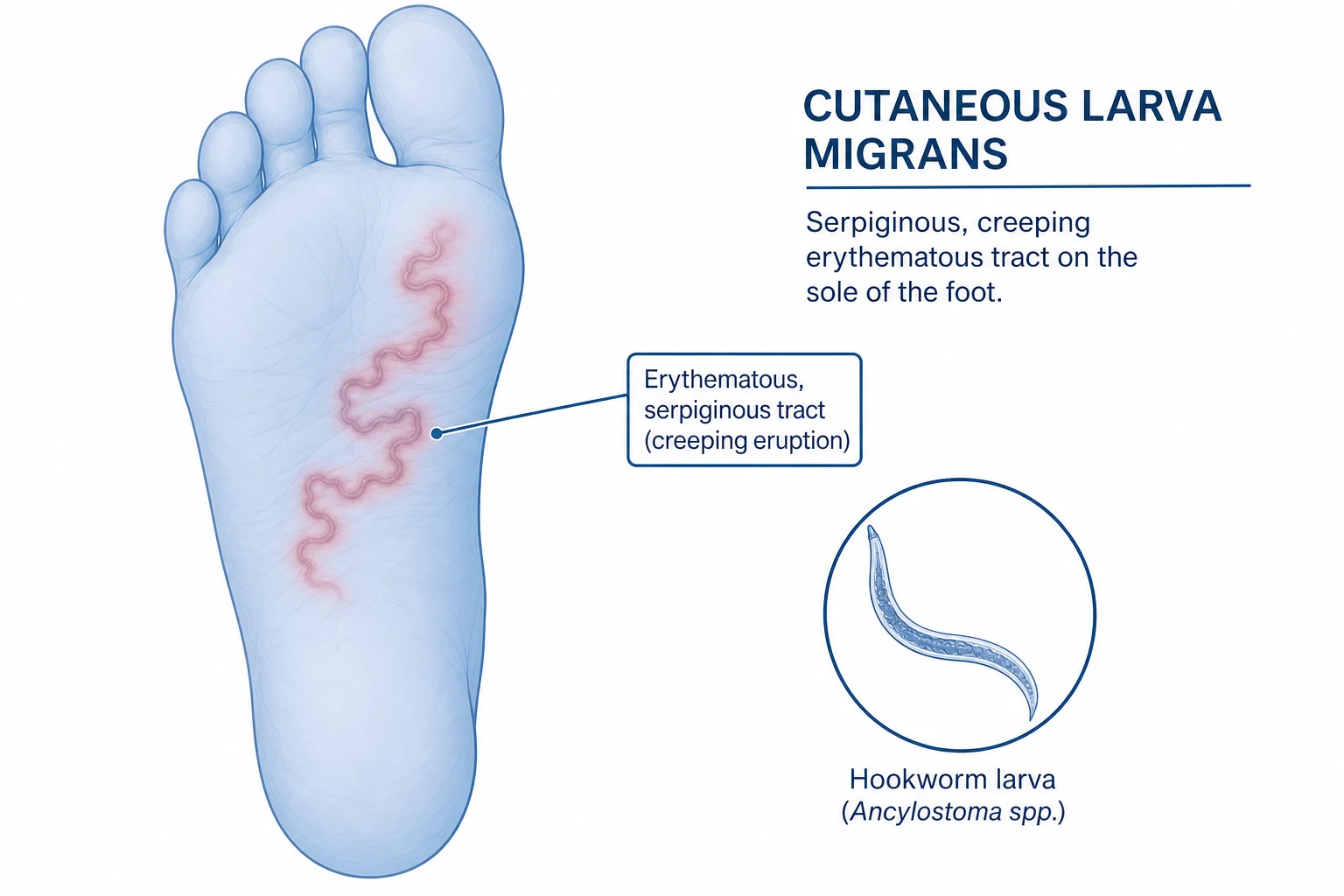
Cutaneous larva migrans (CLM), also known as creeping eruption, is the most common tropically acquired dermatosis in returned travellers, caused by percutaneous penetration and intraepidermal migration of animal hookworm larvae (predominantly Ancylostoma braziliense and A. caninum from dogs and cats). Humans are an accidental (aberrant) host — the larvae cannot penetrate the basement membrane and migrate tortuously within the stratum corneum and upper epidermis, producing the characteristic intensely pruritic, serpiginous, creeping erythematous tract that advances 2–7 mm per day. Distribution favours the feet, buttocks, abdomen, and thighs (areas that contacted contaminated sand or soil). Diagnosis is clinical (travel history + creeping eruption). Treatment of choice is oral ivermectin 200 µg/kg as a single dose, with oral albendazole 400 mg daily for 3–7 days as the second-line alternative. The key fellowship-level differential is larva currens (Strongyloides stercoralis), which migrates up to 5 cm per hour, is recurrent via the autoinfection cycle, and demands a different therapeutic regimen.
On this page & tools
Your progress
Saved locally on this device.
Exam tags
Red flags
Overview and definition
Cutaneous larva migrans (CLM) — also called creeping eruption, hookworm-related cutaneous larva migrans (HrCLM), sandworm disease, and historically plumber's itch — is the most common tropically acquired dermatosis in returned travellers and the single most frequent skin complaint in patients presenting to tropical disease clinics after a beach holiday.[1][3][6] It is caused by the percutaneous penetration and subsequent intraepidermal migration of animal (zoonotic) hookworm larvae — predominantly Ancylostoma braziliense (the dog and cat hookworm of the Americas, Africa, and Asia) and Ancylostoma caninum (the dog hookworm), with smaller contributions from Uncinaria stenocephala (European dog hookworm) and Bunostomum phlebotomum (cattle hookworm, occasionally implicated in agricultural workers).[1][2][8]
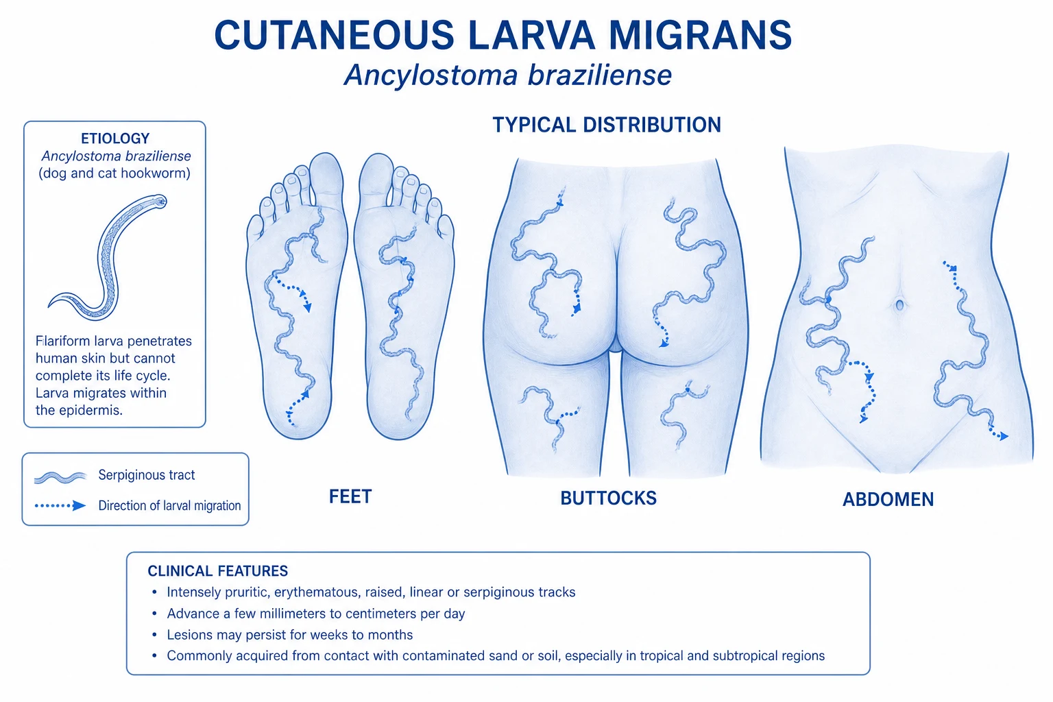
Humans are an accidental (aberrant, dead-end) host. In the natural canine or feline host, the third-stage filariform larva penetrates the skin, enters the venous circulation, traverses the right heart and pulmonary capillaries, is swallowed, and matures into an adult hookworm in the small intestine where it attaches to the mucosa and feeds on blood. In humans, this sequence cannot be completed: the larva releases the proteases, hyaluronidases, and collagenases needed to break down the stratum corneum and slip between keratinocytes, but it lacks the species-specific elastase and basement-membrane ligands required to cross the dermo-epidermal junction. Unable to enter the dermis, the larva is forced to wander laterally within the stratum corneum and upper (spinous) epidermis at a rate of approximately 1–2 cm per day (classically 2–7 mm per day), leaving behind the hallmark serpiginous, intensely pruritic, erythematous tract that gives the disease its name.[1][3][6][7]
[1]The condition is the prototypical member of the larva migrans syndrome — a group of dermatoses in which a metazoan parasite passes through human skin without being able to complete its life cycle. Other members include larva currens (cutaneous Strongyloides stercoralis), visceral larva migrans (Toxocara canis/cati), ocular larva migrans, gnathostomiasis, cutaneous Schistosoma cercarial dermatitis (swimmer's itch), and cutaneous leishmaniasis. Distinguishing between them on geography, exposure, rate of tract migration, and associated systemic features is a recurring fellowship question.[3][6][12]
Classification and clinical variants
CLM is not a single uniform entity. The classical serpiginous creeping eruption is the most recognisable form, but several distinct variants deserve separate mention because they are easily misdiagnosed and have different management implications.[1][6][7]
Classic serpiginous CLM
Hookworm-related folliculitis (HrHF, sand-worm folliculitis)
Bullous CLM
Oral CLM
In addition, several geographical colloquialisms are worth knowing for the viva: the "creeping eruption of the Americas" describes the classic Caribbean, Brazilian, and Gulf Coast disease; "plumber's itch" describes occupational CLM acquired by crawling through contaminated soil under houses; and "sand-worm" is a popular term used in Brazilian tropical medicine. Ocular and visceral extensions (larvae reaching the cornea, lung, or deeper tissues) are exceptionally rare and almost always reported as single case studies.[1][6]
Epidemiology and risk factors
CLM is, by a comfortable margin, the single most common dermatosis in travellers returning from the tropics.[2][3][11] The landmark GeoSentinel surveillance network and the Caumes Parisian cohort both identified CLM as the leading cause of dermatological consultation in returning travellers, accounting for 9–25% of all travel-acquired skin disease depending on the case mix.[3][11] This consistency across continents, decades, and surveillance systems underscores that CLM is the prototypical beach-holiday dermatosis: almost every returning traveller with a creeping skin lesion after a tropical trip has it.
Geographic distribution
The disease mirrors the distribution of infected dogs and cats in warm, humid climates where sanitation is incomplete and free-roaming animals can defaecate on beaches: [1]
- Caribbean basin — Barbados, Jamaica, Dominican Republic, the Bahamas, the US Virgin Islands, and coastal Mexico report the highest returning-traveller rates. Barbados has historically been the single most frequently cited exposure location in North American and European cohorts.[2][3]
- South America — coastal Brazil (especially the beaches of the northeast: Bahia, Pernambuco, Ceará), and the Atlantic coast of Colombia and Venezuela.[2]
- West and Central Africa — beach resorts in Senegal, Ghana, Côte d'Ivoire, Cameroon, and Gabon; also inland foci around rural villages.[1][3]
- Southeast Asia — Thailand (Phuket, Koh Samui, Krabi), Malaysia, Indonesia (Bali), the Philippines, and Vietnam.[2]
- Indian subcontinent — the Goa, Kerala, and Pondicherry coasts are recurring exposure sites for European and Russian travellers; rural and beach foci are also described in Sri Lanka and the Maldives.[2]
- Southeastern United States — Florida, the Gulf Coast (Alabama, Mississippi, Louisiana, Texas), and the Carolinas; domestic US acquisition is increasingly recognised and was highlighted in a 2026 Open Forum Infectious Diseases military health-system analysis.[3]
- Mediterranean Europe — sporadic cases acquired on the Spanish, Italian, French, and Greek coasts, where infected stray dogs and cats still contaminate some beaches.[2]
Exposure risk factors
The reservoir is the faeces of infected dogs and cats, which deposit hookworm ova onto warm, moist, shaded, sandy or loamy soil. The ova hatch into rhabditiform larvae, moult twice, and reach the infective filariform (L3) stage within 5–7 days. Sandy, moist, shaded, faecally-contaminated soil is the ideal reservoir; clay, rocky, or sun-baked terrain is rarely implicated because larvae desiccate rapidly and ova fail to embryonate.[1][2][6]
- Barefoot walking on tropical beaches — the single most important exposure. The dorsum and sole of the foot are the most common site, which is why 50–80% of CLM lesions are on the feet.[2]
- Sitting or lying directly on sand — explains the high frequency of buttock, genital, and abdominal lesions in beachgoers; sand is shifted up under the buttocks, and unprotected skin is exposed to larvae in the upper few millimetres of sand.[3]
- Children playing in sandboxes — domestic sandboxes contaminated by neighbour or stray cats are a classic exposure, particularly in the southeastern US and southern Europe.[2]
- Occupational soil contact — plumbers, electricians, exterminators, gardeners, farmers, and construction workers who crawl under or work on contaminated soil; the historical "plumber's itch" cases in the 19th and early 20th century London were mostly men crawling under houses with sandy crawl-space soil contaminated by dog faeces.[1][6]
The incubation period is short: most patients develop symptoms within 1–6 days of exposure, with a documented range of a few hours to several weeks in cases with very light inocula.[1][3][7] A small but useful clinical point: the incubation may be longer when the larval load is low (light exposure, late-season beach, cooler water-saturated sand), and short when the inoculum is heavy. This explains why some patients first notice the tract on the aeroplane home or in the first few days after returning.
Pathophysiology
The pathogenesis of CLM is a textbook example of an evolutionary mismatch between a parasite that has adapted to a canine or feline host and a human who is not part of that parasite's natural life cycle. The life cycle in the natural host, the molecular basis of the aberrant behaviour in humans, and the host immune response all combine to produce the characteristic clinical picture.[1][3][6][9]

Natural life cycle in dogs and cats
- Adult hookworms live attached to the mucosa of the small intestine of dogs and cats, feeding on blood. The female worms release thousands of ova daily, which are passed in the faeces onto warm, moist, sandy soil.[3]
- Within 1–2 days, the ova hatch into first-stage rhabditiform (L1) larvae, which feed on bacteria and organic debris in the soil and moult twice over 5–7 days to become infective third-stage filariform (L3) larvae.[1][3]
- The filariform L3 larva is the infective stage. It does not feed; it survives in the upper few millimetres of warm, moist, shaded sand for weeks, waiting for a warm-blooded host. When a dog or cat lies on the sand, the larva penetrates intact skin (usually through hair follicles or between stratum-corneum cells), enters the venous circulation, is carried to the lungs, is coughed up and swallowed, and matures into an adult hookworm in the small intestine, completing the cycle in 3–4 weeks.[1][6]
Aberrant behaviour in humans
In humans, the larva successfully penetrates the stratum corneum and releases hydrolytic enzymes — collagenases, hyaluronidases, serine proteases, and metalloproteases — that allow it to slip between keratinocytes and burrow through the upper epidermis. But it lacks the species-specific elastase and the ligands for laminin and collagen IV that the natural hookworm uses to dissolve the lamina lucida of the dermo-epidermal junction. As a result, the larva cannot cross the basement membrane and is forced to migrate laterally within the stratum corneum and superficial spinous layer at a rate determined by the balance between enzymatic advance and the host's local immune response.[1][3][6][7]
The tract is not a tunnel that the larva actively excavates. It is the trail of inflammation left behind as the larva advances a few millimetres to a centimetre each day, with the larva itself usually 1–2 cm ahead of the visible erythematous leading edge. This is why biopsy of the tract itself typically fails to capture the larva — a common viva question and a useful diagnostic pearl. [1]
Migration rate and tract morphology
- Classic rate: 1–2 cm per day, with a commonly cited range of 2–7 mm/day for uncomplicated CLM. The tract advances visibly over hours, which is one of the most striking clinical features — patients frequently photograph or measure the advance.[1][3][6]
- Tract morphology: a raised, erythematous, slightly oedematous, serpiginous or linear track 2–4 mm wide. The leading edge may be more inflamed than the older portion, and a small vesicle or pustule may mark the larva itself.[7]
- Multiple tracts: present in up to 20% of patients, reflecting multiple penetration sites during a single exposure.[1]
Host immune response
The intense pruritus that brings most patients to medical attention is not caused by direct tissue damage. It is the result of a combined type I and type IV hypersensitivity reaction to larval antigens and secreted enzymes: [1]
- Type I (immediate, IgE-mediated): within hours of penetration, mast cells degranulate, releasing histamine, tryptase, leukotrienes, and prostaglandins, producing the wheal-and-flare and intense itching.[3]
- Type IV (delayed, T-cell-mediated): over the next 24–72 hours, antigen-specific T-helper cells (predominantly Th2 with eosinophil recruitment) and macrophages accumulate around the larva, amplifying the inflammation and producing the visible erythematous tract.[1][3]
- Eosinophils are recruited by eotaxin and IL-5, but the local eosinophil load is usually modest and peripheral blood eosinophilia is uncommon in simple CLM, because the systemic antigen load is small. Significant peripheral eosinophilia (>0.5 × 10⁹/L) should prompt investigation for other helminths (Strongyloides, Toxocara, filaria, schistosomiasis).[3][6]
The larvae die within weeks to a few months — typically 4–8 weeks — because they cannot complete their life cycle, cannot find a mate, and are progressively overwhelmed by the host immune response. CLM is therefore self-limiting, but the duration of symptoms is long enough that almost all patients seek treatment, and the treatment is so effective (single-dose ivermectin) that there is no reason to wait for spontaneous resolution.[1][3][4][9]
Clinical presentation
Incubation and prodrome
The first symptom is usually pruritus at the penetration site, beginning within hours to a few days of exposure. A small erythematous papule may mark the entry point but is often missed. The patient then notices a moving thread-like eruption advancing across the skin, often with a visible leading edge slightly ahead of the inflammatory tract.[1][3][7]
Hallmark lesion
The creeping eruption is the pathognomonic finding. It is: [1]
- Serpiginous or linear, often with a snake-like or question-mark configuration when the larva changes direction.
- Erythematous, raised, and 2–4 mm wide, with a sharp leading edge and a fading older portion.
- Intensely pruritic, often worse at night and exacerbated by warmth.
- Slowly advancing at 2–7 mm per day — visible to the patient or to a serial photograph.
- Often accompanied by a trail of vesicles, pustules, or crusting behind the leading edge, particularly if the tract has been scratched.[1][6][7][8]
Distribution
The distribution is dictated by the body surface in contact with the sand: [1]
- Feet — the single most common site, accounting for 50–80% of lesions in most series. The dorsum, sole, lateral border, and web spaces are all vulnerable; barefoot walking is the dominant exposure.[2][3]
- Buttocks and perianal area — the second most common site, reflecting sitting or lying directly on sand.[2]
- Abdomen, lower back, and thighs — contact sites for sunbathers and children playing in sand.[3]
- Hands and forearms — gardeners, plumbers, electricians, and construction workers who handle contaminated soil.
- Genitalia, breasts, and face — less common but well-described, particularly in children and sunbathers.[1][6]
Number, course, and associated features
- Number: usually 1–3 tracts; multiple lesions in 10–20% of patients, occasionally dozens in heavy exposures.
- Course without treatment: the larva dies within 4–8 weeks, the inflammation resolves, the tract fades over weeks, and most patients clear spontaneously within 2–3 months. Pruritus, however, persists throughout.
- Secondary bacterial infection is the most common complication, occurring in 5–25% of patients, particularly with scratching, hot climates, and inadequate hygiene.[1][3][6]
Differential diagnosis
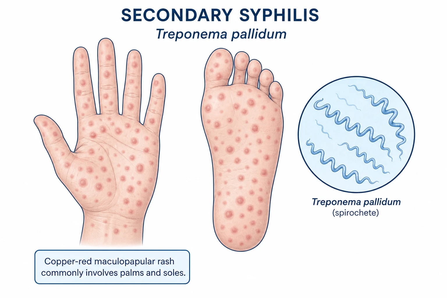
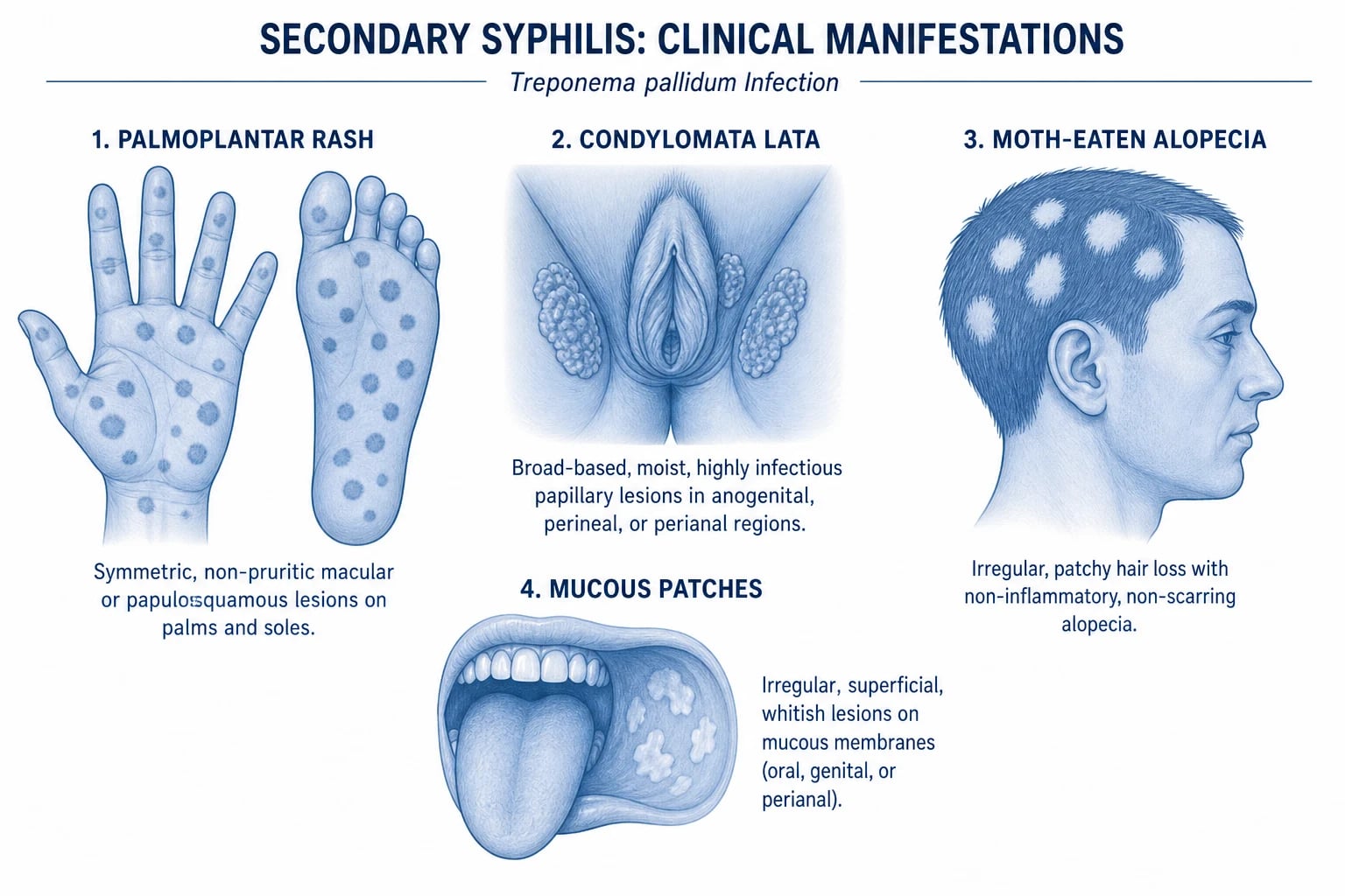
The differential diagnosis of a creeping skin lesion after a tropical trip is broad and includes a number of other "larva migrans" entities, infectious mimics, and non-infectious dermatoses. The two most important distinctions at fellowship level are larva currens (Strongyloides stercoralis) and cutaneous gnathostomiasis, both of which can mimic CLM but require different treatment.[3][6][10][12]
Larva currens (Strongyloides stercoralis)
Gnathostomiasis (Gnathostoma spinigerum)
Cutaneous myiasis (Dermatobia hominis, Cordylobia anthropophaga)
Tinea corporis
Erythema chronicum migrans (Lyme disease)
Scabies
Clinical and bedside assessment
The diagnosis of CLM is clinical and rests on three bedside observations: a recent tropical travel or beach exposure, a characteristic creeping serpiginous tract on a contact site, and the absence of features pointing to an alternative diagnosis. There is no laboratory test that establishes the diagnosis with certainty; the bedside assessment is the diagnostic test.[1][3][5][7]
Focused history
- Travel history: specific countries visited, dates, duration of stay, and the timing of the onset of symptoms relative to the trip. Most patients develop symptoms during the trip or within the first week of returning home.[2][3]
- Exposure history: barefoot walking on beaches, lying or sitting directly on sand, time spent in sandboxes, occupational soil contact, and any contact with stray dogs or cats.
- Accommodation: beach resort, backpacker hostel, rural village, or cruise ship.
- Symptom history: onset of pruritus, time of first noticing the tract, rate of advance, any nocturnal worsening, any associated systemic symptoms.
- Past medical history: atopic dermatitis (predisposes to more severe inflammation), immunosuppression (transplant, HIV, chronic steroids, chemotherapy), and any history of previous helminth infections.
- Drug history: any current anthelmintic, antihistamine, or topical steroid; vaccination status including tetanus.
Focused examination
- Inspect the entire skin with the patient undressed. CLM can be on any contact site, and multiple tracts are present in 10–20% of patients.[1][3]
- Examine the feet, buttocks, abdomen, lower back, and thighs in detail. The interdigital spaces, the plantar surface, and the perianal area are easy to miss.
- Mark the leading edge of the tract with a surgical pen or photograph it with a ruler, and ask the patient to re-present in 24–48 hours if the diagnosis is uncertain — a tract that has visibly advanced 5–20 mm in that interval is diagnostic of a migrating larva.
- Look for secondary infection: impetiginisation, pustules, cellulitis, regional lymphadenitis.
- Dermoscopy (when available) can occasionally visualise the larva as a translucent, comma-shaped or oval structure within the leading edge of the tract (a "larva in a burrow" sign), and can exclude burrowing scabies mites, tinea, and superficial vessels. Dermoscopy is not required for the diagnosis, but it is a useful confirmatory adjunct.[7]
Investigations
The diagnosis of CLM is clinical, and most patients do not need any investigation before treatment.[1][3][5] Laboratory and imaging studies are reserved for atypical presentations, the differential diagnoses discussed above, and patients who fail to respond to first-line therapy.[1][3]
First-line bedside test
- Direct visualisation of the advancing serpiginous tract on a contact site after a relevant exposure. No further investigation is required to start treatment.
- Serial photography of the leading edge over 24–48 hours can be diagnostic when the migration rate is atypical (very slow, or unusually fast and concerning for larva currens). [1]
Laboratory tests
- Full blood count: peripheral eosinophilia is uncommon in simple CLM (it occurs in fewer than 10–20% of cases) because the systemic antigen load is small. Significant eosinophilia (>0.5 × 10⁹/L) should prompt investigation for other helminths: Strongyloides serology, stool O&P for ova and parasites (3 samples on consecutive days, or agar-plate culture for Strongyloides), Toxocara serology, filaria serology, and schistosomiasis serology if the exposure fits.[1][3][6]
- Skin biopsy is rarely needed. When performed for diagnostic uncertainty, it typically shows an epidermal tract with spongiosis, intraepidermal vesiculation, a mixed inflammatory infiltrate with eosinophils, and occasionally a larval cross-section, but the larva is usually ahead of the tract and is captured in fewer than 5% of biopsies.[1][7]
- Strongyloides serology (IgG ELISA) is the test of choice to exclude larva currens in a patient with a rapidly migrating or recurrent tract, particularly with eosinophilia or abdominal symptoms. False positives occur in other helminth infections, so the result must be interpreted in context.[10]
- Stool ova and parasite examination is unhelpful for CLM itself (animal hookworms do not reach the human gut), but is part of the work-up for concomitant helminth infection, particularly Strongyloides, Ascaris, Trichuris, and hookworm in endemic settings.
- Gnathostoma serology (where available) for migratory subcutaneous swellings with marked eosinophilia and a history of raw freshwater fish consumption in Southeast Asia or Mexico.[12]
- HIV serology if the clinical picture raises concern about disseminated strongyloidiasis, particularly in patients with a long-standing recurrent creeping eruption and a CD4-lymphocyte-related risk.
Imaging
Imaging is not required for uncomplicated CLM. Chest radiography may show transient Löffler-like infiltrates in patients with pulmonary symptoms and eosinophilia, but these reflect coincident or concomitant helminth infection (Ascaris, Strongyloides) rather than CLM itself.[3][6]
Management — definitive and stepwise
CLM is a medical, not surgical, condition, and the introduction of oral ivermectin in the 1990s transformed its management. Before ivermectin, treatment options were cryotherapy of the leading edge (painful, unreliable, often unsuccessful because the larva is 1–2 cm ahead of the visible tract), topical thiabendazole (effective but messy and limited by supply), and oral albendazole (effective but slower than ivermectin). The modern approach is unambiguous: single-dose oral ivermectin 200 µg/kg is the drug of choice, with oral albendazole as the second-line alternative.[1][4][5][9]
First-line: oral ivermectin
- Dose: ivermectin 200 µg/kg as a single oral dose (one dose = one treatment course in most cases).[1][4][5][9]
- Cure rate: more than 95% in randomised trials; symptomatic improvement (pruritus and tract progression) usually within 24–48 hours, and complete resolution within 1–2 weeks.[9]
- Mechanism: binds to glutamate-gated chloride channels in nematode motor neurons and pharyngeal muscle, causing paralysis and death of the larva. Mammals are unaffected because they lack these channels.
- Repeat dose: a small proportion of patients (5–10%) require a second dose after 1–2 weeks if pruritus and tract progression persist; this is more common in patients with multiple or heavy-inoculum lesions.[4][5]
- Contraindications and cautions: pregnancy (insufficient data), children under 15 kg body weight (or under 5 years of age in some guidelines), and patients with Loa loa co-infection from Central or West Africa (ivermectin can precipitate encephalopathy in heavily microfilaraemic patients). Take a careful travel history and, in any patient with West or Central African exposure, screen for Loa loa microfilaraemia before giving ivermectin.[3]
Second-line: oral albendazole
- Dose: albendazole 400 mg orally once daily for 3–7 days (some guidelines use 400 mg twice daily for 3 days for heavier disease).[1][4]
- Cure rate: ~80–90% after a 3-day course, ~95% after a 7-day course; slightly slower than ivermectin.
- When to use: pregnancy (albendazole is category C, but post-marketing data are reassuring for short courses in the second and third trimesters, and it is preferred to ivermectin because of better safety data in pregnancy), ivermectin unavailability, or Loa loa co-infection.
- Adverse effects: mild gastrointestinal upset, transient liver enzyme elevation (monitor in patients with pre-existing hepatic disease).
Topical and historic therapies
- Topical thiabendazole 10–15% suspension or cream applied 2–4 times daily for 5–10 days was the mainstay before oral ivermectin became available. It is still useful for very localised, single-tract CLM and in patients who cannot take oral therapy, but it is messy, often unavailable, and inferior to oral ivermectin.[4][9]
- Topical ivermectin has been used off-label for limited lesions but is not a recognised first-line therapy.
- Cryotherapy of the leading edge with liquid nitrogen is a historic approach that targets the visible front of the tract. It is unreliable because the larva is 1–2 cm ahead of the visible erythema; it is painful, can blister normal skin, and frequently fails. It is not recommended unless oral therapy is contraindicated and the lesion is small and single.
Symptomatic and adjunctive measures
- Oral antihistamines (cetirizine 10 mg, loratadine 10 mg, hydroxyzine 25 mg) for pruritus; a non-sedating antihistamine by day and hydroxyzine at night is a useful combination.
- Topical corticosteroids (mild-to-moderate potency, e.g., mometasone furoate 0.1% cream) may be used briefly over inflamed tracts to reduce local inflammation and pruritus, but should not be used over infected or impetiginised skin.
- Secondary bacterial infection is treated with oral flucloxacillin (or erythromycin/clarithromycin if penicillin-allergic); check for MRSA in patients who fail to respond or who have recent hospital contact. Topical mupirocin or fusidic acid is appropriate for localised impetiginisation.
- Tetanus immunisation should be checked and updated in any patient with a contaminated skin lesion in a tropical environment. [1]
Specific subtypes and scenarios
Several CLM-related scenarios deserve separate discussion because they are commonly encountered in exams and have specific management considerations.[1][3][6][12]
Hookworm-related folliculitis (HrHF, sand-worm)
Bullous CLM
Oral CLM
Occupational CLM (plumber's itch)
Co-infection with Strongyloides stercoralis
Patients with a beach exposure in the tropics may have been exposed simultaneously to Strongyloides stercoralis through barefoot skin contact, particularly in rural and agricultural settings. Strongyloides autoinfection can persist for decades after exposure and can cause fatal hyperinfection in patients who later become immunosuppressed. A useful rule of thumb: any patient with a creeping eruption AND eosinophilia, abdominal symptoms, or a West/Central African exposure should be screened for Strongyloides serology and treated for both conditions if positive. Strongyloides requires two consecutive days of ivermectin 200 µg/kg, not single-dose.[10]
CLM acquired in the United States
A 2026 Open Forum Infectious Diseases analysis of the US Military Health System confirmed that CLM is regularly acquired domestically in the southeastern United States, particularly in Florida, the Gulf Coast, and the Carolinas, in patients with no international travel. The implication is that CLM should not be excluded on the basis of absent international travel in patients with a creeping eruption and a beach or sandbox exposure in the southern US.[3]
Complications and pitfalls
CLM is rarely dangerous, but the morbidity from unrelenting pruritus, secondary infection, and the diagnostic delay before correct treatment is significant. Several specific pitfalls deserve emphasis.[1][3][6]
Secondary bacterial infection
Scratching and humid tropical climates predispose to: [1]
- Impetiginisation of the tract, with honey-coloured crusting.
- Cellulitis or erysipelas around the tract, with regional lymphangitis and lymphadenitis.
- Furunculosis if follicular CLM is misdiagnosed as bacterial folliculitis and treated with antibiotics alone.
- Tetanus is a theoretical risk in heavily contaminated wounds; check and update immunisation. [1]
Misdiagnosis and diagnostic delay
CLM is misdiagnosed in 20–40% of cases at first contact, often as: [1]
- Scabies — leading to repeated scabicidal treatments that do not work.
- Tinea corporis — leading to topical antifungal therapy.
- Lyme disease — leading to empirical doxycycline.
- Contact dermatitis, phytodermatitis, or insect bite reaction — leading to topical steroids, which may transiently reduce inflammation but do not kill the larva.
- Larva currens — leading to single-dose ivermectin when two days are needed. [1]
Diagnostic delay is typically 1–3 weeks in primary care and can stretch to 8–12 weeks in patients who present repeatedly with non-resolving "bites" or "eczema."[1][3]
Treatment failure
Failure to respond to single-dose ivermectin within 1–2 weeks should prompt: [1]
- Confirmation of the diagnosis — re-examine for an alternative diagnosis (larva currens, gnathostomiasis, myiasis, Majocchi's granuloma, deep fungal infection with sporotrichoid spread, cutaneous leishmaniasis).[12]
- A second dose of ivermectin 200 µg/kg 1–2 weeks after the first — appropriate for confirmed CLM with multiple or heavy-inoculum lesions.
- Switch to oral albendazole 400 mg daily for 7 days if ivermectin is unavailable, contraindicated, or has failed twice.
- Consider Loa loa co-infection if the patient is from West or Central Africa; ivermectin can precipitate encephalopathy in heavily microfilaraemic patients, and albendazole may be a safer choice.
Specific iatrogenic pitfalls
- Cryotherapy of the leading edge in the mistaken belief that this kills the larva — the larva is 1–2 cm ahead of the visible erythema, and cryotherapy typically blisters normal skin without eradicating the larva. Not recommended.
- Topical steroids alone may transiently reduce inflammation and pruritus but do not kill the larva, and the tract continues to advance.
- Antibiotics alone for a misdiagnosed bacterial folliculitis (when the actual diagnosis is HrHF) do not kill the larva and the eruption continues. [1]
Prognosis and disposition
The prognosis of CLM is excellent with appropriate treatment, and the long-term outlook is determined more by the patient's travel and exposure patterns than by the disease itself. [1]
Factors affecting prognosis
- Inoculum and number of tracts: patients with multiple or large lesions take longer to clear and may need a second ivermectin dose.
- Time to treatment: the earlier ivermectin is given, the shorter the duration of symptoms and the lower the risk of secondary infection.
- Secondary infection: delays tract resolution and increases the risk of scarring.
- Immunosuppression: does not appear to worsen CLM itself, but coexisting Strongyloides infection in an immunosuppressed host can be life-threatening — always consider and screen.
- Re-exposure: patients who return to the same beach environment without prevention are at high risk of recurrence. [1]
Disposition
- Most patients can be managed in the community with a single dose of ivermectin and symptomatic treatment; admission is not required.[1][3]
- Indications for hospital admission are limited to: extensive secondary infection requiring intravenous antibiotics, suspected concomitant Strongyloides hyperinfection (in immunosuppressed patients), diagnostic uncertainty requiring biopsy, and severe allergic reaction to ivermectin.
- Follow-up: most patients do not need formal follow-up; they should be advised to return if the tract has not visibly stopped advancing within 1 week, if new lesions develop, or if secondary infection occurs.
Special populations
Children
CLM is common in children, particularly after sandbox play. The treatment principles are the same as in adults, but ivermectin is generally avoided in children under 15 kg body weight (or under 5 years of age in many guidelines), and albendazole 400 mg daily for 3–7 days is the preferred option. Topical thiabendazole is a useful adjunct in children who cannot swallow tablets. Parental counselling on sandbox hygiene, deworming of household pets, and the avoidance of barefoot play on tropical beaches is essential.[1][3]
Pregnancy and lactation
CLM in pregnancy is uncommon but reported, particularly in long-term residents of endemic regions. Ivermectin is generally avoided in pregnancy because of limited safety data, and albendazole is also conventionally avoided in the first trimester (teratogenicity in animal studies), although recent meta-analyses suggest that inadvertent first-trimester exposure is not associated with a major increase in congenital malformation risk. Topical thiabendazole is a useful option in pregnancy because systemic absorption is minimal. Decisions should be individualised with obstetric and infectious-disease input, and the threshold for treatment is lower than in non-pregnant patients because the pruritus and sleep loss themselves affect pregnancy wellbeing.[3]
Elderly and frail patients
CLM is uncommon in the elderly because exposure patterns differ, but when it occurs, treatment decisions should account for polypharmacy, hepatic and renal function (albendazole is mildly hepatotoxic), and the ability to attend follow-up. Ivermectin remains first-line, with single-dose administration; albendazole is the alternative.[3]
Immunocompromised patients
CLM itself is not more severe in immunocompromised patients, but the differential diagnosis widens considerably to include disseminated strongyloidiasis, atypical fungal infections (sporotrichoid spread, cryptococcosis, histoplasmosis), atypical mycobacterial infection, and cutaneous leishmaniasis. A thorough work-up including skin biopsy, fungal and mycobacterial cultures, Strongyloides serology, and review of the patient's travel and exposure history is warranted. Ivermectin remains the treatment of choice, but the dose may need to be repeated and the patient monitored for treatment failure.[3][10]
Loa loa co-endemicity
In patients exposed in West and Central Africa (Cameroon, Nigeria, Gabon, the Democratic Republic of the Congo, the Republic of the Congo, Angola, Chad, the Central African Republic, Equatorial Guinea), the possibility of Loa loa co-infection must be considered before giving ivermectin. Heavy Loa loa microfilaraemia (>30,000 microfilariae/mL blood) can be associated with ivermectin-induced encephalopathy, a serious adverse event. In any patient from these regions, screen for Loa loa microfilaraemia (daytime blood film for microfilariae) before giving ivermectin; if microfilaraemia is detected, consider albendazole (which does not kill Loa loa microfilariae rapidly) and refer to a tropical-disease specialist.[3]
Evidence, guidelines, and regional differences
Landmark trials and evidence
- Caumes et al. 1993 — the first randomised trial of ivermectin 200 µg/kg single dose versus albendazole 400 mg daily for 3 days in 43 patients with imported CLM. Ivermectin cured 100% of patients (compared with 85% for albendazole) and was associated with faster relief of pruritus.[9]
- Caumes 2000 — a comprehensive review of CLM treatment established oral ivermectin as the drug of choice based on its efficacy, single-dose convenience, and tolerability.[4]
- Davies, Sakuls, Keystone 1993 — a 60-case series from the Toronto tropical disease unit that established the clinical presentation, distribution, and natural history of imported CLM in a North American cohort and informed all subsequent reviews.[8]
- Heukelbach et al. 2008 — a prospective cohort of 146 patients in Brazil that documented the epidemiology, distribution, and clinical course of CLM in an endemic setting, and confirmed the feet as the most common site, the buttocks as the second most common, and a mean tract length of 5–10 cm at presentation.[2]
- Hochedez & Caumes 2007 — a narrative review that established the modern classification of CLM and its place within the larva migrans syndrome.[3]
- Stevens et al. 2015 — the CanTravNet surveillance study of dermatoses in returned Canadian travellers confirmed CLM as one of the top three travel-acquired skin diseases, with the Caribbean and Southeast Asia as the dominant exposure regions.[11]
- Tian et al. 2023 — a seven-case series and literature review of larva currens that re-emphasised the speed of Strongyloides tract migration, its recurrent nature, and the need for two-day ivermectin treatment.[10]
- Hamilton et al. 2018 — a case report and review of imported gnathostomiasis mimicking CLM, with Löffler-like pulmonary infiltrates, illustrating the importance of considering this diagnosis in patients with migratory subcutaneous swellings and marked eosinophilia after raw-fish exposure in Asia.[12]
Guidelines
- American Academy of Dermatology (AAD) and the CDC — both endorse oral ivermectin 200 µg/kg single dose as the treatment of choice for uncomplicated CLM, with oral albendazole as the second-line alternative.
- British Association of Dermatologists (BAD) and the European Centre for Disease Prevention and Control (ECDC) — concordant guidelines with the same first-line recommendation.
- WHO — does not issue a specific CLM guideline, but its anthelmintic policy supports ivermectin and albendazole for soil-transmitted helminths and notes the Loa loa encephalopathy risk. [1]
Regional practice variations
- Europe and North America — ivermectin 200 µg/kg single dose is universally first-line; albendazole is reserved for ivermectin contraindications.[3][4][5]
- Brazil and the Caribbean — ivermectin is widely available and used as first-line; topical thiabendazole is occasionally used for single lesions because of cost and access issues.[2][6]
- India, Sri Lanka, and the Indian subcontinent — albendazole is more commonly first-line because of cost and availability, and CLM in returning travellers is recognised as a frequent presentation of beach exposure in Goa and Kerala.[2]
- West and Central Africa — albendazole is preferred in patients with possible Loa loa co-infection to avoid ivermectin-induced encephalopathy.[3]
Exam pearls and minutiae
CREEPING
Exam application bank (NEET-PG / INICET)
One-line answer
Cutaneous larva migrans (CLM), also known as creeping eruption, is the most common tropically acquired dermatosis in returned travellers, caused by percutaneous penetration and intraepidermal migration of animal hookworm larvae (predominantly Ancylostoma braziliense and A. caninum from dogs and cats). Humans are an accidental (aberrant) host — the larvae cannot penetrate the basement membrane and migrate tortuously within the stratum corneum and upper epidermis, producing the characteristic intensely pruritic, serpiginous, creeping erythematous tract that advances 2–7 mm per day. Distribution favours the feet, buttocks, abdomen, and thighs (areas that contacted contaminated sand or soil). Diagnosis is clinical (travel history + creeping eruption). Treatment of choice is oral ivermectin 200 µg/kg as a single dose, with oral albendazole 400 mg daily for 3–7 days as the second-line alternat [1]
Worked stems (answer without another resource)
Stem 1 — Classic presentation. Map symptoms to mechanism; name the first investigation and first treatment step with dose/route if drug therapy is standard. [1]
Stem 2 — Unstable / complicated. List red flags that force immediate resuscitation, theatre, ICU, antidote, or reperfusion — and what you do in the first 15 minutes. [1]
Stem 3 — Atypical group. Elderly, pregnancy, child, or immunocompromised: how presentation and thresholds change. [1]
Stem 4 — Differential trap. Name the three closest mimics and one discriminator for each. [1]
Stem 5 — Disposition. Who goes home with safety-netting, who is admitted, who needs HDU/ICU/theatre, and what follow-up is mandatory. [1]
Rapid viva checklist
- Definition + classification
- Pathophysiology chain
- Bedside signs / criteria
- Score with exact components (if any)
- Emergency bundle
- Definitive therapy with doses
- Complications of disease and of treatment
- Special populations
- Guideline/trial name if classic
- Three exam traps
Coverage self-check
If you cannot answer any stem above from this page alone, re-read the matching section — the page is intended to be self-sufficient for final-prof and NEET-PG/INICET questions on Cutaneous larva migrans (creeping eruption).
[1]Quick reference: bedside diagnostic checklist
One-line viva summary
"Cutaneous larva migrans is a serpiginous, intensely pruritic, slowly advancing (2–7 mm/day) erythematous tract caused by the intraepidermal migration of animal hookworm larvae (Ancylostoma braziliense > A. caninum) in whom humans are an aberrant host; the diagnosis is clinical, the drug of choice is oral ivermectin 200 µg/kg single dose, and the key differential is larva currens (Strongyloides stercoralis), which migrates centimetres per hour and requires two-day ivermectin." [1]
References
- [1]Palaniappan V, et al. Cutaneous larva migrans Clin Exp Dermatol, 2026.PMID 40795202
- [2]Heukelbach J, et al. Epidemiological and clinical characteristics of hookworm-related cutaneous larva migrans Lancet Infect Dis, 2008.PMID 18471775
- [3]Hochedez P, Caumes E. Hookworm-related cutaneous larva migrans J Travel Med, 2007.PMID 17883464
- [4]Caumes E. Treatment of cutaneous larva migrans Clin Infect Dis, 2000.PMID 10816151
- [5]Kincaid L, et al. Management of imported cutaneous larva migrans: A case series and mini-review Travel Med Infect Dis, 2015.PMID 26243366
- [6]Feldmeier H, et al. Mini review: Hookworm-related cutaneous larva migrans Eur J Clin Microbiol Infect Dis, 2012.PMID 21922198
- [7]Bloomquist RF, Handler EZ, Cohen PR. What's Eating You? Hookworm and Cutaneous Larva Migrans Cutis, 2024.PMID 39787302
- [8]Davies HD, Sakuls P, Keystone JS. Creeping eruption. A review of clinical presentation and management of 60 cases presenting to a tropical disease unit Arch Dermatol, 1993.PMID 8481019
- [9]Caumes E, et al. A randomized trial of ivermectin versus albendazole for the treatment of cutaneous larva migrans Am J Trop Med Hyg, 1993.PMID 8250105
- [10]Tian Y, et al. Larva Currens: Report of Seven Cases and Literature Review Am J Trop Med Hyg, 2023.PMID 36535252
- [11]Stevens MS, et al. Dermatoses among returned Canadian travellers and immigrants: surveillance report based on CanTravNet data, 2009-2012 CMAJ Open, 2015.PMID 25844364
- [12]Hamilton WL, et al. Imported gnathostomiasis manifesting as cutaneous larva migrans and Löffler's syndrome BMJ Case Rep, 2018.PMID 29420245