Dermatology · Medicine
Cutaneous lupus erythematosus
Also known as Cutaneous lupus erythematosus · CLE · Discoid lupus · Subacute cutaneous lupus · DLE
Cutaneous lupus erythematosus (CLE) encompasses all skin manifestations of lupus erythematosus, classified by the 2018 Düsseldorf system into SLE-specific subtypes (ACLE, SCLE, CCLE — DLE, LE profundus/panniculitis, LE tumidus, chilblain LE, mucosal LE) and SLE-non-specific features, plus drug-induced lupus. UV-induced keratinocyte apoptosis with autoantigen exposure (Ro/SSA, dsDNA), Type I interferon signature, complement activation and immune complex deposition at the BMZ drive an interface dermatitis (basal vacuolar degeneration with increased dermal mucin); DIF shows the granular IgG/C3 lupus band. Disease activity and damage are measured with the CLASI score. Management is built on sun protection (SPF 50+) and antimalarials (hydroxychloroquine first-line, ≤5 mg/kg/day with annual retinal screening), escalating to methotrexate, mycophenolate, anifrolumab or belimumab for refractory SLE-associated disease and to thalidomide for refractory DLE. Anti-Ro/SSA-positive pregnancy carries a 1-2 percent risk of neonatal lupus with permanent congenital heart block, halved by preconception hydroxychloroquine. Fellowship-level mastery demands the ACLE–SCLE–DLE clinical spectrum, lupus band histology, hydroxychloroquine retinopathy screening, drug-induced SCLE (terbinafine), and pregnancy planning.
On this page & tools
Your progress
Saved locally on this device.
Exam tags
Red flags
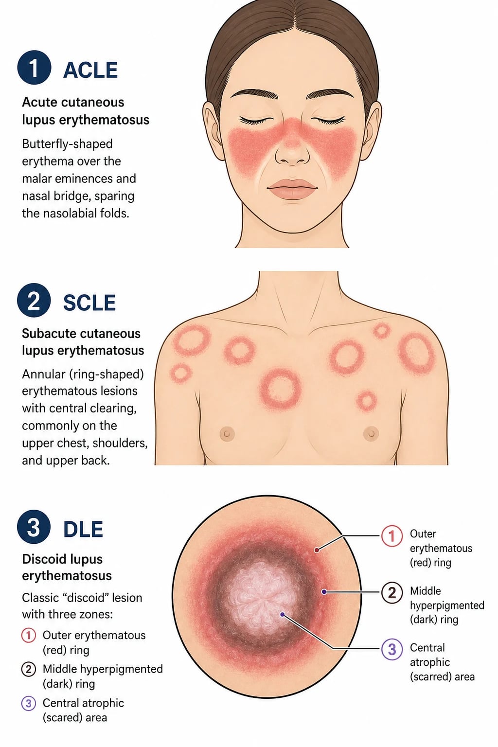
Definition & Classification
Cutaneous lupus erythematosus (CLE) encompasses every dermatological manifestation of lupus erythematosus. The 2018 revised Düsseldorf classification divides lesions into two groups — SLE-specific (also called LE-non-specific historically, now reserved for ACLE, SCLE and CCLE which are histopathologically interface dermatitis) and SLE-non-specific lesions (vasculitis, livedo, non-scarring alopecia, Raynaud's) — and recognises a third category, drug-induced lupus erythematosus (DILE).[1][4]
The SLE-specific CLE group comprises three morphological families based on clinical course, scarring potential and SLE association:[1][9]
| Family | Subtype | Morphology | Photosensitive | Scarring | SLE association |
|---|---|---|---|---|---|
| ACLE (acute) | Localised (malar) or generalised | Oedematous malar erythema, sparing nasolabial folds; or diffuse morbilliform | Yes | No (post-inflammatory dyspigmentation only) | Always present — ACLE is a SLE criterion |
| SCLE (subacute) | Annular/polycyclic OR papulosquamous | Coalescing rings or psoriasiform plaques, sun-exposed V of chest and arms | Highly (UVB-triggered) | No (hypopigmentation) | Anti-Ro/SSA in 70-90 percent; ~50 percent meet SLE criteria |
| CCLE (chronic) | DLE (localised, generalised, hypertrophic, linear) | Discoid scarring plaques, three-zone morphology | Variable | Yes (scarring alopecia, atrophy) | ~5-10 percent (DLE), higher in disseminated DLE |
| CCLE | LE profundus / panniculitis | Deep subcutaneous nodules → atrophic lipoatrophy | No | Yes (atrophy) | ~30-50 percent |
| CCLE | LE tumidus | Smooth urticarial plaques in sun-exposed sites | Highly | No | Less than 5 percent |
| CCLE | Chilblain LE | Acral violaceous nodules, cold-induced | No | Variable | ~20 percent |
| CCLE | Mucosal LE / oral DLE | Discoid plaques of lips, buccal mucosa, palate | No | Yes (scarring) | Variable |
Drug-induced LE is split into (a) systemic DILE — anti-histone antibodies (95 percent), procainamide/hydralazine/isoniazid trigger, spares the skin; (b) drug-induced SCLE — anti-Ro/SSA-positive, classically triggered by terbinafine, hydrochlorothiazide, calcium-channel blockers, TNF inhibitors, proton-pump inhibitors and griseofulvin. A rare drug-induced CCLE pattern occurs with fluorouracil and capecitabine.
Epidemiology [1]
- Incidence: approximately 4 per 100,000 adults per year in European and North American registries; cumulative CLE prevalence of ~40 per 100,000. ACLE is the second most frequent manifestation of SLE after arthritis, and ~70-85 percent of SLE patients develop cutaneous involvement at some point in their disease course.[2]
- Age and sex: peak onset 20-40 years. Female-to-male ratio is ~3:1 for ACLE (SLE-driven), ~2-3:1 for SCLE, and approximately 1:1 for DLE because DLE lacks the same hormonal amplification. Paediatric-onset DLE is rare (~3 percent of all DLE) and warrants complement (C1q, C2, C4) screening.[1][2]
- Ethnicity: DLE is the most common form of CLE in patients with skin of colour (Fitzpatrick IV-VI) — African, Afro-Caribbean, Hispanic and South Asian cohorts have higher rates of cicatricial alopecia and dyspigmentation because the erythema is masked by melanin and the disease is recognised late. ACLE and SCLE occur most commonly in fair-skinned white women.[2]
- Genetic risk factors: HLA-DR3 predisposes to ACLE and CLE generally; HLA-DR4 and HLA-DRB1*0301 are enriched in SCLE; HLA-DRB1*1501 is associated with DLE. C1q deficiency is rare but ~90 percent of homozygous C1q-deficient individuals develop a CLE-like disease by adolescence. C2 and C4 deficiency also increase CLE/SLE risk.[4]
- Environmental risk factors: ultraviolet B (290-320 nm, the most photosensitising band for CLE) and UVA (320-400 nm); smoking — confers a 2-fold risk and reduces hydroxychloroquine efficacy by ~50 percent via inhibition of lysosomal accumulation; drugs for SCLE (see differential and management sections); occupational exposure in outdoor workers, pilots, sailors and drivers.[4][7]
- Drug-induced SCLE proportions: 20-30 percent of all SCLE is drug-induced; >50 culprit drugs reported in VigiBase; terbinafine, hydrochlorothiazide, diltiazem, topical TNF inhibitors and pantoprazole are the most frequent triggers.[7]
CLE numerical anchors
Pathophysiology
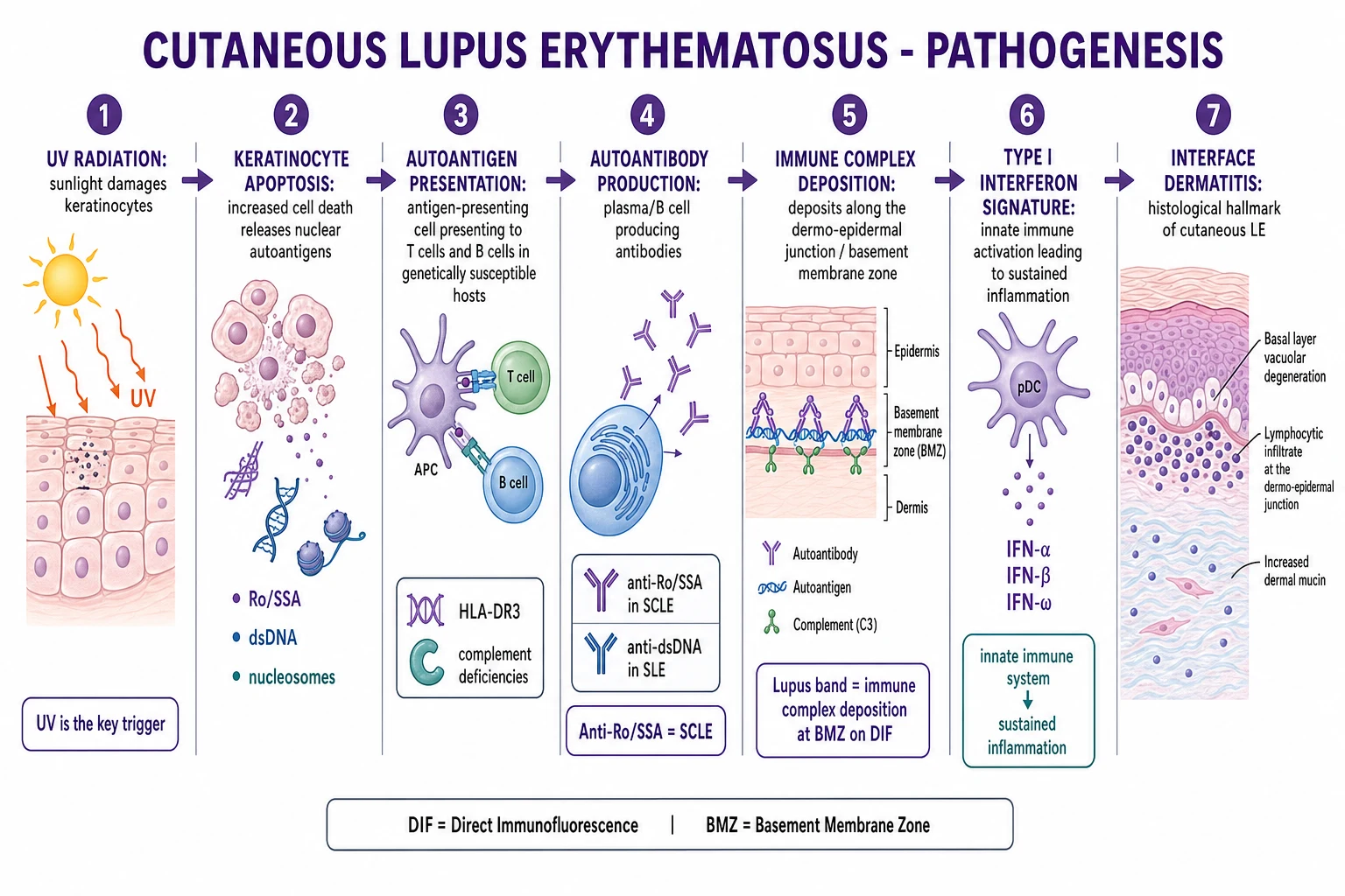
CLE is the prototypical Type I interferon-driven, UV-induced autoimmune skin disease, in which an environmental trigger (UVB) interacts with genetic susceptibility to produce an interface dermatitis centred on the basal layer of the epidermis and the dermo-epidermal junction.[1][4]
The UV-apoptosis-autoantigen loop: UVB (290-320 nm) directly damages keratinocyte DNA and induces apoptosis (programmed cell death with membrane-bleb formation). In susceptible individuals, apoptotic blebs fail to be cleared efficiently ("apoptotic cargo") — a defect in early complement components (C1q, C2, C4) reduces opsonisation — and intracellular autoantigens become exposed on the outer leaflet of the keratinocyte plasma membrane: Ro/SSA (TRIM21, 52 kDa), La/SSB (48 kDa), Sm, RNP, dsDNA, nucleosomes and histones. These autoantigens are visible to antigen-presenting cells and to circulating autoantibodies.[4]
Plasmacytoid dendritic cells (pDCs) and the Type I interferon signature: UV-damaged keratinocytes release CXCL9, CXCL10, CXCL11 and LL-37/DNA complexes that recruit pDCs into the dermis. pDCs are the body's most potent producers of IFN-α and IFN-β; once activated, they drive a sustained interferon-stimulated gene (ISG) response in keratinocytes — MxA, ISG15, IFIT1, IFI44L, OAS1 — which can be visualised by immunohistochemistry and quantified by RT-PCR as the "interferon signature". The Type I IFN pathway is most intense in SCLE and tumid LE lesions.[4]
Autoantibody amplification: Anti-Ro/SSA binds the exposed autoantigen on the keratinocyte surface, marking the cell for antibody-dependent cellular cytotoxicity (ADCC) by natural killer cells and for complement-mediated lysis via the classical pathway. Anti-Ro/SSA immune complexes are then internalised by FcγR-bearing pDCs in a positive-feedback loop, sustaining the interferon response.[1][4]
T-cell contribution: CD4+ T-helper 1 (Th1) cells predominate, with CD8+ cytotoxic lymphocytes at the dermal-epidermal junction directly lysing basal keratinocytes. The perivascular and periadnexal lymphocytic infiltrate extends deep into the dermis — a feature that helps distinguish DLE from superficial interface dermatitides like lichen planus.[4]
Immune complex deposition (the lupus band): Circulating anti-BMZ antibodies and immune complexes deposit at the basement membrane zone — a continuous granular band of IgG, IgA, IgM and C3 on direct immunofluorescence (DIF). The lupus band is positive in lesional skin of ~70-90 percent of active DLE and in non-lesional sun-exposed skin in ~50-80 percent of active SLE, where it reflects systemic immune complex disease.[1]
Genetics and complement: HLA-DR3 and HLA-DR4 confer susceptibility to SCLE; HLA-DRB1*1501 to DLE. Rare but instructive are homozygous C1q deficiency (~90 percent develop a CLE-like disease in childhood), C2 deficiency and C4 deficiency — early complement components are essential for the clearance of apoptotic blebs, so their absence produces a "lupus-like" interface dermatitis even without overt SLE.[4]
Smoking as a pharmacological modifier: Smoking downregulates lysosomal accumulation of hydroxychloroquine and adds a pro-apoptotic, pro-interferon stimulus; smokers require up to twice the dose of HCQ and respond half as often. Smoking cessation therefore improves both the natural history and the response to antimalarial therapy.[4]
The CLE pathway ABCDE-F
Clinical Presentation [1]
CLE manifests across a wide morphological spectrum. The clinical examination must include the entire skin surface, scalp, conchal bowls, oral cavity, nails and periungual nailfold capillary bed in any patient with suspected lupus — partial examination misses scalp DLE scarring and oral lupus plaques that change management.[1][5]
ACLE — Acute cutaneous lupus
- Malar (butterfly) rash: the canonical ACLE lesion — bilateral, symmetrical, oedematous erythema over the cheeks and bridge of the nose with pathognomonic sparing of the nasolabial folds (rosacea and seborrhoeic dermatitis involve those folds). The rash is photosensitive, non-scarring, and resolves over weeks-to-months with treatment of the underlying SLE, frequently leaving post-inflammatory hyperpigmentation, especially in darker skin.[1][2]
- Generalised ACLE: a widespread morbilliform, dusky, photosensitive maculopapular eruption accentuated in sun-exposed sites (upper chest, V of the neck, extensor forearms, dorsum of hands sparing the knuckles — a useful clue against dermatomyositis).[2]
- Bullous ACLE is rare; tense vesicles and bullae on erythematous or normal skin indicate neutrophil-rich subepidermal blistering, anti-Type VII collagen antibodies, and overlap with bullous pemphigoid-like disease.
- ACLE always indicates active SLE; full systemic assessment is mandatory (anti-dsDNA, complement C3/C4, urinalysis, renal function, haematology).[2]
SCLE — Subacute cutaneous lupus
- Annular/polycyclic SCLE (~50 percent): coalescing erythematous rings with raised, slightly scaly advancing edges and central clearing; lesions grow centrifugally over days to weeks, producing polycyclic or figurative patterns on the upper trunk and arms. They do not scar, but may leave long-lasting dyspigmentation — particularly in Fitzpatrick III-VI skin.[1][6]
- Papulosquamous (psoriasiform) SCLE (~50 percent): scaly erythematous plaques resembling psoriasis but without the Auspitz pinpoint-bleed sign, again in sun-exposed sites.[1][6]
- Distribution: V of the chest, upper back, shoulders, extensor forearms, lateral neck — the "photosensitive" distribution. The face is involved in only ~20 percent (distinguishes from ACLE).[4]
- Anti-Ro/SSA positivity in 70-90 percent; anti-La/SSB in 10-20 percent (often co-occurs).[4]
- Drug-induced SCLE is morphologically indistinguishable from idiopathic SCLE — the index trigger is the temporal relationship of drug exposure (latency weeks to months) and resolution within 4-8 weeks of drug withdrawal.[7]
DLE — Discoid lupus erythematosus
- Three-zone morphology of the active discoid plaque is pathognomonic:[1]
- Active raised violaceous border with adherent scale and follicular plugging (keratotic plugs filling hair-follicle openings; the "carpet-tack sign" — when the adherent scale is lifted, the keratotic spicules remain behind, projecting from the underlying follicular openings like carpet tacks).[5]
- Mid-zone of hyperpigmentation, telangiectasia, atrophy and gradual depigmentation.
- Central atrophic scarring with hypopigmented "scarred white" centre — this is permanent.
- Sites of predilection: scalp (scarring alopecia), external ears and conchal bowls (highly characteristic), malar eminences, nasal bridge, periorbital area, vermilion border, neck, V of chest. Mucosal DLE affects the buccal mucosa, hard palate, gingiva and lips with discoid plaques bearing radiating white striae (leukoplakia-like).[1][5]
- Localised DLE = head and neck only (~85 percent of cases, lower SLE progression). Generalised/disseminated DLE = above and below the neck with trunk or extremity involvement (much higher SLE progression ~20 percent).[1]
- Variants of DLE (recognise to avoid misdiagnosis):[1]
- Hypertrophic (verrucous) DLE — thick verrucous plaques on extensor limbs, palms and soles; mimics keratoacanthoma or SCC; biopsy to exclude malignant transformation.
- Linear DLE — follows Blaschko lines, often unilateral, on face or limbs; presents in young adults and may be confused with linear lichen planus or linear psoriasis.
- Comedo-like DLE — central keratinous plugs resembling acne, on the face or trunk.
- Lichenoid DLE — overlap with lichen planus, particularly on the wrists and oral mucosa.
- Dermoscopy of DLE is highly specific: yellow-white follicular plugs, white structureless scarring areas, peripheral pigmented network and red vascular loops; absent follicular ostia distinguish scarring DLE from non-scarring mimics.[5]
LE profundus / Lupus panniculitis
- Deep, firm subcutaneous nodules or plaques with overlying normal or mildly erythematous skin; heals with characteristic "lipoatrophy" — depressed cup-shaped atrophic scars.[1]
- Distribution: face (especially cheeks), upper arms, buttocks, trunk.
- Histology shows lobular panniculitis with hyalinised fat necrosis, a lymphoplasmacytic infiltrate, and often overlying epidermal CLE features (so a deep incisional or wide-punch biopsy including subcutis is essential).
- Differential includes subcutaneous panniculitis-like T-cell lymphoma (SPTCL) — both have lobular panniculitis, but SPTCL shows atypical CD8+ T-cell infiltrates with clonality (TCR rearrangement), haemophagocytosis, and systemic features (fever, cytopenias).
LE tumidus
- Smooth, urticarial-like, non-scarring plaques in sun-exposed sites (face, upper chest, extensor arms), typically without surface scale.[1]
- The most photosensitive CLE subtype on phototesting; strikingly positive cutaneous lupus photosensitivity on minimal erythema dose testing.
- Histology shows abundant dermal mucin deposition with relatively subtle interface change — sometimes called "mucinosis" in some series.
- Excellent response to antimalarials.
Chilblain lupus erythematosus
- Violaceous, cold-induced tender nodules and plaques on the toes, fingers, heels, nose and ears; resemble idiopathic chilblains (perniosis) but persist beyond winter and may ulcerate.[1]
- Anti-Ro/SSA positive in ~50 percent. Differentiation from idiopathic chilblains is critical — careful inspection for underlying interface change, ANA testing, and follow-up.
- Rare familial Aicardi-Goutières-like chilblain LE is associated with TREX1 mutation (gain-of-function) and constitutive type I IFN activation.
Mucosal lupus erythematosus / Oral DLE
- Oral DLE is the most common mucosal CLE; lesions are discoid or lichenoid plaques of the buccal mucosa, palate, gingiva, lip vermilion and occasionally the tongue; radiating white striae and central ulceration are typical.[1]
- Palatal erythema with central ulceration ("palatal discoid lupus") is highly characteristic.
- Nasal septal perforation and xerostomia (Sjögren's overlap) may coexist.
- SCC arising in long-standing oral DLE is reported — biopsy non-healing lesions after 4-6 weeks.
SLE-non-specific cutaneous features (still important)
- Non-scarring diffuse alopecia (lupus hair), periungual capillary dilatation and dropout (best seen with nailfold capillaroscopy), Raynaud's phenomenon, livedo reticularis, leucocytoclastic vasculitis with palpable purpura, urticarial vasculitis, and bullous LE (autoantibodies to Type VII collagen) — these are not CLE-specific but they support systemic disease activity.[2]
Differential Diagnosis
| Mimic (with CLE type) | Distinguishing features |
|---|---|
| Rosacea (vs ACLE) | Papulopustular with telangiectasia, involves the nasolabial folds (ACLE spares them); no autoantibodies; hot flushes, alcohol trigger. |
| Seborrhoeic dermatitis (vs ACLE) | Greasy yellowish scale, eyebrow and nasolabial fold involvement; responds to antifungals; no systemic features. |
| Erysipelas / cellulitis (vs ACLE) | Unilateral, tender, warm, fever, raised inflammatory markers. |
| Pellagra (vs ACLE generalised) | Casal necklace (photosensitive rash on neck), GI symptoms, dementia; niacin deficiency. |
| Dermatomyositis (vs ACLE/heliotrope) | Heliotrope (purple) periorbital rash, Gottron papules on knuckles, proximal muscle weakness, raised CK. |
| Tinea corporis / facei (vs SCLE annular) | Concentric scaling border, central clearing, KOH hyphae positive; fungi on Sabouraud agar. |
| Granuloma annulare (vs SCLE annular) | Skin-coloured to erythematous papules in annular configuration, no scale, no sun distribution, no itch, biopsy shows palisading granulomas. |
| Erythema annulare centrifugum (vs SCLE annular) | Trailing scale inside the advancing edge (the opposite of SCLE); associated with infections, drugs, malignancy; no anti-Ro. |
| Erythema multiforme (vs SCLE annular) | Target lesions with central duskiness/blister, mucosal involvement (lips, oral cavity), often post-infectious (HSV, Mycoplasma). |
| Psoriasis (vs SCLE papulosquamous / vs DLE scalp) | Silvery scale, Auspitz sign, nail pitting, intergluteal involvement; no follicular plugging, no atrophy, no scarring alopecia. |
| Lichen planus (vs DLE / vs mucosal LE) | Violaceous, flat-topped, polygonal papules, Wickham striae in oral cavity; biopsy shows saw-tooth rete ridges and band-like lymphocytic infiltrate; no follicular plugging or hyalinised BMZ. |
| Tinea capitis (vs DLE scalp) | Patchy hair loss with scaling, broken hairs at the surface ("black dots"), KOH hyphae; no scarring unless kerion. |
| Central centrifugal cicatricial alopecia (CCCA) (vs DLE scalp) | Midline parietal/vertex scarring alopecia in women of African descent; no erythema or follicular plugging; biopsy shows premature desquamation of inner root sheath. |
| Frontal fibrosing alopecia (vs DLE frontal scalp) | Band-like frontotemporal alopecia with perifollicular erythema and loss of follicular ostia; biopsy shows lymphocytic scarring alopecia around vellus hairs; brow loss is a clue. |
| Lichen planopilaris (vs DLE scalp) | Perifollicular erythema and scale of the scalp without the three-zone discoid plaque; biopsy shows lichenoid interface around the follicular infundibulum. |
| Folliculitis decalvans (vs DLE scalp) | Pustules around follicular openings, tufted hairs ("tufted folliculitis"); cultures grow Staphylococcus aureus. |
| Polymorphic light eruption (PLE) (vs LE tumidus) | Onset within hours to days of sun exposure, resolves within a week off sun, no autoantibodies, no dermal mucin; biopsy shows spongiosis with perivascular inflammation. |
| Solar urticaria (vs LE tumidus) | True urticaria with weal-and-flare within minutes of sun exposure, transient (under 24 h); antihistamines help. |
| Subcutaneous panniculitis-like T-cell lymphoma (SPTCL) (vs LE panniculitis) | Both have lobular panniculitis — SPTCL has atypical CD8+ T cells with TCR clonality, hemophagocytic syndrome in 20 percent, systemic symptoms; biopsy mandatory if cyclosporine or systemic therapy fails. |
| Idiopathic chilblains (vs chilblain LE) | Winter-only lesions, no anti-Ro, no interface change on biopsy, no progression beyond cold seasons. |
| Aicardi-Goutières syndrome (vs familial chilblain LE) | TREX1 mutation, congenital, type I IFN signature, basal ganglia calcification, glaucoma. |
| SCC arising in chronic DLE (vs hypertrophic DLE) | Ulcerated non-healing nodule, indurated edge, biopsy of the most atypical area is mandatory; chronic DLE supports SCC after years of disease. |
Histopathology
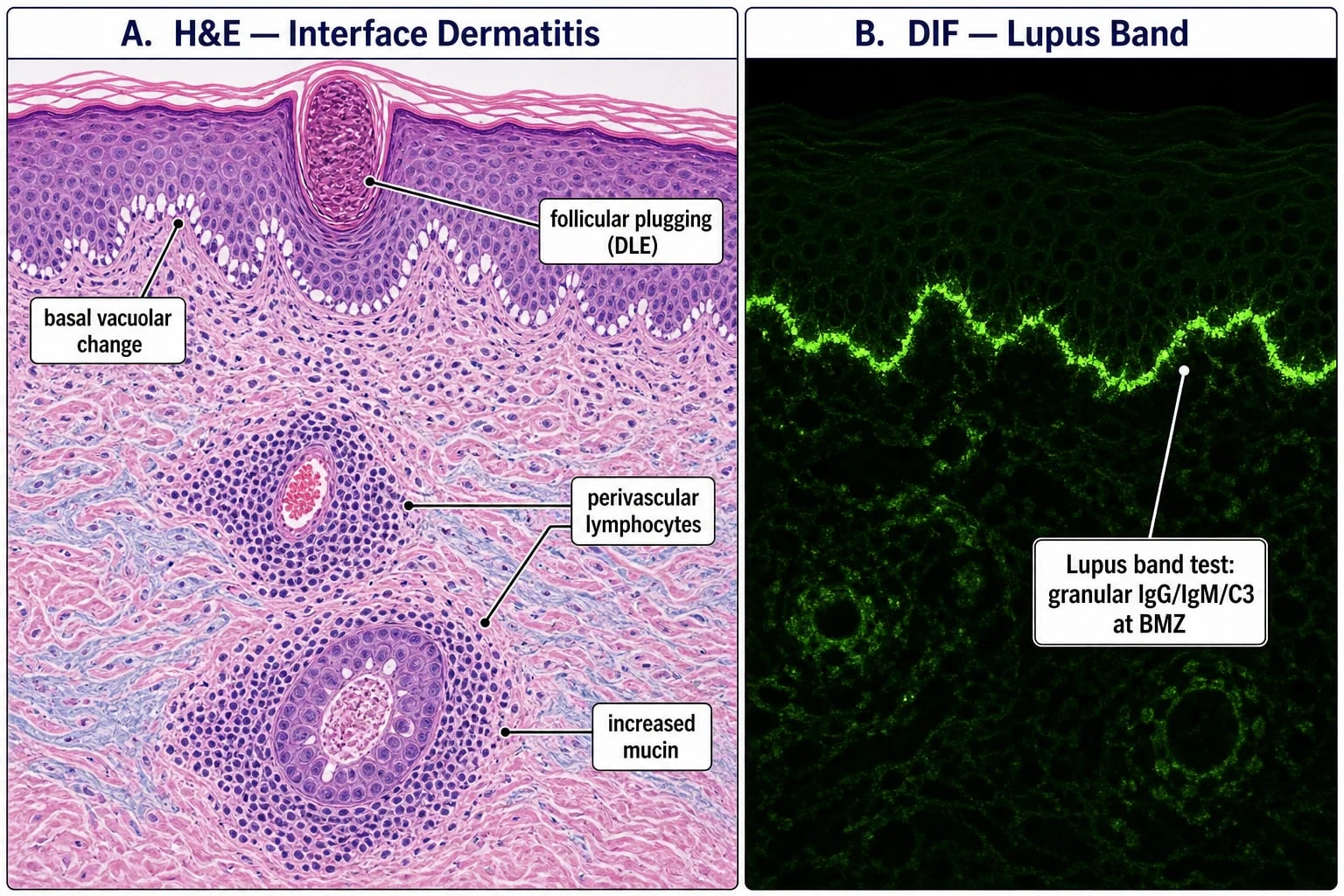
The unifying histological lesion is interface dermatitis (also called "hydropic" or "vacuolar" interface dermatitis) centred on the basal layer. A lesional punch biopsy — taken from the active edge of an established lesion — is the gold standard.[1][4]
Light microscopy (H&E) — common to all CLE subtypes:[1]
- Basal-layer vacuolar degeneration — vacuolisation of basal keratinocytes with pyknosis of nuclei; the lesion that gives "interface dermatitis" its name.
- Hyperkeratosis and follicular plugging — most pronounced in DLE; keratin in dilated follicular infundibula is the histological substrate of the carpet-tack sign.
- Epidermal atrophy in chronic lesions with effacement of rete ridges; acanthosis is occasionally seen in hypertrophic DLE.
- Perivascular and periadnexal lymphocytic infiltrate — extends deep into the dermis (a feature that helps distinguish DLE from lichen planus, which is superficial). Includes CD4+ T-helper and CD8+ T-cytotoxic cells.
- Dermal mucin deposition — glycosaminoglycans (predominantly hyaluronic acid) accumulate between collagen bundles, producing a feathery blue-grey appearance on H&E and staining strongly with colloidal iron or Alcian blue. Dermal mucin is the most useful marker distinguishing CLE from lichen planus and other interface dermatitides. [1]
| CLE subtype | Most useful diagnostic feature |
|---|---|
| ACLE | Subtle interface change + perivascular lymphocytes + dermal mucin (epidermis may appear almost normal). |
| SCLE | Florid interface dermatitis with epidermal necrosis of individual keratinocytes, occasional intraepidermal vesiculation, prominent dermal mucin. |
| DLE | Follicular plugging + epidermal atrophy + hyperkeratosis + thick, basement-membrane-like band at the dermo-epidermal junction (Grenz zone with hyalinised BMZ thickening); deep periadnexal lymphocytic infiltrate. |
| LE profundus / panniculitis | Lobular panniculitis with hyalinised fat necrosis, lymphoplasmacytic infiltrate, occasional lymphoid follicles with germinal centres; superficial and deep biopsy required. |
| LE tumidus | Striking dermal mucin with relatively mild interface change; the infiltrate is predominantly perivascular; follicular plugging is absent. |
| Chilblain LE | Interface change with superficial and deep perivascular lymphocytic vasculitis and fibrinoid vessel-wall change. |
| Oral DLE | Centred on the epithelium-connective tissue interface with parakeratosis, epithelial atrophy, basement-membrane thickening and perivascular inflammation in the lamina propria; mimics lichen planus histologically but the presence of follicular plugging in surrounding skin and DIF findings helps. |
Direct immunofluorescence (DIF) — the "lupus band test":[1]
- Technique: a 4-mm punch from the lesional active edge (positive in ~70-90 percent of active DLE and ~40-50 percent of SCLE) — or from non-lesional sun-exposed skin (e.g., volar forearm; positive in ~50-80 percent of active SLE, ~30 percent in remission); transport in Michel's medium or snap-freeze; cryosection; fluorescein isothiocyanate-labelled anti-human IgG, IgA, IgM and C3.
- Appearance: a continuous, granular band of immunoreactant deposition along the dermo-epidermal junction — usually all of IgG, IgM and C3, sometimes IgA and fibrinogen.
- Pitfall: a false-positive lupus band can occur in sun-damaged non-LE skin, dermatoheliosis, and rosacea; specificity is improved by taking a non-sun-exposed (buttock) non-lesional biopsy.
- Differential from BP: in bullous pemphigoid, DIF shows a linear band of IgG/C3 at the BMZ (salt-split shows the antibody on the epidermal side); in CLE the deposition is granular and is not separated by salt-split. [1]
Immunohistochemistry correlates of the interferon signature: in SCLE and tumid LE, MxA and ISG15 are upregulated in keratinocytes — these can be used as supportive diagnostic stains in equivocal cases.[4]
Investigations
The work-up of any CLE patient combines (1) confirmation of the diagnosis (clinical + skin biopsy + DIF), (2) staging for systemic involvement (autoantibody screen, organ assessment), (3) elimination of drug triggers, and (4) establishment of a disease-activity baseline by CLASI scoring.[2][9]
- Antinuclear antibody (ANA) by IIF on HEp-2 cells: positive in ~70-90 percent of ACLE, ~70 percent of SCLE, and only ~20 percent of isolated DLE. A negative ANA does not exclude CLE — confirm with biopsy.[2]
- Anti-Ro/SSA (TRIM21, 52 kDa + 60 kDa): positive in 70-90 percent of SCLE, ~30 percent of DLE, ~50 percent of Sjögren's and ~95 percent of neonatal lupus. Specific for the anti-Ro/SSA-CLE axis; correlates with photosensitivity and neonatal CHB risk.[4]
- Anti-La/SSB: present in ~10-20 percent of SCLE; usually co-exists with anti-Ro/SSA.
- Anti-U1-RNP: mixed connective tissue disease overlap (Sharp syndrome) with CLE-like skin.
- Anti-Sm (anti-Smith): highly specific for SLE; positive in only 5-20 percent — but counts as a SLE criterion.
- Anti-dsDNA: specific for SLE; rising titres correlate with disease activity and nephritis flare. Use ELISA or Crithidia luciliae IIF to confirm specificity.
- Antiphospholipid antibodies: lupus anticoagulant, anti-cardiolipin, anti-β2-glycoprotein I — request in any patient with thrombotic events, recurrent miscarriage, or features of secondary antiphospholipid syndrome.
- Complement C3 and C4: low in active SLE (consumed by immune complexes); persistently low C4 with normal C3 should raise suspicion for C1q deficiency.
- Full blood count: cytopenias — anaemia of chronic disease, autoimmune haemolytic anaemia (Coombs-positive), leucopenia, lymphopenia, thrombocytopenia — all SLE criteria.
- ESR and CRP: ESR-CRP discordance (ESR up, CRP normal) supports an SLE flare rather than infection.
- Renal assessment: urinalysis with microscopy (proteinuria, RBC casts), urine protein:creatinine ratio, serum creatinine, eGFR; every SLE patient needs renal screening at diagnosis and quarterly thereafter; any active sediment triggers nephrology referral and possible renal biopsy.
- Skin biopsy — H&E and DIF (the lupus-band test): from the active advancing edge of an established lesion (not the healed centre), with a second biopsy from non-lesional sun-exposed skin (forearm) in any patient being assessed for SLE.[1]
- Dermoscopy of DLE: yellow-white follicular plugs, white structureless areas (scarring), peripheral pigmented network, red dots (vascular loops), absent follicular ostia — supports the diagnosis without biopsy.[5]
- Phototesting (UVA + UVB minimal erythema dose) in research settings; useful for confirming photosensitivity in LE tumidus and SCLE where the histology may be subtle.
Autoantibody frequencies by CLE subtype: ANA (ACLE ~80%, SCLE ~70%, DLE ~20%); Anti-Ro/SSA (SCLE 70-90%, neonatal lupus ~95%); Anti-dsDNA (SLE-specific, under 50%); Anti-Sm (SLE, 5-20%); Antihistone (drug-induced SLE ~95%).[2]
Disease activity and damage scoring — CLASI: the Cutaneous Lupus Erythematosus Disease Area and Severity Index measures (a) CLASI-A (activity): erythema, scale, oedema, mucous-membrane involvement, recent alopecia, inflammatory features; (b) CLASI-D (damage): dyspigmentation, scarring, scarring alopecia. CLASI is validated for both DLE and SCLE; a 4-point change in CLASI-A is clinically significant and is the standard primary endpoint in CLE trials.[2]
Management
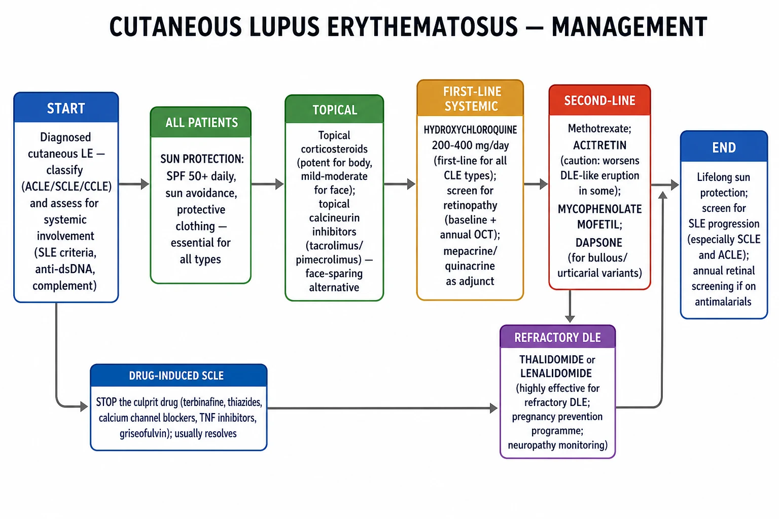
The treatment ladder is built on (1) sun protection + smoking cessation as the foundation, (2) topical therapy for limited disease, (3) hydroxychloroquine as first-line systemic for all CLE types, (4) second-line immunosuppressants in steroid-sparing combination, and (5) biologics and thalidomide for refractory disease. Drug-induced SCLE requires immediate withdrawal of the culprit drug.[1][4][9]
Lifestyle and photoprotection (for every patient)
- Sunscreen: broad-spectrum SPF 50+ with both UVA (PPD ≥ 1/3 of SPF) and UVB protection, applied liberally (2 mg/cm²) and reapplied every 2 hours while outdoors. Reflective physical blockers (zinc oxide, titanium dioxide) are useful in highly photosensitive patients.
- Clothing: UPF 50+ long-sleeved shirts, wide-brimmed hats (>7.5 cm brim), wraparound UV-blocking sunglasses — these reduce UV exposure by 90-99 percent.
- UV films on windows (cars, homes), avoidance of midday sun (10 am-2 pm), and avoidance of reflective surfaces (water, sand, snow).
- Visible light (400-500 nm) and blue light can also trigger photosensitivity in darker skin — tinted sunscreens (iron oxide) or tinted moisturisers may be needed.
- Smoking cessation — smoking doubles the dose requirement of hydroxychloroquine and reduces the response rate by half; current smokers respond in ~30 percent vs ~70 percent in non-smokers.[4][8]
- Vitamin D supplementation 800-1000 IU daily (often deficient due to photoprotection).
Topical therapy (limited disease or adjunctive)
- Topical corticosteroids: potent (clobetasol propionate 0.05 percent) for scalp, body, and acral lesions; mid-potency (mometasone furoate, betamethasone valerate) for the face; limit use to 2-4 weeks to avoid atrophy and telangiectasia (particularly on the face and intertriginous areas).[1]
- Topical calcineurin inhibitors (tacrolimus 0.1 percent ointment, pimecrolimus 1 percent cream): steroid-sparing, particularly on the face and intertriginous CLE; twice-daily application; transient burning is the main side-effect.
- Intralesional triamcinolone acetonide 5-10 mg/mL: useful for thick DLE plaques and hypertrophic DLE; 0.05-0.1 mL per cm²; up to 1 mL per session; avoid skin atrophy.
- Topical retinoids (tretinoin, adapalene): for hypertrophic DLE; can be irritating.
- R-salbutamol 0.5 percent cream (experimental): topical β-2 agonist that inhibits keratinocyte apoptosis — case-series evidence.
First-line systemic therapy
- Hydroxychloroquine (HCQ) 200-400 mg/day (≤5 mg/kg/day based on real body weight, sometimes 6.5 mg/kg for ideal body weight in obese patients): the first-line systemic agent for every CLE subtype. Onset of action 8-12 weeks; full benefit at 6 months. Baseline + annual retinal screening with OCT, fundus autofluorescence, and visual fields after 5 years (or earlier if risk factors — tamoxifen, renal failure, age >65, dose >5 mg/kg/day).[1][8][9]
- Quinacrine (mepacrine) 100 mg/day: added to hydroxychloroquine for partial non-responders; does not cause retinopathy (advantage over chloroquine); may cause yellow discoloration of the skin and sclera at higher doses.
- Chloroquine 250-500 mg/day: occasionally substituted for HCQ in smokers or partial non-responders, but has a higher retinal toxicity rate than HCQ and is contraindicated long-term.
Second-line immunosuppressants (steroid-sparing or antimalarial non-responders)
- Methotrexate 10-25 mg weekly (oral or subcutaneous) with folic acid 5 mg 24-48 h later: well-tolerated; effective for SCLE, generalised DLE, and articular SLE overlap; monitor LFTs every 4-8 weeks, FBC, renal function, hepatitis B/C serology.
- Mycophenolate mofetil (MMF) 2-3 g/day: particularly useful for SLE-associated CLE; pregnancy-compatible if switched pre-conception; main side-effects GI intolerance, cytopenias, teratogenicity.
- Azathioprine 1-2 mg/kg/day: steroid-sparing, fatigue and cytopenia; TPMT assay required before initiation to avoid severe myelosuppression in deficient patients.
- Acitretin 25-50 mg/day (isotretinoin 0.5-1 mg/kg as alternative): effective for hypertrophic DLE and refractory DLE; highly teratogenic — pregnancy prevention for 3 years post-stopping acitretin.
- Dapsone 50-150 mg/day: particularly effective for bullous LE, urticarial vasculitis, SCLE; G6PD screen mandatory (haemolysis in deficiency); methaemoglobinaemia, agranulocytosis, sulphone hypersensitivity syndrome. [1]
Biologics and targeted therapy (refractory SLE-associated CLE)
- Belimumab (anti-BAFF/BLyS) 200 mg SC weekly: approved for SLE with active skin involvement; best evidence in SCLE and ACLE; BLISS-LN and BLISS-52/76 showed significant improvement in mucocutaneous endpoints (SELENA-SLEDAI, BILAG).[4][9]
- Anifrolumab (anti-type I interferon receptor) 300 mg IV every 4 weeks: approved for moderate-to-severe SLE (TULIP-1, TULIP-2); pooled analyses show rapid and significant improvement in CLASI-A and BILAG mucocutaneous domain; most effective CLE biologic in SLE-associated disease.[9]
- Rituximab (anti-CD20) 1 g × 2 weeks every 6 months: off-label for refractory CLE and SLE; benefits some patients, particularly with B-cell driven disease; registry data from the British Isles Lupus Assessment Group (BILAG) suggest a CLASI response in ~50 percent of refractory cases.
- Litifilimab (anti-BDCA2) — pDC depleting antibody in clinical trials with promising CLE outcomes.
- JAK inhibitors (baricitinib, tofacitinib, upadacitinib) — case-series evidence in refractory SCLE and DLE; phase II-III trials ongoing; infectious and thrombotic risk of class labelling.[4]
Refractory DLE pathway
- Thalidomide 50-100 mg/night with mandatory pregnancy-prevention programme (UK Preg-X, US S.T.E.P.S.); neuropathy monitoring at baseline and every 6 months (paresthesia, nerve conduction studies); highly effective with response rates of ~80-90 percent in refractory DLE — most powerful agent for DLE scarring alopecia.
- Lenalidomide 5-10 mg/day: thalidomide analogue with less neuropathy but more myelosuppression and thrombosis risk.
- Apremilast (PDE4 inhibitor) 30 mg BD — small case series in refractory DLE.
- Dapsone — second-line alternative in the refractory pathway.[1]
Drug-induced SCLE — stop the trigger
- Withdraw the likely culprit drug; resolution within 4-8 weeks confirms the diagnosis. Use a temporal probability map (terbinafine, then thiazides, CCB, PPI, TNF inhibitors, statins, antihistamines).
- Treat residual inflammation with topical corticosteroids or short-course antimalarial if withdrawal does not resolve.[7]
Special Populations
CLE behaves differently in pregnancy, childhood, advanced age, and across ethnic skin types. Anticipating these patterns prevents irreversible morbidity.[4][8][10]
Pregnancy and neonatal lupus
Any anti-Ro/SSA-positive woman of child-bearing age is at risk of neonatal lupus through transplacental passage of IgG autoantibodies from around the 16th week of gestation.[10]
- Cutaneous neonatal lupus: ~25 percent of affected neonates develop an annular or macular SCLE-like rash in sun-exposed areas (face, scalp, extremities) within the first weeks of life, often photosensitive, self-resolving by 6-12 months as maternal antibodies clear.
- Neonatal congenital heart block (CHB): the most serious complication, irreversible and usually complete (third-degree); the overall risk in a first anti-Ro/SSA-positive pregnancy is ~1-2 percent, rising to ~20 percent in a subsequent pregnancy after one affected sibling. 70-80 percent of children with CHB ultimately require a permanent pacemaker.[10]
- Other neonatal features: cytopenias, hepatitis, hepatosplenomegaly, macrocephaly with lenticulostriate vasculopathy, hydrops fetalis.
- Obstetric management: serial foetal echocardiography at 16-26 weeks (weekly or fortnightly) for at-risk pregnancies; dexamethasone 4 mg/day (crosses the placenta) when first-degree or second-degree block is detected in utero (controversial).
- Pre-conception hydroxychloroquine 200-400 mg/day reduces recurrence of CHB by ~50 percent (Izmirly, NEJM 2020; EULAR/AHA recommendation), and is safe throughout pregnancy.
- Drug safety in pregnancy: safe — hydroxychloroquine, low-dose prednisolone, azathioprine. Avoid — mycophenolate mofetil (teratogenic; switch to azathioprine pre-conception), methotrexate (teratogenic; wash out 3 months), cyclophosphamide (severe teratogen), retinoids (severe teratogen; acitretin has a 3-year washout), thalidomide (severe teratogen — REMS programme required).
Childhood-onset CLE
Paediatric DLE is rare (~3 percent of all DLE); presentation is similar but subtle — early diagnosis and aggressive therapy reduce scarring alopecia and dyspigmentation. Investigate complement deficiencies (C1q, C2, C4) in any child with CLE; homozygous C1q deficiency mandates family counselling. Hydroxychloroquine is the first-line systemic drug, with retinal surveillance every 6 months (more frequent than adults). Avoid prolonged topical corticosteroids and long-term systemic steroids.[4]
Older adults (>70)
CLE is the presenting feature of SLE in a high proportion of elderly patients; drug-induced SCLE is more frequent (polypharmacy). Consider lower hydroxychloroquine dosing for renal function (eGFR under 30 mL/min/1.73 m²) and annual retinal screening from year 1 rather than year 5. Avoid polypharmacy and review steroid loads in comorbid patients.[8]
Skin of colour
In Fitzpatrick IV-VI skin, the erythema of CLE becomes violaceous or hyperpigmented, and follicular plugging, dyspigmentation and scarring alopecia are more prominent. The clinical threshold for suspicion should be lowered, and dermoscopy, biopsy and DIF are central to avoid delayed diagnosis. Patient education on photosensitivity and use of tinted (visible-light-protective) sunscreens is particularly important.[2]
Occupational and lifestyle factors
Outdoor workers (farmers, builders), pilots, sailors, professional drivers, and people living near the equator or at high altitude have significantly higher UV exposure. UV-protective films on car and home windows, hats, and high-SPF lip balms are essential. Photosensitising medications (doxycycline, tetracyclines, thiazides, amiodarone, NSAIDs, sulfonamides) should be reviewed in anti-Ro/SSA-positive patients.[1]
Prognosis
The prognosis of CLE varies widely with subtype and the presence or absence of systemic disease. The "scarring versus non-scarring" axis predicts most of the long-term morbidity.[1][2][4]
- ACLE: resolves with treatment of the underlying SLE; no cutaneous scarring, but post-inflammatory hyperpigmentation may persist for months, particularly in Fitzpatrick IV-VI skin. Course is dictated by systemic disease activity.
- SCLE (idiopathic): chronic relapsing in ~80 percent; ~50 percent eventually meet ≥4 ACR or 2019 EULAR/ACR SLE criteria. Severe visceral involvement (proliferative nephritis) is uncommon. Hydroxychloroquine controls disease in ~70 percent; relapses on stopping photoprotection are common.
- Drug-induced SCLE: excellent prognosis — resolves within 4-8 weeks of withdrawing the culprit drug; can persist longer if sun exposure is continued.
- DLE (localised): ~85 percent remain skin-limited; permanent scarring alopecia, dyspigmentation, and lip atrophy are the principal sources of morbidity; antimalarials within 6 months of onset preserve hair in active edge lesions.
- Generalised/disseminated DLE: ~20 percent transition to SLE — justifies active surveillance.
- DLE scarring alopecia: irreversible once active disease ceases — early aggressive therapy and minimisation of UV exposure are critical.
- LE profundus/panniculitis: chronic relapsing; lipoatrophy persists once active disease abates; cosmetic surgery is an option for deep atrophy in inactive disease.
- LE tumidus: the most photosensitive subtype — typically highly responsive to antimalarials.
- Bullous LE: responds well to dapsone; healing in 4-8 weeks.
- Chilblain LE: may ulcerate, infect and progress to digital destruction if untreated; respond to antimalarials, calcium-channel blockers and warming.
- Mucocutaneous LE: ~3-5 percent of chronic oral/lip DLE may develop squamous cell carcinoma in long-standing lesions — biopsy non-healing ulcerated DLE at any age.
- Mortality: CLE by itself is rarely fatal; mortality is driven by systemic organ involvement (renal, CNS, cardiovascular) where the patient meets SLE criteria. SLE mortality is now principally driven by cardiovascular disease and infection rather than active lupus per se. [1]
Prognosis at a glance
Evidence, Guidelines & Regional Differences [1]
- EULAR recommendations for SLE management, 2023 update (Fanouriakis et al., Ann Rheum Dis 2024) — directly addressed antimalarial dosing, HCQ ocular monitoring and the role of anifrolumab and belimumab in SLE with cutaneous involvement.[9]
- American Academy of Dermatology (AAD) guidelines for DLE, SCLE and drug-induced lupus — referenced in updated 2024 reviews.[4]
- An Bras Dermatol 2023 (Vale & Garcia): comprehensive etiopathogenic-clinical-therapeutic review of CLE.[1]
- Am J Clin Dermatol 2023 (Niebel): pathogenesis and future therapeutic directions in CLE, including anifrolumab (anti-IFN-α receptor; TULIP-LN), litifilimab (anti-BDCA2 pDC) and JAK inhibitors.[4]
- Best Practice & Research Clinical Obstetrics & Gynaecology 2020 (Brito-Zeron, Izmirly, Ramos-Casals): autoimmune-mediated congenital heart block — evidence base for screening and management.[10]
- Drugs & Aging 2024 (Heinly et al.): CLE-specific considerations in older populations, including dosing adjustments and comorbidity-driven decisions.[8]
- Lancet / NEJM 2011 (BLISS-52) and 2020 (EMBODY) — belimumab and anifrolumab phase III trials — both meet primary endpoints with significant mucocutaneous improvement (SELENA-SLEDAI, BILAG mucocutaneous, CLASI-A).
- ACR/EULAR 2019 classification criteria for SLE — confirmed cutaneous involvement as a major clinical domain (acute cutaneous, chronic cutaneous, oral ulcer, non-scarring alopecia).
- American Academy of Ophthalmology 2016 / 2023 — recommendations on hydroxychloroquine retinal screening — baseline fundus examination with optional adjuncts, then annual screening after 5 years (or sooner if major risk factors).[8]
Prevention
Photoprotection is the cornerstone of primary and secondary prevention. Most CLE flares are triggered by UV exposure, often within hours to days. The most cost-effective preventive intervention is photoprotection.[1][8]
- Daily broad-spectrum SPF 50+ sunscreen on all sun-exposed skin including the ears, lips (SPF 30+ lip balm), neck, and dorsum of hands; reapply every 2 hours when outdoors and after swimming or sweating.
- Physical photoprotection: wide-brimmed hats, UPF 50+ clothing, long sleeves, sunglasses with UV protection, UV-blocking films on car/home windows (most car windscreens block UVB but only allow partial UVA — aftermarket films needed).
- Lifestyle: avoid direct sun exposure between 10 am-2 pm; note that clouds, water and snow all transmit UV and visible light; UVA is not blocked by ordinary glass (so an indoor day does not necessarily spare photosensitive patients).
- Smoking cessation — the single most important modifiable risk factor for treatment response.[4]
- Drug stewardship — careful prescribing of photosensitising drugs (terbinafine, thiazides, CCB, PPI, TNF inhibitors) in known anti-Ro/SSA-positive patients.[7]
- Vaccinations in advance of planned immunosuppression: pneumococcal (PCV13 + PPSV23), influenza (annual, non-live), COVID-19 booster, shingles (Shingrix recombinant — non-live). Avoid live vaccines during biologic therapy.
- Vitamin D 800-1000 IU daily due to sun avoidance.[8]
- Vaccination in pregnancy — anti-Ro/SSA-positive mothers planning pregnancy should receive the recombinant shingles vaccine and annual influenza; mRNA COVID-19 vaccines are safe in CLE/SLE (disease flares rare and no worse than unvaccinated controls).
- Indoor-environment interventions — anti-Ro/SSA-positive office workers can use UV-blocking films, and pilot/cabin crew occupational assessment may inform job suitability.
Exam Pearls
[1]The DLE triad PFA
Drugs that may trigger SCLE TTCCP TNF
Red Flags [1]
Exam application bank (NEET-PG / INICET)
One-line answer
Cutaneous lupus erythematosus (CLE) encompasses all skin manifestations of lupus erythematosus, classified by the 2018 Düsseldorf system into SLE-specific subtypes (ACLE, SCLE, CCLE — DLE, LE profundus/panniculitis, LE tumidus, chilblain LE, mucosal LE) and SLE-non-specific features, plus drug-induced lupus. UV-induced keratinocyte apoptosis with autoantigen exposure (Ro/SSA, dsDNA), Type I interferon signature, complement activation and immune complex deposition at the BMZ drive an interface dermatitis (basal vacuolar degeneration with increased dermal mucin); DIF shows the granular IgG/C3 lupus band. Disease activity and damage are measured with the CLASI score. Management is built on sun protection (SPF 50+) and antimalarials (hydroxychloroquine first-line, ≤5 mg/kg/day with annual retinal screening), escalating to methotrexate, mycophenolate, anifrolumab or belimumab for refractory S [1]
Worked stems (answer without another resource)
Stem 1 — Classic presentation. Map symptoms to mechanism; name the first investigation and first treatment step with dose/route if drug therapy is standard. [1]
Stem 2 — Unstable / complicated. List red flags that force immediate resuscitation, theatre, ICU, antidote, or reperfusion — and what you do in the first 15 minutes. [1]
Stem 3 — Atypical group. Elderly, pregnancy, child, or immunocompromised: how presentation and thresholds change. [1]
Stem 4 — Differential trap. Name the three closest mimics and one discriminator for each. [1]
Stem 5 — Disposition. Who goes home with safety-netting, who is admitted, who needs HDU/ICU/theatre, and what follow-up is mandatory. [1]
Rapid viva checklist
- Definition + classification
- Pathophysiology chain
- Bedside signs / criteria
- Score with exact components (if any)
- Emergency bundle
- Definitive therapy with doses
- Complications of disease and of treatment
- Special populations
- Guideline/trial name if classic
- Three exam traps
Coverage self-check
If you cannot answer any stem above from this page alone, re-read the matching section — the page is intended to be self-sufficient for final-prof and NEET-PG/INICET questions on Cutaneous lupus erythematosus.
[1]References
- [1]Vale ECSD, Garcia LC. Cutaneous lupus erythematosus: a review of etiopathogenic, clinical, diagnostic and therapeutic aspects An Bras Dermatol, 2023.PMID 36868923
- [2]Stull C, Sprow G, Werth VP. Cutaneous Involvement in Systemic Lupus Erythematosus: A Review for the Rheumatologist J Rheumatol, 2023.PMID 36109075
- [3]Olivry T, Linder KE, Banovic F. Cutaneous lupus erythematosus in dogs: a comprehensive review BMC Vet Res, 2018.PMID 29669547
- [4]Niebel D, de Vos L, Fetter T, et al. Cutaneous Lupus Erythematosus: An Update on Pathogenesis and Future Therapeutic Directions Am J Clin Dermatol, 2023.PMID 37140884
- [5]Errichetti E, Stinco G. Dermoscopy in General Dermatology: A Practical Overview Dermatol Ther (Heidelb), 2016.PMID 27613297
- [6]Trayes KP, Savage K, Studdiford JS. Annular Lesions: Diagnosis and Treatment Am Fam Physician, 2018.PMID 30216021
- [7]He Y, Sawalha AH. Drug-induced lupus erythematosus: an update on drugs and mechanisms Curr Opin Rheumatol, 2018.PMID 29870500
- [8]Heinly B, Allenzara A, Helm M, et al. Cutaneous Lupus Erythematosus: Review and Considerations for Older Populations Drugs Aging, 2024.PMID 37991658
- [9]Fanouriakis A, Kostopoulou M, Andersen J, et al. EULAR recommendations for the management of systemic lupus erythematosus: 2023 update Ann Rheum Dis, 2024.PMID 37827694
- [10]Brito-Zeron P, Izmirly PM, Ramos-Casals M, et al. Autoimmune-mediated congenital heart block Best Pract Res Clin Obstet Gynaecol, 2020.PMID 31685414