Dermatology · Medicine
Cutaneous markers of systemic disease
Also known as Cutaneous markers of systemic disease · Skin signs of internal disease · Dermatological manifestations of systemic disease · Skin as a window to systemic disease
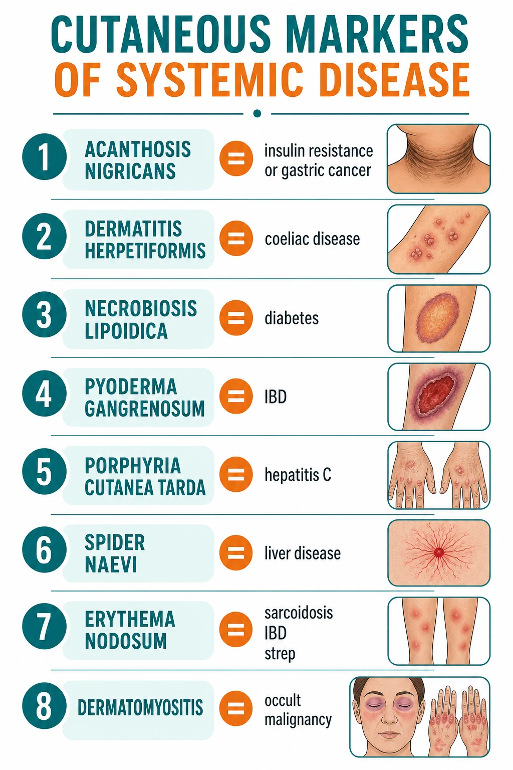
Cutaneous signs that signal underlying systemic disease, organised by organ system. ENDOCRINE: acanthosis nigricans (insulin resistance, metabolic syndrome; or paraneoplastic gastric cancer), necrobiosis lipoidica and granuloma annulare (diabetes), vitiligo (autoimmune thyroid/DM), pretibial myxoedema (Graves), xanthomas (hyperlipidaemia), striae (Cushing). GI/HEPATIC: dermatitis herpetiformis (coeliac), pyoderma gangrenosum (IBD), erythema nodosum (IBD/sarcoid/strep/OCP), lichen planus (HCV), porphyria cutanea tarda (HCV/haemochromatosis), spider naevi/palmar erythema (cirrhosis), Kayser-Fleischer ring (Wilson). RHEUMATOLOGICAL: psoriasis/PsA, Gottron papules/heliotrope (dermatomyositis), malar/butterfly and discoid (SLE), livedo reticularis (APS), Raynaud/sclerodactyly/CREST (systemic sclerosis). HAEMATOLOGICAL: Sweet syndrome (AML). PARANEOPLASTIC: sign of Leser-Trélat (gastric), tripe palms (gastric/lung), dermatomyositis (occult), erythema gyratum repens (lung), necrolytic migratory erythema (glucagonoma). NEURO/GENODERMATOSIS: café-au-lait/Lisch nodules (NF1), ash-leaf/adenoma sebaceum/shagreen (tuberous sclerosis). Nails: clubbing, koilonychia (iron deficiency), half-and-half/Lindsay (renal), Terry (cirrhosis).
On this page & tools
Your progress
Saved locally on this device.
Exam tags
Red flags
Overview & Definition
A cutaneous marker of systemic disease is any skin, nail, hair, or mucosal finding whose presence implies — or substantially raises the probability of — an internal disorder, whether endocrine, gastrointestinal, hepatic, renal, haematological, rheumatological, neurological, infectious, or neoplastic. The marker may be specific (the skin lesion is itself a defining feature of the systemic disease, such as dermatitis herpetiformis in coeliac disease) or non-specific/reactive (a stereotyped cutaneous response that points to a differential, such as erythema nodosum). Some markers are pathognomonic — their presence alone essentially makes the diagnosis (Kayser-Fleischer ring in Wilson disease; adenoma sebaceum in tuberous sclerosis).[1]
The clinical power of this topic is that the skin is examined without a machine, without a needle, and at the bedside in seconds. A trained eye can diagnose insulin resistance from the axillae, coeliac disease from the elbows, occult gastric cancer from the palms, and Wilson disease from the cornea before any blood test returns. The corollary — and the examiner's trap — is that every cutaneous marker must be read as a sentence, not a word: the morphology, the distribution, the tempo of onset, and the patient's age and weight-loss history together determine which systemic disease is being signalled, and how urgently it must be pursued.[1]
Cutaneous markers are biologically universal, but the yield of the triggered workup varies regionally: coeliac serology returns positive more often in populations of European descent; hepatitis C serology is the dominant driver of porphyria cutanea tarda in North America and Europe but haemochromatosis and alcohol dominate elsewhere; and the malignancy implied by a paraneoplastic marker tracks regional cancer epidemiology (gastric adenocarcinoma for Leser-Trélat/tripe palms in high-incidence regions of East Asia and South America; lung cancer for erythema gyratum repens globally). Always interpret the marker against the patient's epidemiology.
Classification — how to organise the catalogue
Cutaneous markers are best classified by the organ system signalled, because that is how the triggered investigation branches. A second axis — mechanistic — separates markers driven by shared pathogenesis (the metabolic, immune, or genetic process affects skin and viscera together) from paraneoplastic markers driven by tumour-derived growth factors.[2]

A working framework
Endocrine
- Acanthosis nigricans (insulin resistance)
- Necrobiosis lipoidica, granuloma annulare (diabetes)
- Vitiligo, alopecia areata (autoimmune polyglandular)
- Pretibial myxoedema (Graves)
- Xanthomas (hyperlipidaemia)
- Striae, moon face (Cushing)
GI / Hepatic
- Dermatitis herpetiformis (coeliac)
- Pyoderma gangrenosum, erythema nodosum (IBD)
- Lichen planus, PCT (hepatitis C)
- Spider naevi, palmar erythema, Terry nails (cirrhosis)
- Kayser-Fleischer ring (Wilson)
- Mucosal pigmentation (Peutz-Jeghers)
Rheumatological
- Psoriasis + nail changes (psoriatic arthritis)
- Gottron papules, heliotrope (dermatomyositis)
- Malar/butterfly, discoid lupus (SLE)
- Livedo reticularis (antiphospholipid syndrome)
- Raynaud, sclerodactyly, calcinosis (systemic sclerosis/CREST)
Renal
- Half-and-half (Lindsay) nails
- Uraemic frost
- Calciphylaxis (calcific uraemic arteriolopathy)
- Acquired ichthyosis
- Pruritus and excoriations
Haematological
- Sweet syndrome (acute myeloid leukaemia)
- Pyoderma gangrenosum (myelodysplasia)
- Thrombophlebitis migrans (pancreatic)
- Generalised pruritus (polycythaemia, lymphoma, Hodgkin)
- Pallor/petechiae (anaemia/thrombocytopenia)
Paraneoplastic
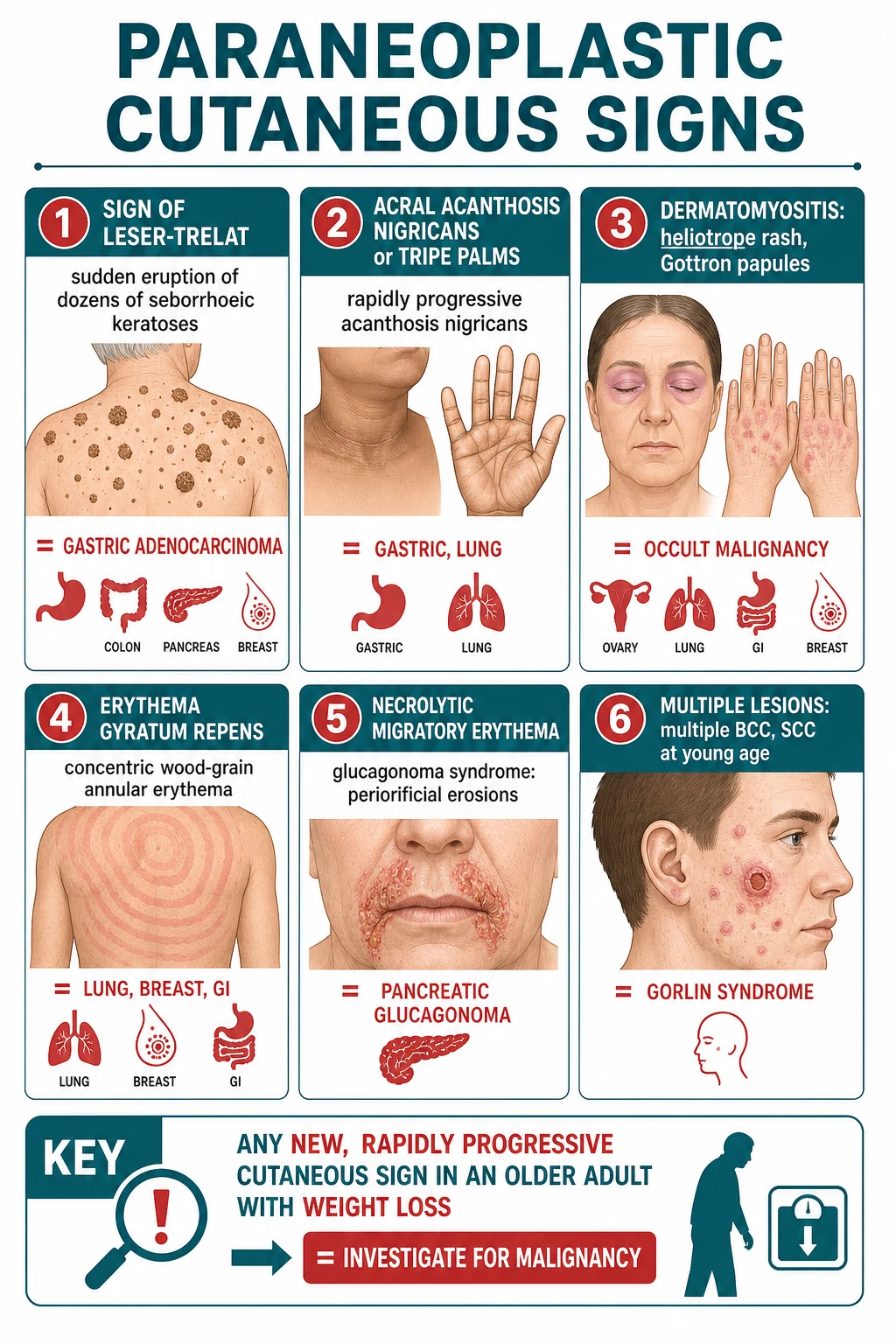
- Malignant acanthosis nigricans (gastric)
- Sign of Leser-Trélat (gastric)
- Tripe palms (gastric/lung)
- Dermatomyositis (occult)
- Erythema gyratum repens (lung)
- Necrolytic migratory erythema (glucagonoma)
- Paget disease (breast/apocrine)
Neuro / Genodermatosis
- Café-au-lait macules, Lisch nodules (NF1)
- Ash-leaf macules, adenoma sebaceum, shagreen patch (tuberous sclerosis)
- Lentiginoses (LEOPARD, Carney, Peutz-Jeghers)
- Incontinentia pigmenti (Blaschko lines)
Epidemiology & risk factors
Cutaneous markers span every age. Genodermatosis markers (café-au-lait macules, ash-leaf spots, incontinentia pigmenti) present in infancy and childhood; atopic and infectious markers dominate childhood and young adults; and metabolic, hepatic, and paraneoplastic markers become progressively more likely with age. The single most important epidemiological pivot is age greater than 50 with unexplained weight loss: in this group a new, rapidly progressive cutaneous sign carries a high probability of underlying malignancy, and the threshold to investigate is low.[2]
Several markers cluster with defined risk profiles. Acanthosis nigricans is common in obesity, type 2 diabetes, and the metabolic syndrome — affecting up to a majority of patients with significant insulin resistance — but the same lesion in a thin, non-diabetic, older adult is malignant until proven otherwise. Porphyria cutanea tarda is triggered by hepatitis C, haemochromatosis, alcohol, oestrogens, and HIV, so its prevalence tracks those exposures. Dermatitis herpetiformis tracks coeliac disease, with the highest prevalence in northern European populations and an HLA-DQ2/DQ8 association. Sweet syndrome and acquired ichthyosis in an adult carry a roughly 20 percent probability of an underlying malignancy and warrant a directed search.[6]
Pathophysiology — why skin mirrors systemic disease
The skin reflects internal disease through a small number of recurring mechanisms, and recognising the mechanism helps predict which marker belongs to which system. [1]
Shared hormonal and metabolic signalling. Insulin resistance drives acanthosis nigricans: hyperinsulinaemia binds insulin-like growth factor 1 (IGF-1) and epidermal growth factor receptors on keratinocytes and fibroblasts, producing hyperproliferation of the squamous epithelium and a velvet-like thickening of flexural skin. The same insulin/IGF axis underlies acrochordons (skin tags) and, controversially, hirsutism in polycystic ovary syndrome.[2] Hyperglycaemia and microangiopathy drive necrobiosis lipoidica and granuloma annulare through collagen degeneration and a granulomatous palisading reaction in the dermis; hyperthyroidism drives pretibial myxoedema through fibroblast-stimulating autoantibodies and glycosaminoglycan deposition.
Immune cross-reactivity. When the same antigen is expressed in skin and viscera, a systemic immune response produces both the rash and the organ disease. Dermatitis herpetiformis is the cutaneous counterpart of coeliac disease: IgA anti-epidermal transglutaminase antibodies deposit at the dermal papillae, producing intensely pruritic vesicles, while the same immune process blunts the duodenal villi. Psoriasis reflects a shared T-helper-17/interleukin-23 inflammatory axis with psoriatic arthritis and metabolic syndrome. Lichen planus shares immune mechanisms with chronic hepatitis C in a subset of patients. [1]
Oestrogen and vascular effects. The spider naevi, palmar erythema, gynaecomastia, and palmar erythema of chronic liver disease reflect failure of hepatic metabolism of circulating oestrogens and vasoactive substances, producing visible capillary proliferation and vasodilation in sun-exposed and palmar skin. [1]
Tumour-derived growth factors (paraneoplastic mechanism). This is the mechanism examiners most reward. Tumours — especially gastric adenocarcinoma — secrete transforming growth factor-alpha (TGF-alpha), epidermal growth factor, and IGF-1 that act on the epidermis to produce acanthosis nigricans, the sign of Leser-Trélat (explosive eruption of seborrhoeic keratoses), tripe palms (velvety palmar thickening), and erythema gyratum repens (concentric advancing rings). The glucagonoma secretes glucagon (and zinc deficiency/amino-acid deficiency is implicated), producing necrolytic migratory erythema; resection of the tumour resolves the eruption. The tumour-directed immune response in dermatomyositis (and the paraneoplastic variant of Sweet syndrome) is presumed to cross-react with regenerating muscle and skin antigens.[3][10]
Embryological and genetic links. Because skin, nervous system, eye, and many viscera share ectodermal and neural-crest origins, genodermatoses present as a constellation of cutaneous pigmentary or hamartomatous lesions with neurological, endocrine, and neoplastic associations — café-au-lait macules and Lisch nodules in neurofibromatosis type 1; hypopigmented ash-leaf macules, angiofibromas (adenoma sebaceum), shagreen patches, and periungual fibromas in tuberous sclerosis; and the lentiginoses in the LEOPARD, Carney, and Peutz-Jeghers syndromes.[14]
Clinical presentation — the catalogue by organ system
This section is the heart of the topic. Each marker is described with its morphology, its characteristic distribution, and the systemic disease it signals. The markers are grouped by the organ system the bedside investigation should target. [1]
Endocrine markers
Acanthosis nigricans
- Velvety, hyperpigmented, hyperkeratotic plaques
- Flexural: axillae, neck, groin, knuckles; can involve palms (tripe palms)
- Benign: obesity, insulin resistance, T2DM, metabolic syndrome
- Malignant (paraneoplastic): gastric adenocarcinoma (90%), sudden onset, weight loss
Necrobiosis lipoidica
- Yellow-brown atrophic plaques with telangiectasia and a violaceous rim
- Bilateral shins (pretibial)
- ~65% have or will develop diabetes; not directly linked to glycaemic control
- Biopsy = necrobiotic granulomas (palisading) in the dermis
Granuloma annulare
- Annular plaques of skin-coloured papules
- Hands, feet, extensor surfaces
- Association with diabetes is debated; disseminated form more often linked
- Histology: foci of necrobiotic collagen and mucin
Vitiligo
- Acquired depigmented macules, symmetrical
- Autoimmune polyglandular association: thyroid disease, T1DM, Addison, pernicious anaemia
- Screen TSH, anti-TPO; glucose
Pretibial myxoedema
- Indurated nodules/plaques on shins (non-pitting)
- Graves disease (thyroid dermopathy); may coexist with thyroid acropachy and ophthalmopathy
- TSH receptor antibodies
Tuberous/xanthomatous
- Xanthomas: eruptive (triglycerides), tendon (familial hypercholesterolaemia), tuberous, palmar (type III)
- Xanthelasma: eyelids; check lipid profile
- Acquired plane xanthomas: paraproteinaemia, myeloma
Striae and the Cushingoid habitus. Purple, broad (more than one centimetre) abdominal striae, a moon face, buffalo hump, central obesity, and easy bruising are the cutaneous face of cushing syndrome (endogenous hypercortisolism or exogenous steroids). Thin, silvery striae distensae are a normal variant of rapid growth or pregnancy and are not pathological. Acne, hirsutism, and acanthosis nigricans complete the metabolic picture in insulin resistance and polycystic ovary syndrome. [1]
Gastrointestinal and hepatic markers
The gut–skin axis is one of the richest sources of examination stems. Several markers are essentially diagnostic of their systemic counterpart. [1]
Dermatitis herpetiformis
- Intensely pruritic herpetiform vesicles on elbows, knees, buttocks, scalp
- PATHOGNOMONIC for coeliac disease (the cutaneous counterpart)
- IgA anti-tissue transglutaminase (tTG-IgA); biopsy skin = neutrophils in dermal papillae
- Direct immunofluorescence: granular IgA at dermal papillae
- Management: strict gluten-free diet + dapsone for itch
Pyoderma gangrenosum
- Painful ulcer with undermined, violaceous edges; pathergy
- Trunks and lower legs; starts as a pustule
- IBD (UC more than Crohn), arthritis, haematological malignancy (MDS, AML)
- Pathergy — do NOT debride; high-dose systemic steroids, ciclosporin, biologics
Erythema nodosum
- Tender, erythematous nodules on shins (panniculitis)
- Bilateral, no ulceration
- IBD, sarcoidosis, streptococcal, drugs (OCP, sulphonamides), TB, pregnancy
- Resolves in weeks; treat the cause; NSAIDs, bed rest
Lichen planus
- Pruritic, polygonal, purple, planar papules (the 5 Ps); Wickham striae
- Wrists, ankles, oral mucosa (reticular white lace)
- Hepatitis C association in a subset; drugs; chronic graft-versus-host
Porphyria cutanea tarda
- Fragile skin, bullae on dorsal hands and sun-exposed sites; hypertrichosis, milia
- Triggered by HEPATITIS C, haemochromatosis, alcohol, oestrogen, HIV
- Screen: HCV serology, iron studies, ferritin, serum/urine porphyrins
- Treat: phlebotomy, low-dose hydroxychloroquine, remove trigger
Spider naevi / palmar erythema
- Central arteriole with radiating vessels; blanches, refills
- More than 5 (or in males/children) suggests chronic liver disease
- Palmar erythema: thenar/hypothenar, sparing the centre
- Mechanism: oestrogen excess (cirrhosis); also pregnancy, thyrotoxicosis
Wilson disease and the Kayser-Fleischer ring. A brown-green copper deposition at the periphery of Descemet membrane, best seen on slit-lamp examination, is essentially pathognomonic for Wilson disease in a patient with neurological or hepatic involvement. It is the prototype of a sign that lives in the skin's neighbourhood (the eye) yet signals a metabolic liver disease, and it is reversible with chelation therapy (penicillamine, trientine, zinc).[13]
Other GI/hepatic markers. Mucocutaneous pigmentation of the lips, buccal mucosa, and digits is the hallmark of Peutz-Jeghers syndrome (STK11 mutation; hamartomatous polyps; increased GI, breast, and ovarian cancer risk). Acrodermatitis enteropathica (periorificial and acral dermatitis with diarrhoea) signals zinc deficiency — classically in total parenteral nutrition without zinc supplementation or in chronic diarrhoea. Jaundice (yellow skin, sclera, and mucous membranes) is the visible endpoint of hyperbilirubinaemia from any hepatic, biliary, or haemolytic cause. Pruritus ani and fistulae point to Crohn disease; glossitis and angular cheilitis point to B12/iron deficiency. [1]
Rheumatological markers
Connective tissue diseases produce some of the most distinctive cutaneous patterns in medicine, and the rash is frequently the presenting feature. [1]
Psoriasis and psoriatic arthritis. Well-demarcated erythematous plaques with silvery scale on extensor surfaces and the scalp, plus nail changes (pitting, onycholysis, oil-drop salmon patches, subungual hyperkeratosis), point to psoriasis; the presence of nail involvement, dactylitis, enthesitis, or asymmetrical large-joint arthritis signals psoriatic arthritis, which affects up to 30 percent of patients with psoriasis and warrants rheumatology referral and a change in management (NSAIDs, DMARDs, IL-17/IL-23/ TNF-alpha biologics). [1]
Dermatomyositis. The pathognomonic cutaneous features are Gottron papules (violaceous papules over the metacarpophalangeal and interphalangeal joints) and the heliotrope rash (violaceous discoloration of the upper eyelids, often with oedema). Supporting features include a V-sign and shawl-sign poikilodermatous erythema in sun-exposed distributions, mechanic's hands (hyperkeratotic, fissured lateral fingers), and periungual telangiectasia with a ragged cuticle. In the adult form, 15 to 25 percent harbour an occult malignancy — ovarian, lung, gastric/colorectal, breast, and nasopharyngeal (the latter especially in Asian populations) — and a structured malignancy workup is mandatory at diagnosis.[3]
Systemic lupus erythematosus. The malar (butterfly) rash is the classic facial erythema sparing the nasolabial folds (a key discriminator from seborrhoeic dermatitis and rosacea, which involve them). Discoid lupus produces scarring coin-shaped plaques with follicular plugging, most on the scalp, face, and ears; only a minority progress to systemic disease. Other SLE markers include photosensitivity, oral ulcers, alopecia, livedo reticularis, and vasculitic lesions. The 2019 EULAR/ACR SLE classification requires an ANA entry criterion plus weighted clinical and immunological criteria (anti-dsDNA, anti-Sm, antiphospholipid, low complement). [1]
Livedo reticularis and antiphospholipid syndrome. A persistent, net-like cyanotic discoloration of the limbs suggests livedo reticularis; when associated with thrombosis (DVT, arterial thrombosis, stroke) or recurrent miscarriage it signals the antiphospholipid syndrome. Livedo is also a feature of the Sneddon syndrome (livedo + stroke) and of cholesterol embolisation after vascular instrumentation. [1]
Systemic sclerosis and CREST. The combination of Raynaud phenomenon (often preceding other features by years), sclerodactyly (tight, shiny digits), facial thickening with a beak-like nose and microstomia, calcinosis cutis (subcutaneous calcium deposits), and mat telangiectasia defines systemic sclerosis. The limited cutaneous form is the CREST syndrome (Calcinosis, Raynaud, Esophageal dysmotility, Sclerodactyly, Telangiectasia), strongly associated with the anti-centromere antibody; diffuse cutaneous disease is associated with anti-Scl-70 (anti-topoisomerase I) and a higher risk of pulmonary fibrosis and renal crisis.[12]
Renal markers
Half-and-half (Lindsay) nails — a proximal pale/white half and a distal pink/brown half occupying roughly 20 to 60 percent of the nail — are seen in around a quarter of chronic kidney disease patients and are a classic examination finding. Uraemic frost (white crystalline urea deposits on the skin in advanced uraemia) is now rare with dialysis but is a named sign. Calciphylaxis (calcific uraemic arteriolopathy) is a devastating vasculopathy of small vessels in dialysis-dependent CKD, presenting as exquisitely painful, retiform purpuric plaques that progress to necrotic ulcers; it carries a high mortality and demands urgent specialist management (wound care, optimisation of calcium-phosphate, sodium thiosulphate). Acquired ichthyosis (dry, scaly skin in adulthood) and intractable pruritus with excoriations are common in advanced CKD. [1]
Haematological markers
Sweet syndrome (acute febrile neutrophilic dermatosis) presents with the abrupt onset of tender, erythematous plaques and nodules (often on the upper limbs and face), fever, arthralgia, and a neutrophil leucocytosis; histology shows a dense dermal neutrophil infiltrate. Roughly a fifth of cases are malignancy-associated, most often with acute myeloid leukaemia and myelodysplasia; it also occurs with solid tumours, inflammatory bowel disease, and as a drug reaction (G-CSF). Sweet syndrome responds dramatically to systemic corticosteroids.[6]
Pyoderma gangrenosum overlaps haematology and gastroenterology, associating with IBD and the myelodysplastic syndromes (and, in the syndrome of pyoderma gangrenosum, acne, and suppurative hidradenitis — PASH). Thrombophlebitis migrans (Trousseau syndrome) — recurrent, migratory superficial venous thromboses in unusual sites — is a hypercoagulable sign of pancreatic cancer (and other mucin-producing adenocarcinomas). Generalised pruritus without a primary skin disease is a classic marker of polycythaemia vera (often aquagenic, after a warm bath), Hodgkin and non-Hodgkin lymphoma, and iron-deficiency anaemia. Pallor, bruising, and petechiae reflect anaemia and thrombocytopenia from marrow failure or infiltration. [1]
Paraneoplastic cutaneous markers
This group — more than any other — is the reason a dermatology or general medicine examiner will probe a candidate's knowledge of the skin. Each marker carries a strong, specific malignancy association, and recognising it can be the first clue to an otherwise occult tumour.[10]
Malignant acanthosis nigricans
- Sudden, rapidly progressive, extensive AN with tripe palms
- ~90% gastric adenocarcinoma; also lung, liver, pancreatic, uterine
- Driven by tumour TGF-alpha/EGF; treat the tumour
Sign of Leser-Trélat
- Explosive, hundreds of seborrhoeic keratoses over weeks–months
- Older adult; ± pruritus
- Gastric adenocarcinoma (most), colon, breast, lung; ± coexists with malignant AN/tripe palms
Tripe palms
- Velvety, rugose palmar thickening (like bovine stomach)
- More than 90% malignancy-associated; lung and gastric
- Often coexists with Leser-Trélat or malignant AN
Dermatomyositis (adult)
- Gottron papules, heliotrope rash, V/shawl sign, mechanic's hands
- 15–25% occult malignancy
- Screen: ovarian, lung, gastric/colorectal, breast, nasopharyngeal (Asian)
Erythema gyratum repens
- Concentric, slowly advancing 'wood-grain' rings; desquamation
- ~80% malignancy; lung most common, then breast, GI, bladder, prostate
- Highly specific; pruritic
Necrolytic migratory erythema
- Migrating erosive, crusted, annular plaques; periorificial, groin, distal limbs
- PATHOGNOMONIC for glucagonoma (alpha-cell pancreatic neuroendocrine tumour)
- Plus diabetes, DVT, weight loss, glossitis; low amino acids/zinc
- Treat: resect tumour; octreotide; zinc/amino-acid replacement
Paget disease
- Eczematous, slowly enlarging plaque of the nipple-areola (mammary Paget) or apocrine skin (extramammary, vulva/scrotum)
- Underlying in situ or invasive adenocarcinoma
- Biopsy any unilateral nipple eczema refractory to treatment
Neurological and genodermatosis markers
Because skin and nervous system share embryological origins, several neurocutaneous (phakomatoses) and genodermatoses present with distinctive cutaneous markers that signal neurological, endocrine, and neoplastic risk. [1]
NF1 (von Recklinghausen)
- Café-au-lait macules (6 or more, >5 mm prepubertal / >15 mm postpubertal)
- Axillary/inguinal freckling (Crowe sign)
- Dermal neurofibromas; Lisch nodules (iris hamartomas)
- Optic glioma, plexiform neurofibroma, sarcomatous change, learning difficulty
Tuberous sclerosis
- Ash-leaf macules (hypopigmented, Wood lamp)
- Angiofibromas (adenoma sebaceum) on the face
- Shagreen patch (connective-tissue naevus, lower back)
- Periungual fibromas (Koenen tumours)
- Epilepsy, cognitive impairment, renal angiomyolipoma, cardiac rhabdomyoma
Lentiginoses
- Multiple lentigines as a marker of multisystem syndromes
- LEOPARD (lentigines, ECG, ocular, pulmonary stenosis, abnormal genitalia, retardation, deafness)
- Carney complex (lentigines, myxomas, endocrine overactivity)
- Peutz-Jeghers (perioral/digital lentigines, GI polyps, cancer risk)
Incontinentia pigmenti
- Blaschkoid vesicular, verrucous, then pigmented stages
- X-linked dominant, lethal in males
- Dental, ocular, neurological anomalies
Incontinentia pigmenti merits a separate note: a female infant with blaschkoid vesicles that progress to verrucous and then whorled hyperpigmented lesions has an X-linked dominant disorder (NEMO/IKBKG mutation, usually lethal in males) with associated dental, ocular, and neurological anomalies, and warrants genetics, ophthalmology, and dental surveillance.[15]
The malignancy implied by paraneoplastic markers, and the specific neurocutaneous associations, follow regional epidemiology. Nasopharyngeal carcinoma is over-represented as the occult tumour in Asian patients with dermatomyositis; gastric adenocarcinoma dominates the Leser-Trélat/malignant acanthosis nigricans/tripe palms triad in regions of high gastric cancer incidence (East Asia, parts of South America); and hepatitis C-driven porphyria cutanea tarda is most prevalent where HCV is endemic. Always weigh the local cancer and infection profile when building the triggered workup.[14]
Nail and hand signs of systemic disease
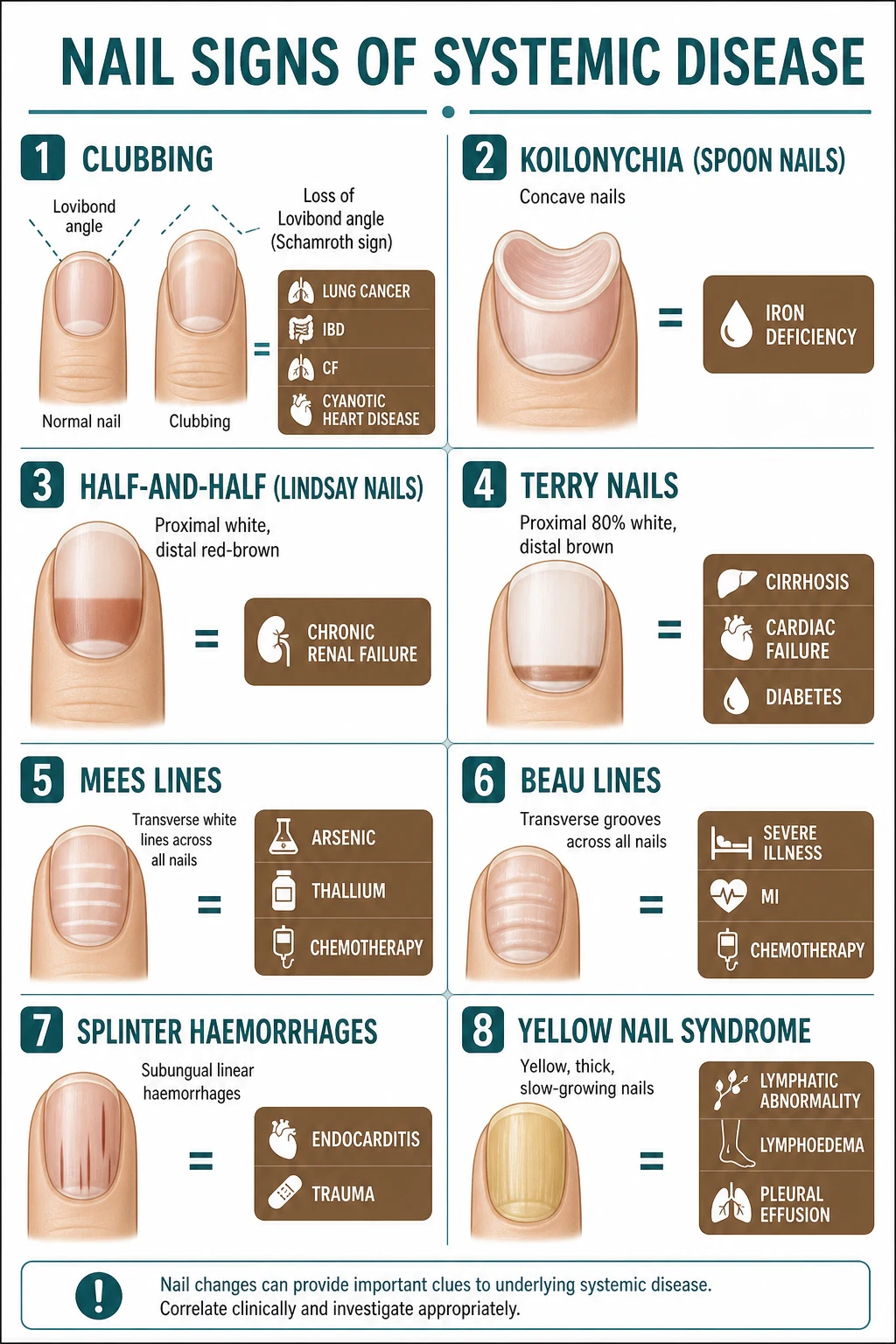
The hands are examined in seconds and yield a disproportionate amount of information. Nail signs, in particular, are high-yield examination material because each has a tight association list. [1]
Clubbing
- Loss of the nail-fold angle (Lovibond angle), floating nail, increased sponginess
- Schamroth window test: absent diamond
- Lung (cancer, bronchiectasis, fibrosis, abscess); cardiac (cyanotic CHD, IE); GI (IBD, cirrhosis, coeliac, malabsorption); PTH
Koilonychia (spoon nails)
- Concave, everted nail plate
- IRON-DEFICIENCY ANAEMIA (classic); also Plummer-Vinson, haemochromatosis, Raynaud
Half-and-half (Lindsay)
- Proximal white, distal pink/brown (20–60%)
- CHRONIC RENAL FAILURE; also Crohn, Kawasaki, Behçet, cirrhosis, pellagra
Terry nails
- Proximal white (>80%) with a narrow distal pink/brown band
- CIRRHOSIS; also chronic cardiac failure, diabetes, hyperthyroidism, ageing
Mees lines
- Multiple transverse white lines that move out with the nail
- ARSENIC, thallium poisoning; chemotherapy; carbon monoxide; renal failure
Beau lines
- Transverse depressions from transient nail-matrix arrest
- Severe systemic illness, high fever, chemotherapy, myocardial infarction, COVID-19
Muehrcke lines
- Paired, transverse white lines that do NOT move (vascular, not matrix)
- Hypoalbuminaemia (nephrotic, hepatic, protein-losing enteropathy); chemotherapy
Splinter haemorrhages
- Linear, distal, subungual haemorrhages
- Trauma (commonest); INFECTIVE ENDOCARDITIS (classical but low specificity); vasculitis; antiphospholipid
Yellow nail syndrome
- Slow-growing, yellow, thickened, onycholytic nails; absent lunulae
- LYMPHOEDEMA + pleural effusion (triad); respiratory tract disease
HANDS
Differential diagnosis — distinguishing the mimics
A rash that looks like one marker may be another; the cost of error is a missed malignancy, a missed coeliac, or inappropriate treatment. Three distinctions recur in examinations. [1]
Pyoderma gangrenosum versus Sweet syndrome versus ecthyma gangrenosum. All three produce erythematous nodules or plaques that ulcerate. Pyoderma gangrenosum is a painful ulcer with undermined, violaceous edges and pathergy (worsens with debridement); it associates with IBD, arthritis, and myelodysplasia. Sweet syndrome produces tender, non-ulcerated plaques with fever and a dermal neutrophil infiltrate; it associates with AML, IBD, and drugs. Ecthyma gangrenosum is a rapidly progressive necrotic ulcer of Pseudomonas septicaemia in an immunocompromised, neutropenic patient — an emergency, not an inflammatory dermatosis. The biopsy and culture distinguish them: PG and Sweet are sterile neutrophilic; ecthyma is infected. [1]
Malar rash (SLE) versus rosacea versus seborrhoeic dermatitis versus heliotrope (dermatomyositis). The SLE malar/butterfly rash spares the nasolabial folds, is photosensitive, and is not greasy. Rosacea involves the nasolabial folds, has telangiectasia, papules, pustules, and flushing, and lacks scarring. Seborrhoeic dermatitis is greasy, scaly, involves the nasolabial folds, eyebrows, and chest. The heliotrope rash of dermatomyositis is periorbital (eyelids), violaceous, and oedematous — a different distribution entirely. [1]
Erythema nodosum versus erythema multiforme versus nodular vasculitis. Erythema nodosum is a panniculitis — tender subcutaneous nodules on the shins that do not ulcerate and resolve with bruising. Erythema multiforme is an epidermal/dermal process with target lesions (three concentric zones) on extensor surfaces and mucosa, often triggered by HSV or drugs. Nodular vasculitis (erythema induratum) ulcerates and involves the calves (posterior), often with TB association. [1]
Clinical and bedside assessment
The structured skin examination turns a catalogue into a diagnostic engine. Begin with the morphology and distribution, then move to the sites of highest yield: the face (malar rash, heliotrope, spider naevi, facial flushing, oral mucosa, Kayser-Fleischer ring at the slit lamp), the hands (clubbing, palmar erythema, Gottron papules, tripe palms, koilonychia, nail signs, periungual telangiectasia, xanthomas), the trunk (striae, spider naevi, café-au-lait, ichthyosis, scalp and nails for psoriasis), and the lower legs (necrobiosis lipoidica, pretibial myxoedema, erythema nodosum, eczema, half-and-half nails, livedo). Two manoeuvres are high-yield: the Schamroth window test for clubbing (the loss of the diamond-shaped window between opposing nailbeds), and the diascopy of a lesion to see whether it blanches (erythema) or remains purpuric (vasculitis).[1]
Ask three questions that reframe any cutaneous sign toward the systemic: How fast did it appear? (an eruption over weeks in an older adult, especially with weight loss, is paraneoplastic until proven otherwise); Is it where a benign cause would put it? (acanthosis in the obese diabetic's axilla is benign; in a thin patient's palms with tripe texture it is malignant); and What else is on examination? (a single finding is a clue; a constellation is a diagnosis — Raynaud plus sclerodactyly plus telangiectasia is CREST; spider naevi plus palmar erythema plus Terry nails plus gynaecomastia is cirrhosis). [1]
Investigations — the triggered workup
The defining principle is that the cutaneous marker triggers a specific, narrow investigation for the systemic disease it signals — not a blanket "bloods and imaging." Each marker has a workup. [1]
Cutaneous marker
- Dermatitis herpetiformis
- Necrobiosis lipoidica / granuloma annulare
- Acanthosis nigricans (thin adult)
- Porphyria cutanea tarda
- Sign of Leser-Trélat / tripe palms
- Dermatomyositis (adult)
- Erythema gyratum repens
- Necrolytic migratory erythema
- Livedo + thrombosis/miscarriage
- Kayser-Fleischer ring
Triggered workup
- tTG-IgA + total IgA; duodenal biopsy (coeliac)
- HbA1c, fasting glucose; lipids
- Upper GI endoscopy (gastric cancer); CT chest/abdomen/pelvis
- HCV serology, HIV, iron studies/ferritin, serum+urine porphyrins
- Upper GI endoscopy (gastric); CT CAP; faecal occult
- CT CAP, mammography, ovarian US ± pelvic MRI, age-appropriate endoscopy; anti-TIF1-gamma (malignancy-associated)
- Chest imaging (lung); CT CAP
- Glucagon level; CT/MRI pancreas (NET); glucose; zinc, amino acids
- Lupus anticoagulant, anticardiolipin, anti-beta-2-glycoprotein I; ANA, dsDNA (APS/SLE)
- Serum + 24h urinary copper, caeruloplasmin; eye exam; LFTs; MRI brain
Skin biopsy and direct immunofluorescence are confirmatory where the diagnosis is uncertain: a punch biopsy of perilesional skin (not the ulcer) is sent for routine histology and a separate sample for direct immunofluorescence. The fluorescence patterns are diagnostic: granular IgA at the dermal papillae in dermatitis herpetiformis; linear IgG/C3 along the basement membrane in bullous pemphigoid; linear IgA along the basement membrane in linear IgA disease; intercellular (fish-net) IgG/C3 in pemphigus vulgaris. A skin biopsy of necrobiotic granulomas in the dermis supports necrobiosis lipoidica and granuloma annulare; a dense dermal neutrophil infiltrate supports Sweet syndrome.[5]
Management — the immediate and the definitive
The guiding principle is that the cutaneous marker is treated by treating the underlying disease; skin-directed therapy is adjunctive. There are, however, a few situations in which the marker is itself the emergency. [1]
Where the marker demands urgent action. Sweet syndrome with high fever is treated with systemic corticosteroids (prednisolone 0.5 to 1 mg/kg/day) after infection is excluded, because untreated it causes significant morbidity and signals a possible acute leukaemia that needs haematology referral within days. Calciphylaxis in a dialysis patient is a dermatologic-nephrology emergency with high short-term mortality, demanding urgent wound care, correction of calcium-phosphate product, and consideration of intravenous sodium thiosulphate. Pyoderma gangrenosum is managed with systemic immunosuppression (high-dose prednisolone, ciclosporin, or a biologic such as infliximab or ustekinumab) and explicit avoidance of surgical debridement, which triggers pathergy and catastrophic worsening.[11]
Definitive, disease-specific management. [1]
Dermatitis herpetiformis (coeliac)
- Strict LIFELONG gluten-free diet (resolves the rash and the enteropathy)
- Dapsone 50–150 mg orally daily for itch while the diet takes effect (months)
- Monitor G6PD, haemoglobin; folate
Necrobiosis lipoidica (diabetes)
- Optimise glycaemic control (note: control does not reliably resolve lesions)
- Topical/intralesional corticosteroid for active inflammation; antiplatelets; wound care
- Compression; excision and grafting reserved for refractory ulceration (recurrence common)
Porphyria cutanea tarda
- Remove the trigger (alcohol, oestrogen, treat HCV, address haemochromatosis)
- Therapeutic phlebotomy (target ferritin near normal); low-dose hydroxychloroquine 200 mg twice weekly
- Sun protection
Malignant acanthosis nigricans / Leser-Trélat / tripe palms
- Treat the underlying malignancy — resect, chemo-/radiotherapy as indicated
- Markers regress with successful tumour treatment
- OGD first (gastric), then CT CAP
Dermatomyositis (adult)
- Malignancy workup at diagnosis and periodically
- Systemic corticosteroids with a steroid-sparing agent (methotrexate, azathioprine, IVIG for refractory/severe)
- Sun protection; physiotherapy
Necrolytic migratory erythema (glucagonoma)
- Resect the pancreatic tumour; octreotide for metastatic/unresectable disease
- Correct zinc, essential fatty acid and amino-acid deficiencies
- Improves dramatically with tumour control
Wilson disease (KF ring)
- Chelation: penicillamine or trientine; oral zinc for maintenance/pre-symptomatic
- Avoid copper-rich foods; liver transplant for decompensation
- Ring fades with treatment
Dapsone
Dose
50–150 mg once daily, titrate to lowest effective
Prednisolone
Dose
0.5–1.0 mg/kg once daily, taper on response over weeks
Specific scenarios — special populations
Pregnancy. Palmar erythema and spider naevi are physiologic in pregnancy (oestrogen), and a single palmar sign in a young pregnant woman is not cirrhosis. Melasma (chloasma), linea nigra, and striae gravidarum are hormonal. Pruritic urticarial papules and plaques of pregnancy (PUPPP) and intrahepatic cholestasis of pregnancy (pruritus without primary skin lesions, raised bile acids, foetal risk) are the dermatoses to separate from systemic disease. Cushingoid striae from hypercortisolism must be distinguished from the common striae of pregnancy, which are physiological. [1]
Children. Genodermatoses dominate — café-au-lait and axillary freckling (NF1), hypopigmented ash-leaf macules and facial angiofibromas (tuberous sclerosis), incontinentia pigmenti along Blaschko lines (female infants). Kawasaki disease (strawberry tongue, polymorphous rash, desquamation of fingers and toes, coronary artery aneurysms) and atopic dermatitis are common; juvenile dermatomyositis has a lower malignancy rate than the adult form and a calcinosis-heavy phenotype. [1]
The elderly. The malignancy pivot dominates. A new acanthosis, Leser-Trélat, tripe palms, erythema gyratum repens, acquired ichthyosis, or unexplained generalised pruritus in an older adult warrants a focused malignancy search (OGD for gastric markers; CT CAP; age-appropriate screening; targeted tests such as mammography, colonoscopy). Senile purpura (Bateman purpura) on the dorsal forearms from dermal atrophy and vascular fragility is benign and must not be confused with a vasculitic or thrombocytopenic process. [1]
The immunocompromised. HIV produces a sequence of cutaneous markers that mirror immune decline — florid seborrhoeic dermatitis, recalcitrant psoriasis, extensive molluscum contagiosum, oral hairy leukoplakia (EBV), Kaposi sarcoma (HHV-8), chronic herpes simplex, and bacillary angiomatosis — and these markers track progression to AIDS.[16]
Complications and pitfalls
The complications are largely those of missing the systemic disease behind the marker. Missing an occult gastric cancer behind a tripe palms or sign of Leser-Trélat, an ovarian cancer behind a dermatomyositis, or a glucagonoma behind a necrolytic migratory erythema converts a potentially resectable tumour into an advanced one. Conversely, over-attribution — labelling a benign acanthosis nigricans as malignant, or a physiological palmar erythema as cirrhosis — drives unnecessary, invasive investigation. The distinguishing features (tempo, weight loss, distribution, age) resolve most of these. [1]
The classic procedural pitfall is surgical debridement of pyoderma gangrenosum. PG worsens with trauma — the pathergy phenomenon — and surgical debridement produces catastrophic, rapidly enlarging ulcers. The rule is never to debride a painful ulcer with undermined violaceous edges in a patient with IBD or a haematological disorder until PG is excluded.[11] A second pitfall is treating the skin and not the disease — applying potent topical steroids to dermatitis herpetiformis will not control it (it is driven by dietary gluten), and glycaemic optimisation alone rarely reverses necrobiosis lipoidica; the skin disease requires its own management alongside the systemic one.
A third pitfall is confusing the marker's natural history with treatment failure. Necrobiosis lipoidica may progress despite excellent glycaemic control (the link to hyperglycaemia is not straightforward); sign of Leser-Trélat can precede detectable malignancy by months, so a negative initial malignancy screen in a high-risk clinical picture warrants repeat investigation rather than reassurance. Terry nails and half-and-half nails can occur in healthy older adults, so a single nail sign without systemic corroboration is not a diagnosis. [1]
Prognosis and disposition
Prognosis tracks the underlying disease. Paraneoplastic markers (acanthosis, Leser-Trélat, tripe palms, erythema gyratum repens) usually regress with successful treatment of the tumour and reappear at recurrence — their behaviour is itself a tumour marker. Necrolytic migratory erythema resolves within weeks of glucagonoma resection. Dermatitis herpetiformis clears on a strict gluten-free diet and predicts the long-term coeliac risks (enteropathy-associated T-cell lymphoma, osteoporosis, other autoimmune disease) that the diet also reduces. Dermatomyositis prognosis is dominated by the malignancy and by interstitial lung disease (especially anti-MDA5 and anti-synthetase antibodies). Calciphylaxis carries a one-year mortality that may exceed 40 percent. [1]
Disposition: most cutaneous markers are managed outpatient by the specialty that owns the systemic disease (gastroenterology for coeliac/IBD/PCT, endocrinology for diabetes/thyroid, rheumatology for the connective tissue diseases, haemato-oncology for Sweet/AML). Admission is reserved for the emergencies — severe Sweet with high fever, calciphylaxis, large PG, or the systemic decompensation of the underlying disease (acute liver failure in Wilson, thyroid storm in Graves with pretibial myxoedema). [1]
Evidence, guidelines, and regional differences
The evidence base is a mixture of large case series and consensus guidelines rather than randomised trials — these are uncommon diseases. The 2019 EULAR/ACR classification criteria for SLE (ANA entry criterion plus additive weighted criteria) have largely superseded the older 1997 ACR and 2012 SLICC criteria for research and clinical classification. The 2017 EULAR/ACR classification for idiopathic inflammatory myopathies (including dermatomyositis) standardised the clinical, histological, and myositis-specific antibody definitions and is used to stratify malignancy risk (anti-TIF1-gamma and anti-NXP2 antibodies carry the highest malignancy risk in adult dermatomyositis).[3]
The ACR/EULAR classification criteria for systemic sclerosis (2013) weight skin thickening, fingertip changes, telangiectasia, abnormal nailfold capillaries, pulmonary arterial hypertension and interstitial lung disease, and SSc-related autoantibodies (anti-centromere, anti-topoisomerase I, anti-RNA polymerase III). The Sydney classification and the revised international criteria for coeliac disease underpin serology-first diagnosis with biopsy confirmation; non-biopsy diagnosis (high-titre tTG-IgA plus anti-endomysial antibody in symptomatic children) is now accepted in paediatric guidance in several regions. The 2008 ACR/EULAR classification for antiphospholipid syndrome (updated 2023) defines clinical and laboratory (lupus anticoagulant, anticardiolipin, anti-beta-2-glycoprotein I) criteria.[12]
[1]The Australasian College of Dermatologists and the Gastroenterological Society of Australia guide coeliac and IBD-related cutaneous management; HCV is a dominant driver of PCT and is screened alongside iron studies and HIV in every new presentation.
Exam pearls and high-yield minutiae
COELIAC
Exam application bank (NEET-PG / INICET)
One-line answer
Cutaneous signs that signal underlying systemic disease, organised by organ system. ENDOCRINE: acanthosis nigricans (insulin resistance, metabolic syndrome; or paraneoplastic gastric cancer), necrobiosis lipoidica and granuloma annulare (diabetes), vitiligo (autoimmune thyroid/DM), pretibial myxoedema (Graves), xanthomas (hyperlipidaemia), striae (Cushing). GI/HEPATIC: dermatitis herpetiformis (coeliac), pyoderma gangrenosum (IBD), erythema nodosum (IBD/sarcoid/strep/OCP), lichen planus (HCV), porphyria cutanea tarda (HCV/haemochromatosis), spider naevi/palmar erythema (cirrhosis), Kayser-Fleischer ring (Wilson). RHEUMATOLOGICAL: psoriasis/PsA, Gottron papules/heliotrope (dermatomyositis), malar/butterfly and discoid (SLE), livedo reticularis (APS), Raynaud/sclerodactyly/CREST (systemic sclerosis). HAEMATOLOGICAL: Sweet syndrome (AML). PARANEOPLASTIC: sign of Leser-Trélat (gastric), trip
Worked stems (answer without another resource)
Stem 1 — Classic presentation. Map symptoms to mechanism; name the first investigation and first treatment step with dose/route if drug therapy is standard. [1]
Stem 2 — Unstable / complicated. List red flags that force immediate resuscitation, theatre, ICU, antidote, or reperfusion — and what you do in the first 15 minutes. [1]
Stem 3 — Atypical group. Elderly, pregnancy, child, or immunocompromised: how presentation and thresholds change. [1]
Stem 4 — Differential trap. Name the three closest mimics and one discriminator for each. [1]
Stem 5 — Disposition. Who goes home with safety-netting, who is admitted, who needs HDU/ICU/theatre, and what follow-up is mandatory. [1]
Rapid viva checklist
- Definition + classification
- Pathophysiology chain
- Bedside signs / criteria
- Score with exact components (if any)
- Emergency bundle
- Definitive therapy with doses
- Complications of disease and of treatment
- Special populations
- Guideline/trial name if classic
- Three exam traps
Coverage self-check
If you cannot answer any stem above from this page alone, re-read the matching section — the page is intended to be self-sufficient for final-prof and NEET-PG/INICET questions on Cutaneous markers of systemic disease.
[1] [1]Self-test: a 58-year-old thin man presents with a three-month history of rapidly progressive velvety darkening of the axillae and palms, 8 kg weight loss, and new 'moles' over the trunk. What is the most important next investigation?
Upper gastrointestinal endoscopy to screen for gastric adenocarcinoma. The combination of rapidly progressive acanthosis nigricans with tripe palms and the explosive new seborrhoeic keratoses of the sign of Leser-Trélat is the classical triad of paraneoplastic acanthosis nigricans from gastric adenocarcinoma — driven by tumour-derived TGF-alpha and EGF. Add a CT chest/abdomen/pelvis. The marker may regress on resection of the tumour.[10]
Self-test: a 45-year-old woman has a six-month history of a violaceous rash over her knuckles and upper eyelids, proximal muscle weakness, and a new facial rash. What must you do at diagnosis?
Screen for occult malignancy. The Gottron papules over the metacarpophalangeal joints and the heliotrope rash on the upper eyelids make the diagnosis of dermatomyositis; 15 to 25 percent of adults with dermatomyositis have an occult malignancy — ovarian, lung, gastric/colorectal, breast, and (especially in Asian populations) nasopharyngeal. Order CT of the chest, abdomen, and pelvis, mammography, age-appropriate endoscopy, ovarian imaging, and myositis-specific antibodies (anti-TIF1-gamma marks the malignancy-associated form). Treat the myositis with systemic corticosteroids and a steroid-sparing agent.[3]
References
- [1]Vella J. Cutaneous Markers of Systemic Disease in the Lower Extremity Clin Podiatr Med Surg, 2016.PMID 27215161
- [2]Ambalal SM. Metabolic Syndrome and Skin: Interactions and Implications Indian J Dermatol, 2022.PMID 36092223
- [3]DeWane ME, Waldman R, Lu J. Dermatomyositis: Clinical features and pathogenesis J Am Acad Dermatol, 2020.PMID 31279808
- [4]Liu M, Li J. Necrobiosis Lipoidica N Engl J Med, 2024.PMID 38169491
- [5]García C, Araya M. [Dermatitis herpetiformis and celiac disease] Rev Med Chil, 2021.PMID 35319687
- [6]Atkins O, Mirvis E, Maynard S, et al. Acute myeloid leukaemia presenting with Sweet syndrome Br J Haematol, 2022.PMID 36052836
- [7]Votquenne N, Richert B. Erythema Gyratum Repens JAMA Dermatol, 2020.PMID 32584932
- [8]Boyce M, Flower C. Tripe Palms N Engl J Med, 2022.PMID 35866753
- [9]Tolliver S, Graham J, Kaffenberger B. A review of cutaneous manifestations within glucagonoma syndrome: necrolytic migratory erythema Int J Dermatol, 2018.PMID 29450880
- [10]Yeh JS, Munn SE, Plunkett TA, et al. Coexistence of acanthosis nigricans and the sign of Leser-Trélat in a patient with gastric adenocarcinoma: a case report and literature review J Am Acad Dermatol, 2000.PMID 10640933
- [11]Rogler G, Singh A, Kavanaugh A, et al. Extraintestinal Manifestations of Inflammatory Bowel Disease: Current Concepts, Treatment, and Implications for Disease Management Gastroenterology, 2021.PMID 34358489
- [12]Ferreli C, Gasparini G, Parodi A, et al. Cutaneous Manifestations of Scleroderma and Scleroderma-Like Disorders: a Comprehensive Review Clin Rev Allergy Immunol, 2017.PMID 28712039
- [13]Arora N, Bhat P, Goel R, et al. Kayser-Fleischer ring QJM, 2020.PMID 31432083
- [14]Bauer AJ, Stratakis CA. The lentiginoses: cutaneous markers of systemic disease and a window to new aspects of tumourigenesis J Med Genet, 2005.PMID 15958502
- [15]Cohen BA. Incontinentia pigmenti Neurol Clin, 1987.PMID 3306331
- [16]Ameen M. Cutaneous markers of HIV infection and progression Curr HIV Res, 2010.PMID 20636278