Dermatology · Medicine
Cutaneous small vessel vasculitis
Also known as Leukocytoclastic vasculitis · LCV · Hypersensitivity vasculitis · Cutaneous small vessel vasculitis
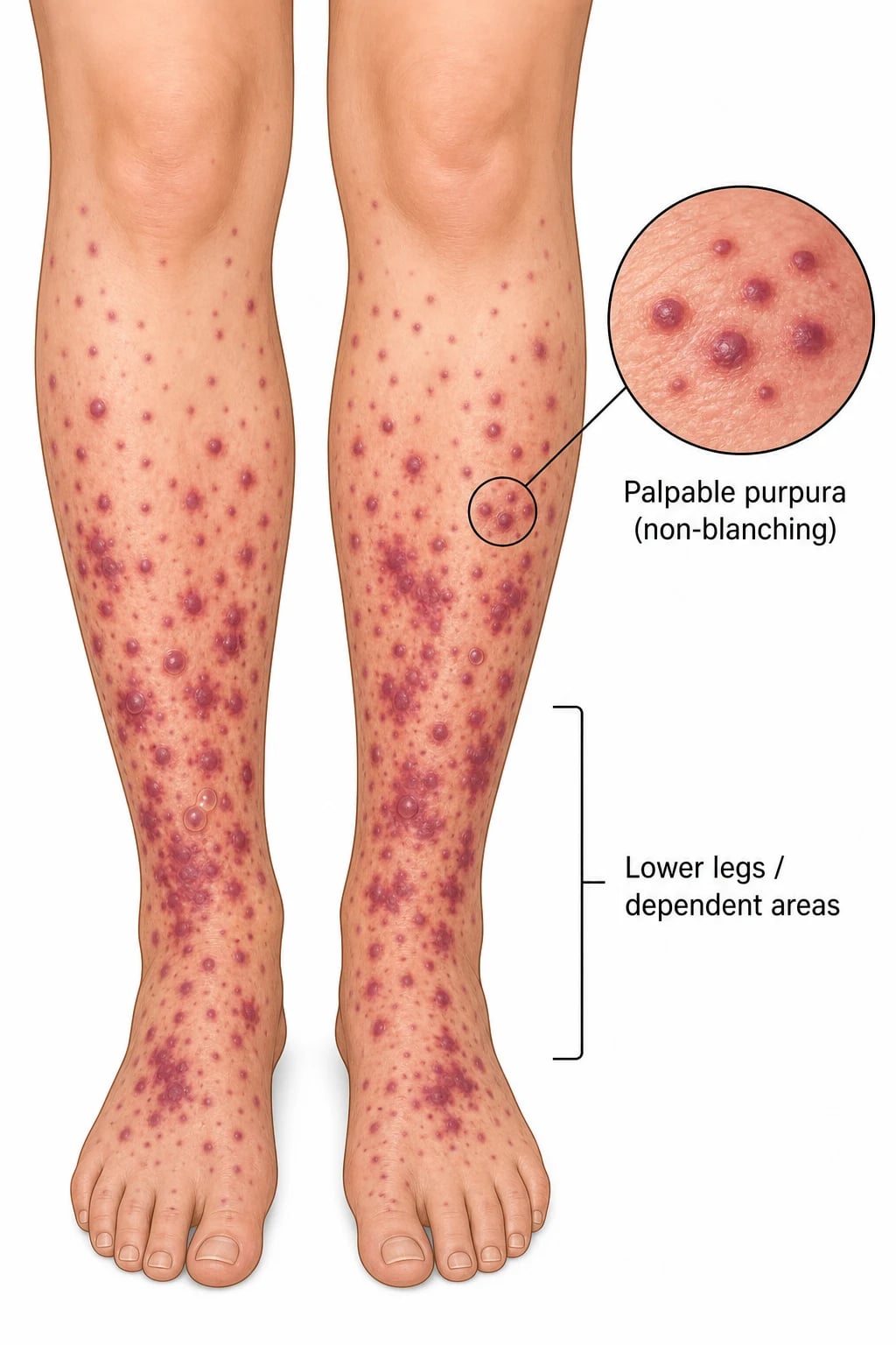
Cutaneous small vessel vasculitis (CSVV), also called leukocytoclastic vasculitis (LCV), is an inflammatory disorder of the post-capillary venules characterised clinically by palpable purpura (non-blanching) on dependent surfaces (especially the lower legs) and histologically by leukocytoclasia, fibrinoid necrosis of vessel walls, and red blood cell extravasation. It may be primary (idiopathic ~50%) or secondary to drugs, infections, autoimmune disease, malignancy, or as IgA vasculitis (Henoch-Schönlein purpura). Diagnosis requires skin biopsy for H&E and direct immunofluorescence (DIF), plus a systemic work-up (urinalysis, complement, ANA, ANCA, hepatitis serology, cryoglobulins). Management ranges from trigger removal and supportive measures (rest, elevation, compression) for skin-limited disease to systemic corticosteroids ± immunosuppressants for severe or systemic disease. Fellowship-level assessment demands mastery of the immune complex pathogenesis, the palpable purpura morphology, the IgA vs IgG/C3 DIF patterns, the IgA vasculitis (HSP) tetrad, urticarial vasculitis (24h lesions, complement), cryoglobulinaemic vasculitis (hepatitis C), and the stepwise management.
On this page & tools
Your progress
Saved locally on this device.
Exam tags
Red flags
Definition & Classification
Cutaneous small vessel vasculitis (CSVV), also called leukocytoclastic vasculitis (LCV) or hypersensitivity vasculitis, is an inflammatory disorder of the post-capillary venules in the superficial dermis, characterised clinically by palpable purpura and histologically by leukocytoclasia (nuclear dust), fibrinoid necrosis of vessel walls, and red blood cell extravasation.[1][2][5]
CSVV may be: [1]
- Primary (idiopathic): ~50% of cases; no identifiable trigger.[3]
- Secondary: drugs (~20%), infections (~20%), autoimmune/connective tissue disease (SLE, RA), malignancy (haematological > solid), or a specific named vasculitis syndrome.[1][5]
Key subtypes/syndromes (examiner-relevant):[5][6]
| Syndrome | Key features | DIF |
|---|---|---|
| IgA vasculitis (Henoch-Schönlein purpura) | Tetrad: palpable purpura + arthralgia + abdominal pain + renal (IgA nephropathy). Children > adults | IgA deposition |
| Urticarial vasculitis | Urticarial lesions >24h, painful/burning, residual hyperpigmentation; hypocomplementaemic variant = SLE overlap | IgG/C3 |
| Cryoglobulinaemic vasculitis | Palpable purpura + arthralgia + weakness (Meltzer triad); hepatitis C association | IgM/IgG/C3 |
| Drug-induced vasculitis | 7-21 days after drug; levamisole (cocaine), hydralazine, propylthiouracil, minocycline, antibiotics | IgG/C3 or pauci-immune |
| Infection-associated | Less than 1 week; bacterial endocarditis, hepatitis B/C, streptococcal, staphylococcal | Variable |
Epidemiology & Risk Factors
- Incidence: cutaneous vasculitis ~5-10 per million per year; CSVV is the commonest pattern encountered in dermatology. IgA vasculitis (HSP) is the commonest childhood vasculitis (~10-20 per 100,000 children per year; peak age 3-10 yr).[1][2]
- Age and sex: cutaneous-only CSVV — slight female predominance, adult-onset; HSP — children > adults (3-10 yr; boys slightly more); cryoglobulinaemic vasculitis — middle-aged adults; urticarial vasculitis — adults 30-50 yr.[2][3]
- Geography: no strong geographic bias for primary CSVV; cryoglobulinaemic vasculitis tracks hepatitis C prevalence (Mediterranean basin, Eastern Europe, Japan, parts of USA); Buruli ulcer (M. ulcerans) — West Africa and Australia; HSP more common in spring and after upper-respiratory infections.
- Risk factors / triggers:
- Drugs (most common cause in adults): penicillins, sulphonamides, NSAIDs, allopurinol, propylthiouracil, hydralazine, minocycline, levamisole (cut cocaine), vaccines, anticoagulants; onset typically 1-3 weeks after drug exposure.[1][5]
- Infections: streptococcal pharyngitis (HSP trigger), hepatitis B (polyarteritis nodosa-like), hepatitis C (cryoglobulinaemia type II/III), HIV, Staph aureus, TB, parvovirus B19.[2][5]
- Autoimmune: SLE (cutaneous vasculitis in 20-30%), rheumatoid arthritis, primary Sjögren syndrome.[5]
- Malignancy (paraneoplastic): haematological (CLL, lymphoma, MDS, paraproteinaemia/cryoglobulinaemia) > solid (lung, colon, breast, prostate); suspect in older adults with no obvious trigger.[5]
- Inflammatory bowel disease: associated with extra-intestinal cutaneous vasculitis.
LCV triggers — remember the four buckets
DRIM
Penicillins, sulphonamides, NSAIDs, allopurinol, hydralazine, PTU, minocycline, levamisole-cut cocaine — onset 1-3 weeks
Strep throat (HSP), hepatitis B, hepatitis C (cryoglobulinaemia), HIV, Staph, TB, parvovirus B19
SLE (20-30% have cutaneous vasculitis), rheumatoid arthritis, Sjögren syndrome, IBD
Haematological > solid; suspect in older adult with new LCV and no other trigger — CLL, lymphoma, MDS, paraproteinaemia
Pathophysiology
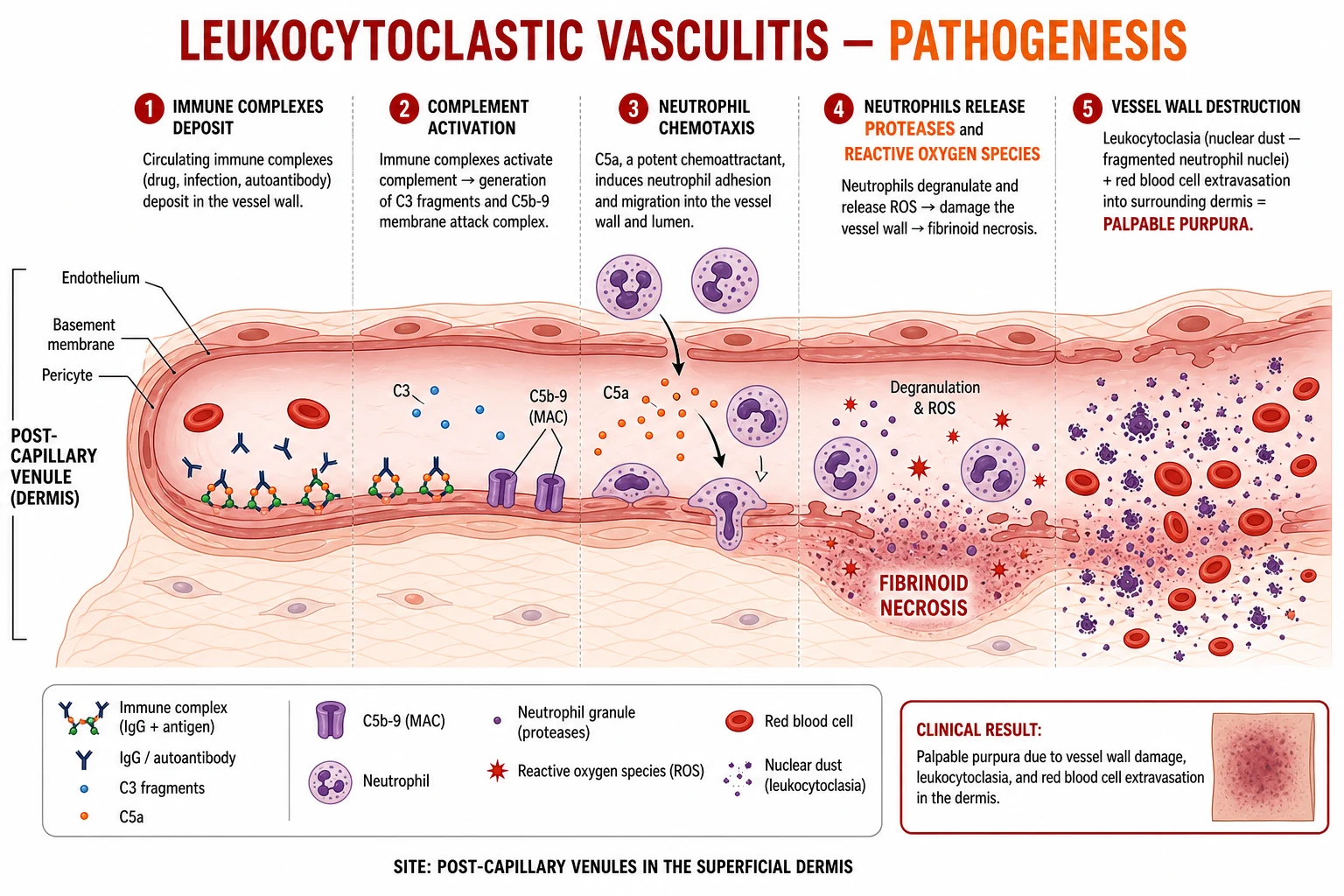
The mechanism is Type III hypersensitivity (immune complex-mediated):[1][5]
- Circulating immune complexes (drug, microbial antigen, autoantibody) deposit in the walls of post-capillary venules (especially in dependent areas — lower legs — due to hydrostatic pressure).
- Complement activation (classical and alternative pathways; C3, C5a, C5b-9 membrane attack complex).
- C5a attracts neutrophils → release of proteases, reactive oxygen species, matrix metalloproteinases.
- Vessel wall damage → fibrinoid necrosis (eosinophilic material replacing the vessel wall).
- Neutrophil death → leukocytoclasia (nuclear dust — fragmented neutrophil nuclei).
- Vessel wall destruction → red blood cell extravasation into the dermis → clinically palpable purpura. [1]
In IgA vasculitis, the immune complexes contain IgA1 (galactose-deficient), which deposits in vessel walls and mesangium, explaining both the skin and renal (IgA nephropathy) involvement.[9][10]
[1]Quick numbers for the examiner
Clinical Presentation [1]
- Hallmark: palpable purpura — non-blanching, deep red-purple macules and papules (2-10 mm), typically on the lower legs and ankles (dependent areas). Lesions may coalesce into plaques, develop bullae over necrotic centres, or ulcerate.[2][3]
- Lesions appear in crops over days; each crop lasts 1-3 weeks; new crops appear as old ones fade.
- Morphology variants: urticarial papules (urticarial vasculitis), nodules, livedo reticularis, ulcers, digital necrosis (severe).
- Systemic involvement (assess for in every patient):[5]
- Renal: haematuria, proteinuria, AKI (check urinalysis).
- GI: abdominal pain, bleeding, perforation (especially IgA vasculitis).
- Joints: arthralgia, arthritis.
- Neurology: mononeuritis multiplex, peripheral neuropathy (severe).
- Pulmonary: haemoptysis (rare; think ANCA-associated).
- Constitutional: fever, malaise, myalgia.
Clinical & Bedside Assessment
Systematic examination is the cornerstone of LCV work-up. The examination answers three questions — (1) is this really an inflammatory vasculitis, (2) how extensive is the skin disease, (3) is there systemic involvement?[5][6]
- Full skin examination — distribution and morphology:
- Distribution: dependent areas (lower legs, ankles), pressure points (sock line, bra strap, waistband), dependent areas of bed-bound patients; check for symmetry.
- Palpability test: every purpuric lesion must be palpated — palpability is the single most useful clinical discriminator between inflammatory (vasculitic) and non-inflammatory purpura.
- Morphology: macular purpura vs papular (palpable) purpura vs vesicular/bullous/ulcerated/necrotic; urticarial papules (urticarial vasculitis); livedo reticularis (vasculopathy, APLS); nodules (PAN, erythema induratum); retiform purpura (levamisole-cut cocaine, cholesterol emboli, calciphylaxis).[2][6]
- Diascopy (glass slide): confirms non-blanching (extravasated RBCs) versus blanching (telangiectasia, vascular lesions).
- Joint examination: synovitis, tenderness, range of motion (IgA vasculitis — arthralgia without effusion; RA overlap).
- Abdominal examination: tenderness (IgA vasculitis — intussusception risk), peritoneal signs, melena on glove.
- Cardiovascular and peripheral: peripheral pulses (livedo, distal ischaemia, digital necrosis), BP (renal involvement, hypertension).
- ENT and respiratory: nasal crusting/ulceration (GPA), sinus tenderness, haemoptysis (DAH), wheeze (EGPA).[6]
- Neurological: peripheral neuropathy, mononeuritis multiplex (foot drop, wrist drop) — flags systemic AAV or cryoglobulinaemic vasculitis.
- Drug history (the most important bedside question): review every new drug (including OTC, herbal, supplements) started in the 1-3 weeks before onset — penicillins, sulphonamides, NSAIDs, allopurinol, propylthiouracil, hydralazine, minocycline, levamisole-cut cocaine.[1][5]
- Infection screen at the bedside: throat inspection (streptococcal), examine for endocarditis stigmata (splinter haemorrhages, Osler nodes, Janeway lesions), examine for skin/soft-tissue infection.
- Urticarial vasculitis bedside clue: wheals lasting over 24 hours, painful or burning rather than pruritic, leaving residual purpura or pigmentation when they resolve.[7]
Specific Subtypes & Clinical Scenarios
IgA vasculitis (HSP)
Most common childhood vasculitis
- **Children 3-10 yr** (peak); ~10-20/100,000/yr; boys slightly > girls
- **Tetrad:** palpable purpura + arthralgia + abdominal pain + renal disease (IgA nephropathy pattern)
- **DIF:** IgA in dermal vessel walls and glomerular mesangium (galactose-deficient IgA1)
- Often follows upper-respiratory infection; usually self-limiting in 4-6 weeks
- **Renal disease dictates prognosis** — 1-2% ESRD; 10-30% have nephritis; monitor urinalysis for 6-12 months
- GI: intussusception (ileo-ileal most common), perforation, bleeding — short steroid course for severe abdominal pain
Cryoglobulinaemic vasculitis
Meltzer triad — think hepatitis C
- **Type II/III cryoglobulins** with rheumatoid factor activity; precipitate on cooling (keep blood warm to lab)
- **Meltzer triad:** palpable purpura + arthralgia + weakness
- **Hepatitis C** in 70-90% of type II; other associations: HIV, autoimmune, lymphoproliferative
- **Complement:** low C4 (classical pathway consumption) — the immunological hallmark
- Treatment: DAA for HCV (cures the vasculitis in most); rituximab for refractory
- Renal disease (MPGN), peripheral neuropathy, digital ischaemia/ulceration in advanced disease
Urticarial vasculitis
Wheals that last and leave marks
- **Urticarial wheals lasting >24 hours** — painful/burning, residual purpura when they resolve
- **Hypocomplementaemic variant** (low C1q, C3, C4) — SLE overlap, severe systemic disease
- **Normocomplementaemic variant** — milder, mostly skin-limited
- **Anti-C1q antibodies** in 60-100% of hypocomplementaemic form
- DIF: C3 ± IgG along basement membrane and vessel wall
- Treatment ladder: antihistamines (limited effect), hydroxychloroquine, dapsone, colchicine; rituximab for hypocomplementaemic/SLE
ANCA-associated vasculitis
Pauci-immune — see AAV topic
- **Pauci-immune** DIF (no immune deposits) — the histological signature
- ANCA positive: PR3 (GPA), MPO (MPA, EGPA); atypical p-ANCA (anti-elastase) in levamisole-cut cocaine
- **Systemic features dominate:** pulmonary-renal syndrome, mononeuritis multiplex, ENT, ocular
- **Emergency:** pulmonary haemorrhage + rapidly progressive glomerulonephritis
- Induction: IV methylpred + rituximab or cyclophosphamide; avacopan steroid-sparing
- Refer to AAV topic for full management
Deep Dive into the Four CSVV Subtypes
The four named subtypes of CSVV that the examiner will test — IgA vasculitis, cryoglobulinaemic vasculitis, urticarial vasculitis, and ANCA-associated vasculitis with cutaneous involvement — are not interchangeable immune-complex reactions. Each has a distinct DIF signature, a distinct complement profile, a distinct set of driver antigens (galactose-deficient IgA1, HCV-IgM rheumatoid factor, anti-C1q, myeloperoxidase/proteinase-3), and a distinct pharmacology ladder. Mistaking one for the other at the bedside delays definitive therapy — most notoriously, an HCV-positive cryoglobulinaemic vasculitis left untreated while the patient is placed on ciclosporin for what is wrongly assumed to be idiopathic LCV.[1][2][3][6]
Subtype 1 — IgA vasculitis (Henoch-Schönlein purpura)
Pathogenesis. IgA vasculitis is driven by galactose-deficient IgA1 immune complexes that deposit in dermal post-capillary venules and in the renal mesangium, activating the alternative complement pathway and recruiting neutrophils. The mesangial deposition explains the IgA-nephropathy phenotype that dictates prognosis. The cutaneous DIF shows IgA in dermal vessel walls (the only vasculitis in which IgA is the dominant deposit); serum IgA is elevated in roughly half of patients at presentation but is non-specific. Triggers include upper-respiratory streptococcal infection (the classic antecedent), CMV, parvovirus B19, and drugs.[9][10]
Clinical tetrad and prognostic stratification. Palpable purpura with IgA in a child aged 3-10 yr is HSP until proven otherwise. The four cardinal features are (1) palpable purpura on the lower legs and buttocks (almost always present), (2) arthralgia/arthritis without effusion (knees and ankles, ~75%), (3) abdominal pain (colicky, peri-umbilical, ~50% — intussusception risk peaks in week 2), and (4) renal disease (haematuria ± proteinuria, 30-50%; nephrotic-range and crescentic glomerulonephritis is the minority that dictates prognosis). Skin-only disease needs no immunosuppression in most children; renal involvement (proteinuria over 0.5 g/day, active sediment, hypertension) needs renal biopsy and ACE inhibitor or ARB ± corticosteroid. The 1-2% ESRD rate is the clinical bottom line for the examiner.[8][9]
Treatment ladder. Step 1 — supportive care (paracetamol for joint/abdominal pain, hydration, leg elevation). Step 2 — for severe abdominal pain or GI bleeding, prednisolone 1 mg/kg/day for 1-2 weeks then taper, which shortens abdominal pain duration and may reduce the need for surgical intervention for intussusception; corticosteroids do not reliably prevent HSP nephritis. Step 3 — for IgA-nephropathy-pattern renal disease, ACE inhibitor or ARB for proteinuria regardless of BP (renoprotective), with corticosteroids for rapidly progressive crescentic GN (rare; biopsy-proven), and azathioprine or mycophenolate for steroid-dependent disease. Step 4 — refractory nephritis: tonsillectomy (especially in Japanese and East Asian cohorts where it reduces relapse rate in IgA-nephropathy-like disease), cyclophosphamide, rituximab, or mycophenolate plus corticosteroid. Monitor urinalysis and blood pressure monthly for 6-12 months — late-onset nephritis is the reason follow-up cannot stop at 6 weeks.[8][9][10]
Subtype 2 — Cryoglobulinaemic vasculitis
Pathogenesis. Cryoglobulins are immunoglobulins that precipitate on cooling (the laboratory specimen must be kept warm at 37 °C). Type I (monoclonal IgM or IgG) is associated with paraproteinaemia/myeloma and causes a hyperviscosity-thrombotic pattern more than an immune-complex vasculitis; Type II (monoclonal IgM rheumatoid factor plus polyclonal IgG) is the HCV-driven mixed cryoglobulinaemia; Type III (polyclonal IgM rheumatoid factor plus polyclonal IgG) is seen in autoimmune disease (SLE, Sjögren). Types II/III form circulating immune complexes that consume complement — classically low C4 with normal or modestly low C3 — and deposit in vessel walls and the renal mesangium, producing a membranoproliferative glomerulonephritis pattern (MPGN).[1][5]
The Meltzer triad and its red flags. The classic Meltzer triad is palpable purpura + arthralgia + weakness (asthenia). Red flags that turn a presentation from "skin-limited" to "vasculitis emergency" include (1) glomerulonephritis (haematuria/proteinuria, MPGN on biopsy), (2) peripheral neuropathy (mononeuritis multiplex — the most common neurological presentation; sensory > motor), (3) digital ischaemia and ulceration, and (4) pulmonary capillaritis with diffuse alveolar haemorrhage. Rheumatoid factor is markedly elevated, C4 is low, and HCV antibodies and HCV RNA confirm 70-90% of type II cases.[5]
Treatment ladder for HCV-positive cryoglobulinaemic vasculitis. Step 1 — direct-acting antiviral (DAA) therapy for the underlying HCV (sofosbuvir/velpatasvir, glecaprevir/pibrentasvir, or equivalent): cures the HCV and resolves the vasculitis in most patients over 3-6 months; this is now the first-line etiological treatment. Step 2 — for severe organ-threatening disease (rapidly progressive glomerulonephritis, severe neuropathy, digital ischaemia, ulceration, or diffuse alveolar haemorrhage), combine DAA + rituximab (rituximab 375 mg/m² IV weekly for 4 weeks — the lymphoma protocol — or 1 g IV on day 1 and day 15 — the AAV/RA protocol); in patients with high cryocrit, plasmapheresis (3-5 exchanges over 10-14 days) is performed first to lower the cryoglobulin burden before rituximab, because sudden B-cell lysis can shower vessels with immune complexes and precipitate a flare. Step 3 — short course of prednisolone 0.5-1 mg/kg/day for severe organ involvement during induction, then taper. Step 4 — for refractory disease: mycophenolate mofetil 1-1.5 g BD, azathioprine 1-3 mg/kg/day, or cyclophosphamide; rituximab maintenance (1 g every 6 months) reduces relapse.[5]
Treatment ladder for HCV-negative cryoglobulinaemic vasculitis. DAA is not indicated; first-line is rituximab (lymphoma protocol as above) plus corticosteroid; steroid-sparing with mycophenolate or azathioprine. Treat the underlying cause (SLE, Sjögren, lymphoproliferative disease). Plasmapheresis for life-threatening disease.[5]
Subtype 3 — Urticarial vasculitis
Pathogenesis. Urticarial vasculitis spans a clinical spectrum from a normocomplementaemic, skin-limited disorder that resembles chronic urticaria to a hypocomplementaemic urticarial vasculitis (HUV) syndrome with anti-C1q autoantibodies, low C1q/C3/C4, systemic features, and overlap with SLE. Anti-C1q antibodies (100% of HUV in some series, 30-60% in others) drive classical-pathway complement consumption; the lesion on DIF is C3 ± IgG along the dermal-epidermal junction (lupus-band-like) and around vessel walls. Histology shows leukocytoclastic vasculitis — the discriminator from ordinary urticaria (no vasculitis, perivascular lymphocytes, no nuclear dust).[7]
The bedside rule. Any urticarial lesion that lasts over 24 hours, is painful or burning rather than pruritic, and leaves residual purpura or post-inflammatory pigmentation when it resolves is urticarial vasculitis — not urticaria. HUV is the form that has arthritis, glomerulonephritis, serositis, obstructive lung disease, and the same autoantibody profile as SLE; many patients fulfil the SLE classification criteria within 5 years.[7]
Treatment ladder. Step 1 — antihistamines (often disappointing, but worth a 2-week trial at up to 4× standard dosing for symptomatic itch). Step 2 — hydroxychloroquine 200-400 mg/day (often the most effective single agent for normocomplementaemic disease; with ophthalmology monitoring). Step 3 — colchicine 0.5-1 mg TDS (anti-neutrophilic; useful for chronic cutaneous disease; titrate from 0.5 mg/day for GI tolerance). Step 4 — dapsone 50-150 mg/day (verify G6PD before starting; aim for haemoglobin ≥10 g/dL after 4 weeks; methaemogloblinemia is common at higher doses). Step 5 — add prednisolone 0.5-1 mg/kg/day for systemic involvement (arthritis, serositis, glomerulonephritis), then taper. Step 6 — refractory hypocomplementaemic disease: mycophenolate mofetil 1-1.5 g BD, azathioprine 1-3 mg/kg/day, or methotrexate 7.5-25 mg weekly; rituximab for SLE-overlap or refractory HUV.[7]
Subtype 4 — ANCA-associated vasculitis with cutaneous involvement
Cutaneous disease is common but secondary. Up to 30-50% of patients with granulomatosis with polyangiitis (GPA, PR3-ANCA) and microscopic polyangiitis (MPA, MPO-ANCA) develop palpable purpura as a presenting sign. The disease is, however, dominated by systemic features — pulmonary-renal syndrome (haemoptysis + rapidly progressive glomerulonephritis), ENT (nasal crusting/ulceration/saddle-nose, sinus disease, subglottic stenosis), ocular (scleritis, peripheral ulcerative keratitis), and mononeuritis multiplex. The DIF is pauci-immune — no or scant immune deposits — the immunopathological signature that sets AAV apart from immune-complex LCV.[1][2][6]
Treatment ladder (summary — see AAV topic for detail). Induction for organ-threatening disease: IV methylprednisolone 500-1000 mg/day for 3 days then prednisolone 1 mg/kg/day plus either rituximab (375 mg/m² IV weekly × 4, or 1 g day 1 + day 15) or cyclophosphamide (IV pulses 15 mg/kg every 2-3 weeks, or oral 2 mg/kg/day). Avacopan (30 mg BD) is now an oral steroid-sparing agent in combination with rituximab or cyclophosphamide. Plasma exchange for refractory disease or pulmonary haemorrhage. Maintenance: azathioprine 2 mg/kg/day, mycophenolate, methotrexate, or rituximab 1 g every 6 months for at least 24-48 months. Cutaneous disease activity broadly tracks the systemic disease but may persist as a low-grade signature during remission.[6]
Subtype pharmacology — drug, dose and target (examiner-ready)
Subtype round-up: the examiner's one-liner for each
- IgA vasculitis (HSP) — galactose-deficient IgA1; tetrad; renal drives prognosis; monitor urinalysis 6-12 months; ACE inhibitor for proteinuria.
- Cryoglobulinemic vasculitis — type II mixed; HCV in 70-90%; Meltzer triad; low C4 is the lab signature; DAA first-line, rituximab + plasmapheresis for severe.
- Urticarial vasculitis — wheals lasting over 24 hours, painful, leaving pigmentation; complement and anti-C1q discriminate HUV from normocomplementaemic form.
- ANCA-associated with skin involvement — pauci-immune DIF; PR3 (GPA), MPO (MPA); systemic features dominate; IV methylpred + rituximab/cyclophosphamide induction.
Differential Diagnosis
| Mimic | Distinguishing features |
|---|---|
| Thrombocytopenic purpura | Non-palpable petechiae/purpura; low platelet count; no biopsy evidence of vasculitis |
| Pigmented purpuric dermatosis | Cayenne pepper macules; non-palpable; chronic; capillaritis on histology (no fibrinoid necrosis) |
| Meningococcaemia | Rapid onset, febrile, septic; petechiae + purpura; gram-negative diplococci on blood culture |
| Warfarin necrosis | Early in therapy; large necrotic plaques; not palpable purpura |
| Antiphospholipid syndrome | Livedo reticularis, thrombosis, recurrent miscarriage; anticardiolipin/anti-β2-GPI |
| Trauma / dependent purpura | History; non-palpable; resolves quickly |
Investigations
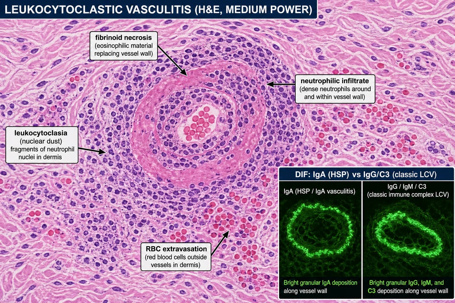
Skin biopsy (punch, from an early lesion less than 24-48h old):[5][6]
- H&E: leukocytoclastic vasculitis — fibrinoid necrosis of vessel walls, neutrophilic infiltrate, nuclear dust (leukocytoclasia), RBC extravasation.
- DIF (from a newer lesion, less than 24h old): the most important diagnostic discriminator —
Systemic work-up (every patient):[5]
- Urinalysis (haematuria, proteinuria — renal involvement) — the single most important screening test.
- Bloods: FBC, U&E, LFTs, CRP/ESR.
- Complement: C3 and C4 (low → cryoglobulinaemia, SLE, urticarial vasculitis).
- Autoantibodies: ANA, ANCA (excludes ANCA-associated), RF, anti-C1q (urticarial vasculitis).
- Infection screen: hepatitis B surface antigen, hepatitis C antibody, HIV.
- Cryoglobulins (if complement low or hepatitis C positive — must keep warm).
- Serum protein electrophoresis (if paraproteinaemia suspected).
- Chest X-ray (if pulmonary involvement or ANCA positive).
- Age-appropriate malignancy screen (if no other trigger identified in older patients).[5]
Management
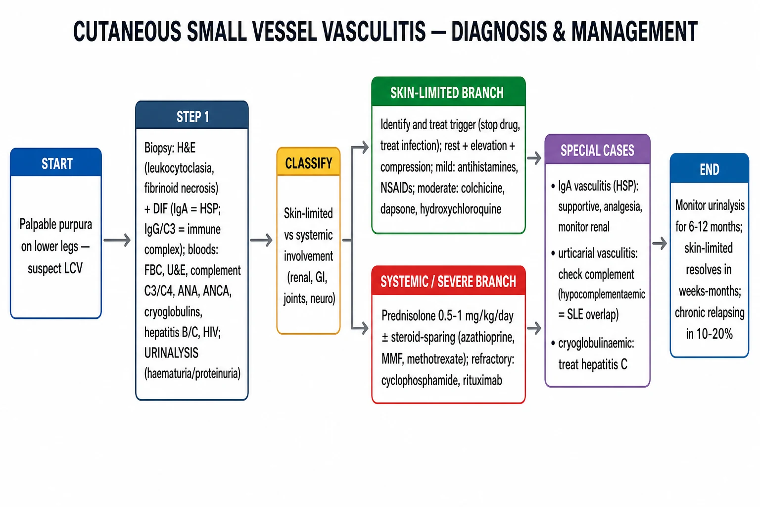
Step 1 — Identify and remove trigger
- Stop all potentially causative drugs (especially those started 1-3 weeks before onset).[5]
- Treat any identified infection (hepatitis C for cryoglobulinaemia; streptococcal; endocarditis).[5]
- Manage underlying disease (SLE, RA, malignancy).
Step 2 — Skin-limited disease (mild-moderate)
- Rest, leg elevation, compression stockings (reduces hydrostatic pressure → reduces new lesions).
- Antihistamines (for pruritus).
- NSAIDs (for arthralgia; avoid if renal involvement).
- Colchicine (0.6 mg BD) — anti-neutrophilic; useful for chronic/recurrent skin-limited LCV.[5]
- Dapsone (50-100 mg; check G6PD) — especially for IgA vasculitis skin involvement.[3]
- Hydroxychloroquine — for chronic skin-limited disease.[5]
Step 3 — Systemic or severe disease
- Systemic corticosteroids: prednisolone 0.5-1 mg/kg/day for systemic involvement (renal, GI, neuro).[5]
- Steroid-sparing agents: azathioprine, mycophenolate mofetil, methotrexate for chronic or relapsing disease.[5]
- Refractory: cyclophosphamide (pulse IV), rituximab (especially for cryoglobulinaemic and IgA vasculitis).[5]
Special subtype management
- IgA vasculitis (Henoch-Schönlein purpura): supportive care; analgesia for arthralgia/abdominal pain; short course of corticosteroids for severe GI or renal involvement; monitor urinalysis and BP for 6-12 months (delayed renal involvement can occur).[8][9]
- Urticarial vasculitis: antihistamines (limited effect); hydroxychloroquine, dapsone, colchicine; systemic steroids or rituximab for hypocomplementaemic/SLE-overlap disease.[7]
- Cryoglobulinaemic vasculitis: treat underlying hepatitis C (direct-acting antivirals — usually cures the vasculitis); rituximab for refractory.[5]
Special Populations
- Children (IgA vasculitis/HSP): the commonest childhood vasculitis; usually self-limiting (4-6 weeks); renal follow-up for 6-12 months; corticosteroids for severe GI/renal involvement; prognosis generally excellent.[8]
- Elderly: higher risk of underlying malignancy; drug-induced more common; screen for myelodysplasia if persistent.[5]
- Pregnancy: rare; manage with topical and mild systemic agents; avoid teratogenic immunosuppressants.[5]
Prognosis
- Skin-limited CSVV: excellent; resolves in weeks to months; chronic relapsing course in ~10-20%.[3]
- Systemic involvement (renal, GI, neuro) worsens prognosis; IgA vasculitis has long-term renal risk.[9]
- Cryoglobulinaemic vasculitis prognosis driven by hepatitis C response.[5]
Complications & Pitfalls
The downstream consequences of CSVV range from trivial post-inflammatory hyperpigmentation to life-threatening organ failure. The examiner expects you to anticipate each one.[1][5][9]
- Renal — the dominant determinant of prognosis:
- IgA vasculitis: nephritis in 30-50% of children (haematuria, proteinuria, nephrotic-range, rapidly progressive GN); 1-2% progress to ESRD; 10-30% have chronic kidney disease.
- Cryoglobulinaemic vasculitis: MPGN pattern; proteinuria and slowly progressive renal impairment.
- ANCA-associated vasculitis: rapidly progressive glomerulonephritis — the classical "pauci-immune crescentic GN."
- Pulmonary haemorrhage: in pauci-immune LCV / AAV — life-threatening diffuse alveolar haemorrhage (DAH); mortality high without plasma exchange and induction immunosuppression.
- GI complications of IgA vasculitis (HSP):
- Intussusception — ileo-ileal most common (vs ileo-colic in idiopathic childhood intussusception); ultrasound shows the classic "target sign."
- Bowel wall haematoma, perforation, ischaemia, massive GI bleeding — surgical emergency.
- Steroids reduce abdominal pain duration and may reduce intussusception risk but do not prevent nephritis.
- Skin complications: ulceration, secondary bacterial infection, post-inflammatory hyperpigmentation (months to years); chronic relapsing course in ~10-20% of skin-limited disease.[3]
- Peripheral neuropathy: mononeuritis multiplex in cryoglobulinaemic and ANCA-associated vasculitis — irreversible if treatment is delayed.
- Pitfalls (frequent exam traps):[5][6]
- Misdiagnosing as thrombocytopenic purpura, scurvy, or vasculopathy — delays biopsy and the work-up.
- Missing an underlying malignancy in an older adult with new LCV and no other trigger — investigate aggressively.
- Missing the drug trigger — re-challenge can be fatal, especially with allopurinol, propylthiouracil, hydralazine.
- Biopsy of an OLD lesion (>48-72 h) — the neutrophilic infiltrate has already faded and histology may be non-diagnostic. Always biopsy a fresh purpuric lesion.
- Chronic corticosteroid dependence — introduce a steroid-sparing agent (azathioprine, mycophenolate, methotrexate) early in chronic relapsing disease.
IgA vasculitis (HSP) — natural history and follow-up timeline
Evidence, Guidelines & Regional Differences
- No single international guideline; management is based on expert consensus, the Chapel Hill Consensus Classification, and EULAR/PRES/PRINTO criteria for IgA vasculitis.[5][9]
- Chapel Hill nomenclature classifies CSVV by vessel size and dominant immune complex type.[6]
- Emerging: COVID-19-associated CSVV (Olson 2025); rituximab for refractory IgA vasculitis.[4][9]
Prevention
- Avoid re-exposure to identified drug triggers; document in medical record.
- Treat underlying infections (hepatitis C) and autoimmune disease.
- Compression stockings reduce recurrence in dependent purpura. [1]
Viva vignette: a 7-year-old boy with palpable purpura on the legs, knee pain and 48 hours of colicky abdominal pain. What is your single most important next investigation and why?
This is the IgA vasculitis (HSP) tetrad — palpable purpura + arthralgia + abdominal pain + (imminent) renal involvement. The single most important next investigation is a urinalysis with microscopy (looking for haematuria and proteinuria) plus blood pressure measurement, because (1) renal involvement is silent until late but dictates long-term prognosis, (2) you must establish a baseline before considering corticosteroids, and (3) you must risk-stratify the need for renal biopsy (proteinuria >0.5 g/day or active urinary sediment). The next steps are a punch biopsy of a fresh lesion (under 24-48 h) purpuric lesion for H&E and DIF, an abdominal ultrasound if intussusception is suspected (ileo-ileal), and supportive management — analgesia, IV fluids, and a short course of corticosteroids if the abdominal pain is severe. Avoid NSAIDs given the renal risk. Counsel the family about the 6-12 month renal follow-up schedule.
Exam Pearls
[1]Red Flags
Exam application bank (NEET-PG / INICET)
One-line answer
Cutaneous small vessel vasculitis (CSVV), also called leukocytoclastic vasculitis (LCV), is an inflammatory disorder of the post-capillary venules characterised clinically by palpable purpura (non-blanching) on dependent surfaces (especially the lower legs) and histologically by leukocytoclasia, fibrinoid necrosis of vessel walls, and red blood cell extravasation. It may be primary (idiopathic ~50%) or secondary to drugs, infections, autoimmune disease, malignancy, or as IgA vasculitis (Henoch-Schönlein purpura). Diagnosis requires skin biopsy for H&E and direct immunofluorescence (DIF), plus a systemic work-up (urinalysis, complement, ANA, ANCA, hepatitis serology, cryoglobulins). Management ranges from trigger removal and supportive measures (rest, elevation, compression) for skin-limited disease to systemic corticosteroids ± immunosuppressants for severe or systemic disease. Fellowshi
Worked stems (answer without another resource)
Stem 1 — Classic presentation. Map symptoms to mechanism; name the first investigation and first treatment step with dose/route if drug therapy is standard. [1]
Stem 2 — Unstable / complicated. List red flags that force immediate resuscitation, theatre, ICU, antidote, or reperfusion — and what you do in the first 15 minutes. [1]
Stem 3 — Atypical group. Elderly, pregnancy, child, or immunocompromised: how presentation and thresholds change. [1]
Stem 4 — Differential trap. Name the three closest mimics and one discriminator for each. [1]
Stem 5 — Disposition. Who goes home with safety-netting, who is admitted, who needs HDU/ICU/theatre, and what follow-up is mandatory. [1]
Rapid viva checklist
- Definition + classification
- Pathophysiology chain
- Bedside signs / criteria
- Score with exact components (if any)
- Emergency bundle
- Definitive therapy with doses
- Complications of disease and of treatment
- Special populations
- Guideline/trial name if classic
- Three exam traps
Coverage self-check
If you cannot answer any stem above from this page alone, re-read the matching section — the page is intended to be self-sufficient for final-prof and NEET-PG/INICET questions on Cutaneous small vessel vasculitis.
[1]References
- [1]Shavit E, Alavi A, Sibbald RG. Vasculitis-What Do We Have to Know? A Review of Literature Int J Low Extrem Wounds, 2018.PMID 30501545
- [2]DeHoratius DM. Cutaneous small vessel vasculitis Postgrad Med, 2023.PMID 36524408
- [3]Lotti T, Ghersetich I, Comacchi C, Jorizzo JL. Cutaneous small-vessel vasculitis J Am Acad Dermatol, 1998.PMID 9810883
- [4]Olson KO, Patel S, Pathak P, et al. Cutaneous small vessel vasculitis in the COVID-19 era: a systematic review Skin Health Dis, 2025.PMID 40365256
- [5]Fraticelli P, Benfaremo D, Gabrielli A. Diagnosis and management of leukocytoclastic vasculitis Intern Emerg Med, 2021.PMID 33713282
- [6]Frumholtz L, Laurent-Roussel S, et al. Cutaneous Vasculitis: Review on Diagnosis and Clinicopathologic Correlations Clin Rev Allergy Immunol, 2021.PMID 32378145
- [7]Gu SL, Jorizzo JL. Urticarial vasculitis Int J Womens Dermatol, 2021.PMID 34222586
- [8]Reamy BV, Servey JT, Williams PM. Henoch-Schönlein Purpura (IgA Vasculitis): Rapid Evidence Review Am Fam Physician, 2020.PMID 32803924
- [9]Pillebout E, Sunderkötter C. IgA vasculitis Semin Immunopathol, 2021.PMID 34170395
- [10]Song Y, Huang X, Yu G, et al. Pathogenesis of IgA Vasculitis: An Up-To-Date Review Front Immunol, 2021.PMID 34858429