Dermatology · Medicine
Cutaneous T-cell lymphoma
Also known as Cutaneous T-cell lymphoma · CTCL · Mycosis fungoides · MF · Sezary syndrome · SS · Alibert-Bazin syndrome
Cutaneous T-cell lymphoma (CTCL) is a heterogeneous group of extranodal non-Hodgkin lymphomas of skin-homing mature T cells, dominated by mycosis fungoides (MF) (~50 percent of CTCL; indolent course progressing from patch to plaque to tumour to erythroderma) and Sezary syndrome (SS) (~3-5 percent; aggressive leukaemic variant with erythroderma + lymphadenopathy + circulating Sezary cells). MF is the prototypical great mimicker of eczema, psoriasis and tinea corporis, often misdiagnosed for years before a biopsy secures the diagnosis. Histology shows epidermotropism with Pautrier microabscesses; immunophenotyping reveals a clonal CD4+ T-cell population with aberrant loss of CD7 and CD26; PCR for T-cell receptor gene rearrangement confirms clonality. Management is stage-driven: skin-directed therapies for early disease (topical corticosteroids, mechlorethamine, bexarotene, narrowband UVB / PUVA, total skin electron beam therapy) and escalating systemic therapy (extracorporeal photopheresis, bexarotene, interferon, HDAC inhibitors, mogamulizumab, brentuximab vedotin) for advanced or refractory disease, with allogeneic stem cell transplantation the only potentially curative option in advanced disease.
On this page & tools
Your progress
Saved locally on this device.
Exam tags
Red flags
Quick recall — SEZARY
Definition and Classification
Cutaneous T-cell lymphoma (CTCL) is a heterogeneous group of extranodal non-Hodgkin lymphomas of skin-homing mature T cells, accounting for roughly 75-80 percent of all primary cutaneous lymphomas. The classification follows the WHO-EORTC scheme (Willemze 2005, PMID 15692063, with updates in the 2018 WHO classification of haematolymphoid tumours), which integrates clinical, histological, immunophenotypic and molecular criteria to define distinct disease entities rather than relying on histology alone.[1][6][7]
The two dominant and clinicopathologically best-characterised subtypes are mycosis fungoides (MF) and Sezary syndrome (SS). Other important CTCL variants include the primary cutaneous CD30+ lymphoproliferative disorders (lymphomatoid papulosis [LyP] and primary cutaneous anaplastic large cell lymphoma [pcALCL]), subcutaneous panniculitis-like T-cell lymphoma (SPTCL), primary cutaneous CD4+ small/medium T-cell lymphoproliferative disorder, primary cutaneous gamma-delta T-cell lymphoma, and extranodal NK/T-cell lymphoma, nasal type (often EBV-associated, aggressive, usually presents in the upper aerodigestive tract but may involve skin).[6][7]
Frequency
- MF: approximately 50 percent of all CTCL — the commonest single entity
- SS: approximately 3 to 5 percent of CTCL — uncommon
- Both are dominated by the CD4+ T-helper phenotype
- Malignant clone is a skin-homing central memory T cell
Clinical course
- MF: indolent; years to decades from patch to tumour
- SS: aggressive; median survival 2 to 4 years
- Diagnostic delay in MF averages 3 to 6 years
- Relapse is the rule across all stages
Skin disease
- MF: patch then plaque then tumour then erythroderma (sequential)
- SS: erythroderma (more than 80 percent BSA) from onset
- MF favours a sun-protected bathing-trunk distribution
- Tumour-stage disease may herald large cell transformation
Blood and nodes
- MF: usually absent or minimal clone by PCR
- SS: Sezary cells; CD4:CD8 ratio greater than 10; clone positive
- MF: dermatopathic nodes (variable by stage)
- SS: generalised lymphadenopathy at presentation
Prognosis
- MF T1: 10-year overall survival greater than 95 percent
- MF T4: 10-year overall survival approximately 40 percent
- SS: median overall survival 2 to 4 years with modern therapy
- Large cell transformation halves expected survival
Pathophysiology
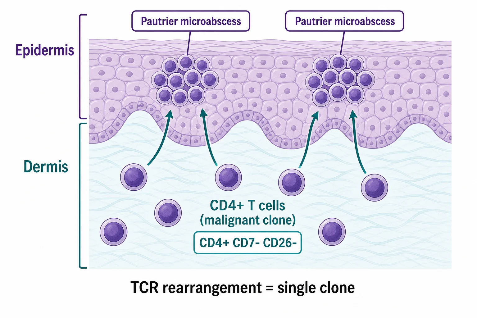
The pathophysiology of CTCL is best understood for mycosis fungoides and Sezary syndrome. Several converging biological themes underpin disease initiation and progression.[3][6]
Cell of origin. The malignant clone in MF and SS is a skin-homing CD4+ central memory T cell expressing the cutaneous lymphocyte antigen (CLA), the chemokine receptors CCR4 and CCR7, and the memory marker CD45RO. These cells normally recirculate between skin and lymph nodes; in CTCL, the clone is locked into the skin compartment by aberrant homing receptor expression and progressively dominates the cutaneous immune landscape.[3]
Epidermotropism and Pautrier microabscesses. The defining histological hallmark is epidermotropism — migration of atypical lymphocytes into the epidermis without the surrounding intercellular oedema (spongiosis) that characterises eczema. Where clusters of atypical cells aggregate within the epidermis, they form Pautrier microabscesses, the pathognomonic collection of three or more atypical lymphocytes in a small intraepidermal vesicle. The malignant cells also display cerebriform nuclei — small, hyperconvoluted brain-like nuclei best appreciated at high magnification and in semi-thin sections.[1][3]
Aberrant immunophenotype. On immunohistochemistry and flow cytometry, the malignant clone is CD3+, CD4+, CD45RO+, CD2+, with frequent aberrant loss of CD7 (in up to 70-80 percent of cases) and CD26 (the dipeptidyl peptidase IV). Loss of CD26 is the more sensitive marker for the Sezary cell population in peripheral blood. CD30 expression emerges with large cell transformation and defines the CD30+ lymphoproliferative disorders.[3][5]
Clonality. A single dominant T-cell receptor (TCR) gene rearrangement — most often of the beta or gamma chain — is detected by PCR or next-generation sequencing. This monoclonality distinguishes MF from reactive dermatitides in which the TCR repertoire is polyclonal or oligoclonal. The same dominant clone can be tracked in skin, blood and lymph node, allowing molecular staging and disease monitoring.[6]
Cytokine skewing and immunosuppression. As disease progresses there is a progressive shift from a Th1 (cell-mediated, IFN-gamma) to a Th2 (humoral, IL-4 / IL-5 / IL-13) cytokine profile. This Th2 dominance explains the peripheral eosinophilia, elevated IgE, and progressive cellular immune dysfunction that predisposes to secondary cutaneous infections (Staphylococcus aureus, herpes simplex, molluscum contagiosum) which are the most common immediate causes of death in advanced CTCL.[1][5]
Genomic evolution and transformation. Recurrently mutated genes include TP53 (associated with large cell transformation), STAT3 / STAT5B (constitutive JAK-STAT activation), CDKN2A (cell-cycle checkpoint loss), DNMT3A (DNA methylation), and TET2 (DNA demethylation). Loss of CDKN2A and TP53 herald large cell transformation, the most adverse biological event in MF, with median survival of approximately 2 years post-transformation.[5][6]
Clinical Presentation
The clinical picture of CTCL is dominated by mycosis fungoides, but the spectrum includes several variants, each with its own morphology and prognostic implications. [1]
MF clinical stages — PPT-E
-
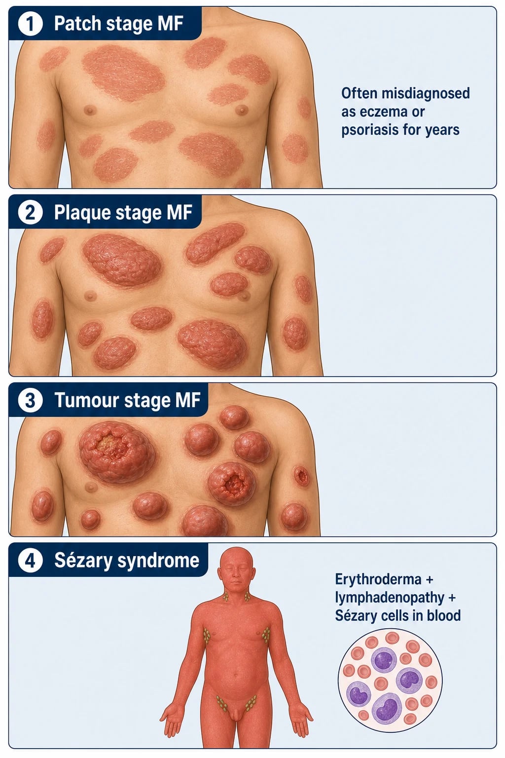
Patch stage (T1 < 10 percent BSA; T2 >= 10 percent). The earliest and most easily missed presentation. Flat, scaly, red-brown, variably atrophic patches favour the bathing-trunk distribution (buttocks, hips, lower abdomen, lower back, breasts, axillae) — areas typically spared by sun and chronic eczema. The patches may be mildly pruritic or asymptomatic, and the patient may carry a long-standing label of eczema or psoriasis before a biopsy is performed. The classic trap is that a chronic, treatment-resistant eczematous or psoriasiform eruption in a sun-protected distribution persisting or progressing over years is MF until proven otherwise.[1][4]
-
Plaque stage (T1-T2). Raised, infiltrated, well-demarcated red-brown plaques. Patches and plaques often coexist. The surface may show poikiloderma (atrophy, telangiectasia, mottled hyper- and hypopigmentation), particularly in chronic plaque disease, which is a strong clinical clue. [1]
-
Tumour stage (T3). Dome-shaped red-brown or violaceous nodules, often ulcerating and prone to secondary bacterial infection. Tumours may arise within pre-existing plaques or de novo. The appearance of tumours is a clinical red flag for large cell transformation (defined as CD30+ large cells comprising more than 25 percent of the dermal infiltrate) and should always prompt a fresh biopsy.[1][5]
-
Erythroderma (T4). Generalised redness and scaling of more than 80 percent BSA, often intensely pruritic. Erythroderma in CTCL may be erythrodermic MF (without blood involvement) or, when accompanied by lymphadenopathy and a circulating clone, Sezary syndrome. Erythrodermic patients are at high risk of high-output cardiac failure, hypothermia, dehydration, electrolyte disturbance and sepsis.[1][6]
Sezary syndrome — the diagnostic triad
Sezary syndrome is the leukaemic variant of CTCL, defined by the diagnostic triad of:[1][3][5]
- Erythroderma (more than 80 percent BSA; red, scaling, intensely pruritic, often with ectropion, palmoplantar keratoderma, onychodystrophy and patchy alopecia).
- Generalised lymphadenopathy (often with dermatopathic changes on biopsy, sometimes with partial or complete effacement).
- Sezary cells in the peripheral blood — atypical cerebriform T cells (CD4+CD7-CD26-) at more than 1000 cells per microlitre, or a CD4:CD8 ratio greater than 10, with a demonstrable T-cell clone by PCR.[3][8]
A monoclonal TCR gene rearrangement in the blood is required by the current WHO-EORTC / ISCL criteria and is the most reliable single laboratory marker of leukaemic involvement.[8]
Other CTCL variants — clinical pearls
Hypopigmented MF
- Hypopigmented scaly patches, often in children and dark-skinned patients
- Biopsies are subtle — a high index of suspicion is required
- Excellent prognosis; treat with NB-UVB phototherapy
- Commonest MF variant in patients with skin of colour
Folliculotropic MF
- Follicular papules, acneiform lesions, alopecia (often scalp and beard)
- Predominantly head and neck distribution
- Deep biopsy is essential because the infiltrate is peri- and intrafollicular
- Prognosis worse than classic patch MF; TSEBT or systemic therapy
Pagetoid reticulosis (Woringer-Kolopp)
- Solitary, well-demarcated, scaly or crusted plaque, usually on distal extremity
- Localised form with excellent prognosis
- Treated with local radiotherapy (20 to 30 Gy)
- Histology shows epidermal infiltration by atypical CD4+CD30- lymphocytes
Granulomatous slack skin
- Pendulous, lax skin folds in axillae and groins
- Granulomatous infiltrate with elastophagocytosis on biopsy
- Rare variant; may be associated with lymphomatoid papulosis
- Indolent course; localised radiotherapy or surgical excision
Tumour-stage MF
- Dome-shaped nodules, often ulcerate
- Rule out large cell transformation on every fresh biopsy
- Brentuximab vedotin for CD30+ disease (ALCANZA trial)
- TSEBT for diffuse tumour burden; localised radiotherapy for individual nodules
Erythrodermic MF / SS
- More than 80 percent BSA red, scaling, pruritic
- SS if blood Sezary cells more than 1000 per microlitre and CD4:CD8 greater than 10
- ECP first-line for SS; supportive care (warming, fluids, antipruritics)
- Monitor for high-output cardiac failure and sepsis
Histopathology
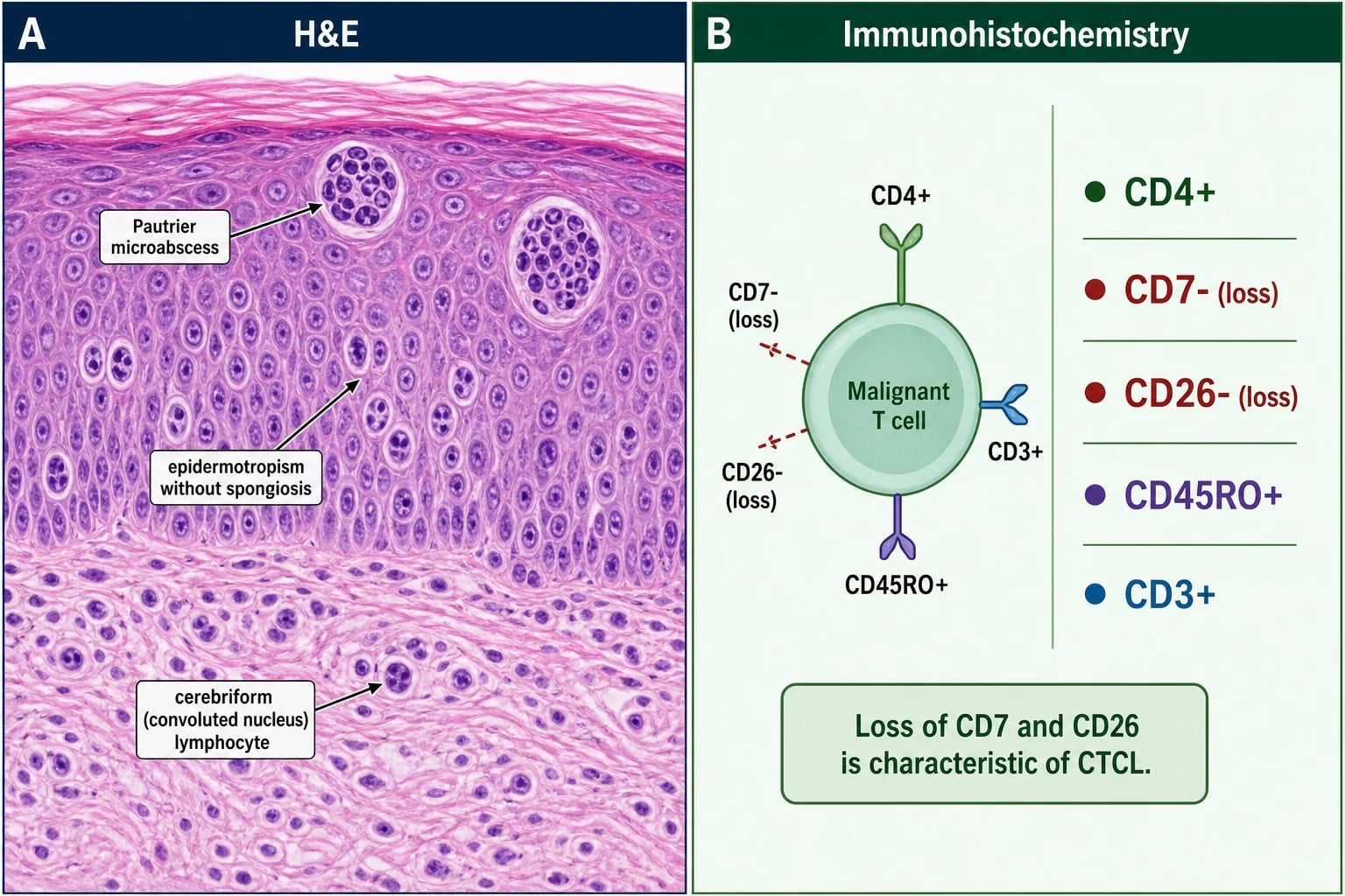
The histological diagnosis of MF rests on a combination of architectural, cytological and immunophenotypic features, ideally with molecular confirmation. Because early MF histology is often subtle and may be confused with chronic dermatitis, a single negative biopsy does not exclude the diagnosis — multiple biopsies over time may be required.[1][4][6]
Architectural features:
- Epidermotropism — atypical lymphocytes within the epidermis, particularly along the basal layer and in the lower spinous layer, in the absence of spongiosis.
- Pautrier microabscesses — three or more atypical lymphocytes within a small intraepidermal vesicle; seen in 20-30 percent of MF biopsies but pathognomonic when present.
- Haloed lymphocytes — atypical lymphocytes surrounded by a clear halo within the epidermis (artefactual retraction).
- Band-like (lichenoid) dermal infiltrate of atypical lymphocytes, especially in plaque and tumour stages. [1]
Cytological features:
- Cerebriform nuclei — small, hyperconvoluted nuclei (brain-like or anaplastic); in the Sezary cell they are best appreciated in peripheral blood films (the Lutzner cell in skin and the Sezary cell in blood are morphologically similar).
- Hyperchromasia, irregular contours, mitoses in advanced lesions.
- Large cell transformation — large cells (>=4x the size of a small lymphocyte) with vesicular nuclei and prominent nucleoli, comprising more than 25 percent of the dermal infiltrate; associated with worse prognosis.[1][5]
Immunohistochemistry:
- CD3+, CD4+, CD45RO+, CD2+ — the typical mature T-helper phenotype.
- Aberrant loss of CD7 (in up to 70-80 percent of cases) and CD26 — diagnostic clues.
- CD30 expression emerges with large cell transformation and is the diagnostic hallmark of CD30+ lymphoproliferative disorders.[5]
- A CD4:CD8 ratio of more than 4-6:1 in the dermal infiltrate is suggestive.
Molecular pathology:
- PCR for TCR gene rearrangement (beta or gamma chain) demonstrates a single dominant peak in 60-80 percent of patch MF, 80-90 percent of plaque MF, and over 95 percent of tumour MF and SS. False negatives are more common in early patch disease, where the clonal population is sparse.[6]
Pitfalls in early MF. The histological differential of early patch MF includes chronic spongiotic dermatitis (eczema), psoriasis, lichenoid drug eruption, connective tissue disease, and pityriasis lichenoides chronica. The key discriminators are the absence of spongiosis, the presence of atypical cerebriform lymphocytes in the epidermis, and a dominant TCR clone. A clinicopathological correlation conference, with the dermatologist and pathologist reviewing the slides side-by-side, is the gold standard for resolving equivocal cases.[4]
Differential Diagnosis
CTCL mimics are legion. Recognising them prevents the most common errors: over-diagnosis of eczema as MF, and under-diagnosis of MF as chronic eczema. [1]
Eczema / atopic dermatitis
- Spongiosis on histology; responds to topical steroids; no TCR clone
- No atypical cerebriform cells; no Pautrier microabscesses
- Distribution favours flexures (acute) or lichenified plaques (chronic)
- No bathing-trunk predilection
Psoriasis
- Silvery scale, extensor distribution, nail pitting
- Munro microabscesses on histology; no atypical infiltrate
- Often family history; arthropathy in a minority
- Responds to vitamin D analogues, tar, biologics
Tinea corporis
- KOH positive for hyphae; annular leading edge with central clearing
- PAS-positive on biopsy; no clonal TCR
- Responds to topical antifungals within 2 to 4 weeks
- May coexist with MF — biopsy any atypical or recalcitrant lesion
Pityriasis rosea
- Herald patch then Christmas-tree distribution on trunk
- Self-limiting within 6 to 12 weeks
- No clonal TCR; spongiotic histology
- EBV and HHV-6/7 have been implicated (controversial)
Drug eruption (morbilliform / lichenoid)
- Drug timeline (2 to 6 weeks from new drug)
- Eosinophils in infiltrate; resolves on drug withdrawal
- No Pautrier microabscesses
- Symmetric morbilliform pattern favours drug
Parapsoriasis (small / large plaque)
- Considered a precursor or early MF by some authorities
- Biopsy may be indeterminate; clone may be present in large plaque
- Long-term follow-up required (progression to MF in 10 to 30 percent)
- Often treated as early MF
Pityriasis lichenoides chronica (PLC) / varioliformis acuta (PLEVA)
- Waxing / waning eruption; histology shows lichenoid infiltrate with parakeratosis
- Clonal TCR absent in most cases; distinct entity
- Responds to tetracyclines, phototherapy, topical steroids
- Not associated with progression to CTCL
Lupus erythematosus (discoid / tumid / subacute)
- Photosensitivity, ANA, anti-Ro/La
- Interface dermatitis with follicular plugging
- No Pautrier microabscesses; responds to antimalarials
- DIF: granular immunoglobulin and complement at the basement membrane
Adult T-cell leukaemia/lymphoma (ATLL)
- HTLV-1 serology positive
- Hypercalcaemia; lytic bone lesions
- Aggressive course; endemic in Japan, Caribbean, West Africa
- Distinguished from SS by HTLV-1 serology and clinical context
Primary cutaneous anaplastic large cell lymphoma (pcALCL)
- Solitary or grouped ulcerating nodules without preceding patches
- CD30+ in more than 75 percent of cells
- ALK negative in most primary cutaneous cases
- Brentuximab vedotin (ALCANZA) in advanced disease
Pitfall — adult T-cell leukaemia/lymphoma (ATLL). In erythrodermic patients from HTLV-1 endemic areas (southern Japan, Caribbean, West Africa, parts of South America, Iran), an HTLV-1 serology is mandatory. ATLL shares the erythroderma and circulating atypical lymphocytes of SS, but is distinguished by positive HTLV-1 serology, frequent hypercalcaemia, lytic bone lesions, and the absence of the chronic psoriasiform prodrome of MF. [1]
Pitfall — pseudolymphoma. Drug-induced or actinic reticuloid pseudolymphomas may show a dense cutaneous lymphoid infiltrate with CD4+ predominance and even small monoclonal TCR peaks, but lack Pautrier microabscesses, lack the architectural features of MF, and resolve on withdrawal of the offending drug. [1]
Investigations
A structured workup guides staging, treatment selection, and prognosis.[2][6][8]
Skin-directed investigations:
- Multiple skin biopsies — at least two, taken from the most indurated or representative lesions, avoiding previously treated or ulcerated sites if possible. Each biopsy should be sent for H&E, immunohistochemistry (a CTCL-oriented panel: CD2, CD3, CD4, CD5, CD7, CD8, CD20, CD25, CD26, CD30, CD45RO, TIA-1, granzyme B, ALK-1) and PCR for TCR gene rearrangement (BIOMED-2 protocol).[6]
- Direct immunofluorescence — generally not required, but useful to exclude lupus or autoimmune bullous disease when the differential includes these.
- Image capture / body chart — serial clinical photographs and body charts document disease extent and response (mSWAT scoring, see below).
Blood investigations:
- Full blood count with differential — look for lymphocytosis, eosinophilia, and Sezary cells on the blood film.
- Peripheral blood flow cytometry — quantifies the CD4+CD7- and CD4+CD26- populations; reports absolute Sezary cell count, CD4:CD8 ratio, and the proportion of aberrant CD4+ T cells.
- PCR for TCR gene rearrangement on peripheral blood — defines B0 (no clone), B1 (clonal but low-level), and B2 (clonal, with Sezary count more than 1000 per microlitre or CD4:CD8 greater than 10) categories per ISCL/EORTC.[8]
- Serum LDH, soluble IL-2 receptor (sCD25), and IgE — prognostic markers in advanced disease; sCD25 is incorporated into a prognostic index for SS.[5]
- HTLV-1 serology — in patients from endemic areas, those with hypercalcaemia, or those with rapidly progressive erythroderma, to exclude ATLL.
- HIV serology — relevant in patients with atypical aggressive CTCL or severe opportunistic infection.
Imaging and tissue staging:
- Lymph node examination — palpate all peripheral groups. Excisional biopsy of any suspicious node (>=1.5 cm, hard, fixed, or progressively enlarging) for histology and TCR clonality.
- Contrast-enhanced CT of neck, chest, abdomen and pelvis — for nodal and visceral staging in stage IIB or higher, or in any patient with palpable lymphadenopathy.
- FDG-PET/CT — more sensitive than CT for nodal and visceral involvement in advanced disease; recommended for stage IIB and above, and increasingly for restaging after therapy.
- Bone marrow biopsy — not routine, but indicated in stage IV disease or in the workup before allogeneic transplantation.[2][6]
Optional and emerging:
- TCR high-throughput sequencing (NGS) — quantitative tracking of the malignant clone at very low levels; useful in blood staging and for monitoring minimal residual disease.
- Gene expression profiling — distinguishes SS from inflammatory erythroderma with high accuracy in research settings.
- Whole-body integrated PET/MRI — under investigation for combined staging with lower radiation dose. [1]
TNMB Staging
The current staging system is the ISCL/EORTC revision of Olsen 2007 (PMID 17540844), which is integrated into the AJCC 8th edition and used worldwide. The four components are the T (skin), N (node), M (visceral metastasis), and B (blood) categories, which are combined into clinical stage IA through IVA2/IVB.[6][8]
T1
- Limited patch / plaque, less than 10 percent BSA
- Stage IA when combined with N0M0B0
- 10-year overall survival greater than 95 percent
T2
- Generalised patch / plaque, 10 percent BSA or more
- Stage IB when combined with N0M0B0
- 10-year disease-specific survival approximately 85 percent
T3
- Tumour(s) (one centimetre diameter or more)
- Stage IIB
- TSEBT is the most effective single skin-directed modality
T4
- Erythroderma (more than 80 percent BSA)
- Stage III when combined with N0-2M0B0-1
- ECP first-line if B2 (Sezary syndrome)
N0-N3
- N0: no clinically abnormal nodes
- N1: clinically abnormal, dermatopathic (N1a) / with clone (N1b)
- N2: clinically normal nodes, dermatopathic with clone
- N3: clinically abnormal nodes, partial or complete effacement
M0 / M1
- M0: no visceral involvement
- M1: visceral involvement (with confirmation)
- Imaging: CT or FDG-PET/CT for staging
B0 / B1 / B2
- B0: no blood involvement (Sezary cells less than 250 per microlitre, no clone)
- B1: low blood involvement (clone positive, Sezary less than 1000)
- B2: high blood involvement (Sezary 1000 or more per microlitre, or CD4:CD8 greater than 10 with clone)
Stage IVB
- M1 disease with visceral involvement
- Median 5-year overall survival less than 15 percent
- Multi-agent chemotherapy and transplant evaluation
Management
The treatment of CTCL is stage-driven, escalating from skin-directed therapy in early disease to multimodal systemic therapy in advanced disease. The 2023 EORTC consensus (Latzka 2023, PMID 37890355) is the principal European guideline, broadly concordant with the US NCCN guideline (current 2024 version).[2][5][6]
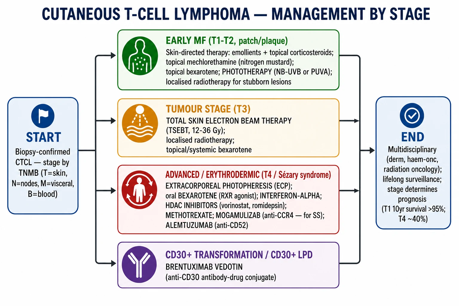
Early MF (stages IA-IIA) — skin-directed therapy
Skin-directed therapy aims to clear the malignant clone from the skin while minimising systemic toxicity. Combination skin-directed therapy is more effective than single-modality treatment and may defer the need for systemic therapy for years. [1]
Emollients and bathing
- Universal; reduce scale and pruritus
- Urea- or salicylic-acid-based preparations are most effective
- Twice-daily application after bathing
Topical corticosteroids (class I-III)
- First-line; potent steroids (clobetasol, betamethasone dipropionate) for plaques
- Mid-potency for face and folds
- Response in 60 to 80 percent of patch / plaque disease
Topical mechlorethamine (nitrogen mustard)
- CTCL-specific; 0.02 percent gel or compounded ointment
- Daily for 6 to 12 months
- Main toxicity is contact dermatitis (10 to 20 percent)
Topical carmustine (BCNU)
- Alternative to mechlorethamine; reserved for refractory disease
- Haematological monitoring required
- Risk of irritant dermatitis and hyperpigmentation
Topical bexarotene 1 percent gel
- RXR retinoid; useful on focal lesions
- Mild local irritation; can combine with phototherapy
- Avoid in pregnancy (teratogenic)
Narrowband UVB (NB-UVB, 311 nm)
- First-line phototherapy for patch-predominant disease
- Two to three sessions per week
- Cumulative dose 20 to 50 J per square centimetre
- Remission in 50 to 80 percent
PUVA (psoralen + UVA, 320-400 nm)
- Plaque-predominant or NB-UVB-refractory disease
- Oral 8-MOP two hours before UVA
- Better than NB-UVB for plaques
- Risk of photoageing and non-melanoma skin cancer
Localised radiotherapy / orthovoltage
- Single-plaque disease, pagetoid reticulosis
- 20 to 30 Gy in 2 to 3 Gy fractions
- 90 percent durable control
Tumour stage (IIB) and refractory early disease
- Total skin electron beam therapy (TSEBT) — the most effective single skin-directed modality for diffuse disease. A standard course is 12 Gy over 6-9 weeks; higher doses (24-36 Gy) give more durable remissions but more cutaneous toxicity. Overall response rates exceed 90 percent in stage IB-IIA, with complete response in 60-75 percent. TSEBT is typically followed by maintenance skin-directed therapy (PUVA, topical nitrogen mustard) to extend remission.[2][5]
- Localised radiotherapy for individual tumour nodules.
- Brentuximab vedotin — an anti-CD30 antibody-drug conjugate; standard of care for CD30+ CTCL (ALCANZA trial, Prince 2017, PMID 28600132). In the ALCANZA trial, brentuximab vedotin produced an objective response lasting at least 4 months (ORR4) of 56 percent versus 13 percent for physician's choice of methotrexate or bexarotene, with median progression-free survival of 16.7 versus 3.5 months. Peripheral neuropathy is the dose-limiting toxicity.[9]
- Mogamulizumab — a defucosylated humanised monoclonal antibody against CCR4, with enhanced antibody-dependent cellular cytotoxicity. The pivotal MAVORIC trial (Kim 2018, PMID 30100375) showed superior progression-free survival over vorinostat in relapsed/refractory MF/SS; the response rate in Sezary syndrome was 37 percent, with the drug now preferred in SS.[10]
- Low-dose methotrexate (10-25 mg weekly) is a well-tolerated systemic option in plaque / tumour-stage disease.
- Gemcitabine and liposomal doxorubicin are reasonable options for advanced disease.
Advanced disease — erythrodermic MF and Sezary syndrome (stage III-IVA1)
- Extracorporeal photopheresis (ECP) — first-line systemic treatment for Sezary syndrome. White cells are collected by apheresis, exposed to 8-methoxypsoralen and UVA extracorporeally, and reinfused. The mechanism involves induction of apoptosis in malignant T cells, which is immunogenic, and modulation of the immune response to the malignant clone. Best responses are seen in SS with a circulating clone, and ECP is often combined with bexarotene or interferon.[2][5]
- Oral bexarotene — a synthetic RXR retinoid; standard dose 300 mg per square metre per day. Side effects include hypertriglyceridaemia, central hypothyroidism, leucopenia, and headaches; concomitant fenofibrate, levothyroxine, and lipid-lowering therapy are required. Often combined with PUVA or ECP.[2]
- Interferon-alpha — 3-9 million units SC three times weekly; response rates 50-70 percent in advanced disease; flu-like syndrome and cytopenias are common. Often combined with ECP or bexarotene.[2]
- HDAC inhibitors — vorinostat (400 mg orally daily) and romidepsin (14 mg per square metre IV on days 1, 8, 15 of a 28-day cycle). Thrombocytopenia, fatigue, nausea, and QT prolongation are common. Approved for relapsed/refractory CTCL; less effective than mogamulizumab head-to-head in MAVORIC.[5][10]
- Mogamulizumab — anti-CCR4 monoclonal; particularly effective in Sezary syndrome (37 percent response in MAVORIC). Drug rash (15 to 25 percent) is an important adverse effect and may mimic disease progression.[10]
- Alemtuzumab — anti-CD52 monoclonal; high response rates in heavily pretreated SS but significant immunosuppression (CMV reactivation, opportunistic infection); low-dose SC alemtuzumab (10-15 mg three times weekly) reduces toxicity.[5]
Stage IVB (visceral) and refractory advanced disease
- Brentuximab vedotin for CD30+ disease, mogamulizumab for blood-dominant disease, pralatrexate (antifolate), gemcitabine, liposomal doxorubicin, and multi-agent chemotherapy (e.g. CHOP-like regimens) are options.
- Allogeneic haematopoietic stem cell transplantation is the only potentially curative option in advanced disease and should be considered in patients under 65 with chemosensitive disease and a suitable donor. Reduced-intensity conditioning regimens have reduced transplant-related mortality while preserving graft-versus-lymphoma effects.[2][5]
- CD30+ transformation / pcALCL — brentuximab vedotin is the standard first-line salvage (ALCANZA); localised radiotherapy for solitary lesions.[9]
- Newer therapies — cemiplimab (anti-PD-1) and tenalisib (PI3K inhibitor) have shown activity in early-phase trials for relapsed/refractory CTCL and may be options on clinical trial.[5]
Supportive care
- Antipruritic therapy — emollients, sedating antihistamines at night, gabapentin or pregabalin, low-dose mirtazapine, or low-dose naltrexone.
- Antibiotic prophylaxis in advanced disease — consider topical mupirocin to nares for S. aureus decolonisation; oral doxycycline for suppression of recurrent cellulitis.
- Skin care — gentle soap substitutes, bland emollients, and avoidance of irritants.
- Psychosocial support — the chronic, visible, and pruritic nature of CTCL has a profound impact on quality of life; access to specialist nurses, psychological support, and patient organisations (Lymphoma Action, Cutaneous Lymphoma Foundation) is essential. [1]
Subtypes and Special Scenarios
Patch-stage MF (T1-T2)
- Flat, scaly, sun-protected patches; mSWAT less than 10
- T1 10-year survival greater than 95 percent
- Skin-directed therapy: class I steroids, NB-UVB, mechlorethamine
Plaque-stage MF (T1-T2)
- Raised infiltrated plaques
- PUVA, bexarotene, TSEBT for refractory disease
- Prognosis intermediate
Tumour-stage MF (T3)
- Dome-shaped ulcerating nodules
- TSEBT, brentuximab (CD30+), gemcitabine, liposomal doxorubicin
- Prognosis median 4 to 5 years
Erythrodermic MF (T4)
- Generalised erythroderma
- ECP, bexarotene, interferon, mogamulizumab
- Monitor for high-output cardiac failure and sepsis
Sezary syndrome (B2)
- Triad: erythroderma + lymphadenopathy + Sezary cells
- ECP first-line; mogamulizumab for refractory disease
- Median survival 2 to 4 years
Folliculotropic MF
- Follicular papules, alopecia, head and neck distribution
- Deep biopsy required
- Poor prognosis; TSEBT or systemic therapy
Hypopigmented MF
- Hypopigmented patches in children and dark-skinned patients
- NB-UVB first-line
- Excellent prognosis
Pagetoid reticulosis (Woringer-Kolopp)
- Solitary, scaly plaque, distal extremity
- Localised form; radiotherapy curative
Granulomatous slack skin
- Pendulous lax skin folds
- Elastophagocytosis on biopsy
- Rare; may be associated with LyP
Large cell transformation (CD30+ more than 25 percent)
- Worse prognosis
- Brentuximab vedotin
- Chemotherapy and transplant evaluation
Lymphomatoid papulosis (LyP)
- Recurrent, self-resolving, asymptomatic papulonodular eruption
- Histology CD30+; benign course in most cases
- Less than 5 percent progress to lymphoma; watchful waiting
Primary cutaneous anaplastic large cell lymphoma (pcALCL)
- Solitary or grouped ulcerating CD30+ tumours without preceding patches
- ALK negative in most primary cutaneous cases
- Brentuximab vedotin (ALCANZA) for advanced disease
- Prognosis excellent (5-year DSS greater than 90 percent)
Subcutaneous panniculitis-like T-cell lymphoma (SPTCL)
- Subcutaneous nodules, often legs
- Alpha-beta T-cell phenotype; usually indolent
- Systemic steroids, ciclosporin; avoid aggressive chemotherapy
Primary cutaneous gamma-delta T-cell lymphoma
- Subcutaneous or dermal nodules, often ulcerate
- Aggressive; multi-agent chemotherapy, transplant
- Worse prognosis than SPTCL
Primary cutaneous CD4+ small/medium T-cell LPD
- Solitary nodule on face or upper trunk
- Usually indolent; local excision / radiotherapy
- Watch-and-wait acceptable for small lesions
Prognosis
The prognosis of CTCL varies dramatically with stage at diagnosis. Stage IA disease is largely curable with skin-directed therapy, while stage IVB disease has a 5-year survival below 15 percent. Risk stratification informs both treatment intensity and surveillance schedule.[2][6]
Adverse prognostic factors:
- Advanced T stage (T3, T4)
- Blood involvement (B2)
- Elevated serum LDH
- Elevated soluble IL-2 receptor (sCD25)
- Age greater than 60
- Large cell transformation
- Folliculotropic variant
- Failure to achieve complete response after first-line therapy
- Peripheral eosinophilia (a marker of Th2 cytokine skewing in advanced disease) [1]
Composite prognostic index (CLIPi, Cutaneous Lymphoma International Prognostic index). A four-factor index incorporating early-stage (T3/T4) versus advanced-stage (N3/M1), age, and LDH stratifies patients into low / intermediate / high risk groups with distinct 5-year overall survival.[2]
Relapse is the rule. Even in early-stage MF, relapse rates approach 30-50 percent within 5 years of achieving clinical remission, particularly when skin-directed therapy is discontinued. Maintenance therapy (intermittent phototherapy, intermittent topical nitrogen mustard) is often continued indefinitely. [1]
Complications
[1]Special Populations
- Paediatric. Hypopigmented MF is the most common CTCL variant in children and in patients with dark skin. NB-UVB phototherapy is the first-line treatment. PUVA, bexarotene, systemic agents, and TSEBT are reserved for refractory disease. The prognosis is generally excellent.
- Pregnancy. CTCL may flare during pregnancy, although data are limited. NB-UVB is safe. Systemic retinoids (teratogenic), methotrexate (teratogenic), and interferon (use with caution) are generally avoided. ECP and TSEBT are acceptable. Treatment decisions are made jointly with the patient, obstetrician, and haemato-oncologist.
- Elderly. The highest incidence population; comorbidities frequently limit systemic therapy options. Topical and phototherapy-based approaches are preferred. Treatment of CTCL must be balanced against the patient's functional status and life expectancy.
- HIV-positive. CTCL may pursue a more aggressive course in HIV. Antiretroviral therapy should be optimised. Distinguish from ATLL, which is associated with HTLV-1 rather than HIV.
- Transplant recipients on immunosuppression. Increased incidence of CTCL; consider reduction of immunosuppression and switch to mTOR inhibitors (sirolimus) which have been associated with disease regression in some reports.
- Skin of colour. Hypopigmented MF is more common. Diagnostic delay is common because the hypopigmented patches are subtle and the differential (pityriasis alba, post-inflammatory hypopigmentation, vitiligo) is broad. Biopsy of any persistent hypopigmented patch in a sun-protected distribution is mandatory.
- Occupational exposures. Chronic exposure to aromatic hydrocarbons, pesticides, and certain industrial chemicals has been associated with CTCL in some epidemiological studies, although the data are inconsistent. Patients with possible exposure should be counselled and, where possible, exposure reduced. [1]
Evidence, Guidelines, and Regional Differences
Willemze 2005, Blood (PMID 15692063)
- WHO-EORTC classification — the foundational classification of primary cutaneous lymphomas
- Updated in 2018
- Combines clinical, histological, immunophenotypic and molecular criteria
Olsen 2007, Blood (PMID 17540844)
- ISCL / EORTC TNMB staging revision
- Current international standard; integrated into AJCC 8th edition
- Formal blood (B) category introduced
Prince 2017, Lancet (ALCANZA, PMID 28600132)
- Brentuximab vedotin in CD30+ CTCL
- ORR4 56 percent vs 13 percent for physician's choice
- PFS 16.7 vs 3.5 months
Kim 2018, Lancet Oncol (MAVORIC, PMID 30100375)
- Mogamulizumab in relapsed / refractory MF / SS
- Superior PFS over vorinostat
- Response 37 percent in Sezary syndrome
Latzka 2023, Eur J Cancer (PMID 37890355)
- EORTC consensus recommendations 2023
- The principal European treatment guideline
- Stage-driven, escalation strategy
Dummer 2021, Nat Rev Dis Primers (PMID 34446710)
- Comprehensive disease primer
- Covers epidemiology, pathophysiology, diagnosis, and management
Weiner and Rook 2024, Hematol Oncol Clin N Am (PMID 39079789)
- Concise review of contemporary CTCL management
- Practical treatment algorithms
Sheern 2025, Clin Exp Dermatol (PMID 40721285)
- Recent review emphasising practical management of MF
- Diagnostic delay and biopsy indications
Regional differences. The major guidelines are broadly concordant on treatment principles, but access to specific therapies varies:
- Mogamulizumab and brentuximab vedotin are approved in the US, EU, UK, Japan, and Australia, but are not universally reimbursed.
- TSEBT is available in major radiotherapy centres in the US, UK, EU, Australia, and Japan; centralisation in regional centres is the norm.
- ECP is available in most tertiary centres in the US, UK, EU, Australia, and Japan; limited availability in lower- and middle-income countries.
- NICE technology appraisals in the UK have approved mogamulizumab for SS and brentuximab vedotin for CD30+ CTCL under specific criteria.
- Indian subcontinent — methotrexate and interferon are widely used first-line systemic agents for cost reasons; TSEBT and ECP are concentrated in tertiary cancer centres.
- Sub-Saharan Africa and Latin America — CTCL is under-reported; pigmented skin may delay recognition; access to TCR PCR and flow cytometry is limited. [1]
Prevention
There is no primary prevention for CTCL. The most important secondary preventive strategy is early biopsy of any chronic eczematous or psoriasiform eruption that does not respond to standard therapy, particularly in a sun-protected (bathing-trunk) distribution. Diagnostic delay from first symptom to confirmed diagnosis averages 3-6 years; reducing this delay is the single most important prognostic intervention in early MF.[4]
In established CTCL, prevention of complications centres on:
- Secondary infection prevention — antiseptic washes, mupirocin to nares, prompt treatment of impetiginised lesions.
- Photoprotection for PUVA / NB-UVB treated patients to limit cumulative UV damage and NMSC risk.
- Avoidance of immunosuppressants that may drive progression (cyclosporine, azathioprine, TNF inhibitors).
- Surveillance for second malignancies — annual full skin examination, age-appropriate cancer screening, vigilance for Hodgkin lymphoma in long-term CTCL survivors. [1]
Sun protection does not influence CTCL development (unlike non-melanoma skin cancer), but is important to limit photoageing and treatment-related NMSC risk. [1]
Exam Pearls
[1]Skin-directed therapy armamentarium — STaMP
Systemic therapy by line — Beware the Ladder
Red Flags
Exam application bank (NEET-PG / INICET)
One-line answer
Cutaneous T-cell lymphoma (CTCL) is a heterogeneous group of extranodal non-Hodgkin lymphomas of skin-homing mature T cells, dominated by mycosis fungoides (MF) (~50 percent of CTCL; indolent course progressing from patch to plaque to tumour to erythroderma) and Sezary syndrome (SS) (~3-5 percent; aggressive leukaemic variant with erythroderma + lymphadenopathy + circulating Sezary cells). MF is the prototypical great mimicker of eczema, psoriasis and tinea corporis, often misdiagnosed for years before a biopsy secures the diagnosis. Histology shows epidermotropism with Pautrier microabscesses; immunophenotyping reveals a clonal CD4+ T-cell population with aberrant loss of CD7 and CD26; PCR for T-cell receptor gene rearrangement confirms clonality. Management is stage-driven: skin-directed therapies for early disease (topical corticosteroids, mechlorethamine, bexarotene, narrowband UVB /
Worked stems (answer without another resource)
Stem 1 — Classic presentation. Map symptoms to mechanism; name the first investigation and first treatment step with dose/route if drug therapy is standard. [1]
Stem 2 — Unstable / complicated. List red flags that force immediate resuscitation, theatre, ICU, antidote, or reperfusion — and what you do in the first 15 minutes. [1]
Stem 3 — Atypical group. Elderly, pregnancy, child, or immunocompromised: how presentation and thresholds change. [1]
Stem 4 — Differential trap. Name the three closest mimics and one discriminator for each. [1]
Stem 5 — Disposition. Who goes home with safety-netting, who is admitted, who needs HDU/ICU/theatre, and what follow-up is mandatory. [1]
Rapid viva checklist
- Definition + classification
- Pathophysiology chain
- Bedside signs / criteria
- Score with exact components (if any)
- Emergency bundle
- Definitive therapy with doses
- Complications of disease and of treatment
- Special populations
- Guideline/trial name if classic
- Three exam traps
Coverage self-check
If you cannot answer any stem above from this page alone, re-read the matching section — the page is intended to be self-sufficient for final-prof and NEET-PG/INICET questions on Cutaneous T-cell lymphoma.
[1]References
- [1]Lee H. Mycosis fungoides and Sézary syndrome Blood Res, 2023.PMID 37105561
- [2]Latzka J, Assaf C, Bagot M, et al. EORTC consensus recommendations for the treatment of mycosis fungoides/Sézary syndrome - Update 2023 Eur J Cancer, 2023.PMID 37890355
- [3]Larocca C, Kupper T. Mycosis Fungoides and Sézary Syndrome: An Update Hematol Oncol Clin North Am, 2019.PMID 30497668
- [4]Sheern C, Levell NJ, Craig PJ, et al. Mycosis fungoides: a review Clin Exp Dermatol, 2025.PMID 40721285
- [5]Weiner DM, Rook AH. Cutaneous T-cell Lymphoma Hematol Oncol Clin North Am, 2024.PMID 39079789
- [6]Dummer R, Vermeer MH, Scarisbrick JJ, et al. Cutaneous T cell lymphoma Nat Rev Dis Primers, 2021.PMID 34446710
- [7]Willemze R, Jaffe ES, Burg G, Cerroni L, Berti E, et al. WHO-EORTC classification for cutaneous lymphomas Blood, 2005.PMID 15692063
- [8]Olsen E, Vonderheid E, Pimpinelli N, Willemze R, Kim Y, et al. Revisions to the staging and classification of mycosis fungoides and Sezary syndrome: a proposal of the International Society for Cutaneous Lymphomas (ISCL) and the cutaneous lymphoma task force of the European Organization of Research and Treatment of Cancer (EORTC) Blood, 2007.PMID 17540844
- [9]Prince HM, Kim YH, Horwitz SM, Dummer R, Scarisbrick J, et al. Brentuximab vedotin or physician's choice in CD30-positive cutaneous T-cell lymphoma (ALCANZA): an international, open-label, randomised, phase 3, multicentre trial Lancet, 2017.PMID 28600132
- [10]Kim YH, Bagot M, Pinter-Brown L, Rook AH, Porcu P, et al. Mogamulizumab versus vorinostat in previously treated cutaneous T-cell lymphoma (MAVORIC): an international, open-label, randomised, controlled phase 3 trial Lancet Oncol, 2018.PMID 30100375