Dermatology · Medicine
Cutaneous tuberculosis
Also known as Cutaneous tuberculosis · Lupus vulgaris · Scrofuloderma · Warty tuberculosis · Tuberculids · Tuberculosis verrucosa cutis
Cutaneous tuberculosis is infection of the skin and subcutaneous tissue by Mycobacterium tuberculosis (rarely M. bovis). It is classified by the route of infection: exogenous inoculation (primary tuberculous chancre in a non-sensitised host; TB verrucosa cutis / warty TB — a hyperkeratotic warty plaque on the hand of a previously sensitised, BCG-vaccinated host), contiguous spread from an underlying focus (scrofuloderma — a tuberculous cervical lymph node breaks down to the skin via sinus tracts; orificial TB — painful ulcers at mucocutaneous orifices from advanced pulmonary/intestinal disease), haematogenous spread (lupus vulgaris — soft apple-jelly nodules on the face with a long-term squamous cell carcinoma risk; miliary TB — widespread papules and pustules in the immunosuppressed; metastatic tuberculous gumma), and tuberculids (papulonecrotic tuberculid, erythema induratum of Bazin, lichen scroculosorum — a hypersensitivity reaction to haematogenous TB antigens in which the skin is sterile). Histology shows caseating granulomas. Treatment is standard RIPE anti-TB therapy (rifampicin, isoniazid, pyrazinamide, ethambutol) for 6 months.
On this page & tools
Your progress
Saved locally on this device.
Exam tags
Red flags
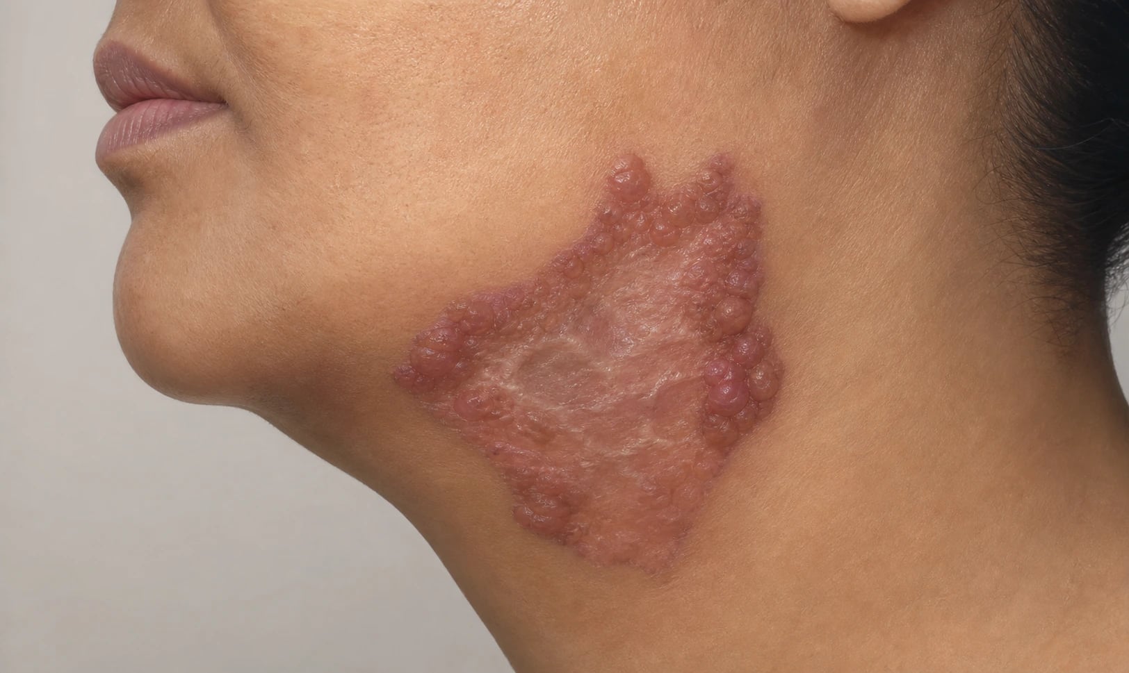
Overview & Definition
Cutaneous tuberculosis is a chronic granulomatous infection of the skin and subcutaneous tissue caused by Mycobacterium tuberculosis and, less often, M. bovis (including the vaccine strain M. bovis BCG in immunodeficient infants).[1][4] It is one of the "great imitators" of dermatology: its morphology ranges from a painless warty plaque to a destructive facial ulcer to a symmetric crop of necrotic papules, and a single clinical appearance can sit on either end of the immunity spectrum.[2] What unifies these disparate appearances is a single organism and a single histological signature — the caseating granuloma — and a single management principle: standard anti-tuberculous therapy.
The clinical form the disease takes is determined by two forces acting together: the route by which the organism reaches the skin (exogenous inoculation, contiguous extension from an underlying focus, or haematogenous/lymphatic seeding) and the host's cell-mediated immune state (whether the patient is tuberculin-naïve, sensitised, or anergic).[1][3] A strong cell-mediated response in a BCG-vaccinated adult tends to localise the organism into paucibacillary, granulomatous forms such as warty TB or lupus vulgaris, in which acid-fast bacilli are scarce on biopsy. A weak or anergic response — as in advanced HIV, malnutrition, or overwhelming disseminated disease — produces multibacillary forms such as scrofuloderma, orificial TB, and miliary TB of skin, in which organisms are abundant. The tuberculids occupy a special position: the skin itself is sterile (no organisms on stain or culture), and the eruption is a hypersensitivity reaction to haematogenously disseminated TB antigens from a distant, often occult, focus.[8]
Classification
Cutaneous tuberculosis is classified by the route by which the organism reaches the skin combined with the host's immune state. The widely used schema recognises four groups: exogenous (inoculation) infection, contiguous spread from an underlying focus, haematogenous/lymphatic spread, and the tuberculids.[1][3]
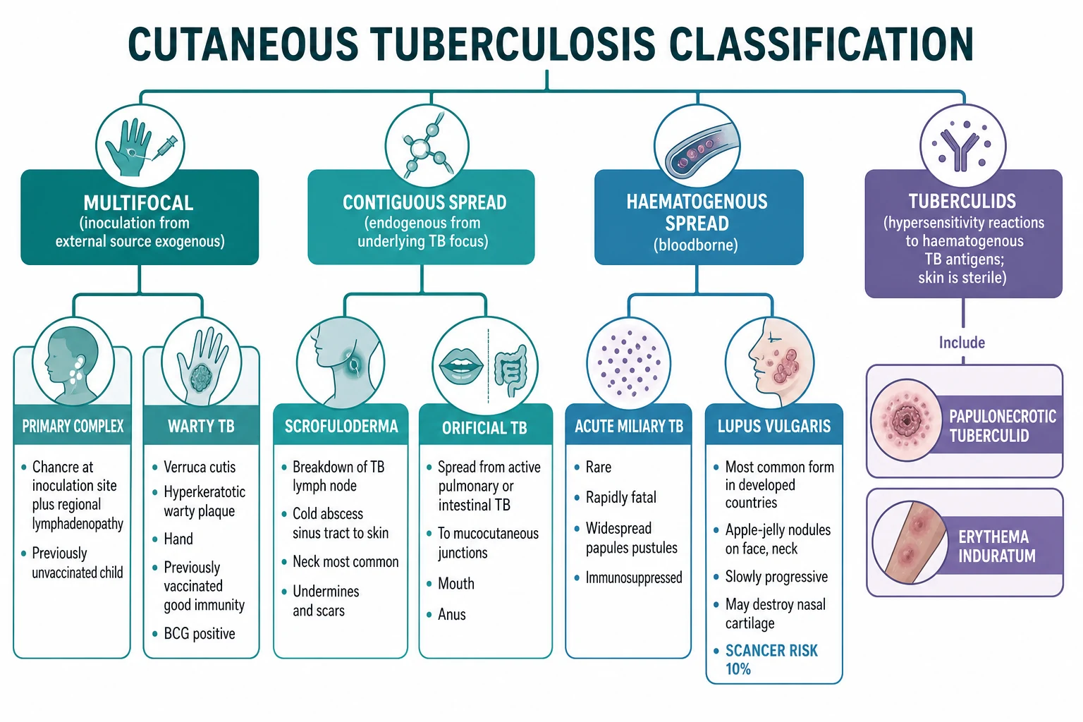
Exogenous (inoculation from an external source)
In exogenous cutaneous TB, M. tuberculosis (or M. bovis) enters through a break in the skin — an abrasion, a wound, a laboratory or autopsy accident, or contact with contaminated material. The form the lesion takes depends on whether the host has been previously sensitised by infection or BCG vaccination.[3][5]
- Primary tuberculous chancre. In a tuberculin-negative host with no prior immunity, inoculation produces a painless ulcer with a granular base and an undermined edge at the inoculation site (commonly a hand or finger), accompanied by regional lymphadenopathy 2 to 4 weeks later — the primary inoculation complex, the cutaneous analogue of the Ghon focus. It is seen most often in children and heals spontaneously over months, sometimes leaving a scar; the tuberculin skin test converts to positive as immunity develops.
- Tuberculosis verrucosa cutis (warty TB). In a host with good cell-mediated immunity (previously infected or BCG-vaccinated, tuberculin-positive), the same inoculation produces a hyperkeratotic, verrucous, warty plaque with a violaceous inflammatory rim on the dorsum of the hand, a finger, the knee, or the foot. It grows slowly over years, is painless, and tends not to ulcerate. It is paucibacillary, reflecting strong host containment of the organism.[5]
Contiguous spread (from an underlying TB focus)
When tuberculosis in a deeper structure extends directly into the overlying skin, the clinical form reflects the underlying disease.[1]
- Scrofuloderma. A tuberculous lymph node (most often a cervical node, less often axillary or inguinal) enlarges, softens, and finally breaks down through the skin, discharging caseous material through bluish, undermined, sinus tracts. Over time the sinus tracts heal with characteristic bridged, keloidal, or papery scarring. Scrofuloderma is the commonest form of cutaneous TB in children in endemic regions such as India, where the organism is usually M. tuberculosis (in contrast to atypical mycobacterial cervical adenitis seen in high-income countries).[3][5]
- Orificial tuberculosis. Tuberculosis from an active pulmonary source spreads to the oral mucosa and lips, or from an intestinal source to the anal and genital mucocutaneous junctions, producing painful, shallow, ragged ulcers with undermined bluish edges. Its presence indicates advanced systemic tuberculosis and poor host immunity, and it carries a poor prognosis.[5]
Haematogenous spread (bloodborne)
Haematogenous seeding of the skin produces a spectrum from indolent to fulminant, again modulated by immunity.[1]
- Lupus vulgaris. The classic, indolent form. Soft, reddish-brown, translucent plaques and nodules appear most often on the face (nose, cheeks, ear lobes), neck, and buttocks. On diascopy (pressing a glass slide against the lesion), the nodules show the pathognomonic apple-jelly (yellow-brown) colour. The plaque enlarges centrifugally over years, leaving central scarring while remaining active at the edges; it may destroy underlying nasal and auricular cartilage. Lupus vulgaris is paucibacillary and is the commonest form of cutaneous TB in many developed-country series.[2][5]
- Acute miliary tuberculosis of skin. A rare, fulminant form of disseminated TB in which tiny, widespread erythematous papules, pustules, vesicles, petechiae, and small ulcers seed the skin, accompanied by systemic toxicity. It occurs in immunosuppressed patients (advanced HIV) and in young children, is multibacillary, and is rapidly fatal if untreated.[5]
- Metastatic tuberculous abscess (tuberculous gumma). Firm, subcutaneous, cold abscesses that arise from haematogenous spread in malnourished or immunosuppressed patients; they may rupture to form sinus tracts.[6]
Tuberculids (hypersensitivity reactions)
The tuberculids are a group of cutaneous eruptions that arise not from organisms in the skin but from a hypersensitivity reaction to haematogenously disseminated M. tuberculosis antigens.[8] Three diagnostic criteria define a tuberculid: the skin lesion is sterile (negative AFB stain and culture), the patient is strongly tuberculin-positive, and the eruption resolves with anti-TB therapy.[8]
- Papulonecrotic tuberculid. Symmetric, dusky-red, necrotic papules on the extensor surfaces of the limbs and the buttocks, which crust and ulcerate and then heal over weeks with varioliform (pock-like) scarring. Crops recur over months.[9][10]
- Erythema induratum of Bazin (nodular vasculitis). Tender, erythematous nodules and plaques on the calves (posterior lower legs) that may ulcerate and scar. Histology shows a lobular panniculitis with vasculitis.[8]
- Lichen scrofulosorum. Tiny, grouped, follicular or perifollicular keratotic papules, skin-coloured to brown, on the trunk — seen especially in children with underlying TB; resolves with ATT.[8]
Epidemiology & Risk Factors
Cutaneous tuberculosis is uncommon even where tuberculosis is common. It accounts for roughly 1 to 2 percent of extrapulmonary tuberculosis cases, and extrapulmonary disease in turn accounts for only a minority of all TB.[1][3] It is therefore a disease seen most often in high-burden, developing countries — South-East Asia, sub-Saharan Africa, and the Western Pacific — and in pockets of vulnerable people in high-income settings.
The risk factors are those of tuberculosis in general operating through impaired cell-mediated immunity: HIV/AIDS, malnutrition, poverty and overcrowding, alcoholism, diabetes mellitus, chronic renal failure, malignancy, and iatrogenic immunosuppression — notably anti-TNF (tumour necrosis factor) therapy, which has produced a well-described reactivation signal for latent TB including cutaneous forms.[4][6] Occupational exposure matters for the exogenous forms: laboratory workers handling cultures, pathologists at autopsy, and (for M. bovis) those handling infected meat or unpasteurised dairy.[3]
HIV co-infection deserves special emphasis because it both increases the incidence of cutaneous TB and shifts its pattern. As CD4 counts fall, the disease becomes more multibacillary and more likely to disseminate as miliary TB, and the morphology grows atypical, mimicking common dermatoses and so delaying diagnosis.[4] BCG vaccination exerts an opposite, protective effect: by boosting cell-mediated immunity it lowers the risk of disseminated and miliary disease and biases any cutaneous TB that does occur toward the paucibacillary, contained forms such as warty TB.[3]
Pathophysiology
The clinical form of cutaneous TB is the visible product of two interacting variables: the route by which the organism reaches the skin and the host's cell-mediated immune response to it.[1]
The organism. Mycobacterium tuberculosis is a slow-growing, obligate aerobe with a waxy, lipid-rich cell wall (mycolic acid) that resists drying and acids and that gives it the property of acid-fastness on Ziehl-Neelsen staining. The same cell wall drives the intense, delayed-type hypersensitivity granulomatous response that defines the pathology.[4]
Route of spread. Exogenous inoculation deposits the organism directly in the dermis through broken skin; contiguous spread delivers it from an underlying lymph node, bone, joint, or viscus; haematogenous and lymphatic spread seed it from a distant focus. The route sets the anatomical distribution — hand in warty TB, neck in scrofuloderma, face in lupus vulgaris, symmetric limbs in papulonecrotic tuberculid, calves in erythema induratum.[1][3]
Host immunity and the bacillary load. The single most rewarding concept is that bacillary load is the inverse of cell-mediated immunity. A strongly tuberculin-positive, BCG-vaccinated host mounts a brisk Th1 (interferon-gamma, macrophage-activating) response that walls off the organism into well-formed granulomas in which few bacilli survive — the paucibacillary forms (lupus vulgaris, warty TB), in which AFB stains are usually negative and PCR may be needed. A weak or anergic host cannot contain the organism; bacilli multiply freely and the lesions teem with organisms — the multibacillary forms (primary chancre, scrofuloderma, orificial TB, miliary TB), in which AFB stains are positive.[3][5]
The histological hallmark — caseating granulomas. The organising pathology is the caseating (necrotising) granuloma: a central area of caseation (cheese-like, structureless necrosis) surrounded by epithelioid macrophages, Langhans giant cells (formed by macrophage fusion, with a horseshoe rim of peripheral nuclei), and an outer cuff of lymphocytes. This pattern is the morphological signature of a vigorous Th1 response to a persistent intracellular pathogen.[1]
The tuberculid mechanism. In a tuberculid, a distant (often occult) TB focus seeds antigens — and sometimes scant bacilli — into the skin haematogenously. The eruption is a type III (immune-complex) and type IV (delayed-type hypersensitivity) reaction to these antigens, producing a necrotising vasculitis and granulomatous response in which the skin itself is sterile on stain and culture. The defining triad is therefore sterile skin, strong tuberculin positivity, and resolution with anti-TB therapy.[8]
Clinical Presentation
Each form of cutaneous TB has a recognisable morphology and tempo, and examiners reward a precise description. The high-yield presentations are described below.[1][3]
Lupus vulgaris
A patient — usually an adult woman — develops a slowly enlarging, soft, reddish-brown, translucent plaque on the face (nose, cheeks, ear lobes), neck, or buttocks. The plaque is studded with deeper-seated, gelatinous nodules that, on diascopy with a glass slide, reveal the pathognomonic apple-jelly (yellow-brown) colour. The lesion expands slowly over years to decades, healing with central atrophic scarring while remaining active at its advancing edge. Continued activity destroys nasal and auricular cartilage (a collapsed nasal bridge is a classic late sign), and longstanding lesions carry a well-described risk of squamous cell carcinoma (Marjolin ulcer), which must be suspected in any ulcerative or nodular change within a chronic lupus vulgaris plaque.[2][5]
Scrofuloderma
A child or young adult in an endemic region develops a firm, bluish, subcutaneous swelling overlying a cervical lymph node that enlarges and then breaks down, discharging caseous, purulent material through one or more sinus tracts with bluish, undermined edges and surrounding induration. Over months the tracts heal with bridged, keloidal, or papery scarring, producing the characteristic puckered neck. Axillary and inguinal nodes, and underlying bone or joint, may be involved.[3]
Tuberculosis verrucosa cutis (warty TB)
A previously sensitised, BCG-vaccinated adult (often with occupational exposure) develops a hyperkeratotic, verrucous, warty plaque on the dorsum of a hand or finger, a knee, or the foot, with an erythematous to violaceous inflammatory rim. The plaque grows slowly, is painless, and tends not to ulcerate. It is the cutaneous form that best reflects a strong host response.[5]
Orificial tuberculosis
A patient with advanced pulmonary or intestinal tuberculosis develops painful, shallow, ragged ulcers with undermined bluish edges at a mucocutaneous orifice — the mouth and lips (from pulmonary disease), or the anal and genital orifices (from intestinal disease). The pain, the location at an orifice, and the systemic setting distinguish it from a chancre or an aphthous ulcer.[5]
Primary tuberculous chancre
A child, or an adult with no prior TB exposure (often occupational), develops a painless ulcer with a granular base and undermined edge at an inoculation site, with regional lymphadenopathy appearing 2 to 4 weeks later (the primary inoculation complex). The lesion heals spontaneously over months, and the tuberculin skin test converts to positive.[3]
Acute miliary tuberculosis of skin
An immunosuppressed patient (advanced HIV) or a young child develops widespread tiny erythematous papules, pustules, vesicles, and petechiae that may progress to small ulcers, accompanied by fever and systemic toxicity. This is a manifestation of disseminated miliary TB and is a medical emergency.[5]
Metastatic tuberculous abscess (gumma)
A malnourished or immunosuppressed patient develops firm, subcutaneous, "cold" abscesses that may rupture to form sinus tracts — the result of haematogenous spread.[6]
The tuberculids
- Papulonecrotic tuberculid — symmetric, dusky-red, necrotic papules on the extensor limbs and buttocks, appearing in crops, crusting and ulcerating, and healing with varioliform (pock-like) scars.[9][10]
- Erythema induratum of Bazin — tender, erythematous nodules and plaques on the calves that may ulcerate and scar; a lobular panniculitis with vasculitis.[8]
- Lichen scrofulosorum — tiny, grouped, follicular or perifollicular keratotic papules on the trunk, seen especially in children with underlying TB.[8]
Differential Diagnosis
Cutaneous tuberculosis is the "great imitator" of dermatology, and the differential is form-specific. The discipline is to pair each form with the mimics that share its morphology, and to distinguish them on histology, AFB/PCR, and systemic/serological context.[2]
The can't-miss considerations are squamous cell carcinoma arising in chronic lupus vulgaris (Marjolin — biopsy any change), miliary TB in the immunosuppressed patient with atypical widespread papules (a systemic emergency), and atypical (non-tuberculous) mycobacteria — M. marinum (swimming-pool/fish-tank granuloma), M. fortuitum/chelonae/abscessus (rapid growers, post-procedure abscesses), and M. scrofulaceum/avium (paediatric cervical adenitis) — which can mimic any cutaneous TB form and are distinguished only by culture and PCR.[4][6]
Clinical & Bedside Assessment
The focused assessment serves four goals: characterise the lesion, find the underlying focus, assess the immune status, and stage the extent of disease.[1][3]
Characterise the lesion. Perform diascopy (press a glass slide firmly against the lesion) on any chronic facial plaque — the apple-jelly sign confirms lupus vulgaris. Examine the surface: a hyperkeratotic warty plaque on the hand suggests warty TB; undermined bluish sinus tracts on the neck suggest scrofuloderma; painful undermined ulcers at an orifice suggest orificial TB; symmetric necrotic papules on extensor limbs suggest a papulonecrotic tuberculid; tender nodules on the calves suggest erythema induratum. [1]
Find the underlying focus. Examine the cervical, axillary, and inguinal lymph nodes for lymphadenitis and sinus tracts (scrofuloderma). Examine the mouth, anus, and genitalia for orificial TB. Auscultate the chest and look for pulmonary TB. Palpate the abdomen for hepatosplenomegaly and seek bony tenderness or joint swelling (osteoarticular TB seeding scrofuloderma or gumma). [1]
Assess the immune status. Look explicitly for stigmata of HIV (oral candidiasis, generalized lymphadenopathy, seborrhoeic dermatitis, wasting) — HIV testing is mandatory in every case. Note malnutrition, alcoholism, and diabetes, and ask about anti-TNF therapy.[4]
Stage the extent. A patient with cutaneous TB may have limited localised disease or disseminated miliary disease; the distinction changes the urgency. Map all lesions, measure them, and photograph them for serial monitoring of the response to therapy. [1]
Investigations
The diagnosis of cutaneous tuberculosis rests on tissue-based demonstration of the organism or its genome, supported by the immune response and the systemic workup. Because the paucibacillary forms are often smear-negative, histology plus culture plus PCR together give the highest yield, and a negative AFB stain never excludes the diagnosis.[1][5]
Histopathology and microbiology of a skin biopsy
A punch or incisional biopsy of lesional skin (taken through the active edge) is the single most important investigation and is divided for three simultaneous tests:[1]
- Histopathology — the caseating (necrotising) granuloma (epithelioid macrophages, Langhans giant cells, central caseation, lymphocytic cuff) is the hallmark. AFB are usually scanty or absent in the paucibacillary forms (lupus vulgaris, warty TB) and abundant in the multibacillary forms (primary chancre, scrofuloderma, orificial TB, miliary TB).
- AFB stain (Ziehl-Neelsen) and mycobacterial culture — culture on Lowenstein-Jensen solid medium or in liquid (MGIT) medium is the gold standard, confirming species and allowing drug-susceptibility testing, but takes 2 to 6 weeks.
- PCR / NAAT (GeneXpert MTB/RIF) — a rapid molecular test that both detects M. tuberculosis complex DNA and identifies rifampicin resistance within hours; WHO-endorsed and now first-line.[4]
For the tuberculids, the skin is sterile — AFB stain, culture, and tissue PCR are negative — and the diagnosis rests on the clinical morphology, a strongly positive tuberculin test/IGRA, histology consistent with a hypersensitivity vasculitis or granulomatous reaction, and resolution with anti-TB therapy.[8]
Immunological and systemic tests
- Tuberculin skin test (Mantoux/TST) — positive in most forms (a strongly positive reaction supports the diagnosis); it is negative in the primary chancre (before immunity develops) and in anergic miliary TB.[5]
- Interferon-gamma release assay (IGRA, e.g. QuantiFERON-TB Gold) — measures in-vitro interferon-gamma release in response to M. tuberculosis-specific antigens; like the TST it does not distinguish latent from active disease, and in BCG-vaccinated populations it avoids cross-reactivity.[4]
- HIV test — mandatory in every case of cutaneous TB.[4]
- Chest X-ray (and sputum AFB/PCR if there is pulmonary disease) — to screen for and stage pulmonary TB.[5]
- CT or MRI — to define an underlying lymph-node, bone/joint, or abdominal focus in scrofuloderma or gumma, and to stage disseminated disease.
Management — Resuscitation
Most forms of cutaneous TB are not dermatological emergencies, but two scenarios demand immediate action.[5]
Acute miliary TB of skin is the dermatological face of a systemic emergency. A patient with disseminated miliary TB and widespread cutaneous papules and pustules is at risk of miliary spread to the lungs, meninges, and other organs. The immediate steps are admission, respiratory isolation, a rapid systemic workup (blood cultures, sputum, chest imaging, lumbar puncture if neurological signs), and prompt initiation of anti-TB therapy without waiting for full microbiological confirmation.[5]
Orificial TB signals advanced systemic tuberculosis and demands urgent full evaluation for a pulmonary or intestinal source and immediate anti-TB therapy.[5]
In every case, cutaneous tuberculosis is a statutorily notifiable disease in many jurisdictions (including India under the National TB Elimination Programme). Management is never complete without notification, contact tracing, and the offer of directly observed therapy to secure adherence and protect public health.[3]
Management — Definitive & Stepwise
The definitive treatment of cutaneous tuberculosis is standard anti-tuberculous therapy — the same regimen used for pulmonary disease — because the organism in the skin is the same organism and the goal is its eradication.[1][5]
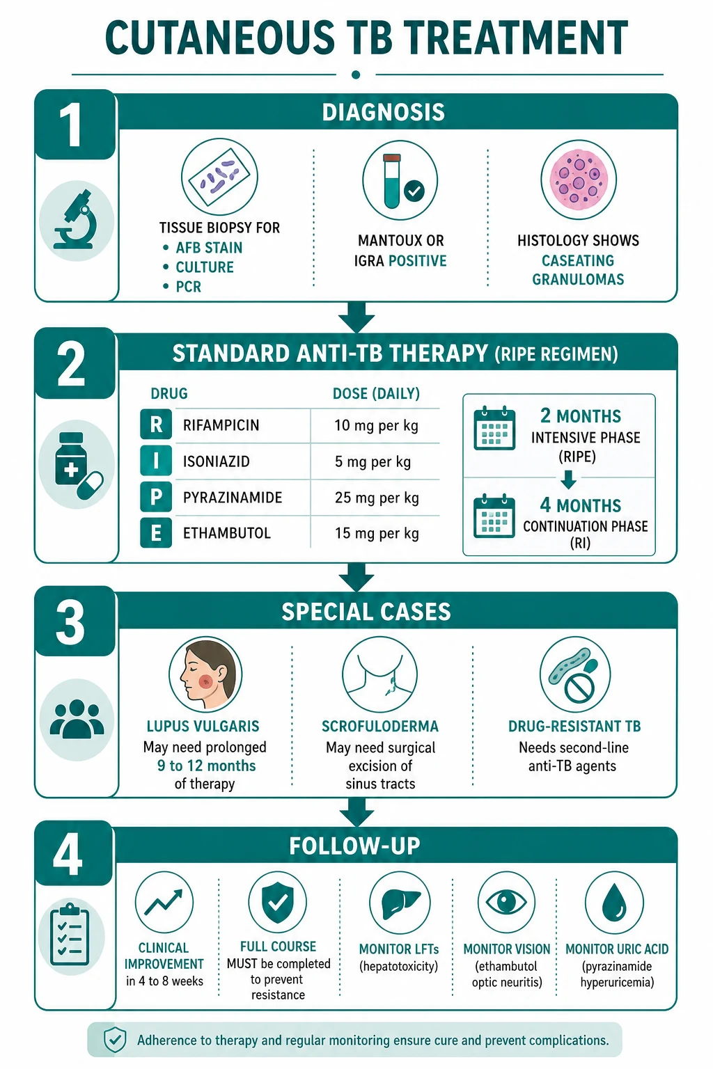
The RIPE regimen
The WHO standard category I regimen for drug-susceptible tuberculosis combines four drugs daily for the intensive phase, then two for the continuation phase.[5]
| Drug | Daily dose | Phase | Key adverse effects |
|---|---|---|---|
| Rifampicin | 10 mg/kg (max 600 mg) | Both phases (6 months) | Hepatotoxicity; orange-red staining of body fluids (urine, tears, secretions); potent CYP450 inducer (reduces oral contraceptive and warfarin efficacy) |
| Isoniazid | 5 mg/kg (max 300 mg), with pyridoxine 10 to 25 mg | Both phases (6 months) | Hepatotoxicity; peripheral neuropathy (prevented by pyridoxine/B6) |
| Pyrazinamide | 25 mg/kg (max 2 g) | Intensive only (2 months) | Hepatotoxicity; hyperuricaemia and gout |
| Ethambutol | 15 mg/kg (some regimens 15 to 20) | Intensive only (2 months) | Optic (retrobulbar) neuritis — monitor visual acuity and colour vision |
The intensive phase runs for 2 months with all four drugs, followed by a 4-month continuation phase with rifampicin and isoniazid alone — 6 months in total.[5] Some authorities extend the continuation phase to 9 months for lupus vulgaris and the tuberculids, where the response can be slower.[2][8]
Pyridoxine (vitamin B6) 10 to 25 mg daily is given alongside isoniazid to prevent the peripheral neuropathy caused by isoniazid's interference with B6 metabolism — particularly important in pregnancy, malnutrition, diabetes, alcoholism, HIV, and renal failure.[5]
Monitoring and adverse effects
Monitoring during RIPE therapy targets the predictable toxicities:[5]
- Baseline and monthly liver function tests — rifampicin, isoniazid, and pyrazinamide are all hepatotoxic; a transient asymptomatic rise in transaminases is common, but a rise to more than three to five times the upper limit of normal with symptoms (or five times without symptoms) mandates stopping the offending drug(s).
- Visual acuity and colour vision (Ishihara plates) at baseline and periodically — ethambutol causes a dose-dependent retrobulbar optic neuritis that can be irreversible; the drug is stopped at the first visual symptom.
- Serum uric acid — pyrazinamide inhibits renal excretion of urate and can precipitate gout. [1]
Surgical and adjunctive measures
Scrofuloderma and large tuberculous gummas may need surgical debridement or drainage, or excision of chronic sinus tracts, after medical therapy has controlled the infection.[5] Reconstructive surgery for the disfiguring scarring of lupus vulgaris and scrofuloderma is deferred until disease is quiescent.
Drug-resistant tuberculosis
If culture or GeneXpert shows rifampicin resistance (a reliable proxy for multidrug-resistant TB, MDR-TB), the standard RIPE regimen is not adequate. MDR/XDR-TB requires prolonged second-line therapy under specialist guidance — fluoroquinolones (moxifloxacin, levofloxacin), second-line injectable aminoglycosides (amikacin), ethionamide, cycloserine, para-aminosalicylic acid, and the newer agents bedaquiline and delamanid, with linezolid and clofazimine as important components of shorter all-oral regimens.[6]
The tuberculids
Tuberculids resolve with standard anti-TB therapy once the underlying focus is treated; symptomatic erythema-induratum nodules may additionally need rest, compression, and occasionally NSAIDs for pain.[8]
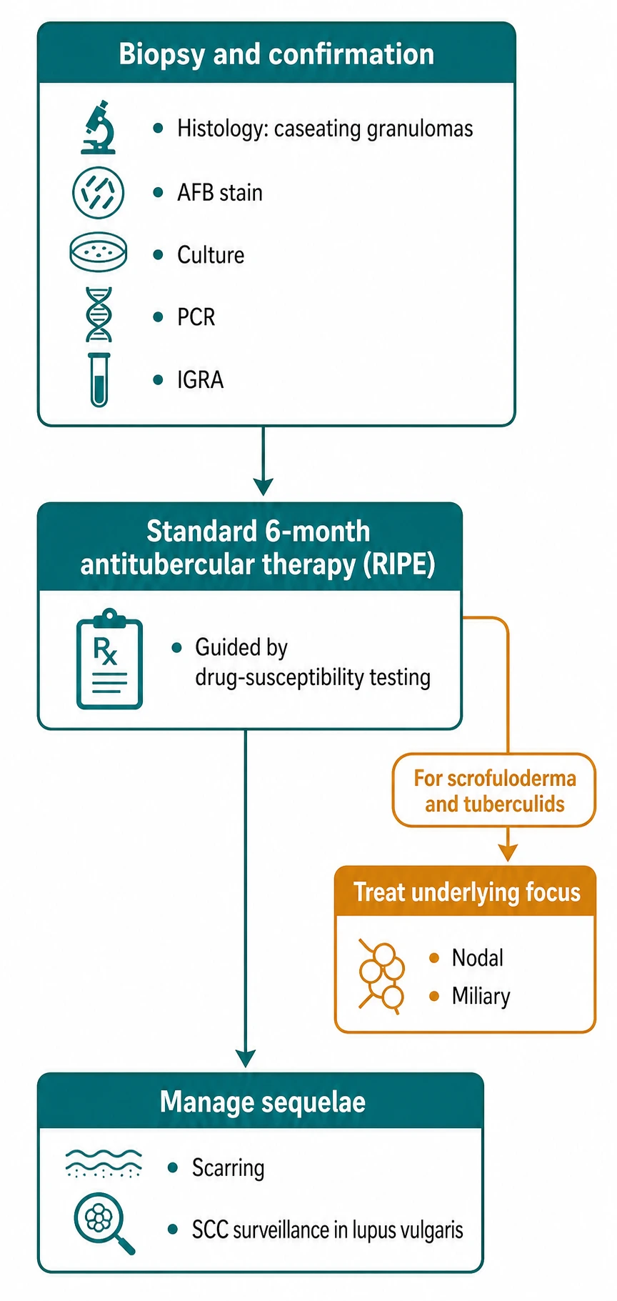
RIPE therapy and its pitfalls — RIPE
Specific Subtypes & Scenarios
Lupus vulgaris — the indolent facial plague
Lupus vulgaris is the prototype of a paucibacillary, immunity-contained cutaneous TB. Its danger is twofold: relentless destruction of facial cartilage over years, and the long-term risk of squamous cell carcinoma (Marjolin) in chronic lesions. Management is prolonged anti-TB therapy (6 months standard, sometimes extended to 9 to 12 months), with biopsy of any nodular, ulcerative, or verrucous change within a chronic plaque to exclude malignant transformation, and reconstructive surgery deferred until the disease is quiescent.[2][5]
Scrofuloderma in children
In endemic regions, scrofuloderma is the commonest form of cutaneous TB and reflects breakdown of a tuberculous cervical lymph node. In India the organism is usually M. tuberculosis; in high-income countries the same clinical picture in a child is more often an atypical mycobacterium (M. avium, M. scrofulaceum), which is distinguished by culture and managed by surgical excision rather than prolonged ATT.[3] Tuberculous scrofuloderma is treated with standard ATT, with surgical debridement or excision of sinus tracts as an adjunct once medical control is achieved.
HIV-associated cutaneous TB
HIV co-infection increases the incidence of cutaneous TB and biases it toward multibacillary and disseminated (miliary) forms, with atypical morphology that mimics common dermatoses and so delays diagnosis.[4] Management pairs anti-TB therapy with antiretroviral therapy (ART), coordinated to avoid the rifampicin–protease-inhibitor interaction (rifampicin is a potent CYP3A4 inducer that lowers protease-inhibitor levels; rifabutin is substituted where appropriate) and to anticipate the immune reconstitution inflammatory syndrome (IRIS), in which recovering immunity unmaskingly intensifies the TB inflammation as ART takes effect.[4]
The tuberculids — sterile-skin reactions
Papulonecrotic tuberculid, erythema induratum of Bazin, and lichen scrofulosorum share the triad of sterile skin, strong tuberculin positivity, and resolution with ATT.[8] The task is always to find and treat the underlying focus — pulmonary, lymph-node, or occult — and to confirm the diagnosis by the response to therapy. Papulonecrotic tuberculid must be distinguished from PLEVA and leukocytoclastic vasculitis; erythema induratum from erythema nodosum (which is septal, on the anterior shin, and non-ulcerative).[9][10]
BCG-related cutaneous disease
BCG vaccination normally produces a local pustule that heals with scarring, but in immunodeficient infants (severe combined immunodeficiency, chronic granulomatous disease, HIV) it can cause disseminated BCG infection with cutaneous and systemic disease, requiring specialist mycobacterial management with anti-TB agents active against M. bovis BCG.[4]
Complications & Pitfalls
The complications of cutaneous TB are determined by the form and the host.[1][5]
- Scarring and disfigurement — the face, neck, and ears bear the brunt; lupus vulgaris and scrofuloderma leave prominent, often keloidal scarring with significant cosmetic and psychological impact.
- Cartilage destruction — lupus vulgaris can destroy nasal and auricular cartilage over years.
- Squamous cell carcinoma (Marjolin) — chronic lupus vulgaris plaques carry a well-described long-term risk of malignant transformation; any change within a plaque demands biopsy.[2]
- Disseminated and miliary disease — in the immunosuppressed, cutaneous TB may be the presenting feature of fatal miliary TB or TB meningitis.[4]
- Drug toxicity — hepatotoxicity, ethambutol optic neuritis, isoniazid neuropathy, and pyrazinamide hyperuricaemia are predictable and monitored.[5]
- Drug resistance — inadequate or interrupted therapy selects MDR/XDR-TB.[6]
- Post-TB sequelae in children — survivors of childhood TB may suffer bronchiectasis, skeletal deformity, and impaired lung function, underscoring the importance of complete, supervised treatment.[11]
Exam application bank (NEET-PG / INICET)
One-line answer
Cutaneous tuberculosis is infection of the skin and subcutaneous tissue by Mycobacterium tuberculosis (rarely M. bovis). It is classified by the route of infection: exogenous inoculation (primary tuberculous chancre in a non-sensitised host; TB verrucosa cutis / warty TB — a hyperkeratotic warty plaque on the hand of a previously sensitised, BCG-vaccinated host), contiguous spread from an underlying focus (scrofuloderma — a tuberculous cervical lymph node breaks down to the skin via sinus tracts; orificial TB — painful ulcers at mucocutaneous orifices from advanced pulmonary/intestinal disease), haematogenous spread (lupus vulgaris — soft apple-jelly nodules on the face with a long-term squamous cell carcinoma risk; miliary TB — widespread papules and pustules in the immunosuppressed; metastatic tuberculous gumma), and tuberculids (papulonecrotic tuberculid, erythema induratum of Baz
Worked stems (answer without another resource)
Stem 1 — Classic presentation. Map symptoms to mechanism; name the first investigation and first treatment step with dose/route if drug therapy is standard. [1]
Stem 2 — Unstable / complicated. List red flags that force immediate resuscitation, theatre, ICU, antidote, or reperfusion — and what you do in the first 15 minutes. [1]
Stem 3 — Atypical group. Elderly, pregnancy, child, or immunocompromised: how presentation and thresholds change. [1]
Stem 4 — Differential trap. Name the three closest mimics and one discriminator for each. [1]
Stem 5 — Disposition. Who goes home with safety-netting, who is admitted, who needs HDU/ICU/theatre, and what follow-up is mandatory. [1]
Rapid viva checklist
- Definition + classification
- Pathophysiology chain
- Bedside signs / criteria
- Score with exact components (if any)
- Emergency bundle
- Definitive therapy with doses
- Complications of disease and of treatment
- Special populations
- Guideline/trial name if classic
- Three exam traps
Coverage self-check
If you cannot answer any stem above from this page alone, re-read the matching section — the page is intended to be self-sufficient for final-prof and NEET-PG/INICET questions on Cutaneous tuberculosis.
[1]The classic pitfalls are: (1) mistaking cutaneous TB for a common dermatosis — the "great imitator" trap — and delaying biopsy; (2) relying on a negative AFB stain to exclude the paucibacillary forms (lupus vulgaris, warty TB); (3) failing to test for HIV in every case; (4) diagnosing a tuberculid without excluding the fungal and bacterial mimics (and without seeking the underlying focus); (5) giving an inadequate ATT duration, inviting relapse and resistance; and (6) overlooking the rifampicin–oral contraceptive interaction, with unintended pregnancy.[2][5]
Prognosis & Disposition
The prognosis of cutaneous tuberculosis is good with appropriate anti-TB therapy for most forms.[5] Lupus vulgaris, warty TB, scrofuloderma, and the tuberculids respond well to standard RIPE therapy, although scarring and cosmetic deformity may persist and require later reconstruction.
The poorer-prognosis forms are those that signal advanced or disseminated disease or immunosuppression. Orificial TB reflects advanced systemic TB; miliary TB of skin is a manifestation of disseminated disease that is rapidly fatal if untreated; and drug-resistant TB and HIV co-infection each independently worsen the outcome.[4][6]
Disposition is shared between dermatology and the infectious-disease/TB service. Most patients are managed as outpatients on directly observed therapy (DOTS); admission is reserved for miliary and orificial TB, for systemically unwell patients, and for the initiation of complex MDR-TB regimens. Every case carries the public-health obligations of notification and contact tracing.[3]
Special Populations
Children
Paediatric cutaneous TB is dominated by scrofuloderma (cervical lymphadenitis), the primary chancre, and lichen scrofulosorum.[3] Anti-TB therapy is dosed by weight (the mg/kg values above), and pyridoxine is given with isoniazid. Children are at particular risk of TB meningitis and miliary disease, so any child with cutaneous TB needs a careful systemic evaluation, and survivors need follow-up for post-TB sequelae — bronchiectasis, skeletal deformity, and impaired pulmonary function.[11]
Pregnancy
Anti-TB therapy is generally safe and indicated in pregnancy, because untreated TB endangers both mother and fetus. Rifampicin, isoniazid, and ethambutol are considered safe; pyrazinamide has less established safety data but is widely used in the intensive phase. Pyridoxine is supplemented. Aminoglycosides (streptomycin, amikacin) are contraindicated because of fetal ototoxicity. Breastfeeding is permitted on ATT.[5]
HIV co-infection
HIV co-infection demands coordinated anti-TB and antiretroviral therapy, attention to the rifampicin–protease-inhibitor interaction (rifabutin substitution), and vigilance for immune reconstitution inflammatory syndrome (IRIS) as ART restores immunity.[4]
Immunocompromised and anti-TNF therapy
Patients on anti-TNF agents (infliximab, adalimumab, etanercept) carry a markedly elevated risk of TB reactivation, including cutaneous forms; all should be screened for latent TB (IGRA ± chest imaging) before biologic therapy and treated for latent infection when positive.[4][6]
Evidence, Guidelines & Regional Differences
The global standard for drug-susceptible tuberculosis is the WHO six-month RIPE regimen, and cutaneous tuberculosis is treated identically to pulmonary disease on the principle that the organism is the same.[1][5] The GeneXpert MTB/RIF assay is WHO-endorsed as a first-line rapid diagnostic because it simultaneously detects M. tuberculosis complex DNA and rifampicin resistance within hours.[4]
Controversies remain where the evidence is thin. The optimal duration of therapy for lupus vulgaris and the tuberculids is debated (standard 6 months versus an extended 9 months for slower-responding disease);[2][8] the diagnostic weight of a positive Mantoux/IGRA in a BCG-vaccinated population is a perennial problem that IGRA (which avoids BCG cross-reactivity) only partially resolves;[4] and the role of surgical adjuncts in scrofuloderma varies by centre.[5]
Prevention
BCG vaccination at birth is the cornerstone of prevention at the population level. It does not prevent infection but substantially reduces the risk of disseminated (miliary) TB and TB meningitis in children — the fatal forms — and biases any cutaneous TB that does occur toward the contained, paucibacillary forms.[3] Individual prevention rests on prompt diagnosis and complete treatment of infectious pulmonary cases, contact tracing and screening, and treatment of latent TB infection (with isoniazid or shorter rifamycin-based regimens) in high-risk contacts and before anti-TNF therapy.[4]
Exam Pearls
Classify cutaneous tuberculosis by route of infection, and give the prototype lesion of each group.
Exogenous inoculation — primary tuberculous chancre (painless ulcer + regional lymphadenopathy in a non-sensitised host) and warty TB/verrucosa cutis (hyperkeratotic warty plaque on the hand in a sensitised host). Contiguous spread — scrofuloderma (cervical TB lymph node breakdown with sinus tracts) and orificial TB (painful ulcers at mucocutaneous orifices from advanced pulmonary/intestinal disease). Haematogenous spread — lupus vulgaris (apple-jelly nodules on the face), acute miliary TB of skin (widespread papules/pustules in the immunosuppressed), and metastatic tuberculous gumma (cold subcutaneous abscess). Tuberculids — papulonecrotic tuberculid, erythema induratum of Bazin, and lichen scrofulosorum (sterile-skin hypersensitivity reactions).[1][3]
A 45-year-old woman has a slowly enlarging, soft, reddish-brown plaque on the nose for 8 years. Diascopy shows apple-jelly nodules. What is the diagnosis, the key risk, and the management?
The diagnosis is lupus vulgaris, a paucibacillary haematogenous cutaneous tuberculosis confirmed by the apple-jelly sign on diascopy. The key risk is long-term squamous cell carcinoma (Marjolin ulcer) arising in the chronic plaque — any nodular, ulcerative, or verrucous change demands biopsy. Management is skin biopsy for histology (caseating granulomas), AFB stain, culture, and GeneXpert MTB/RIF; HIV testing; chest X-ray; and standard RIPE anti-TB therapy (rifampicin, isoniazid, pyrazinamide, ethambutol for 2 months intensive, then rifampicin plus isoniazid for 4 months continuation, 6 months total), sometimes extended to 9 to 12 months for lupus vulgaris, with reconstructive surgery deferred until the disease is quiescent.[2][5]
What three criteria define a tuberculid, and why is the skin sterile?
A tuberculid is defined by: (1) a sterile skin lesion — negative AFB stain, mycobacterial culture, and tissue PCR; (2) a strongly positive tuberculin test or IGRA, confirming exposure to TB antigens; and (3) resolution of the eruption with anti-TB therapy, establishing the aetiological link. The skin is sterile because the eruption is a type III/IV hypersensitivity reaction to haematogenously disseminated M. tuberculosis antigens (and sometimes scant bacilli) from a distant, often occult focus — the inflammation is driven by antigen, not by multiplying organisms in the skin. The underlying focus must always be sought and treated.[8]
Reproduce the standard anti-TB regimen and the monitoring it requires.
The WHO standard RIPE regimen: rifampicin 10 mg/kg, isoniazid 5 mg/kg with pyridoxine 10 to 25 mg, pyrazinamide 25 mg/kg, and ethambutol 15 mg/kg daily for the 2-month intensive phase, then rifampicin and isoniazid alone for the 4-month continuation phase (6 months total). Monitoring requires baseline and monthly liver function tests (all three of rifampicin, isoniazid, pyrazinamide are hepatotoxic), visual acuity and colour vision with Ishihara plates for ethambutol optic neuritis, and serum uric acid for pyrazinamide-induced hyperuricaemia. Patients should be warned that rifampicin stains urine and secretions orange-red and reduces the efficacy of the oral contraceptive, and that isoniazid causes peripheral neuropathy prevented by pyridoxine.[5]
Cutaneous tuberculosis rewards the candidate who can read the immunity from the morphology: a warty plaque on the hand of a vaccinated adult, an apple-jelly nasal plague with a long SCC shadow, a child's neck broken into sinuses, a symmetric crop of necrotic papules with sterile skin. Hold the classification by route, the caseating granuloma, and the six-month RIPE regimen, and the topic is yours.[1]
References
- [1]Kaul S, Kaur I, Mehta S, et al. Cutaneous tuberculosis. Part I: Pathogenesis, classification, and clinical features J Am Acad Dermatol, 2023.PMID 35149149
- [2]Chen Q, Chen W, Hao F. Cutaneous tuberculosis: A great imitator Clin Dermatol, 2019.PMID 31178102
- [3]Bravo FG, Gotuzzo E. Cutaneous tuberculosis Clin Dermatol, 2007.PMID 17350496
- [4]Franco-Paredes C, Marcos LA, Henao-Martínez AF, et al. Cutaneous Mycobacterial Infections Clin Microbiol Rev, 2018.PMID 30429139
- [5]Hill MK, Sanders CV. Cutaneous Tuberculosis Microbiol Spectr, 2017.PMID 28233513
- [6]Gardini G, Gregori N, Matteelli A, et al. Mycobacterial skin infection Curr Opin Infect Dis, 2022.PMID 35067521
- [7]Tobin EH, Warda K, Gropper C, et al. Cutaneous Tuberculosis 2026.PMID 29489274
- [8]Dhattarwal N, Ramesh V. Tuberculids: A Narrative Review Indian Dermatol Online J, 2023.PMID 37266079
- [9]Palaniappan V, Sadhasivamohan A, Karthikeyan K. Papulonecrotic tuberculid Clin Exp Dermatol, 2022.PMID 35724330
- [10]Zhang LW, Wu J, Chen T. Papulonecrotic tuberculid CMAJ, 2024.PMID 38346781
- [11]Igbokwe V, Ruby LC, Sultanli A, et al. Post-tuberculosis sequelae in children and adolescents: a systematic review Lancet Infect Dis, 2023.PMID 36963920