Dermatology · Medicine
Dermatitis herpetiformis
Also known as Dermatitis herpetiformis · DH · Duhring disease · coeliac disease of the skin
Dermatitis herpetiformis (DH) is the cutaneous manifestation of gluten-sensitive enteropathy (coeliac disease), characterised by intensely pruritic grouped papulovesicles on extensor surfaces, caused by IgA deposition at dermal papillae tips and neutrophilic microabscesses. Virtually all patients have associated coeliac disease, often subclinical. Diagnosis rests on direct immunofluorescence of perilesional skin (granular IgA at dermal papillae), supported by histology, serology (IgA anti-tTG, EMA, anti-TG3) and coeliac work-up. Management combines a strict lifelong gluten-free diet (disease-modifying, clears skin over months) with dapsone (rapid symptomatic control within 24 to 72 hours). Long-term surveillance targets coeliac complications, including osteoporosis, micronutrient deficiencies and enteropathy-associated T-cell lymphoma.
On this page & tools
Your progress
Saved locally on this device.
Exam tags
Red flags
Overview & Definition
Dermatitis herpetiformis (DH) is an autoimmune subepidermal blistering disease that is now understood to be the skin manifestation of coeliac disease (gluten-sensitive enteropathy) rather than a separate entity. The name derives from the clinical resemblance of the grouped vesicles to herpes simplex, but the condition is not viral. Pathologically, it is defined by the deposition of granular IgA immune complexes at the tips of dermal papillae, which triggers neutrophil chemotaxis and the formation of papillary dermal microabscesses that progress to subepidermal vesicles.[3][7]
The clinical hallmark is an eruption of symmetrical, intensely pruritic, grouped small papulovesicles on extensor surfaces — the elbows, knees, buttocks, scalp, shoulders, and sacrum. Because the itch is so severe, the lesions are often scratched into erosions and excoriations, so the primary vesicles may be absent by the time the patient presents. A focused history and examination, combined with the correct biopsy, almost always secure the diagnosis.[1]
[1]Every patient diagnosed with DH should therefore be evaluated for coeliac disease, even if gastrointestinal symptoms are absent. Conversely, patients with known coeliac disease should be questioned about pruritic extensor rashes, because early recognition of DH can prevent years of misdiagnosis and inappropriate topical steroid use.[2][6]
Classification
DH is not usually divided into rigid subtypes, but several clinical patterns are important for exams and for clinical reasoning:[1][3]
- Classical DH — the typical presentation with grouped papulovesicles, excoriations, and extensor distribution.
- Latent / preclinical DH — patients with coeliac disease who develop IgA deposits at the dermal papillae but have minimal or no skin lesions. This group may become symptomatic after a gluten challenge or during periods of stress.
- DH with normal or negative coeliac serology — a small minority of patients have normal anti-tTG or EMA titres despite villous atrophy on duodenal biopsy; diagnosis still rests on DIF and histology.
- IgA-deficient DH — rare. Total IgA deficiency produces falsely negative IgA-based coeliac serology; diagnosis requires IgG-based tests or duodenal biopsy.
- Drug-induced DH-like eruption — rare reports with medications such as iodine or non-steroidal anti-inflammatory drugs; the eruption is clinically and histologically DH-like but the underlying coeliac association is absent or uncertain, and resolution follows drug withdrawal.
- Refractory DH — skin disease persisting despite strict gluten-free diet and adequate dapsone. This requires reassessment of diagnosis, dietary adherence, and occasionally escalation to immunosuppressive therapy. [1]
A useful diagnostic distinction is between DH and linear IgA bullous dermatosis. Both are neutrophilic subepidermal blistering diseases and both respond to dapsone, but they differ in immunofluorescence pattern, associated autoantigen, and systemic associations. This distinction is high-yield for exams and is discussed in detail below.[7]
Epidemiology & Risk Factors
DH shares the epidemiology of coeliac disease but is rarer and shows a strong male predominance. The condition is most common in populations of Northern European descent and in regions where wheat consumption is high. The prevalence is estimated at roughly 10 to 100 per 100,000 population, though precise figures vary widely because many cases are misdiagnosed as eczema or scabies.[1][3]
Key risk factors and associations include:[1][2][3]
- HLA-DQ2 and HLA-DQ8 — the same haplotypes that confer coeliac disease susceptibility. HLA-DQ2 is present in about 90% of patients and HLA-DQ8 in most of the remainder. A negative result for both makes the diagnosis very unlikely.
- Coeliac disease — the strongest association. Virtually all patients with DH have at least latent coeliac disease, and approximately 15 to 25% of patients with coeliac disease will develop DH.
- Autoimmune endocrinopathies — autoimmune thyroid disease, type 1 diabetes mellitus, and Addison disease occur more frequently than in the general population.
- Down syndrome — a well-recognised association with both coeliac disease and DH.
- Selective IgA deficiency — found more often in patients with coeliac disease and can produce false-negative IgA serology.
- Family history — first-degree relatives of patients with coeliac disease or DH have an increased risk of gluten-related disease and should be screened with serology.
- Gluten ingestion — a modifiable environmental factor. The disease is driven by dietary gluten; periods of high intake or deliberate gluten challenge can precipitate or worsen rash. [1]
Pathophysiology
DH is an IgA-mediated autoimmune disease that begins in the gut and manifests in the skin. The central steps are:[3][4][7]
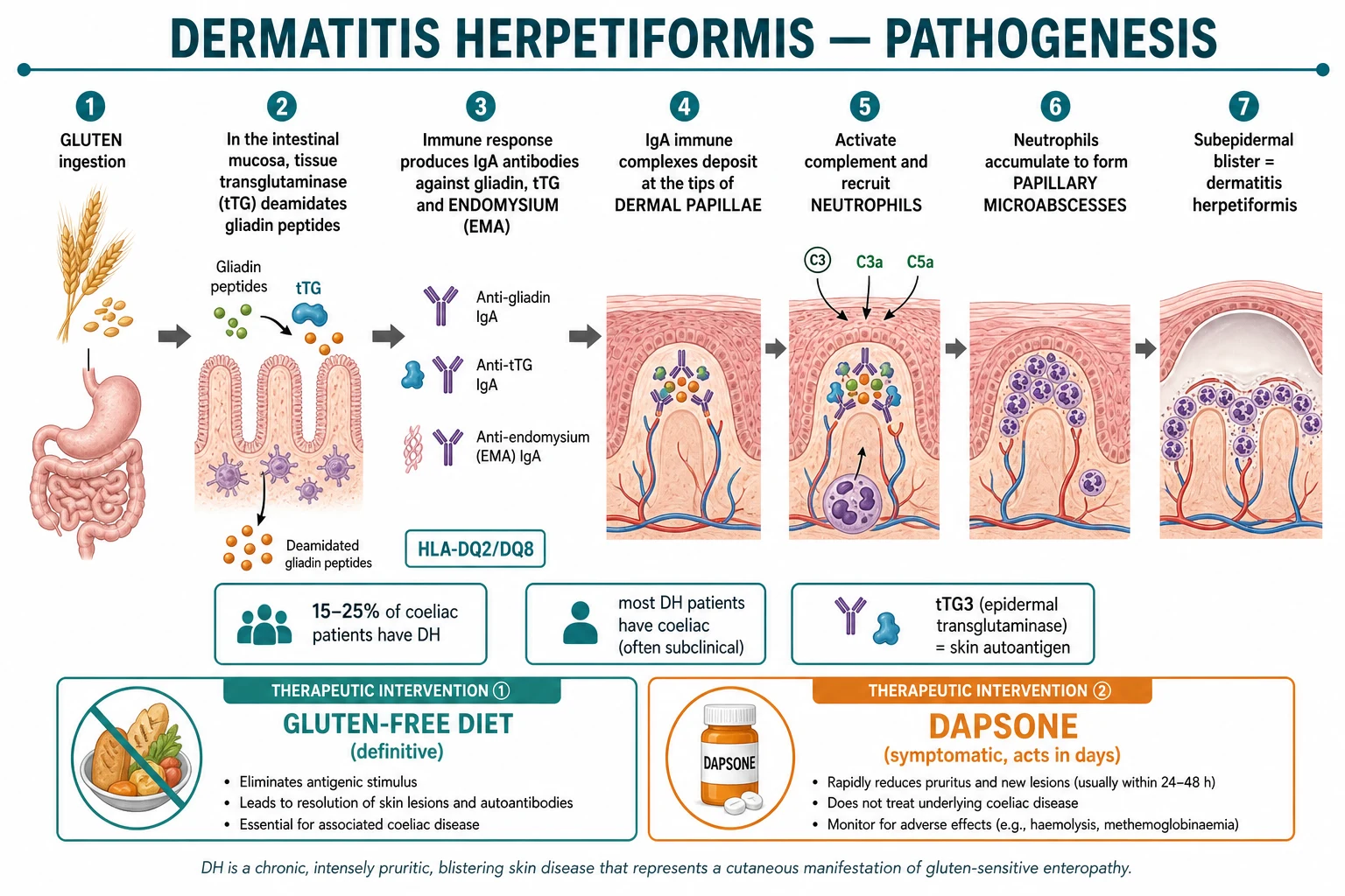
- Gluten ingestion and intestinal deamidation — Ingested gluten proteins, particularly gliadin, are deamidated by tissue transglutaminase 2 (tTG2) in the small-intestinal mucosa. Deamidation converts glutamine to glutamic acid, increasing the affinity of gliadin peptides for HLA-DQ2 and HLA-DQ8 molecules on antigen-presenting cells.
- Adaptive immune response — Gliadin peptide–HLA complexes activate gluten-specific CD4-positive T cells. These T cells drive B cells to produce IgA antibodies against gliadin, tTG2, and endomysium (the extracellular matrix surrounding smooth-muscle fibres, which contains tTG2). IgA anti-tTG2 and anti-endomysial antibodies (EMA) are the classic serological markers of coeliac disease.
- Skin autoantigen cross-reactivity — The critical skin autoantigen is epidermal transglutaminase (also called transglutaminase 3 or eTG/TG3). Circulating IgA antibodies cross-react with TG3, and TG3–IgA complexes preferentially deposit at the tips of dermal papillae, where capillary loops and TG3 expression are abundant. Some patients also have serum IgA anti-TG3 antibodies, which can be used as a disease-specific marker.
- IgA deposition and complement activation — Granular IgA deposits at the dermal papillae activate the alternative complement pathway, generating C3a and C5a. These anaphylatoxins recruit neutrophils from the surrounding vasculature.
- Neutrophilic microabscesses and blister formation — Neutrophils accumulate within the dermal papillae, forming papillary microabscesses. Release of neutrophil proteases and reactive oxygen species damages the basement membrane, leading to a subepidermal split and the formation of tense, clear vesicles. The earliest histological sign is the neutrophilic microabscess, even before a visible blister appears.
- Therapeutic targets — The gluten-free diet removes the antigenic stimulus and gradually allows IgA deposits to clear from the skin. Dapsone acts quickly by inhibiting neutrophil chemotaxis, myeloperoxidase activity, and the respiratory burst, thereby reducing microabscess formation and relieving itch within 24 to 72 hours. [1]
Clinical Presentation
DH is primarily a disease of the skin, but clues to the underlying coeliac disease are often present. The history should focus on the character, timing, distribution, and severity of the itch, together with any gastrointestinal or systemic symptoms.[1][3]
Skin findings
The hallmark lesions are symmetrical, grouped, small papulovesicles on extensor surfaces. The most frequently affected sites are:[1]
- Elbows (often the first site, involved in over 90% of patients)
- Knees
- Buttocks and sacrum
- Scalp
- Shoulders and scapular areas
- Less commonly: forearms, ankles, and trunk [1]
The lesions are typically tense, clear or haemorrhagic vesicles on an erythematous base, arranged in clusters or herpetiform groups of a few millimetres. The intense pruritus often leads to excoriations, crusting, and post-inflammatory hyperpigmentation, so intact vesicles may be sparse. In long-standing disease, the skin may show lichenified plaques, papules, and excoriations without obvious blisters. Some patients report the lesions burning or stinging before they become itchy. [1]
Itch
The itch is characteristically intense and often nocturnal. It is frequently described as the worst the patient has ever experienced. Patients may scratch the skin until it bleeds, and the sleep disturbance can be profound. The severity of itch does not always correlate with the number of visible lesions. [1]
Systemic features of coeliac disease
Although many patients with DH have no bowel symptoms, a directed history often reveals subtle evidence of coeliac disease:[2]
- Bloating, diarrhoea, steatorrhoea, or constipation
- Iron-deficiency anaemia that is refractory to oral iron
- Fatigue, weight loss, or failure to thrive in children
- Oral aphthous ulcers
- Dental enamel hypoplasia
- Peripheral neuropathy or ataxia (rare, related to gluten ataxia or vitamin B12 deficiency)
- Reduced bone mineral density or fractures [1]
Atypical presentations
Children
DH before puberty
- Itch and excoriations may dominate over vesicles
- May present with grouped papules rather than blisters
- Associated failure to thrive, short stature, or dental enamel defects
- Dapsone dosing is weight-based and G6PD must be checked
Elderly
Late-onset or long-standing disease
- Lesions may be more widespread and less obviously grouped
- Coeliac disease may present with anaemia or osteoporosis rather than diarrhoea
- Higher risk of drug toxicity and nutrient deficiencies
- Need careful dapsone dose adjustment and monitoring
Dark skin
Post-inflammatory pigmentary change
- Vesicles may be harder to see against pigmented skin
- Excoriations and post-inflammatory hyperpigmentation may be the most visible findings
- A high index of suspicion is needed; biopsy is diagnostic
Pregnancy
May flare or present de novo
- Pruritus may be mistaken for polymorphic eruption of pregnancy
- GFD is essential and safe; dapsone generally considered safe
- Monitor haemoglobin closely because physiological anaemia and dapsone haemolysis can compound
Differential Diagnosis
The differential diagnosis of an intensely pruritic papulovesicular eruption includes common eczematous and infestational conditions, as well as other autoimmune blistering diseases. The key distinguishing features are distribution, the presence or absence of grouped vesicles, and the results of skin biopsy with direct immunofluorescence.[3][7]
Bullous pemphigoid
Large tense bullae, eosinophils
- Older patients; large tense bullae on urticarial plaques
- Trunk and flexures more than extensor surfaces
- DIF: linear IgG and C3 along the basement membrane zone
- Pruritus is present but grouped small vesicles are not typical
Linear IgA bullous dermatosis
String-of-pearls, linear IgA
- Often drug-induced (vancomycin, NSAIDs, captopril)
- Classic annular string-of-pearls vesicles
- DIF: linear IgA along the basement membrane, not granular at papillae
- Both are dapsone-responsive but the DIF pattern separates them
Scabies
Mite infestation, burrows
- Finger webs, wrists, genitalia, axillae; household contacts itchy
- Burrows, papules, and nodules; no IgA deposits
- Skin scraping may show mites, eggs, or faeces
- Extensor distribution is not typical; responds to scabicides
Atopic dermatitis
Flexural, history of atopy
- Flexural eczema, lichenification, xerosis, personal or family atopy
- No grouped vesicles on extensor surfaces, no dermal IgA deposits
- May be pruritic but lesions are eczematous, not blistering
Contact dermatitis
Exposure history, eczema
- Acute or chronic eczematous change related to contactant
- Distribution matches exposure rather than extensor surfaces
- Patch testing positive; no IgA deposition at dermal papillae
Prurigo simplex / nodularis
Excoriated nodules
- Discrete pruritic papules and nodules with excoriations
- No primary vesicles; biopsy is non-specific
Clinical & Bedside Assessment
A focused examination in suspected DH should confirm the characteristic distribution and morphology, look for evidence of scratching, and search for clues of underlying coeliac disease.[1]
Skin examination: [1]
- Inspect the extensor surfaces in good light, including the elbows, knees, buttocks, sacrum, shoulders, and scalp. Use a dermatoscope if available; small vesicles may be visible only on dermoscopy.
- Look for grouped papulovesicles, excoriations, crusts, erosions, and post-inflammatory hyperpigmentation.
- Palpate for tender papules at the dermal papillae, which can precede vesiculation.
- Record the distribution as symmetrical or asymmetrical; DH is typically symmetrical.
- Check for secondary infection if lesions are heavily excoriated. [1]
General examination: [1]
- Assess for pallor (iron-deficiency or folate-deficiency anaemia).
- Check for glossitis, angular cheilitis, or aphthous ulcers.
- Measure height and weight in children; plot growth charts to detect failure to thrive.
- Look for peripheral oedema from hypoproteinaemia in severe malabsorption.
- Examine the thyroid for goitre or nodules.
- Test for dermatitis herpetiformis–associated neuropathy if symptoms are present. [1]
Bedside diagnostic principle: the correct biopsy site is perilesional, uninvolved skin adjacent to a lesion, not the lesion itself. Inflammation within an established blister can destroy the diagnostic IgA deposits and yield a false-negative DIF. [1]
Investigations
The diagnostic work-up of DH has three parallel aims: confirm the skin disease, evaluate the underlying coeliac disease, and screen for complications. A combination of biopsy, serology, duodenal histology, and HLA typing is usually required.[3][4][7]
Skin biopsy and direct immunofluorescence
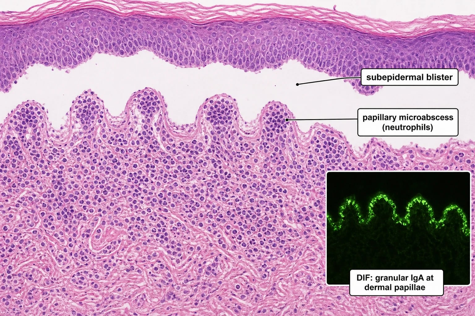
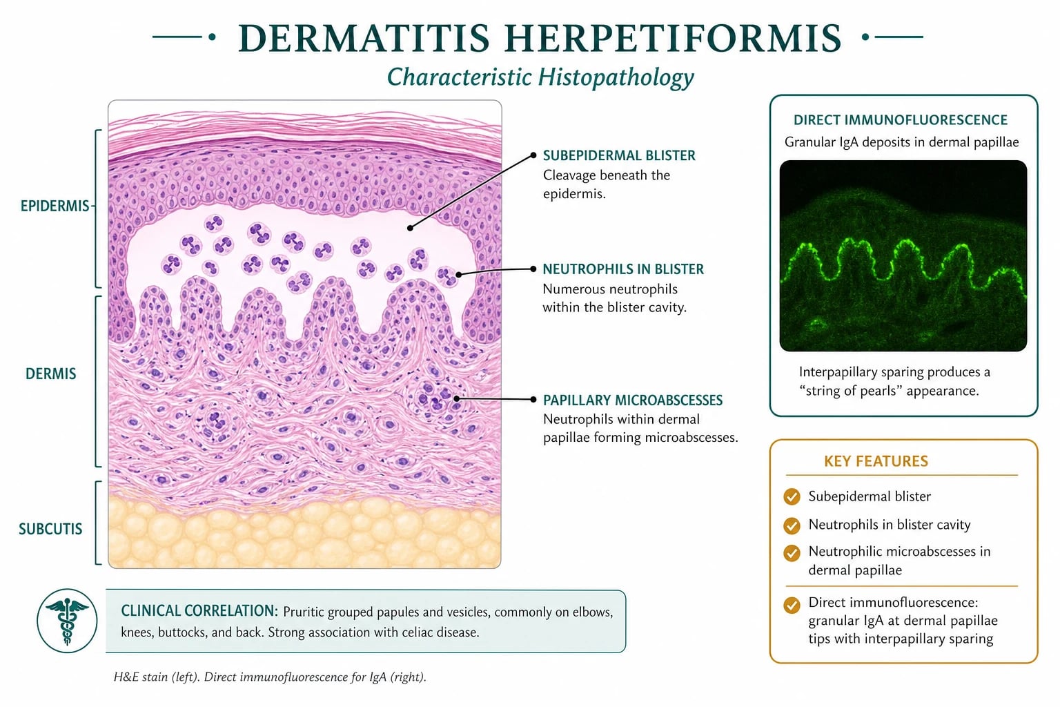
- Direct immunofluorescence (DIF) of perilesional skin — the gold standard for DH. The biopsy should be taken from clinically normal-appearing skin immediately adjacent to an active lesion (typically within 1 cm). DIF shows granular IgA deposits at the tips of dermal papillae. The sensitivity is approximately 90 to 95%.[3][7]
- Histopathology of a lesional biopsy — not the diagnostic gold standard but supportive. Early lesions show neutrophilic microabscesses within the dermal papillae; established lesions show a subepidermal blister with a neutrophilic infiltrate. Eosinophils are usually sparse.
- Why not biopsy the blister? The inflammatory process within an established blister disrupts and destroys the IgA deposits. A DIF taken from the blister itself can be falsely negative.
Serology for coeliac disease and DH
- IgA tissue transglutaminase antibody (anti-tTG IgA) — the first-line serological test for coeliac disease. Highly sensitive and specific, but may be negative in a minority of DH patients with normal mucosa or in those on a gluten-free diet.
- IgA endomysial antibody (EMA) — highly specific for coeliac disease; often used as a confirmatory test. It is usually positive when villous atrophy is present.
- IgG deamidated gliadin peptide antibodies (DGP-IgG) — useful when total IgA is deficient, because standard IgA-based tests are falsely negative.
- Total serum IgA — must be measured to exclude selective IgA deficiency, which occurs more frequently in coeliac disease than in the general population. If IgA deficiency is present, rely on IgG-based tests or duodenal biopsy.
- Anti-epidermal transglutaminase (anti-TG3) IgA — a more disease-specific antibody for DH. It is not universally available but may be positive when standard coeliac serology is normal. [1]
Duodenal biopsy and Marsh classification
A duodenal biopsy is the gold standard for diagnosing coeliac disease. The modified Marsh-Oberhuber classification grades the histological changes:[2]
| Marsh stage | Histological features |
|---|---|
| Marsh 0 | Normal mucosal architecture; villous:crypt ratio normal; no increase in intraepithelial lymphocytes (IELs) |
| Marsh 1 | Increased IELs (typically 30 or more per 100 enterocytes) with preserved villous architecture |
| Marsh 2 | Increased IELs plus crypt hyperplasia; villi still normal in height |
| Marsh 3a | Increased IELs, crypt hyperplasia, and partial villous atrophy (mild) |
| Marsh 3b | Increased IELs, crypt hyperplasia, and subtotal villous atrophy (moderate) |
| Marsh 3c | Increased IELs, crypt hyperplasia, and total villous atrophy (severe) |
| Marsh 4 | Total villous atrophy with normal crypt height and normal IEL count (hypoplastic; rare) |
The diagnosis of coeliac disease is established when characteristic serology is present together with villous atrophy (Marsh 3a to 3c). A patient with DH and positive DIF may still require a duodenal biopsy because the presence of villous atrophy determines the need for strict long-term dietary management and surveillance. [1]
HLA typing
HLA-DQ2 and HLA-DQ8 testing is not required for diagnosis but is useful for excluding coeliac disease when the diagnosis is uncertain. A negative result for both haplotypes makes coeliac disease and DH very unlikely. It is particularly helpful in patients already on a gluten-free diet, in atypical presentations, and in family screening.[2][6]
Baseline laboratory and metabolic work-up
At diagnosis, every patient with DH should have:[2][4]
- Full blood count and film — iron-deficiency, folate-deficiency, or B12-deficiency anaemia.
- Iron studies, ferritin, folate, vitamin B12 — replace deficiencies.
- Calcium, phosphate, alkaline phosphatase, vitamin D — bone metabolism.
- Liver function tests and renal function — baseline before dapsone and to detect associated liver disease.
- Thyroid function and autoantibodies — autoimmune thyroid disease association.
- Glucose and HbA1c — screen for type 1 diabetes in appropriate patients.
- Bone mineral density (DEXA scan) — at diagnosis and periodically because of malabsorption-related osteoporosis. [1]
What if the DIF is negative?
A negative DIF does not completely exclude DH if the clinical picture is classic. The most common reason for a false-negative result is an inappropriately chosen biopsy site. Skin taken from the centre of an ulcerated or excoriated lesion, from skin remote from active disease, or from a patient who has been strictly gluten-free for a prolonged period may show no IgA deposits. In this situation, repeat the biopsy from perilesional skin adjacent to a fresh papule or vesicle, ideally within 24 hours of onset, and ensure the sample is snap-frozen or transported in Michel medium according to local laboratory practice.[3][7]
A small proportion of patients with otherwise typical DH have negative or only focal IgA on DIF. Look for an alternative explanation: the patient may be on a GFD, may have taken systemic corticosteroids or dapsone before the biopsy, or the diagnosis may actually be linear IgA bullous dermatosis, bullous pemphigoid, or scabies. If serology is positive and the clinical picture is characteristic, a trial of strict GFD and dapsone may be justified even with an equivocal DIF, provided the case has been discussed at a specialist dermatology multidisciplinary meeting.[3]
Biopsy technique
For DIF, the dermatologist should select a fresh, non-erosive papule or the edge of a small vesicle and take a 4 mm punch biopsy from the clinically normal skin immediately next to it. The specimen must include the dermal papillae and upper dermis. Avoid the base of an ulcer or the roof of a ruptured blister. Send the sample in normal saline for immunofluorescence studies or place it in Michel transport medium if there is a delay to processing. For histology, a separate punch biopsy from an intact vesicle or early papule is preferred, because older lesions may show only secondary inflammation and ulceration. [1]
Management — Resuscitation & Immediate Control
DH is rarely a life-threatening emergency, but the pruritus can be overwhelming and the risk of diagnostic delay is high. The immediate priorities are to relieve itch, confirm the diagnosis, and exclude dapsone contraindications before starting the drug.[3]
First 24 to 72 hours in newly suspected DH
Confirm the diagnosis
Take a perilesional skin biopsy for DIF and a lesional biopsy for histology; send coeliac serology (anti-tTG IgA, EMA, total IgA)
Start dapsone
Check G6PD status first; if normal, start dapsone 50 mg once daily and titrate to 100 to 200 mg daily as needed
Dietary counselling
Introduce a strict gluten-free diet immediately; explain that GFD is disease-modifying but takes months to clear the skin
Safety monitoring
FBC, reticulocytes, methaemoglobin level, LFTs, renal function; warn about signs of haemolysis
Coeliac work-up
Arrange duodenal biopsy, DEXA scan, micronutrient panel, thyroid and diabetes screening
Symptomatic itch relief: [1]
- Sedating antihistamines (for example hydroxyzine or chlorphenamine at night) can help sleep but do not treat the underlying disease.
- Topical corticosteroids (potent or superpotent) provide modest anti-inflammatory relief but are not a substitute for dapsone or GFD.
- Cooling emollients and wet dressings can reduce pruritus and prevent excoriation. [1]
The most effective immediate relief is dapsone, but it must be started safely. The next section details dosing and monitoring. [1]
Management — Definitive & Stepwise
DH management is built on two pillars: the gluten-free diet (GFD) is the definitive disease-modifying therapy, and dapsone is the rapid symptomatic bridge. Both are almost always required at diagnosis, because the diet takes months to work while the rash needs immediate control.[3][4]
Gluten-free diet
Definitive, disease-modifying
- Removes the trigger for IgA and TG3 immune responses
- Heals coeliac enteropathy and prevents long-term complications
- Skin improves over weeks to months; full clearance may take 6 to 12 months
- Lifelong and strict; even trace gluten can reactivate the rash
Dapsone
Rapid symptomatic control
- Inhibits neutrophil chemotaxis and myeloperoxidase
- Dramatic itch reduction within 24 to 72 hours
- Does not treat enteropathy or remove the antigenic trigger
- Tapered as GFD takes effect; many patients can stop after 6 to 12 months
Gluten-free diet
A strict, lifelong gluten-free diet is the cornerstone of treatment. Gluten-containing grains to avoid are wheat, barley, rye, and oats (unless certified gluten-free because of cross-contamination risk). Patients should be referred to a specialist dietitian for education about label reading, cross-contamination, and hidden gluten in medications and cosmetics.[2][5]
The expected timeline is:[3]
- Days 1 to 7 — no change in skin while the immune response is still active; dapsone is needed for symptomatic control.
- Weeks 4 to 8 — gradual reduction in new lesion formation and pruritus.
- Months 3 to 6 — substantial clearing; dapsone can often be tapered if the diet is strict.
- Months 6 to 12 — most patients achieve near-complete skin remission.
- Long term — ongoing GFD is required to maintain remission, heal the intestine, and reduce the risk of enteropathy-associated T-cell lymphoma. [1]
Patient adherence is the single most important determinant of outcome. Dietary lapses, even accidental, can cause flares within hours to days. Common hidden sources of gluten include wheat-based thickeners in sauces and soups, malt vinegar, soy sauce, some processed meats, communion wafers, and cross-contaminated oats. Medications and supplements may also contain gluten as a binder or filler, and pharmacists should review these. Patients benefit from repeated dietitian review, membership of a coeliac society, and clear written advice on label reading. Even a single dietary mistake can reactivate the rash and delay the time to dapsone withdrawal. [1]
Patient adherence is the single most important determinant of outcome. Dietary lapses, even accidental, can cause flares within hours to days. Common hidden sources of gluten include wheat-based thickeners in sauces and soups, malt vinegar, soy sauce, some processed meats, communion wafers, and cross-contaminated oats. Medications and supplements may also contain gluten as a binder or filler, and pharmacists should review these. Patients benefit from repeated dietitian review, membership of a coeliac society, and clear written advice on label reading. Even a single dietary mistake can reactivate the rash and delay the time to dapsone withdrawal. [1]
Practical counselling points: [1]
- Read labels for wheat, barley, rye, malt, and oats not labelled gluten-free.
- Avoid cross-contamination in shared toasters, fryers, and chopping boards.
- Choose naturally gluten-free staples such as rice, maize, quinoa, buckwheat, and certified gluten-free oats.
- When eating out, ask about ingredients, marinades, and thickening agents.
- Maintain a food and symptom diary to identify accidental exposures. [1]
A motivated patient with access to a specialist dietitian is far more likely to achieve rapid skin clearance and long-term remission than a patient given only a list of forbidden foods. [1]
Dapsone
Dapsone is a sulfone antimicrobial with potent anti-inflammatory effects on neutrophils. It is the drug of choice for rapid symptom control in DH.[3]
Dosing and titration: [1]
- Start dapsone 50 mg once daily in adults with normal G6PD.
- Increase to 100 mg once daily after 1 to 2 weeks if needed.
- Most patients require 100 to 200 mg daily; doses above 200 mg daily are rarely needed and increase toxicity.
- The response is usually dramatic: itch improves within 24 to 72 hours, and new lesions stop appearing within days.
- Once the skin is controlled and the GFD has been strict for several months, dapsone can be tapered by 25 to 50 mg every 4 to 8 weeks and eventually stopped in many patients. [1]
Mandatory monitoring:[3][4][8]
- G6PD deficiency screening must be performed before starting dapsone. In G6PD deficiency, dapsone can cause severe, life-threatening haemolysis.
- Full blood count and reticulocyte count — regularly, especially in the first three months. Dapsone causes dose-dependent haemolysis even in patients with normal G6PD.
- Methaemoglobin level — dapsone oxidises haemoglobin to methaemoglobin. Cyanosis, dyspnoea, or fatigue may signal methaemoglobinaemia.
- Liver function and renal function — baseline and periodic monitoring.
- Peripheral neuropathy — rare with dapsone; ask about sensory symptoms.
- Dapsone hypersensitivity syndrome — fever, rash, lymphadenopathy, hepatitis; stop immediately. [1]
Adverse effects:[8]
- Haemolytic anaemia (dose-related, worse with G6PD deficiency)
- Methaemoglobinaemia (cyanosis, headache, fatigue)
- Agranulocytosis (rare but serious; typically in first 3 months)
- Hepatitis, cholestatic jaundice
- Peripheral neuropathy
- Dapsone hypersensitivity syndrome
- Nausea, headache, skin rash [1]
Drug interactions and special cautions: Dapsone is metabolised by the liver and its levels can be increased by inhibitors such as cimetidine. Concurrent use of folate antagonists, including trimethoprim and methotrexate, may increase haematological toxicity. Patients should avoid oxidant drugs and chemicals that can precipitate haemolysis, particularly in those with borderline G6PD activity. Dapsone also causes a clinically insignificant mild haemolysis in many patients, which is usually compensated by increased reticulocyte production; a falling haemoglobin or rising reticulocyte count outside the expected range warrants dose reduction or temporary cessation. [1]
Alternatives to dapsone
If dapsone is not tolerated, contraindicated, or fails, options include:[3][4]
- Sulfapyridine — structurally related to dapsone; sometimes used in patients who cannot tolerate dapsone. It is less effective and can cause similar haematological toxicity.
- Sulfasalazine — an alternative sulfonamide with anti-inflammatory effects; may be useful in selected patients.
- Tetracyclines — minocycline or doxycycline, sometimes combined with nicotinamide, can help in mild or partial cases, though evidence is less robust than for dapsone.
- Topical corticosteroids — useful for symptomatic relief of localised areas but do not control the disease.
- Immunosuppressants — for refractory DH despite strict GFD and dapsone, options include azathioprine and ciclosporin, but these are second-line and require specialist supervision. [1]
Refractory DH
True refractory DH is uncommon and almost always due to continued gluten exposure or an incorrect diagnosis. Before escalating therapy, the clinician should:[3]
- Reconfirm the diagnosis by reviewing the DIF and histology.
- Reassess dietary adherence with a dietitian and consider serological markers (anti-tTG IgA, EMA) as indirect measures of gluten exposure.
- Reconsider linear IgA bullous dermatosis or other blistering diseases if the DIF pattern is atypical.
- Check for infection, thyroid disease, or malignancy that can worsen itch or inflammation.
- If adherence is perfect and the diagnosis is secure, consider adding or switching to azathioprine or ciclosporin under specialist care. [1]
Specific Subtypes & Scenarios
Paediatric DH
DH can occur in children, although it is less common than in adults. The presentation is dominated by intense pruritus, excoriations, and grouped papules or vesicles on extensor surfaces. Children are more likely to have failure to thrive, short stature, dental enamel defects, and anaemia. Management follows the same principles: GFD, dapsone with weight-based dosing, and G6PD testing before dapsone. Growth and nutrition should be monitored closely.[1]
Pregnancy and lactation
DH may flare during pregnancy or present for the first time. The GFD remains essential and is safe. Dapsone crosses the placenta, but it has been used safely for many years in DH and leprosy; most specialists consider it acceptable in pregnancy when needed. Folate supplementation should be given because dapsone can interfere with folate metabolism. Breastfeeding is generally considered safe with dapsone at usual doses, but the infant should be monitored for haemolysis or jaundice. Haemoglobin should be monitored closely because pregnancy-related anaemia and dapsone haemolysis can compound.[3]
Selective IgA deficiency
Patients with selective IgA deficiency cannot produce IgA antibodies, so standard IgA-based coeliac serology (anti-tTG IgA, EMA) will be falsely negative. In this situation, use IgG-based tests (anti-DGP IgG, anti-tTG IgG) and duodenal biopsy. The DIF of skin is unaffected by IgA deficiency because the IgA deposits are present in the skin even if serum IgA antibodies are absent. Total serum IgA should always be measured at the initial evaluation.[2]
DH with normal coeliac serology
A subset of patients with DH have normal anti-tTG IgA and EMA but still show villous atrophy on duodenal biopsy. This occurs because the immune response in the skin can be more prominent than the mucosal response. In such cases, the diagnosis of coeliac disease is established by biopsy, and the patient should be managed with a GFD. Anti-TG3 antibodies may be positive when standard coeliac serology is negative.[3]
Drug-induced DH-like eruption
Rarely, medications such as iodine, potassium iodide, or NSAIDs can produce a DH-like rash. The eruption is usually clinically and histologically similar to DH, but the underlying coeliac association is absent. A detailed drug history is essential. The rash resolves after withdrawal of the offending drug, and dapsone may be used temporarily for severe symptoms.[3]
Complications & Pitfalls
The complications of DH arise either from the skin disease, the underlying coeliac disease, or its treatment. The most important pitfalls are missing the diagnosis, missing the coeliac association, and mismanaging dapsone.[3][4]
Disease-related complications
- Osteoporosis and osteopenia — from chronic malabsorption of calcium and vitamin D; all patients should have a DEXA scan at diagnosis.
- Iron-deficiency anaemia — common and often refractory to oral iron until the GFD is established.
- Folate, B12, and vitamin D deficiency — can cause anaemia, fatigue, neuropathy, and bone disease.
- Enteropathy-associated T-cell lymphoma (EATL) — a rare but serious complication of coeliac disease. The risk is reduced by strict adherence to a GFD.
- Other autoimmune diseases — thyroid disease, type 1 diabetes, Addison disease, Sjögren syndrome, and autoimmune liver disease occur more frequently. Small-bowel adenocarcinoma — increased risk in untreated coeliac disease, though rare.
- Dermatitis herpetiformis–specific complications — chronic excoriations can lead to secondary infection, scarring, and post-inflammatory pigmentary change. [1]
Bone health: Because DH patients often have undiagnosed coeliac disease with long-standing malabsorption, low bone mineral density is common at presentation. A baseline DEXA scan identifies osteoporosis or osteopenia early. Ensure adequate calcium and vitamin D replacement, weight-bearing exercise, and smoking cessation. Repeat DEXA every one to two years until density stabilises, then less frequently. [1]
Malignancy surveillance: Enteropathy-associated T-cell lymphoma is the malignancy most strongly linked to coeliac disease. Although the absolute risk is low, it is higher in patients with persistent villous atrophy and poor GFD adherence. Maintain a low threshold for investigating new abdominal pain, weight loss, night sweats, or lymphadenopathy. Strict GFD reduces the risk substantially. [1]
Treatment-related complications
- Dapsone-induced haemolysis — even in patients with normal G6PD; risk increases with dose and is more severe in the elderly. Always monitor FBC and reticulocytes.
- Methaemoglobinaemia — presents with cyanosis, headache, fatigue, and dyspnoea. Measure methaemoglobin if suspected.
- Agranulocytosis — usually occurs within the first 12 weeks; patients should be warned to report fever or sore throat promptly.
- Dapsone hypersensitivity syndrome — a rare but potentially fatal drug reaction with fever, rash, lymphadenopathy, and hepatitis. [1]
Classic diagnostic and management pitfalls
- Biopsying the lesion for DIF — always take perilesional uninvolved skin.
- Starting topical steroids alone — they provide only symptomatic relief and do not alter the disease course or the coeliac risk.
- Forgetting to check total IgA — missing IgA deficiency can lead to false reassurance from negative coeliac serology.
- Not evaluating for coeliac disease — virtually every patient with DH has coeliac disease; the work-up must not be skipped even if asymptomatic.
- Starting dapsone without checking G6PD — can precipitate life-threatening haemolysis.
- Attributing refractory disease to treatment failure before excluding gluten exposure — dietary lapses are the commonest cause of persistent rash. [1]
Prognosis & Disposition
The prognosis of DH is excellent if the patient maintains a strict gluten-free diet. Most patients achieve long-term skin remission, and many are able to stop dapsone after 6 to 12 months of dietary adherence. The underlying coeliac enteropathy also heals, and the risk of complications such as EATL and osteoporosis decreases substantially.[3][5]
Expected timelines
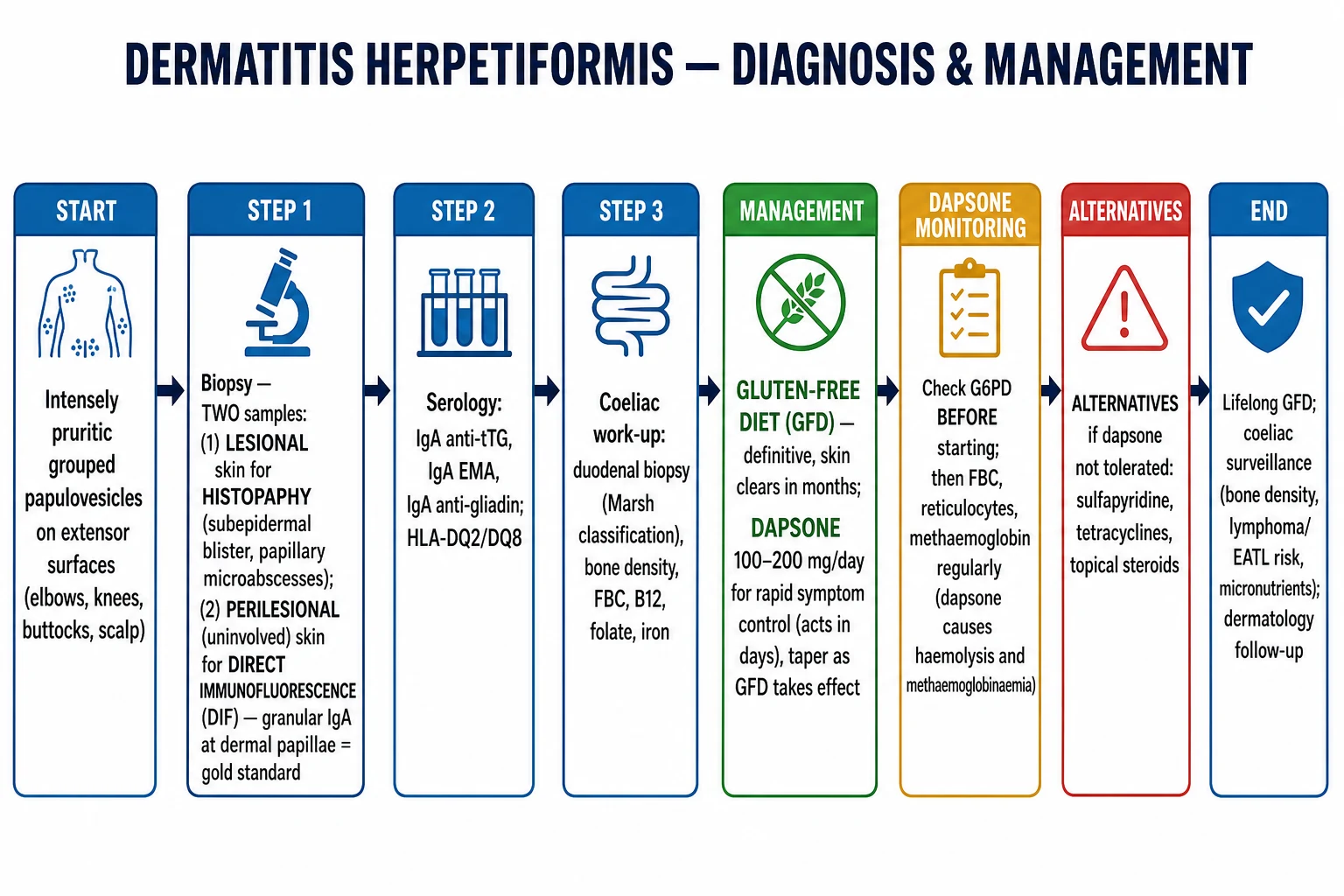
- Dapsone response — 24 to 72 hours for itch reduction; new lesions stop forming within days.
- GFD skin response — weeks to months; full clearance often 6 to 12 months.
- Serological normalisation — anti-tTG IgA and EMA may fall within 3 to 12 months of strict GFD, but they can remain detectable for longer.
- Mucosal healing — duodenal histology may take 1 to 2 years to normalise. [1]
Long-term follow-up
Patients with DH require lifelong follow-up by both dermatology and gastroenterology. A practical surveillance schedule includes:[2][4]
- Annual review of symptoms, diet adherence, and physical examination.
- Periodic coeliac serology as a marker of gluten exposure (for example every 6 to 12 months).
- Repeat duodenal biopsy if symptoms recur or if there is concern about refractory coeliac disease.
- DEXA scan at diagnosis and every 1 to 2 years until bone density is stable.
- FBC, iron studies, folate, B12, vitamin D, calcium, and alkaline phosphatase annually or as clinically indicated.
- Thyroid function and diabetes screening at diagnosis and periodically.
- Low threshold for investigating abdominal symptoms, weight loss, or lymphadenopathy because of the EATL risk. [1]
Special Populations
Children and adolescents
DH in children is managed similarly to adults, but with additional attention to growth, nutrition, and psychosocial impact. Dapsone dosing is weight-based and G6PD testing is mandatory. The GFD must be nutritionally adequate for growth; dietitian input is essential. School and social activities require careful education to avoid gluten exposure.[1]
Pregnancy and breastfeeding
GFD is safe and essential. Dapsone is generally considered acceptable when needed, but folate supplementation is important. Monitor haemoglobin closely. Sulfapyridine and sulfasalazine are alternatives if dapsone is contraindicated, but each has its own safety profile in pregnancy. Topical steroids and antihistamines can be used for symptomatic relief.[3]
Elderly patients
Late-onset DH is increasingly recognised. Elderly patients may have more subtle skin findings, more severe pruritus, and more comorbidities. They are at higher risk of dapsone toxicity, particularly haemolysis and methaemoglobinaemia. Start with lower dapsone doses, monitor frequently, and pay careful attention to drug interactions and renal function. [1]
Immunocompromised patients
Patients with HIV, transplant recipients, or those on immunosuppressive therapy may have atypical presentations. The underlying coeliac disease may be more difficult to diagnose. Management should be multidisciplinary, with careful monitoring of drug interactions and infection risk. [1]
Evidence, Guidelines & Regional Differences
The management of DH is guided by coeliac disease guidelines and dermatology expert reviews. The key international documents are:[2][3]
- European Society for the Study of Coeliac Disease (ESsCD) 2019 guideline — covers diagnosis and management of coeliac disease and related gluten disorders, including DH.
- British Society of Gastroenterology (BSG) and American College of Gastroenterology (ACG) coeliac disease guidelines — include recommendations for serological testing, duodenal biopsy, and GFD.
- Autoimmune blistering disease guidelines — reinforce the role of DIF in the diagnosis of subepidermal blistering diseases. [1]
In Europe, North America, and Australasia, gluten-free products are widely available, but the cost and quality vary. In many low- and middle-income countries, wheat-based staples (chapati, bread, noodles) make a strict GFD challenging. Cultural dietary counselling and local food knowledge are essential. Dapsone is generally available and affordable, but G6PD deficiency is more prevalent in some populations (for example parts of Africa, the Mediterranean, and South-East Asia), making pre-treatment G6PD testing especially important.
Controversies and areas of evolving evidence
- The necessity of routine duodenal biopsy in every DH patient — some experts accept a diagnosis of coeliac disease based on positive serology and DIF alone, but guidelines still recommend biopsy when feasible to grade mucosal damage and guide surveillance.
- Optimal duration of dapsone — while many patients can stop dapsone after 6 to 12 months of strict GFD, some require long-term low-dose therapy. The decision is individualised based on symptoms, diet adherence, and serology.
- Anti-TG3 testing — not universally available, but it may become a more disease-specific serological test for DH in the future.
- HLA testing — useful for exclusion but not required for diagnosis. [1]
Exam Pearls
DH in five letters
PAPUL
Intensely itchy grouped lesions.
Gluten-driven IgA-mediated disease.
Small tense vesicles on extensor surfaces.
Perilesional skin for DIF, not the blister.
EATL is reduced by strict GFD.
Exam application bank (NEET-PG / INICET)
One-line answer
Dermatitis herpetiformis (DH) is the cutaneous manifestation of gluten-sensitive enteropathy (coeliac disease), characterised by intensely pruritic grouped papulovesicles on extensor surfaces, caused by IgA deposition at dermal papillae tips and neutrophilic microabscesses. Virtually all patients have associated coeliac disease, often subclinical. Diagnosis rests on direct immunofluorescence of perilesional skin (granular IgA at dermal papillae), supported by histology, serology (IgA anti-tTG, EMA, anti-TG3) and coeliac work-up. Management combines a strict lifelong gluten-free diet (disease-modifying, clears skin over months) with dapsone (rapid symptomatic control within 24 to 72 hours). Long-term surveillance targets coeliac complications, including osteoporosis, micronutrient deficiencies and enteropathy-associated T-cell lymphoma.
Worked stems (answer without another resource)
Stem 1 — Classic presentation. Map symptoms to mechanism; name the first investigation and first treatment step with dose/route if drug therapy is standard. [1]
Stem 2 — Unstable / complicated. List red flags that force immediate resuscitation, theatre, ICU, antidote, or reperfusion — and what you do in the first 15 minutes. [1]
Stem 3 — Atypical group. Elderly, pregnancy, child, or immunocompromised: how presentation and thresholds change. [1]
Stem 4 — Differential trap. Name the three closest mimics and one discriminator for each. [1]
Stem 5 — Disposition. Who goes home with safety-netting, who is admitted, who needs HDU/ICU/theatre, and what follow-up is mandatory. [1]
Rapid viva checklist
- Definition + classification
- Pathophysiology chain
- Bedside signs / criteria
- Score with exact components (if any)
- Emergency bundle
- Definitive therapy with doses
- Complications of disease and of treatment
- Special populations
- Guideline/trial name if classic
- Three exam traps
Coverage self-check
If you cannot answer any stem above from this page alone, re-read the matching section — the page is intended to be self-sufficient for final-prof and NEET-PG/INICET questions on Dermatitis herpetiformis.
[1]Self-test: Which biopsy site gives the gold-standard DIF finding in DH?
Direct immunofluorescence of perilesional, uninvolved skin adjacent to an active lesion shows granular IgA deposits at the tips of dermal papillae. Biopsying the blister itself can destroy the IgA deposits and give a false-negative result.
References
- [1]Salmi TT. Dermatitis herpetiformis Clin Exp Dermatol, 2019.PMID 31093998
- [2]Al-Toma A, Volta U, Auricchio R, et al. European Society for the Study of Coeliac Disease (ESsCD) guideline for coeliac disease and other gluten-related disorders United European Gastroenterol J, 2019.PMID 31210940
- [3]Reunala T, Hervonen K, Salmi T. Dermatitis Herpetiformis: An Update on Diagnosis and Management Am J Clin Dermatol, 2021.PMID 33432477
- [4]Nguyen CN, Kim SJ. Dermatitis Herpetiformis: An Update on Diagnosis, Disease Monitoring, and Management Medicina (Kaunas), 2021.PMID 34441049
- [5]Aljada B, Zohni A, El-Matary W. The Gluten-Free Diet for Celiac Disease and Beyond Nutrients, 2021.PMID 34836247
- [6]Zingone F, Bai JC, Cellier C, et al. Celiac Disease-Related Conditions: Who to Test? Gastroenterology, 2024.PMID 38460606
- [7]van Beek N, Holtsche MM, Atefi I, et al. State-of-the-art diagnosis of autoimmune blistering diseases Front Immunol, 2024.PMID 38903493
- [8]Hu Y, Kong L, Liu Y, et al. Dapsone-induced methemoglobinemia and hemolysis in a woman without G6PD deficiency presenting with idiopathic urticaria Hematology, 2022.PMID 36444994