Dermatology · Medicine
Dermatofibroma
Also known as Dermatofibroma (DF) · Benign fibrous histiocytoma · Sclerosing haemangioma · Histiocytoma (cutis)
Dermatofibroma (DF; benign fibrous histiocytoma of the skin) is a common benign dermal tumour of fibroblast-like and histiocyte-like (fibrohistiocytic) cells arranged in a storiform pattern. Classic presentation: firm 3-10 mm reddish-brown dermal papule on the lower legs of a young-to-middle-aged adult woman, tethered to the overlying skin but freely mobile over the subcutis. The DIMPLE SIGN (Fitzpatrick / herniation sign) — central dimpling on LATERAL compression — is the bedside pathognomonic clue. Dermoscopy shows a central white scar-like patch with a peripheral delicate ('busy') pigment network. Histology: storiform spindle cells, epidermal acanthosis, and fenestrated collagen at the margins; immunohistochemistry is Factor XIIIa positive and CD34 negative (the key discriminator from dermatofibrosarcoma protuberans, which is CD34 positive with a COL1A1-PDGFB fusion). Management is reassurance and observation; surgical excision (narrow margin) is reserved for symptomatic, cosmetic, or diagnostically uncertain lesions.
On this page & tools
Your progress
Saved locally on this device.
Exam tags
Red flags
Overview & Definition
Dermatofibroma (DF) — also called benign fibrous histiocytoma of the skin, and historically sclerosing haemangioma or histiocytoma cutis — is one of the commonest benign soft-tissue lesions of the skin. It is a discrete, firm, dermal nodule composed of spindle-shaped fibroblast-like and histiocyte-like cells (collectively, fibrohistiocytic cells) arranged in a short intersecting storiform (cartwheel) pattern, embedded in a collagenous stroma. The lesion is bounded by the dermis: it is tethered to the overlying epidermis but, crucially, freely mobile over the deeper subcutaneous fat — a relationship that produces the pathognomonic dimple sign at the bedside.[1][2]
Although the term "benign fibrous histiocytoma" is sometimes used for soft-tissue lesions elsewhere, in dermatology "dermatofibroma" refers to the cutaneous (superficial dermal) entity. The deep (muscle) and atypical/borderline variants are histologically related but behave differently and are discussed separately below. Dermatofibroma is examined at every level — NEET-PG and INICET for the classic dimple-sign stem, and fellowship boards (FRCDerm, ABD, MRCP SCE, RANZCD) for the dermatofibroma-versus-dermatofibrosarcoma-protuberans (DFSP) immunohistochemical axis and for the histological variants.[1]
[1]Classification & Clinical Variants
Most dermatofibromas are the classic (common) type, but several morphological and histological variants are recognised. These variants matter because they change the clinical appearance (and therefore the differential) and, for cellular and atypical forms, the local recurrence risk. The figure below maps the principal clinical phenotypes. [1]
The clinically and histologically important subtypes are:[1][5]
- Classic (common) DF — the firm brown papule of the lower leg; the default.
- Aneurysmal / haemosiderotic DF — blood-filled cavernous spaces and haemosiderin make the lesion dark blue-black; it mimics a vascular tumour or nodular melanoma and is the variant most often biopsied.[3]
- Atrophic DF — the lesion loses substance and presents as a depressed, wrinkled, slightly pigmented plaque; more common in older adults.[4]
- Epithelioid DF — a dome-shaped, often vascular-appearing nodule composed of epithelioid cells with eosinophilic cytoplasm; a subset harbour ALK rearrangements.[5]
- Cellular DF — densely cellular, may extend into the subcutis in a honeycomb fashion, and has a higher local recurrence rate (about 20 to 30 percent) after excision; recurrent gene fusions (for example MYADM::PRKCG) have now been reported.
- Atypical (pseudosarcomatous) DF — scattered atypical cells and occasional mitoses; considered borderline with low-grade malignant potential; complete excision is required.
- Rare variants — lipidised, clear-cell, signet-ring, hemosiderotic, and palisading subtypes.
Epidemiology & Risk Factors
Dermatofibroma is common. It is predominantly a lesion of young to middle-aged adults (peak in the twenties to fifties) and shows a clear female predominance of up to roughly 4 to 1. The lower extremities are by far the commonest site — around 70 percent of lesions occur on the legs (especially the shins) — followed by the upper limbs, the trunk, and the shoulders; the palms, soles, and face are uncommon sites.[2]
Headline numbers
In roughly a fifth of cases the patient recalls a preceding local injury — an insect bite, a thorn prick, folliculitis, a ruptured hair follicle, or minor trauma such as a vaccination site — which is the basis for the traditional "reactive" interpretation (see Pathophysiology).[1]
Multiple (eruptive) dermatofibromas — defined as several to dozens of lesions appearing over weeks to months — are uncommon (about 10 percent of patients have more than one lesion, but genuinely eruptive disease is rarer) and should prompt a search for an underlying trigger: immunosuppression (notably HIV and solid-organ transplant recipients), autoimmune disease (systemic lupus erythematosus), myelodysplastic syndromes and leukaemia, atopy, and rarely an inherited predisposition. Children are less often affected than adults, but eruptive disease in adolescents is recognised. Pregnancy can enlarge existing lesions or generate new ones, consistent with a hormonal influence.[1] Prevalence and incidence. Dermatofibroma is one of the most frequently encountered benign skin tumours in primary care and dermatology clinics; surveys of dermatology outpatients report a point prevalence of several percent, and the lifetime prevalence is high enough that most people will have at least one lesion. Solitary lesions are the rule (about 90 percent of patients have a single lesion), and patients with more than one are still usually limited to a handful. Familial clustering of multiple dermatofibromas has been reported but is rare and does not follow a clear Mendelian pattern.[1]
Paediatric disease. Dermatofibroma is distinctly less common in prepubertal children than in adults, and a solitary lesion in a young child should lower the threshold for dermoscopy and, if atypical, biopsy — chiefly to exclude a Spitz naevus. Eruptive disease in older children and adolescents, however, is well described and is managed as in adults. The sex predilection becomes evident after puberty, again pointing to a hormonal contribution to growth.[1]
Pathophysiology: Reactive Proliferation or Neoplasm?
Whether dermatofibroma is a reactive process or a true neoplasm has been debated for decades, and the modern answer is that it is probably both, with the balance varying by subtype. The traditional view holds that dermatofibroma is a reactive fibrohistiocytic proliferation in response to a dermal injury — which explains the firmness (abundant collagen deposition), the history of a preceding trigger in many cases, and the absence of malignant transformation in classic lesions.[1]
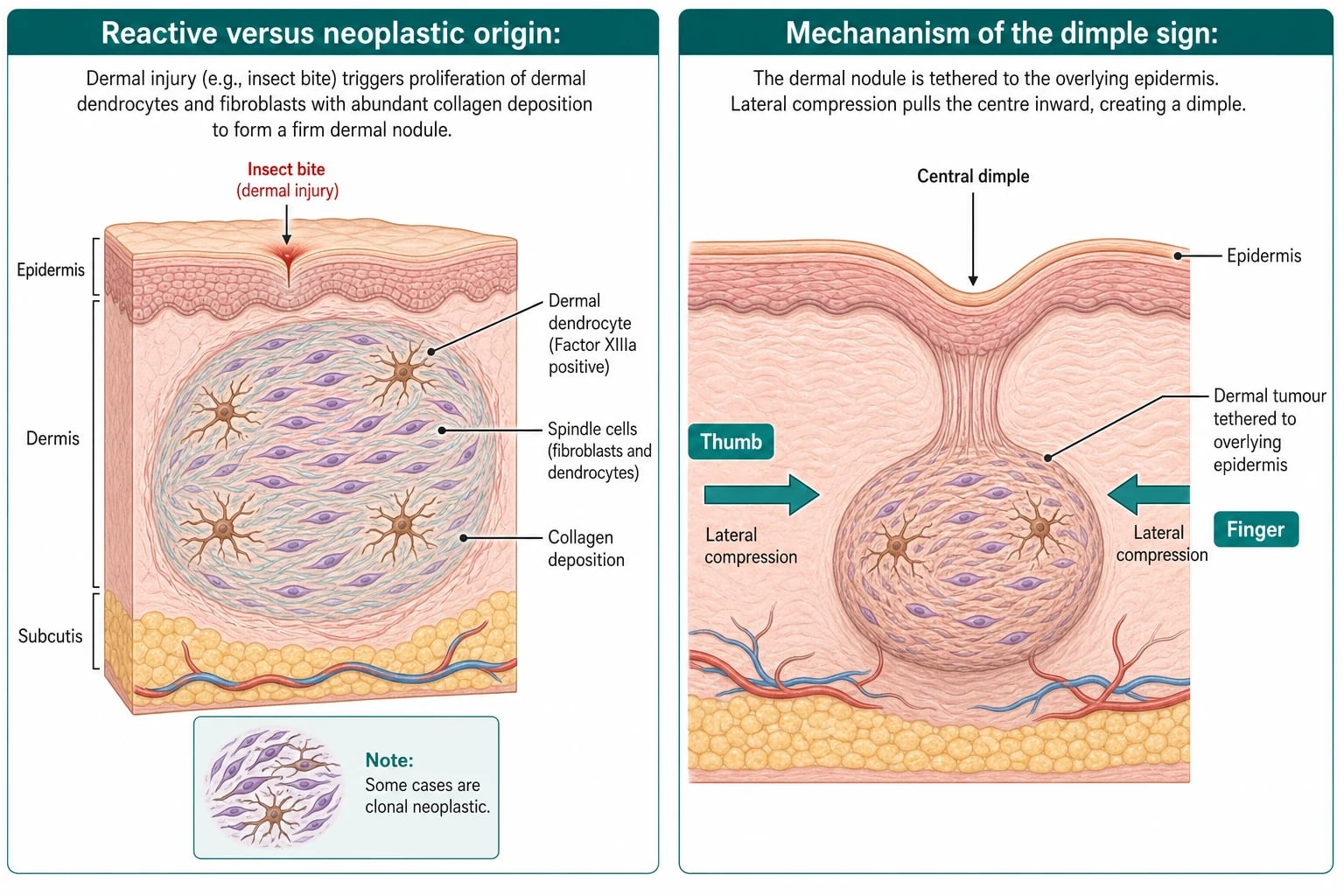
The cell of origin is the dermal dendrocyte — a Factor XIIIa-positive dermal dendritic phagocyte — which explains the defining immunohistochemical profile. Against a purely reactive model, however, clonality studies have demonstrated non-random X-chromosome inactivation and recurrent cytogenetic abnormalities (for example monosomy 7 and trisomy 16) in a subset of lesions, and recurrent gene fusions (such as MYADM::PRKCG) have now been identified in cellular dermatofibroma. The current synthesis is that most classic lesions are reactive, while cellular and atypical variants carry neoplastic, clonal features — a distinction that matters because the cellular/atypical forms are the ones that recur locally and, very rarely, metastasise.[1][4]
The cellular and molecular cascade. After a dermal injury, local inflammation recruits Factor XIIIa-positive dermal dendrocytes, fibroblasts, and macrophages, which release transforming growth factor-beta (TGF-beta) and other profibrotic cytokines. These drive fibroblast activation and abundant type I and type III collagen deposition in the dermis, producing the firm, sclerotic centre that characterises the lesion. The overlying epidermis responds with reactive acanthosis and basal-layer hyperpigmentation — a bystander hyperproliferation that produces the peripheral pigment network on dermoscopy. Vascular endothelial proliferation and, in the aneurysmal variant, haemorrhage into the lesion explain the red and blue-black colouration and the haemosiderin deposition. In cellular and atypical variants the balance shifts toward autonomous proliferation: increased cellularity, infiltration of the subcutis, and — in a subset — driver gene fusions, which is the molecular basis for the higher local recurrence of these subtypes.[1][4]
Why the lesion is firm. The firm, button-like consistency reflects abundant collagen deposition in the dermis together with the compact storiform arrangement of spindle cells; this firmness is itself a useful clinical clue (a melanocytic naevus, by contrast, is soft and compressible). [1]
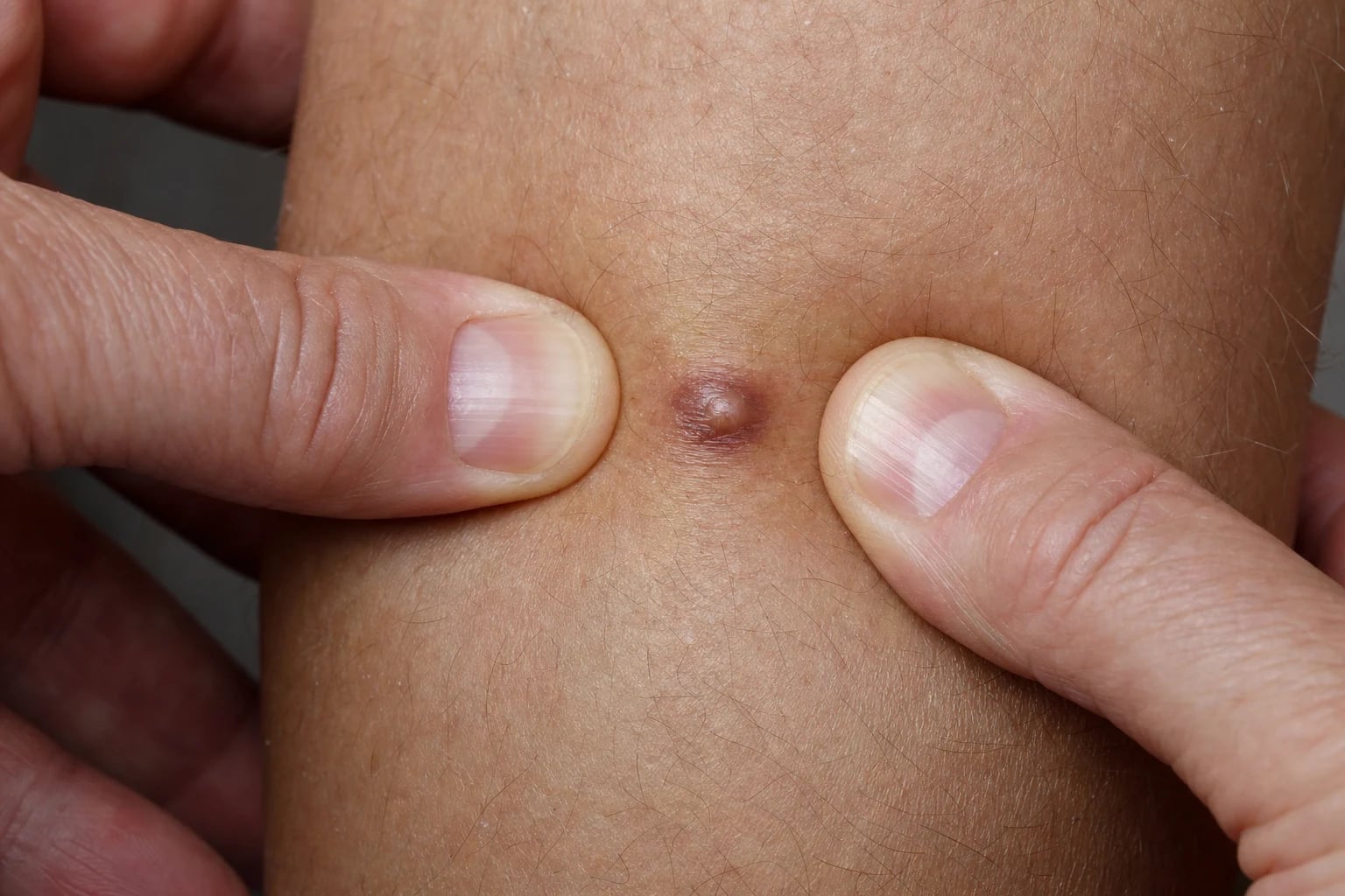
Mechanism of the dimple sign. The tumour lies in the dermis and is tethered to the overlying epidermis but not to the deeper subcutaneous fat. When the lesion is compressed laterally (sideways) between thumb and forefinger, the tethered epidermis is pulled inward and the centre invaginates, producing the dimple. A melanocytic naevus, which is not tethered in this way, instead protrudes or everts on compression — the bedside distinction that earns marks.[1]
Why the aneurysmal variant looks dark. Blood-filled cavernous spaces and haemosiderin deposition within the lesion produce the dark blue-black colour that mimics melanoma; dermoscopically this subtype shows a polymorphous mixture of colours and structures rather than the classic central white patch.[3]
Clinical Presentation
The classic dermatofibroma is a firm, 3 to 10 mm (occasionally up to 1 to 2 cm) dermal papule or nodule, round or oval, projecting slightly above the skin surface. The colour ranges from skin-coloured through light brown, reddish-brown, pink, purple to dark brown; a reddish or brown halo is sometimes present. The lesion is fixed to the overlying skin (you cannot move it independently of the epidermis) but mobile over the deeper subcutaneous tissue — it is not fixed to fascia or bone.[2]
The classic lesion at a glance
The single most useful bedside sign is the dimple sign (also called the Fitzpatrick sign or herniation sign). To elicit it, the examiner compresses the lesion laterally between thumb and index finger (not vertically); a positive sign is central dimpling/invagination of the lesion. This is highly specific for dermatofibroma and distinguishes it from a melanocytic naevus, which protrudes on compression. The dimple sign is positive in the great majority of classic dermatofibromas, and its demonstration is a fellowship-viva staple.[1]
Most dermatofibromas are asymptomatic. When symptoms occur they are usually mild tenderness or itch, often provoked by clothing friction at the waistband or sock line. The aneurysmal and cellular variants may be genuinely painful. The natural history is of stability: a lesion grows slowly over months to its final size and then persists unchanged for years or decades. A minority involute, leaving a slightly depressed, hyperpigmented scar; rapid growth or change is atypical and should prompt biopsy.[1]
Atypical presentations that examiners test deliberately include: an aneurysmal lesion (dark vascular nodule mimicking melanoma), an atrophic lesion (depressed, in an older adult), a giant lesion (larger than 5 cm), subungual, palmar, plantar, or facial lesions (rare sites that lower the threshold for biopsy), and eruptive/multiple lesions (dozens of papules, which trigger a systemic work-up).[1]
Each atypical presentation carries its own trap. The aneurysmal variant is dark blue-black and often tender; because it lacks the classic central white patch on dermoscopy it is the variant most often biopsied, chiefly to exclude nodular melanoma.[3] The atrophic variant, commoner in older adults, presents as a depressed, anetodermic plaque that can be mistaken for a morphea or an atrophic scar; histology shows loss of dermal substance with a flattened lesion. Giant dermatofibromas (over 5 cm) are rare and, by their size, breach the diagnostic ceiling for a "classic" lesion and warrant biopsy to exclude DFSP. Lesions at unusual sites — subungual (a painless nail-bed nodule), palmar or plantar (where the dimple sign is harder to elicit), or facial — should be examined with dermoscopy and a lower threshold for biopsy, because DFSP and other sarcomas also occur at these sites. Eruptive or multiple lesions demand a systemic work-up rather than local treatment.[1]
Differential Diagnosis
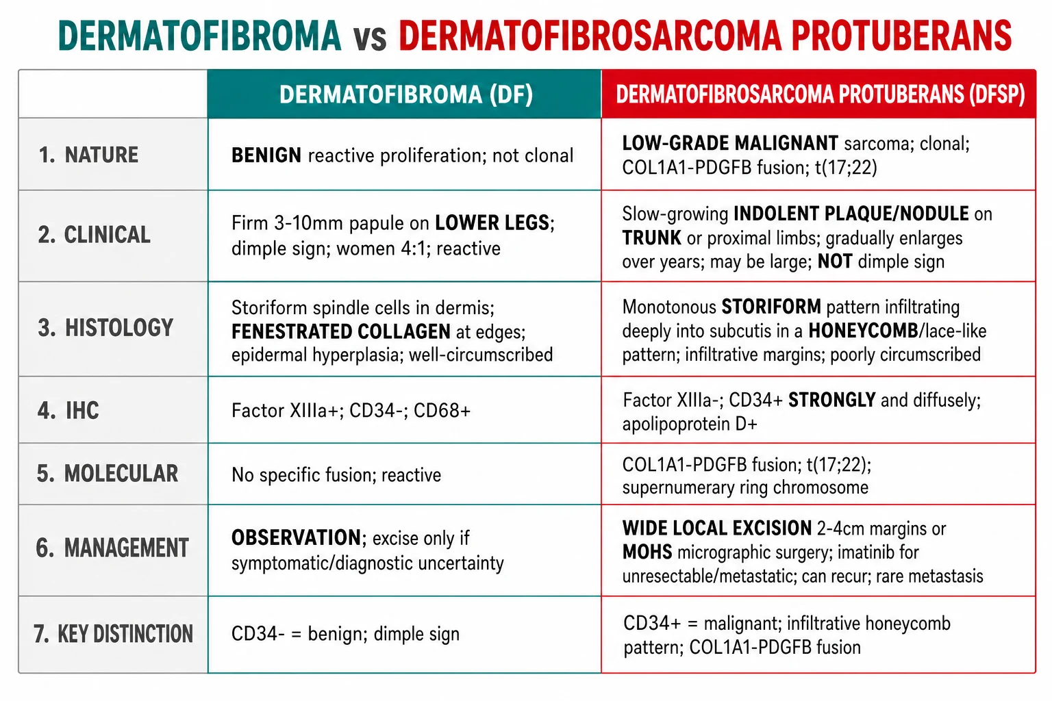
The differential is wide because dermatofibroma is common and brown. The must-not-miss mimic is dermatofibrosarcoma protuberans (DFSP); the everyday mimics are the melanocytic naevus, the neurofibroma, and the keloid. The distinguishing features are summarised below.[1]
Dermatofibroma
Benign; observe
- **Firm** 3-10 mm papule, **lower leg**; **dimple sign positive**
- **CD34 negative**, **Factor XIIIa positive**, stromelysin-3 positive
- Stable for years; no metastasis in classic form
- Tethered to skin, mobile over subcutis
Dermatofibrosarcoma protuberans
Low-grade sarcoma
- **Slow-growing plaque** on the **trunk** that becomes protuberant
- **CD34 positive** (strong diffuse), **Factor XIIIa negative**, COL1A1-PDGFB fusion
- Honeycomb infiltration into subcutis; recurs locally
- **Mohs** or wide local excision; imatinib if unresectable
Melanocytic naevus
Soft; everts on compression
- **Soft, compressible**; **protrudes/everts** on lateral compression (no dimple)
- Pigment network on dermoscopy; naevomelanocytes on histology
- Not tethered to skin in the same way
Neurofibroma
Soft; buttonhole sign
- **Soft, skin-coloured** papule; **buttonhole sign** (invaginates through the dermis on pressure)
- May be multiple (NF1); axillary freckling, cafe-au-lait macules
- S100 positive, SOX10 positive on histology
Keloid / hypertrophic scar
History of trauma
- **History of trauma/surgery**; keloid extends **beyond** the wound margins
- Collagen bundles parallel to the surface; chest, earlobe, shoulder
- Often pruritic or painful
Other mimics
Less common
- **Nodular melanoma** — rapid, blue-black, no dimple; S100 positive
- **Mastocytoma** — **Darier sign** positive (wheals on rubbing)
- **Leiomyoma** — **painful**; desmin positive; from arrector pili muscle
- **Sebaceous hyperplasia** — central dell, yellow, on the face
The single most important mimic to exclude is DFSP, because it is a low-grade sarcoma that recurs locally and very rarely metastasises; misdiagnosed as a dermatofibroma it is under-excised and undertreated. Any lesion that is large (over 2 cm), plaque-like, on the trunk, growing, or recurrent after excision should be biopsied with immunohistochemistry to settle the distinction.[1] Other mimics worth a sentence. A Spitz naevus in a child or young adult is a fast-growing pink-red dome-shaped nodule that, like a dermatofibroma, can be firm — but it lacks the dimple sign and shows characteristic spindle or epithelioid melanocytes and "rain shower" vessels on dermoscopy; biopsy is mandatory because of its melanocytic origin. A pyogenic granuloma is a friable, bleeding, rapidly growing red nodule (often after minor trauma) rather than a stable firm brown papule. A basal cell carcinoma can superficially mimic the acanthotic, pigmented epidermis overlying a dermatofibroma on a shave biopsy, which is why a superficial shave is to be avoided.[6] A solitary mastocytoma in a child is distinguished by a positive Darier sign (urtication on rubbing), and a cutaneous leiomyoma (from arrector pili muscle) by its characteristic pain on minor trauma or cold, with desmin positivity on histology.
The three bedside brown-papule manoeuvres are worth holding together. Lateral compression of a dermatofibroma gives central dimpling (the dimple sign); firm compression of a neurofibroma lets the lesion buttonhole (invaginate through a dermal defect); and compression of a naevus makes it protrude or evert. Three manoeuvres, three diagnoses — a compact viva answer.[1]
Clinical & Bedside Assessment
Dermatofibroma is, in the first instance, a clinical diagnosis made at the bedside. The focused assessment has four components.[1]
1. Inspect and palpate the lesion. Confirm the firm dermal consistency, the colour (light brown to reddish-brown to purple), the size (typically under 1 cm), and the site (lower leg). Establish that the lesion is tethered to the overlying skin but mobile over the subcutis — the latter rules out a deep structure such as a sarcoma fixed to fascia. [1]
2. Elicit the dimple sign. Compress the lesion laterally between thumb and index finger. A positive sign (central dimpling) is the bedside hallmark; demonstrate it to the patient and document it. A naevus everts; a neurofibroma gives the buttonhole sign (the whole lesion invaginates through a dermal defect on firm pressure) — three different bedside manoeuvres for three different lesions. [1]
3. Dermoscopy. Examine with a dermatoscope (polarised or non-polarised). The classic pattern (see Investigations) of a central white scar-like patch and a peripheral delicate pigment network confirms the diagnosis in a typical lesion. [1]
4. General examination when multiple lesions are found. If there are several to dozens of dermatofibromas, examine for stigmata of systemic lupus erythematosus, signs of immunosuppression (oral candidiasis, generalized lymphadenopathy), and features of a haematological disorder (pallor, bruising, splenomegaly), and arrange the appropriate blood tests (see Special Populations). [1]
Document the lesion with a scaled photograph and record the site, size, and colour so that change can be detected at review. Give the patient a safety-net: return urgently if the lesion grows beyond about 2 cm, changes colour, ulcerates, or becomes painful — features that mandate biopsy. [1]
Investigations & Dermoscopy
For a classic lesion with a positive dimple sign and characteristic dermoscopy, no investigation is required — the diagnosis is clinical and biopsy is avoided (a biopsy of a classic dermatofibroma leaves a scar that may be more conspicuous than the lesion itself). Investigation is reserved for atypical lesions (over 2 cm, growing, painful, plaque-like, on the trunk, recurrent, or with atypical colour), where biopsy with immunohistochemistry is mandatory to exclude DFSP, atypical fibrous histiocytoma, or melanoma.[1]
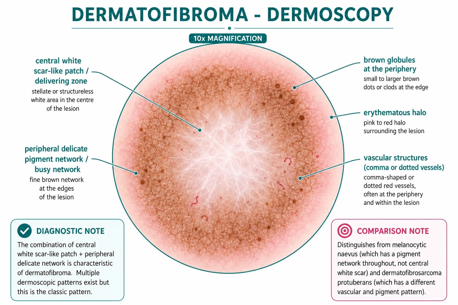
The classic dermoscopic pattern combines two features:[1]
- A central white scar-like patch (also called the "central white patch") — a stellate, structureless white area in the centre of the lesion, corresponding to the collagenous centre and epidermal hyperplasia.
- A peripheral delicate pigment network (the "busy network") — a fine brown network around the edges of the lesion, corresponding to basal-layer hyperpigmentation overlying the tumour. [1]
Additional features include brown globules at the periphery, an erythematous halo, and small vessels (comma, dotted, or polymorphous). The central white patch is found in roughly 80 percent of classic dermatofibromas and is the most specific single dermoscopic feature. At least ten dermoscopic patterns have now been described, and the pattern is far less reliable in the aneurysmal/haemosiderotic subtype, which shows a polymorphous combination of colours — brown, red, and black structureless areas, brown globules, and a mixed vascular pattern — and is frequently biopsied because it mimics melanoma.[3] Recognised dermoscopic patterns (in approximate order of frequency) include the classic central white patch with peripheral network, a homogeneous pattern, a multicomponent / polymorphous pattern, a predominantly vascular pattern (with dotted, comma, or polymorphous vessels), and a rare starburst pattern; the aneurysmal subtype is recognised by its mixture of brown, red, and black structureless areas with a mixed vascular network.[3] The practical message for the exam is that the central white patch is the one feature to name, but its absence does not exclude a dermatofibroma — particularly in the aneurysmal, atrophic, and cellular variants.
Dermoscopy technique. Both polarised (non-contact) and non-polarised (contact) dermoscopy are acceptable; polarised dermoscopy better displays the white scar-like central structure, while non-polarised may better resolve the peripheral pigment network. Apply ultrasound gel for contact dermoscopy. Compare the lesion with surrounding skin: the relative hypopigmentation of the centre against the pigmented network at the edge is a useful internal control. [1]
When dermoscopy is ambiguous but biopsy is undesirable, reflectance confocal microscopy (RCM) and optical coherence tomography (OCT) are non-invasive adjuncts used in specialist centres; they are not required for the routine case. [1]
Histopathology
Histology is obtained only when a lesion is excised (for symptoms, cosmesis, or diagnostic uncertainty) or biopsied (for atypical features). The histological appearance confirms the diagnosis and excludes DFSP and melanoma. [1]
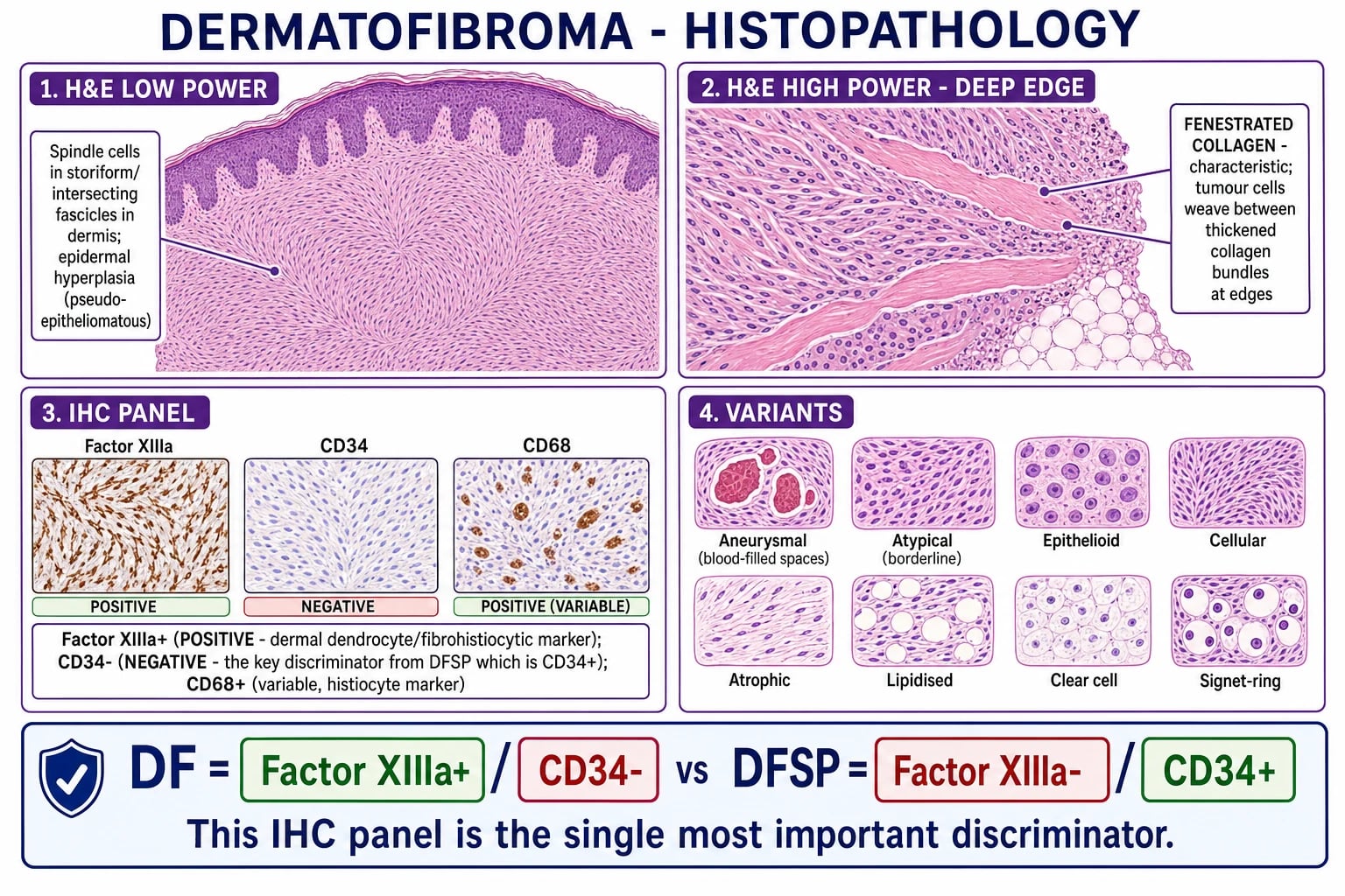
The defining features of classic dermatofibroma are:[1][6]
- Spindle cells in a storiform (cartwheel) pattern in the dermis, with no significant atypia and no significant mitotic activity.
- Epidermal changes — acanthosis and pseudo-epitheliomatous hyperplasia with basal-layer hyperpigmentation (sometimes called "dirty feet" hyperpigmentation), which can superficially mimic a basal cell carcinoma on a shallow shave.[6]
- Fenestrated collagen at the deep and lateral margins — tumour cells entrap and weave between thickened collagen bundles — a characteristic low-power finding.
- A grenz zone of normal papillary dermis between the epidermis and the tumour in many lesions.
The immunohistochemical profile is the key discriminator from DFSP:[1]
- Factor XIIIa — positive (the marker of the dermal dendrocyte of origin; confirms fibrohistiocytic lineage).
- CD34 — negative (the single most important negative marker; DFSP is CD34 positive).
- Stromelysin-3 — positive (a relatively specific positive marker for dermatofibroma).
- CD68 — variably positive (histiocyte marker).
- HMGA1 and podoplanin (D2-40) — positive in most lesions; useful adjuncts. [1]
Two practical biopsy pitfalls: (1) a superficial shave may show only the acanthotic epidermis with basal hyperpigmentation and be misread as a basal cell carcinoma — always biopsy to the dermis (punch or excisional).[6] (2) The edge of a cellular dermatofibroma can infiltrate the subcutis in a honeycomb pattern that resembles DFSP; CD34 staining is then essential to resolve the distinction (DF is CD34 negative; DFSP is CD34 positive). Cellular dermatofibromas are also more hypocellular in older patients, which can make histological assessment more difficult.[4]
Management
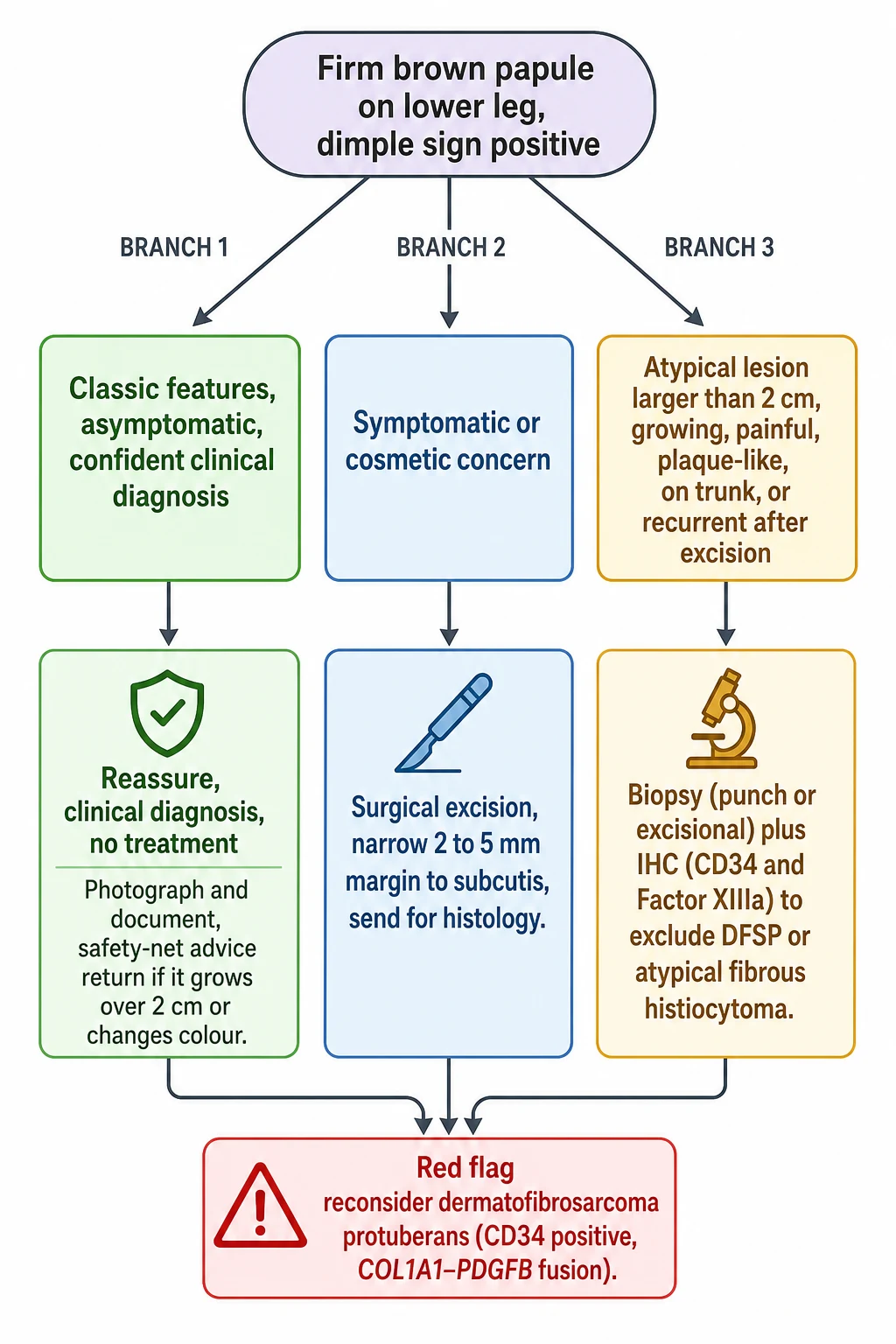
Dermatofibroma needs no time-critical intervention — it is benign. The first step at the initial consultation is therefore reassurance: explain that the lesion is a common benign growth, that it will not turn into cancer, and that no treatment is needed. Document and photograph the lesion, and provide written safety-net advice (return if it grows beyond about 2 cm, changes colour, ulcerates, or becomes painful). The only same-day action required is a biopsy when any feature suggests DFSP, atypical fibrous histiocytoma, or melanoma.[1][2]
Definitive management — stepwise
The default is observation. Intervention is reserved for specific indications, and the lesion is never treated "just in case".[1]
-
Observation / reassurance — for a classic lesion with a confident clinical diagnosis. This is the correct management for the great majority of patients.
-
Surgical excision — indicated when the lesion is symptomatic (pain or irritation from clothing), a cosmetic concern to the patient, or there is diagnostic uncertainty (atypical clinical or dermoscopic features). Excise with a narrow margin of about 2 to 5 mm, down to the subcutis, and send the specimen for histology. The histology both confirms the diagnosis and excludes DFSP in any atypical case. Surgical technique. Mark the lesion before local anaesthetic infiltration (which distorts the margins). Use an elliptical excision oriented along the relaxed skin tension lines of the lower leg (usually vertical on the shin), with a 2 to 5 mm clinical margin around the lesion. Carry the excision down to and including the subcutis so that the entire dermal tumour is removed en bloc; a superficial shave that leaves the deep dermal base behind is the commonest cause of recurrence. Achieve haemostasis and close with non-absorbable sutures (lower-leg skin is under tension and heals slowly). Always send the specimen for histology with a clinical note stating the pre-operative diagnosis and any atypical feature — so the pathologist stains for CD34 and Factor XIIIa where indicated. On a lower leg, counsel the patient that the scar may be more visible than the original lesion, and that wound healing can take 2 to 3 weeks. [1]
-
Biopsy with immunohistochemistry — for any lesion with red-flag features (over 2 cm, growing, painful, plaque-like, on the trunk, recurrent). A punch or excisional biopsy (not a superficial shave) is sent for CD34 and Factor XIIIa staining. If DFSP is confirmed, refer to the sarcoma pathway (Mohs or wide local excision; imatinib for unresectable/metastatic disease targeting the COL1A1-PDGFB fusion).
-
Cellular or atypical (borderline) fibrous histiocytoma — complete excision with clear margins and close follow-up for local recurrence (the cellular variant recurs in about 20 to 30 percent of cases).[1]
Treatments that do NOT work (and why)
- Cryotherapy flattens the surface but does not remove the dermal component; recurrence is common and it leaves hypopigmentation — not recommended.[1]
- Intralesional corticosteroid injection may shrink the lesion slightly but is not curative and is painful — not recommended.
- Shave excision alone is inadequate — the dermal base is left behind and the lesion recurs. If surgery is undertaken, it should be a complete excision to the subcutis.
Avoid biopsy of a classic lesion. A biopsy scar on the lower leg can be more conspicuous than the dermatofibroma itself; the diagnosis of a classic lesion is clinical. Reserve biopsy for atypical cases where the histology will change management.[1]
Follow-up. A classic dermatofibroma that is observed needs no scheduled review — the patient re-presents only if the lesion changes (safety-net above). A classic lesion that is completely excised also needs no follow-up once the wound has healed and histology confirms the diagnosis. After excision of a cellular variant, review at 6 and 12 months to detect local recurrence, then discharge if stable. After an atypical (borderline) variant, review at 6 and 12 months, then annually for 2 to 3 years, because recurrence can be late. If DFSP is confirmed on histology, follow-up follows the sarcoma pathway (5 to 10 years of clinical surveillance). Any recurrence after excision should be re-biopsied with CD34 and Factor XIIIa to exclude DFSP misdiagnosed as a dermatofibroma. [1]
Specific Subtypes & Scenarios
Aneurysmal / haemosiderotic dermatofibroma. Blood-filled cavernous spaces and haemosiderin deposition produce a dark blue-black, sometimes painful nodule that clinically mimics nodular melanoma, a pyogenic granuloma, or a vascular tumour. Dermoscopy is polymorphous and unreliable (mixed brown, red, and black structureless areas with brown globules and a mixed vascular pattern); the central white patch is often absent.[3] Biopsy is usually required to exclude melanoma; histology shows cavernous vascular spaces within an otherwise typical dermatofibroma, and immunohistochemistry (S100 negative, Factor XIIIa positive) confirms the diagnosis.
Cellular dermatofibroma. A densely cellular variant that may extend into the subcutis in a honeycomb pattern (raising the differential of DFSP until CD34 staining resolves it). It carries a higher local recurrence rate (about 20 to 30 percent) after excision, and recurrent gene fusions such as MYADM::PRKCG have now been reported — supporting a neoplastic basis for this subtype. Complete excision with clear margins and follow-up is required.[1]
Atypical (pseudosarcomatous / borderline) fibrous histiocytoma. Scattered atypical cells and occasional mitoses place this variant in the borderline category with low-grade malignant potential. It requires complete excision and monitoring for local recurrence; distant metastasis is very rare but described. [1]
Epithelioid dermatofibroma (epithelioid fibrous histiocytoma). A dome-shaped, often vascular-appearing nodule composed of epithelioid cells with abundant eosinophilic cytoplasm. A subset harbour ALK rearrangements, which can be demonstrated immunohistochemically.[5]
Atrophic dermatofibroma. The lesion loses substance and presents as a depressed, wrinkled, slightly pigmented plaque; it is more common in older adults and can be confused with an atrophic scar or a morphea-type lesion.[4]
Giant, subungual, palmar, plantar, and facial dermatofibromas are rare; their unusual site lowers the threshold for biopsy to exclude DFSP or another malignancy. [1]
Eruptive / multiple dermatofibromas. The appearance of several to dozens of lesions over weeks to months should prompt a search for an underlying systemic trigger — immunosuppression (HIV, solid-organ transplant), autoimmune disease (SLE), myelodysplasia or leukaemia, and atopy. Investigate with HIV serology, an autoimmune screen (ANA, double-stranded DNA), and a full blood count with film. [1]
Complications & Pitfalls
Complications of dermatofibroma itself are uncommon, because the classic lesion is benign and stable. The clinically important issues are local recurrence, the diagnostic pitfall of confusing dermatofibroma with DFSP, and the morbidity of unnecessary procedures.[1]
- Local recurrence after incomplete excision. Classic dermatofibroma recurs in about 5 to 15 percent of cases after incomplete excision; the cellular variant recurs in 20 to 30 percent. Recurrence presents as a new firm papule within or adjacent to the scar.
- The classic diagnostic error. Misdiagnosing DFSP as a dermatofibroma (or vice versa) leads to under-excision of a low-grade sarcoma with local recurrence and undertreatment. The safeguard is immunohistochemistry — CD34 and Factor XIIIa — on any atypical or recurrent lesion.
- The superficial-shave pitfall. A shave biopsy may show only acanthotic epidermis with basal-layer hyperpigmentation and be misread as a basal cell carcinoma.[6] Always biopsy to the dermis (punch or excisional).
- Procedural morbidity. Excision on the lower leg (the commonest site) leaves a scar that can be slow to heal and more conspicuous than the original lesion; weigh this against the (usually cosmetic) indication before operating.
- Cosmetic and psychological morbidity. A visible persistent lesion, or a surgical scar on the leg, can distress the patient; reassurance and realistic counselling about scarring are part of management.
- Rare aggressive behaviour. Truly metastasising dermatofibroma is essentially confined to deep, cellular, or atypical/borderline variants; classic cutaneous dermatofibroma does not metastasise.
Comorbidities & Associated Conditions
A solitary classic dermatofibroma is not a marker of systemic disease and requires no comorbidity screen. The comorbidity axis comes into play only with multiple or eruptive lesions, where an underlying condition is driving dysregulated fibrohistiocytic proliferation.[1]
- Immunosuppression — HIV infection, solid-organ transplantation, and long-term immunosuppressive therapy are the strongest associations; eruptive dermatofibromas may even be a presenting feature of previously undiagnosed HIV.
- Autoimmune disease — systemic lupus erythematosus is the classic autoimmune association, and an ANA should be sent when multiple lesions appear.
- Haematological disease — myelodysplastic syndromes and, less often, leukaemia and lymphoma have been linked to eruptive dermatofibromas; a full blood count with blood film is a reasonable screen.
- Atopy and inflammatory skin disease — an association with atopic dermatitis has been reported, though the link is weaker than for immunosuppression or lupus.
- Pregnancy — a physiological state in which existing lesions enlarge and new lesions appear, attributable to hormonal effects on fibrohistiocytic cells. [1]
The practical implication: a patient who suddenly develops many dermatofibromas warrants HIV serology, a full blood count with film, an autoimmune screen (ANA, double-stranded DNA), and a focused history for immunosuppression and constitutional symptoms. The lesions themselves are managed individually, but the underlying condition is the priority. [1]
Prognosis & Disposition
The prognosis of classic dermatofibroma is excellent: it is benign, stable for years to decades, may involute, and does not undergo malignant transformation. No long-term follow-up is needed for a classic lesion, whether observed or completely excised. The cellular and atypical variants have a low risk of local recurrence (about 20 to 30 percent for cellular) after incomplete excision and very rarely metastasise; these warrant complete excision and a period of surveillance.[1]
Disposition. Classic dermatofibroma is managed in primary care with safety-net advice. An atypical lesion requiring biopsy or excision is referred to secondary care (dermatology); if DFSP or another sarcoma is confirmed on histology, the patient enters the sarcoma multidisciplinary team pathway (Mohs or wide local excision; imatinib for unresectable/metastatic disease). The safety-net for any conservatively managed lesion is to return urgently if it grows beyond about 2 cm, changes colour, ulcerates, or becomes painful.[1]
Special Populations
Eruptive / multiple dermatofibromas. The sudden appearance of multiple lesions is the scenario that most changes the work-up. Investigate for HIV and other immunosuppression, systemic lupus erythematosus (ANA, double-stranded DNA), myelodysplastic syndromes and leukaemia (full blood count with film), and atopy. Treat the lesions individually as above (most are observed).[1]
Pregnancy. Existing dermatofibromas may enlarge and new lesions may appear, consistent with hormonal influence. Management is conservative; defer excision if the only indication is cosmetic, and re-assess post-partum. [1]
Children and adolescents. Dermatofibroma is less common in children than in adults but is recognised, including eruptive disease in adolescents. Management is identical — observe unless atypical. [1]
Older adults. The atrophic variant is more common, and cellular lesions may be more hypocellular (making histology harder).[4] Lower-leg excisions heal more slowly in older skin; weigh the scar against the benefit.
Immunosuppressed patients (solid-organ transplant, chemotherapy) have a higher rate of multiple and atypical dermatofibromas; maintain a lower threshold to biopsy a changing lesion. [1]
Evidence, Guidelines & Regional Differences
Dermatofibroma is a global entity and there is no region-specific drug guideline — because no drug is first-line (the lesion is structural, not inflammatory or infective). Regional practice differs mainly in the threshold to biopsy or excise and in the referral pathway.[1][2]
Global consensus — dermatofibroma is a clinical diagnosis; observation is the default. No pharmacotherapy is indicated. Excision is reserved for symptomatic, cosmetic, or diagnostically uncertain lesions; biopsy for atypical features.
Australia / New Zealand (RANZCD). Surgical excision with a 2 to 5 mm margin for symptomatic or atypical lesions; histopathology is mandatory on any excised lesion to exclude DFSP.
Evidence and controversies
The defining contemporary controversy is the reactive versus neoplastic origin. The traditional reactive model is supported by the history of a preceding injury in many cases; the neoplastic model is supported by clonality studies (non-random X-chromosome inactivation; cytogenetic abnormalities such as monosomy 7 and trisomy 16) and by recurrent gene fusions (for example MYADM::PRKCG in cellular dermatofibroma). The synthesis is that classic lesions are largely reactive while cellular and atypical variants carry clonal, neoplastic features — and these are the variants that recur and very rarely metastasise.[1]
Zaballos and colleagues (2023) performed the largest morphological study of dermoscopy of the aneurysmal / haemosiderotic subtype (110 cases), showing that this variant displays a polymorphous combination of colours and structures in which the classic central white patch is often absent — explaining why this subtype is the one most often biopsied because it mimics melanoma.[3]
Jordan and colleagues (2026) showed in a retrospective study of 307 cases that dermatofibroma hypocellularity increases with patient age — a histological feature that can complicate interpretation in older patients and underscores the need for immunohistochemistry in equivocal cases.[4]
The dermatofibroma-versus-DFSP immunohistochemical revolution (CD34 negative versus CD34 positive; Factor XIIIa positive versus negative), together with the identification of the COL1A1-PDGFB fusion and the efficacy of imatinib (a PDGFR tyrosine-kinase inhibitor) for unresectable or metastatic DFSP, has transformed the management of the differential — even though the management of classic dermatofibroma itself has not changed.[1] Nomenclature and history. The lesion has carried several names — sclerosing haemangioma (for its vascular-appearing, sclerotic histology), histiocytoma (for its histiocytic cells), dermatofibroma (the clinical term), and benign fibrous histiocytoma (the modern pathological term). The persistence of multiple names reflects the historical reactive-versus-neoplastic debate: "histiocytoma" implied a neoplasm of histiocytes, while "dermatofibroma" implied a fibrous reactive lesion of the dermis. Contemporary pathology favours benign fibrous histiocytoma as the unifying term, with "dermatofibroma" retained for the common cutaneous form.
Why there is no drug therapy. Unlike inflammatory or infective skin lesions, dermatofibroma is a structural dermal nodule of collagen and spindle cells; there is no inflammatory or microbial target for a topical or systemic drug to act on. This is why cryotherapy and intralesional corticosteroid — the only "medical" options tried — fail (they do not remove the dermal component), and why definitive treatment, when required, is surgical. The single pharmacological story in the dermatofibroma literature is the success of imatinib for the differential (DFSP), which works because DFSP's COL1A1-PDGFB fusion drives constitutive PDGFR signalling — a target dermatofibroma itself does not have. [1]
Exam Pearls
Bedside brown-papule manoeuvres
DIMPLE
Dermatofibroma — central dimpling on LATERAL compression (positive dimple sign)
Investigate the trigger — insect bite, trauma, folliculitis precede ~20%
Tethered to skin, mobile over subcutis — rules out a deep sarcoma
Paucity of treatment needed — observe; never biopsy a classic lesion
Lower legs (~70%), women > men — the classic stem
Exclude DFSP — CD34 positive, COL1A1-PDGFB — in any atypical/recurrent lesion
Self-test: a firm brown papule on the shin
A 34-year-old woman presents with a 7 mm firm reddish-brown papule on her shin that has been stable for two years. On lateral compression between thumb and finger the centre of the lesion dimples inward. What is the diagnosis, what confirms it, and what is the management? [1]
Answer. The diagnosis is a dermatofibroma (benign fibrous histiocytoma), established clinically by the firm dermal papule on the lower leg of a young woman with a positive dimple sign. Dermoscopy (central white scar-like patch with a peripheral delicate pigment network) confirms it. The lesion is benign; management is reassurance and observation — no biopsy, no excision — with safety-net advice to return if it grows, changes colour, ulcerates, or becomes painful.
Red Flags
Exam application bank (NEET-PG / INICET)
One-line answer
Dermatofibroma (DF; benign fibrous histiocytoma of the skin) is a common benign dermal tumour of fibroblast-like and histiocyte-like (fibrohistiocytic) cells arranged in a storiform pattern. Classic presentation: firm 3-10 mm reddish-brown dermal papule on the lower legs of a young-to-middle-aged adult woman, tethered to the overlying skin but freely mobile over the subcutis. The DIMPLE SIGN (Fitzpatrick / herniation sign) — central dimpling on LATERAL compression — is the bedside pathognomonic clue. Dermoscopy shows a central white scar-like patch with a peripheral delicate ('busy') pigment network. Histology: storiform spindle cells, epidermal acanthosis, and fenestrated collagen at the margins; immunohistochemistry is Factor XIIIa positive and CD34 negative (the key discriminator from dermatofibrosarcoma protuberans, which is CD34 positive with a COL1A1-PDGFB fusion). Management is re
Worked stems (answer without another resource)
Stem 1 — Classic presentation. Map symptoms to mechanism; name the first investigation and first treatment step with dose/route if drug therapy is standard. [1]
Stem 2 — Unstable / complicated. List red flags that force immediate resuscitation, theatre, ICU, antidote, or reperfusion — and what you do in the first 15 minutes. [1]
Stem 3 — Atypical group. Elderly, pregnancy, child, or immunocompromised: how presentation and thresholds change. [1]
Stem 4 — Differential trap. Name the three closest mimics and one discriminator for each. [1]
Stem 5 — Disposition. Who goes home with safety-netting, who is admitted, who needs HDU/ICU/theatre, and what follow-up is mandatory. [1]
Rapid viva checklist
- Definition + classification
- Pathophysiology chain
- Bedside signs / criteria
- Score with exact components (if any)
- Emergency bundle
- Definitive therapy with doses
- Complications of disease and of treatment
- Special populations
- Guideline/trial name if classic
- Three exam traps
Coverage self-check
If you cannot answer any stem above from this page alone, re-read the matching section — the page is intended to be self-sufficient for final-prof and NEET-PG/INICET questions on Dermatofibroma.
[1]References
- [1]Wan L, Park A, Almatroud L, et al. Dermatofibroma: Reappraisal and Updated Review Clin Cosmet Investig Dermatol, 2025.PMID 40785832
- [2]Wilson JL. Benign Skin Tumors Prim Care, 2025.PMID 40835288
- [3]Zaballos P, Alvarez-Salafranca M, Llambrich A, et al. Dermoscopy of haemosiderotic/aneurysmal dermatofibroma: A morphological study of 110 cases J Eur Acad Dermatol Venereol, 2023.PMID 36251407
- [4]Jordan C, Hodges W, Russell L, et al. Dermatofibroma Hypocellularity Is Associated With Patient Age: A Retrospective Study of 307 Cases J Cutan Pathol, 2026.PMID 42252143
- [5]Felty CC, Linos K. Epithelioid Fibrous Histiocytoma: A Concise Review Am J Dermatopathol, 2019.PMID 30289773
- [6]Stanoszek LM, Wang GY, Harms PW. Histologic Mimics of Basal Cell Carcinoma Arch Pathol Lab Med, 2017.PMID 29072946