Dermatology · Medicine
Dermatofibrosarcoma protuberans
Also known as Dermatofibrosarcoma protuberans (DFSP) · DFSP · Bednar tumour (pigmented variant) · Progressive and recurrent dermatofibroma (historical, obsolete) · Darier-Ferrand tumour (European eponym)
Dermatofibrosarcoma protuberans (DFSP) is a low-to-intermediate grade malignant fibroblastic tumour of skin, a cutaneous sarcoma of dermal fibroblast origin. Classic presentation: a slow-growing, indurated, blue-red-brown plaque or nodule on the trunk of a 30-50 year old, which may have an atrophic centre and progress to a protuberant multinodular mass. Histology: monotonous storiform (cartwheel) pattern of CD34-positive spindle cells infiltrating subcutaneous fat in a honeycomb pattern. IHC: CD34+ (strong, diffuse), Factor XIIIa- (versus dermatofibroma, which is CD34- and Factor XIIIa+). Molecular hallmark: COL1A1-PDGFB fusion from t(17;22) or a supernumerary ring chromosome r(17;22) - constitutive PDGFR activation - the target of imatinib. Mohs micrographic surgery is the standard of care (1 percent recurrence) versus wide local excision with 2-3 cm margins (10-20 percent recurrence). Imatinib is reserved for unresectable, recurrent, or metastatic disease, with fibrosarcomatous transformation raising metastatic risk to 15-30 percent.
On this page & tools
Your progress
Saved locally on this device.
Exam tags
Red flags
Overview & Definition
Dermatofibrosarcoma protuberans (DFSP) is a low-to-intermediate grade malignant fibroblastic tumour of skin - a rare cutaneous sarcoma (incidence roughly 0.8 to 4.5 cases per million per year) that arises in the dermis from CD34-positive fibroblastic cells. The WHO classification of soft-tissue and bone tumours (4th edition, 2024) places it in the category of intermediate-grade fibroblastic/myofibroblastic tumours that are locally aggressive and very rarely metastasising - except in the fibrosarcomatous variant, which is upgraded to a frankly malignant behaviour profile.[3][6]
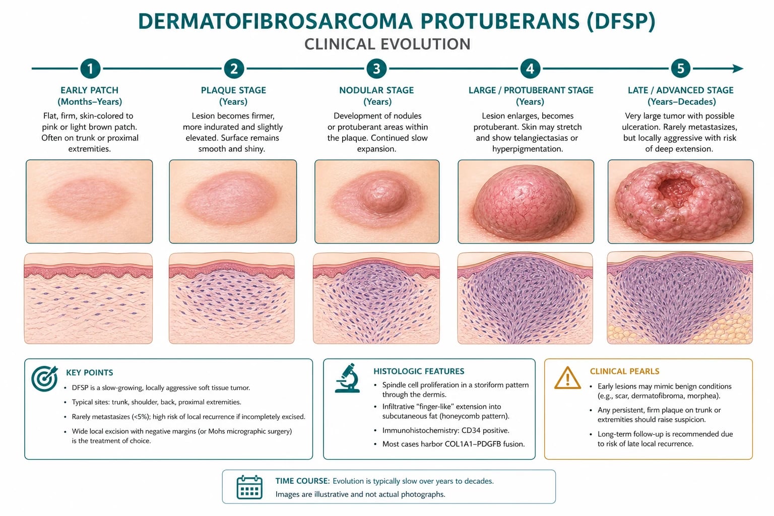
The clinical hallmark is a slow-growing, indurated, blue-red-brown plaque or nodule (sometimes with an atrophic or depressed centre) that progresses - often over years - to a protuberant, multinodular, sometimes ulcerated mass. Most lesions sit on the trunk (chest, abdomen, back, shoulder girdle, proximal thigh) and most patients are 30 to 50 years old at diagnosis. The lesion is locally infiltrative: spindle cells crawl along connective-tissue septa into subcutaneous fat in a honeycomb or lace-like pattern that extends well beyond the clinical margin and is invisible to the naked eye. That subclinical extension is why standard wide local excision fails so often, why Mohs micrographic surgery is the standard of care, and why imatinib - a small-molecule inhibitor of the PDGFR-beta tyrosine kinase encoded by the fusion partner of the pathognomonic t(17;22) COL1A1-PDGFB fusion gene - is the dominant targeted therapy in unresectable, recurrent or metastatic disease.[1][3][4]
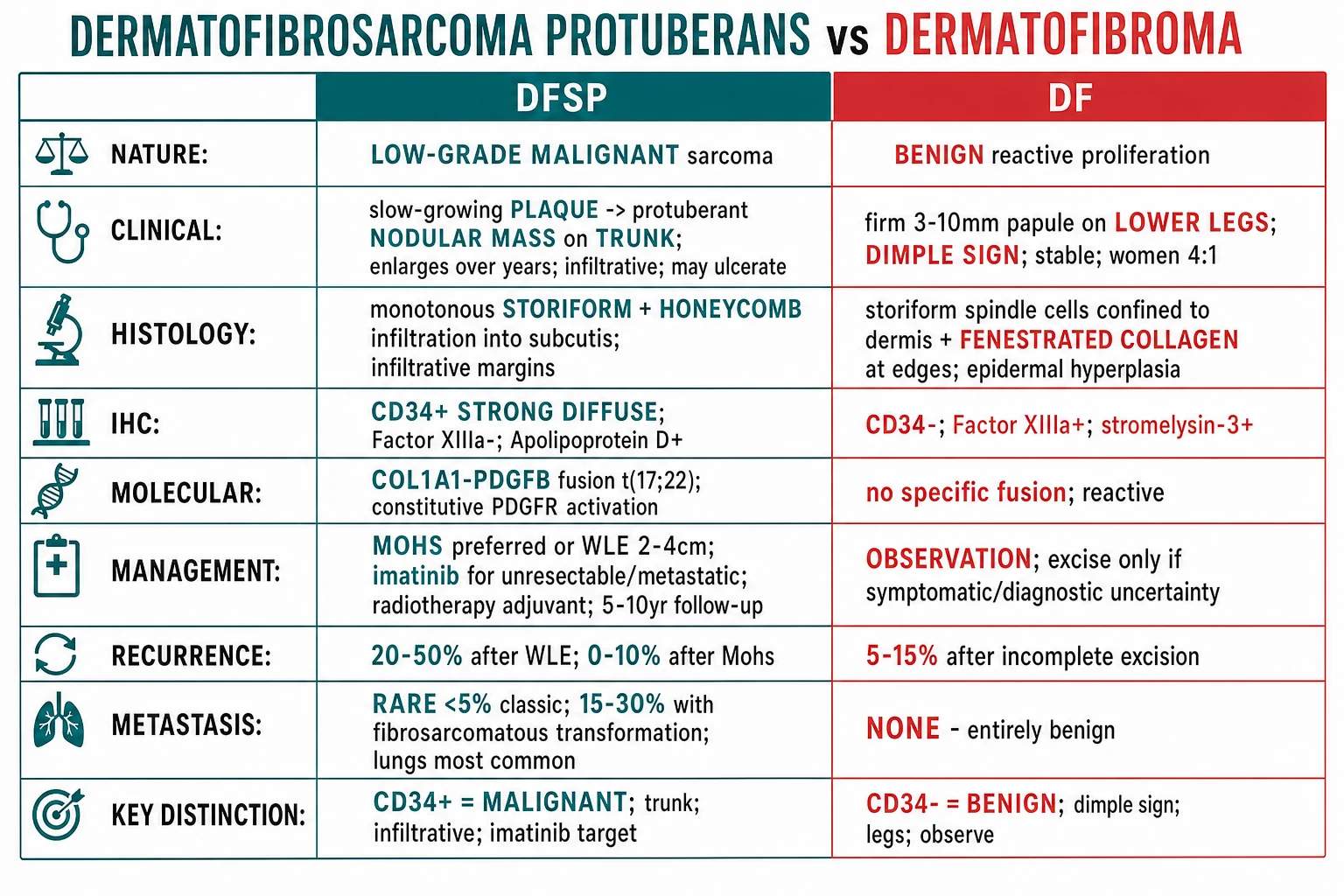
DFSP is best understood by contrast with its principal mimic, dermatofibroma (benign fibrous histiocytoma): DF is small, on the lower leg, has a positive dimple sign, and is CD34-negative / Factor XIIIa-positive; DFSP is large, on the trunk, plaque-like, infiltrative, and is CD34-positive / Factor XIIIa-negative. The immunohistochemistry (and, when needed, FISH or RT-PCR for the fusion) is what closes the diagnosis. [1]
[1]DFSP at a glance
Classification
DFSP is a single clinicopathological entity with a small number of well-defined morphological and behavioural subtypes. The classification matters because the fibrosarcomatous variant - approximately 10-15 percent of cases - carries a meaningfully higher metastatic risk, and the pigmented (Bednar) variant is a histological curiosity that does not change prognosis.[3][6]
- Low-to-intermediate grade; uniform spindle cells; storiform pattern
- CD34+ diffuse; honeycomb fat infiltration; under 5 mitoses/10 HPF
- Locally infiltrative; under 5 percent metastasis; Mohs cures >99 percent
- Higher-grade areas: increased cellularity, >5 mitoses/10 HPF, herringbone pattern
- CD34 may be LOST in high-grade areas; fusion still present
- Metastatic risk 15-30 percent; treat aggressively (wider margins, adjuvant RT, consider imatinib)
- Melanin-laden dendritic cells scattered among spindle cells
- S100+/HMB45+/Melan-A+ in the pigmented cells (not the tumour cells)
- No prognostic difference from classic DFSP; mainly seen in patients with skin of colour
- Prominent myxoid stroma; mimics other myxoid tumours (myxoid liposarcoma, myxofibrosarcoma)
- CD34+ and t(17;22) fusion confirm DFSP
- Prognosis similar to classic DFSP; histology is the trap
- Depressed or atrophic plaque rather than a protuberant mass; easily missed
- CD34+ and t(17;22) fusion; behaves like classic DFSP
- Diagnostic delay is common - consider DFSP in any chronic depressed truncal plaque
Epidemiology & Risk Factors
DFSP accounts for roughly 1 percent of all soft-tissue sarcomas and approximately 18 percent of all cutaneous soft-tissue sarcomas, making it the single most important cutaneous sarcoma to recognise at the bedside. The annual incidence is 0.8 to 4.5 cases per million population worldwide, with higher reported rates in studies with active cancer-registry capture. The lesion presents across a wide age range - from congenital (rare) to the ninth decade - but the median age at diagnosis is 38 to 45 years, and 70-80 percent of patients are diagnosed between 20 and 50 years of age.[3][6]
Demographics
Risk factors are largely host-related rather than environmental:[1][2][6]
- Prior trauma at the site (surgical scar, burn, vaccination site, insect bite, blunt trauma) is reported in 10-20 percent of cases and is biologically plausible given the fibroblastic, scar-like behaviour of the tumour. A history of DFSP arising in a chronic burn scar or at the site of a vaccination is well documented.
- Skin of colour is over-represented in several large series, and the Bednar (pigmented) variant is more common in patients of African descent; the precise driver is unclear.
- Acral DFSP is uncommon but described; it may behave less favourably because of anatomic constraints on margin.
- Congenital DFSP (typically the plaque-like variant) and paediatric DFSP account for roughly 1-2 percent of cases each, often present at birth or in early childhood, and are often initially misdiagnosed as a vascular birthmark or plaque.
- No consistent occupational, environmental, or chemical exposure has been validated as a risk factor.
- Family history is not a recognised risk factor; the t(17;22) fusion is acquired (somatic), not germline, in the vast majority of cases. [1]
European interdisciplinary guideline 2024 (Saiag et al., EJC 2025): Mohs micrographic surgery is the standard of care for localised disease; wide local excision with 2-3 cm margins to the deep fascia is the alternative; imatinib 400 mg/day is first-line for unresectable/recurrent/metastatic disease; adjuvant radiotherapy (50-60 Gy) for positive margins if further surgery is not feasible.[1]
Pathophysiology
The molecular engine of DFSP is the t(17;22)(q22;q13) translocation (or, in roughly two-thirds of cases, a supernumerary ring chromosome r(17;22) that contains amplified material from chromosomes 17 and 22). The translocation fuses the COL1A1 gene on 17q22 - a highly expressed collagen gene - with the PDGFB gene on 22q13 - the gene that encodes platelet-derived growth factor beta. The COL1A1 promoter drives constitutive overexpression of PDGFB, which in turn activates the PDGFR-beta receptor on the same cell in an autocrine loop. PDGFR-beta signalling then drives proliferation, survival, and stromal recruitment through RAS-MAPK and PI3K-AKT pathways, producing the slowly growing, locally infiltrative, scar-like tumour that defines DFSP.[1][3][6][8]
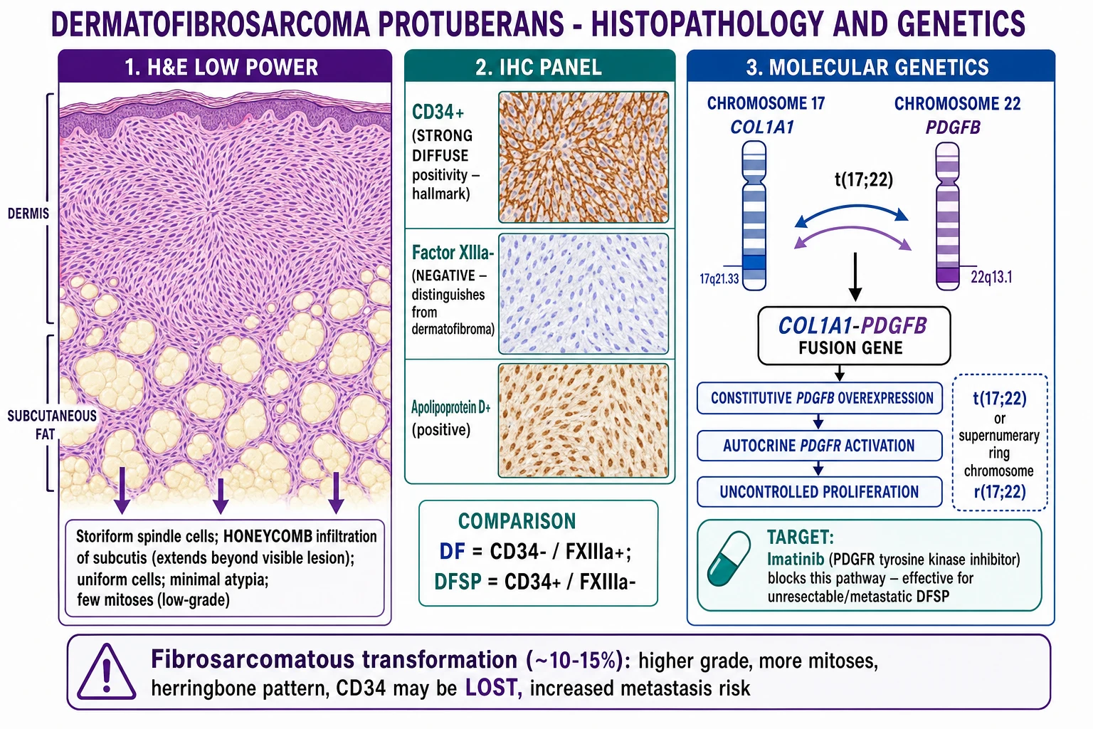
Key pathophysiological points for the examiner: [1]
- The fusion is somatic and tumour-specific. It is not found in normal skin, in dermatofibroma, or in the overlying epidermis. Detection by FISH, RT-PCR, or next-generation sequencing is diagnostic when histology is ambiguous.
- Variant fusion partners are increasingly recognised. The vast majority of DFSPs carry a COL1A1-PDGFB fusion, but rare COL1A2-PDGFB, COL1A1-PDGFA, EMILIN2-PDGFB, TPM3-PDGFRB and other fusions have been described, particularly in fibrosarcomatous and CD34-negative examples. These rare variants still predict imatinib sensitivity when PDGFR is the 3' partner.[8]
- The same fusion is found in giant cell fibroblastoma (GCF) of childhood, which is now regarded as the paediatric and juvenile form of the same entity. GCF and DFSP share the COL1A1-PDGFB fusion, the same CD34+ immunoprofile, and a similar locally infiltrative behaviour. A combined entity - giant cell fibroblastoma / DFSP - is listed in the WHO 4th edition.[6]
- The honeycomb infiltration reflects the tumour's preference for connective-tissue planes: spindle cells crawl along the septa between subcutaneous fat lobules, producing the lace-like pattern that defines the deep margin of the lesion. This is the anatomic basis of the subclinical extension that defeats standard wide excision.
- The slow growth rate reflects a low mitotic index, a low mutational burden, and an absence of the high-grade driver mutations seen in frankly malignant sarcomas. Ki-67 is typically low (5-10 percent) in classic DFSP and rises sharply in the fibrosarcomatous areas (often >20 percent).
- Loss of CD34 staining in the fibrosarcomatous component does not mean the fusion is gone - the fusion persists, and imatinib remains effective in many FS-DFSPs.
- Telomerase activation, p53 overexpression, and p16 loss have been described in FS-DFSP and may be the molecular events that drive the transformation; they are not, however, validated as diagnostic or prognostic biomarkers in routine practice.
CD34
Clinical Presentation
The presentation of DFSP is, in the early years, insidious - a small, firm, skin-coloured, pink, red, or blue-brown plaque or nodule that the patient and their family physician may dismiss as a cyst, a lipoma, a keloid, a scar, or a dermatofibroma. Over months to years the lesion enlarges slowly, thickens, and develops multinodular or protuberant surface change; satellite nodules may appear at the periphery; and the lesion may eventually ulcerate (a late and ominous sign). The natural history gives the tumour its name: protuberans (Latin: "to push forward").[3][6]
Headline clinical features
Site distribution (cumulative from pooled series):[3][5][6]
- Trunk (~50 percent): chest, abdomen, back, flank, shoulder girdle, groin.
- Proximal extremities (~20-35 percent): shoulder, upper arm, thigh, buttock.
- Head and neck (~10-15 percent): scalp, face (especially cheek), neck. Higher recurrence risk because of anatomic constraints on margin.
- Acral sites (hands, feet) and genitalia are uncommon but recognised (vulvar DFSP is described in the MITO rare-cancer-group series).[6]
Atypical presentations - exactly the ones examiners test: [1]
- Atrophic DFSP - a depressed, sclerotic plaque that looks like morphea, atrophoderma, or a burn scar; frequently missed.
- Plaque-stage DFSP - the earliest stage, before the protuberant phase; may be present for 10-20 years before diagnosis.
- Giant DFSP - neglected lesions that have grown to >10-20 cm and may ulcerate or fungate.
- Congenital / paediatric DFSP - present at birth or in early childhood; often plaque-like and misdiagnosed as a vascular birthmark; histologically identical to adult DFSP and shares the fusion.
- Recurrent DFSP - a "scar" or "keloid" that recurs at the site of a previous excision; this is the classic stem that the examiner uses to test whether you thought of DFSP.
- Multiple DFSP - very rare; in the context of Gorlin syndrome or prior radiotherapy, multiple primary lesions are recognised.
- DFSP in Black skin - more often plaque-stage at presentation (less protuberant) and more often of the Bednar (pigmented) variant; diagnostic delay is the rule, not the exception.[2]
- DFSP in pregnancy - may enlarge rapidly due to hormonal and stromal influences; imatinib is generally avoided in pregnancy.
Differential Diagnosis
The single most important differential is dermatofibroma (benign fibrous histiocytoma), and the immunohistochemical profile - CD34+ in DFSP, CD34- in DF - is the discriminator. Beyond DF, the lesion must be distinguished from other dermal and subcutaneous spindle-cell tumours, from scar and keloid, and from the mimics of protuberant nodular disease (cyst, lipoma, dermatofibrosarcoma, melanoma).[1][3][4]
- Site: trunk > extremities; head/neck 10-15 percent
- Size: >2 cm plaque or nodule; may be large
- IHC: CD34+ diffuse, Factor XIIIa-, stromelysin-3-
- Molecular: COL1A1-PDGFB t(17;22)
- Management: Mohs (1 percent recurrence) or WLE 2-3 cm (10-20 percent recurrence); imatinib for advanced
- Course: locally infiltrative; metastasis under 5 percent (15-30 percent with FS transformation)
- Site: lower legs 70 percent; trunk 20-30 percent
- Size: usually under 1 cm; rarely >2 cm
- IHC: CD34-, Factor XIIIa+, stromelysin-3+
- Molecular: no COL1A1-PDGFB fusion; rare fusions in cellular DF (MYADM::PRKCG)
- Management: observation; excision if symptomatic/atypical
- Course: stable for years; no malignant transformation
Other entities to consider in the differential (with the discriminating feature for each):[3][4][5]
- Keloid / hypertrophic scar - history of trauma; extends beyond wound margins (keloid); thick hyalinised collagen bundles on histology; CD34- in both; do not confuse the "scar recurrence" pattern of DFSP for a keloid.
- Dermatomyofibroma - plaque on the shoulder or upper trunk of young adults; CD34-, smooth-muscle actin+; superficial plaque rather than infiltrative nodule.
- Solitary fibrous tumour (SFT) - deeper (subcutis, deep soft tissue, pleura); CD34+, STAT6+, NAB2-STAT6 fusion; the IHC pattern overlaps with DFSP on CD34 alone - request STAT6.
- Desmoplastic melanoma - S100+, SOX10+; CD34-; atypical spindle cells in a sclerotic stroma; commonly on sun-damaged skin; high index of suspicion in older patients.
- Atypical fibroxanthoma (AFX) - sun-damaged skin of the elderly (head/neck); CD34-, CD10+, S100-, with marked pleomorphism and atypical mitoses; behaves indolently if completely excised but is a close histologic mimic of pleomorphic dermal sarcoma.
- Pleomorphic dermal sarcoma (PDS) - the frankly malignant counterpart of AFX; deep, infiltrative, often CD34-; high-grade pleomorphic sarcoma of sun-damaged skin; behaves like a high-grade sarcoma.
- Cutaneous leiomyosarcoma - pilar or angioleiomyosarcoma; desmin+, smooth-muscle actin+; CD34-; painful, on extensor surfaces.
- Nodular fasciitis - rapid growth (weeks, not years); CD34-, smooth-muscle actin+, MYH9-USP6 fusion; spontaneously regresses.
- Morphea / scleroderma plaque - sclerotic, ivory, with lilac rim; CD34 loss in dermis is a known feature; not infiltrative clinically.
- Morpheaform (sclerosing) basal cell carcinoma - indurated scar-like plaque on the face; p63+, CK5/6+, BerEP4+; CD34-; biopsy mandatory.
- Nodular melanoma - rapid growth, blue-black, S100+, SOX10+, HMB45+, Melan-A+; not CD34+.
- Cutaneous B-cell lymphoma - persistent dermal nodule; CD20+, CD79a+; clonal B-cell population.
- Dermatofibrosarcoma vs dermatofibroma is the central axis; the rest of the list is a series of diagnostic traps once the central IHC pattern is established. [1]
Clinical & Bedside Assessment
The clinical assessment of a suspected DFSP follows the same structure as any cutaneous tumour assessment - history, inspection, palpation, bedside manoeuvre, and surface imaging - but with three particular emphases: the duration (years, not weeks), the morphology (plaque with atrophic centre or protuberant nodule), and the distribution (trunk and proximal extremities). [1]
History - duration (median 3-10 years; often "present for as long as I can remember"), change over time, prior biopsy or excision, prior trauma at the site, prior radiotherapy, prior dermatofibroma diagnosis, family history of similar lesions (rare), symptoms (usually asymptomatic, occasionally tender, rarely painful), and red-flag features (rapid growth, ulceration, fixation to deep structures, satellite nodules, lymphadenopathy). [1]
Inspection - in good light, with a ruler. The classic lesion is a firm, indurated, blue-red-brown plaque that is tethered to the overlying skin (the skin does not glide over the lesion as it would over a lipoma) and is slightly mobile over the deep fascia. The centre may be atrophic or depressed (a key clue), and the periphery may show satellite papules or nodules (a feature that almost always indicates DFSP rather than a benign mimic). Late lesions are protuberant, multinodular, and may be ulcerated. [1]
Palpation - confirm the induration, the skin tethering, the deep mobility, and assess for regional lymphadenopathy (uncommon at presentation; more common in advanced or fibrosarcomatous disease). Note the size, the depth, the borders, and the relationship to deep structures (fascia, muscle, periosteum, perichondrium). [1]
Bedside manoeuvre - there is no specific bedside sign for DFSP (unlike the dimple sign for dermatofibroma), but the fixation to overlying skin and mobility over deep fascia is the most useful clinical finding. Document the lesion photographically with a ruler; mark the clinical margin with a surgical pen before biopsy to assist the surgeon. [1]
Surface imaging - dermoscopy is increasingly reported but is not as discriminating as it is for melanocytic lesions. Reported patterns include a delicate pigment network (when pigment is present), structureless pink-white areas, arborising vessels (in larger lesions), and multiple small yellow-pink globules (corresponding to the storiform nests). The absence of a pathognomonic dermoscopic pattern is a feature; do not rely on dermoscopy to exclude DFSP. Ultrasound (high-frequency 15-22 MHz) can demonstrate a dermal/subcutaneous hypoechoic, often lobulated mass with increased vascularity on Doppler; it is useful for measuring the deep extent in the clinic and for monitoring recurrence. [1]
What not to miss on examination - regional lymph nodes (rarely involved at presentation; palpate in advanced disease), signs of metastasis (pulmonary, bony, soft-tissue - in advanced FS-DFSP), and a full skin survey for multiple primary DFSP (rare but described). [1]
Investigations
Diagnosis is histological. The clinical picture raises suspicion, the biopsy confirms it, and the IHC and molecular work-up closes it.[1][3][4]
Biopsy technique
Histology
Immunohistochemistry
Molecular confirmation
Imaging for staging
Baseline labs before imatinib
DFSP
Management - Resuscitation
DFSP is a chronic, slowly progressive malignancy. There is no resuscitation pathway in the conventional sense - the disease does not present as an emergency. The only time-critical element is diagnostic: a recurrent nodule at a previous excision site, a "scar" or "keloid" that has been growing for years, or a plaque with satellite nodules should prompt biopsy without delay (2-week-wait skin-cancer pathway in the UK; expedited dermatology referral in the US/EU). The "resuscitation" of the diagnostic pathway is prompt biopsy and sarcoma-MDT discussion; the disease itself is not a resuscitation scenario. [1]
Management - Definitive & Stepwise
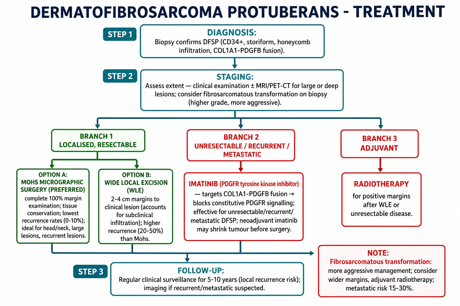
The 2024 European interdisciplinary guideline (Saiag et al., EJC 2025), the NCCN guideline for dermatofibrosarcoma, and the EADO/EORTC consensus agree on the same stepwise framework:[1][3][4][6][7]
Step 1 - Localised primary disease
Step 2 - Recurrent or unresectable disease
Step 3 - Fibrosarcomatous transformation (FS-DFSP)
Step 4 - Metastatic disease
Surgery
- Mohs micrographic surgery (MMS) - the standard of care. The technique allows complete 100 percent margin examination using horizontal frozen sections (or, increasingly, immunostained MMS with CD34 to highlight tumour cells at the margin). The combination of complete margin examination and tissue conservation makes MMS ideal for DFSP. Reported 5-year local control rates are 95-100 percent, with recurrence rates of 0-2 percent in pooled series.[1][3][5]
- Wide local excision (WLE) with 2-3 cm peripheral margins to the deep fascia (or the next anatomical barrier - periosteum, perichondrium) remains the alternative. The traditional 2-3 cm margin (not 4-5 cm as sometimes cited in older texts) reflects the current consensus. The recurrence rate after WLE is 10-20 percent at 5 years, reflecting the subclinical infiltration missed by bread-loaf sectioning. Immunostaining of the WLE specimen with CD34 at the margin is recommended to reduce the rate of false-negative margins.[1][3][4]
- Reconstruction - depends on defect size and site. Small defects can be closed primarily; larger defects need local flaps, skin grafts, or - for very large defects - free tissue transfer. The 2022 scoping review of reconstruction options for recurrent DFSP (Mago et al.) notes that the reconstructive ladder is determined by the size, site, depth, and previous surgical history; multidisciplinary planning with a reconstructive surgeon is essential for head-and-neck, acral, and recurrent disease.[6]
Imatinib (systemic targeted therapy)
- Mechanism - small-molecule tyrosine kinase inhibitor of PDGFR-alpha, PDGFR-beta, BCR-ABL, and KIT. In DFSP, the relevant target is the PDGFR-beta encoded by the PDGFB portion of the COL1A1-PDGFB fusion.
- Dose - 400 mg orally once daily (or 400 mg twice daily for resistant disease) with food and a large glass of water. The 400 mg daily dose is the registration dose for DFSP.
- Indication - unresectable, recurrent, or metastatic DFSP (FDA 2006, EMA 2006). Neoadjuvant use in large or anatomically constrained DFSP is well described but off-label in many jurisdictions.
- Efficacy - objective response rate ~50 percent; disease control (response + stable disease) ~80 percent in pooled series. Median time to response 2-3 months; median duration of response 6-24 months in older series, longer with maintenance.
- Toxicity - oedema (especially periorbital), nausea, diarrhoea, myelosuppression (neutropenia, thrombocytopenia), hepatotoxicity, fatigue, muscle cramps, hypophosphataemia. Less common: cardiac failure (rare, baseline echo recommended in cardiac comorbidity), hypothyroidism (TSH every 3 months), hepatitis B reactivation.
- Monitoring - FBC and LFTs every 2 weeks for the first 2 months, then monthly; weight, oedema, and symptom check at every visit. Continue imatinib until progression or unacceptable toxicity; consider maintenance in responders, with periodic re-imaging (every 3 months).[1][4][7]
Radiotherapy
- Adjuvant for positive margins after WLE when re-excision is not feasible, for unresectable primary disease, and as definitive therapy for inoperable FS-DFSP.
- Dose - 50-60 Gy in 2 Gy fractions (or equivalent hypofractionated schedules in palliative settings).
- Outcome - improves local control after subtotal resection; effect on overall survival is uncertain. Useful in head-and-neck DFSP where margin clearance is anatomically constrained.[5]
Follow-up
- Clinical review every 6 months for 5 years, then annually for a further 5 years (total 10 years of follow-up). Local recurrence can be very late, and the 5-10 year window is when the late recurrences present.
- MRI or ultrasound of the primary site at each review, with comparison to baseline post-operative imaging.
- CT chest annually in fibrosarcomatous, large, or recurrent disease.
- Patient education - return promptly for any new nodule at the surgical site, any change in the scar, or any new truncal plaque. Photographs with a ruler at each visit are invaluable.[1][3]
Specific Subtypes & Scenarios
- Plaque-stage DFSP - the earliest clinical form, before the protuberant phase. Often present for 10-20 years; biopsy often delayed because the lesion is small and flat. Mohs is curative.
- Fibrosarcomatous DFSP (FS-DFSP, ~10-15 percent of cases) - higher-grade areas within a DFSP; cellular atypia, >5 mitoses per 10 HPF, herringbone pattern. CD34 may be LOST in the high-grade areas (but the fusion persists). Metastatic risk 15-30 percent (versus under 5 percent in classic DFSP). Management: wider margins (2-3 cm or more), adjuvant radiotherapy, consider imatinib (response less predictable), CT chest and MRI staging.[1][3][6]
- Bednar tumour (pigmented DFSP, ~1-5 percent of cases) - melanin-laden dendritic cells scattered among the spindle cells. S100+ in the pigmented cells (not the tumour cells). More common in patients of African descent. Prognosis is identical to classic DFSP; the pigmentation is a histological curiosity, not a prognostic marker.[2]
- Myxoid DFSP - prominent myxoid stroma; can mimic myxoid liposarcoma, myxofibrosarcoma, and superficial angiomyxoma. CD34+ and t(17;22) fusion confirm the diagnosis.
- Atrophic DFSP - depressed, sclerotic plaque rather than a protuberant mass; often confused with morphea, atrophoderma, or a burn scar. CD34+ and the fusion are diagnostic.
- Giant cell fibroblastoma (GCF) of childhood - the paediatric equivalent of DFSP; shares the COL1A1-PDGFB fusion, the CD34+ immunoprofile, and the locally infiltrative behaviour. The WHO 4th edition lists GCF and DFSP as a combined entity. Paediatric presentation with a dermal/subcutaneous nodule or plaque should raise GCF as well as DFSP; biopsy and IHC close the diagnosis.[6]
- Congenital DFSP - rare; present at birth or in the first year; may be plaque-like and pigmented; biopsy often delayed because of confusion with vascular birthmark. Histology and fusion confirm.
- DFSP in pregnancy - may enlarge rapidly due to hormonal and stromal influences; imatinib is generally avoided in pregnancy (teratogenic); defer definitive surgery to the second trimester if possible, and to the postpartum period for non-urgent cases. Mohs is feasible in the second trimester with appropriate positioning and obstetric support.
- DFSP in Black skin - plaque-stage at presentation is more common (less protuberant), Bednar variant is more common, and diagnostic delay is the rule. The 2024 Alchorne review in Anais Brasileiros de Dermatologia is a high-yield reference for the cultural, clinical, and IHC features of DFSP in skin of colour.[2]
- DFSP of the vulva - rare; the MITO rare-cancer-group review (Mancari et al., 2024) summarises the limited series. Wide local excision with 1-2 cm margins and gynaecological-oncology input is the typical pathway; imatinib for unresectable or recurrent disease.
- DFSP of the face/scalp - the head and neck account for 10-15 percent of DFSPs; recurrence is higher (up to 30 percent in some series) because of the anatomic constraints on margin; Mohs is strongly preferred; adjuvant radiotherapy is used more liberally.
- Radiation-induced DFSP - rare but described; latency 10-30 years after radiotherapy; behaves like de novo DFSP; FISH for the fusion is diagnostic (and excludes post-radiation angiosarcoma, which is MYC-amplified and CD31+).
- Multiple primary DFSP - very rare; reported in the context of Gorlin syndrome, Li-Fraumeni syndrome (p53 germline), and prior radiotherapy. Full staging and genetic counselling are recommended.
Complications & Pitfalls
- Local recurrence - the dominant complication. Driven by subclinical extension that defeats standard WLE. Recurrence is most common in the first 3 years, but late recurrence (5-10+ years) is well documented. Recurrence is more frequent in head-and-neck DFSP and in fibrosarcomatous disease.
- Metastasis - less than 5 percent in classic DFSP; 15-30 percent with FS-DFSP. Lungs are the most common site (haematogenous spread); lymph nodes are involved in 5-10 percent of metastatic cases (unusually high for a "sarcoma"); bone is a late site.
- Surgical complications - wound dehiscence, infection, bleeding, nerve injury (head and neck, perineal), flap or graft failure, lymphoedema (groin and lower limb), and the need for revision reconstruction.
- Imatinib toxicity - oedema (often periorbital), nausea, diarrhoea, myelosuppression, hepatotoxicity, fatigue, muscle cramps; less commonly cardiac failure, hypothyroidism, hepatitis B reactivation. Drug interactions via CYP3A4 (avoid azoles, macrolides, grapefruit).
- Radiation-induced complications - dermatitis, hypopigmentation, telangiectasia, atrophy, fibrosis; in the head and neck: xerostomia, mucositis, taste change; rare: secondary malignancy after 10-20 years.
- Cosmetic and functional morbidity - large truncal defects, scars on visible areas, sensory change, and the psychological burden of a chronic, recurrent malignancy.
- The classic pitfalls (examiners will test these):
- Misdiagnosis as dermatofibroma - the most common diagnostic error; the lesion is on the trunk, the lesion is large, or the lesion recurs after excision. Always re-biopsy with CD34.
- Inadequate biopsy - a shave biopsy misses the deep infiltration and may miss the FS component; punch or incisional biopsy is mandatory.
- Standard WLE with 1 cm margin - inadequate; the subclinical honeycomb infiltration extends 1-3 cm beyond the visible margin in 80-90 percent of cases. Use 2-3 cm or Mohs.
- Inadequate margin assessment - bread-loaf sectioning examines under 1 percent of the margin; the rest is unchecked. Mohs or immunostained WLE is required.
- CD34 loss in the FS component - the fusion is still there; imatinib is still worth trying. Don't dismiss imatinib in FS-DFSP on the basis of CD34 loss.
- Failure to stage the lungs in FS-DFSP, recurrent disease, or large primary tumours. [1]
Prognosis & Disposition
- Local control with Mohs is 95-100 percent at 5 years; with WLE (2-3 cm margins), 80-90 percent at 5 years. Recurrence is the rule, not the exception, after inadequate surgery.
- Disease-specific survival for localised DFSP is >99 percent at 5 years and remains >95 percent at 10 years. For metastatic DFSP, 5-year survival is 10-30 percent in older series, improving with imatinib.
- Prognostic factors (in order of importance):[1][3][6]
- Fibrosarcomatous transformation - metastatic risk 15-30 percent.
- Positive margins after surgery - higher recurrence.
- Size - lesions >5 cm have higher recurrence and metastatic risk.
- Head and neck location - higher recurrence because of anatomic constraints.
- Depth - invasion of deep fascia or muscle worsens prognosis.
- Patient age and immune status - younger patients with intact immunity do better; immunocompromised patients have higher recurrence.
- Fusion variant - classic COL1A1-PDGFB predicts imatinib response; rare fusions may be less predictable.
- Disposition - sarcoma MDT for all DFSPs; plastic surgery and dermatology surgery for reconstruction; medical oncology for advanced or metastatic disease; clinical genetics for the very rare familial or syndromic cases.
- Long-term safety net - patient education on self-examination, photographic surveillance, and prompt review of any new nodule. Annual review for 10 years.
Special Populations
- Paediatric DFSP - rare; typically the plaque-stage or giant cell fibroblastoma (GCF) phenotype. Histology and fusion identical to adult DFSP. Mohs or WLE with 1-2 cm margins in children, with paediatric surgery input. Imatinib is generally avoided in young children because of growth-plate effects.
- Pregnancy - DFSP may enlarge rapidly; imatinib is teratogenic and generally avoided. Mohs surgery is feasible in the second trimester with appropriate positioning; defer definitive surgery to the second trimester or postpartum.
- Elderly - head and neck DFSP more common (sun-exposed skin); AFX and pleomorphic dermal sarcoma are the main differential; Mohs is often preferable to limit defect size; imatinib toxicity (oedema, fatigue) may be more pronounced.
- Immunocompromised - higher incidence of multiple lesions, atypical histology, and recurrence; lower threshold to biopsy; imatinib drug interactions with calcineurin inhibitors (cyclosporine, tacrolimus) need close monitoring.
- Skin of colour - higher incidence of Bednar variant; higher rate of diagnostic delay; the 2024 Alchorne review is a high-yield reference for the cultural and clinical context.[2]
- Gorlin syndrome - rare association with multiple DFSPs; PTCH1 germline mutation; consider in any patient with multiple DFSPs and additional features of the syndrome (palmar pits, odontogenic keratocysts, basal cell carcinomas).
- Anticoagulated patients - imatinib has antiplatelet effects via PDGFR inhibition on megakaryocytes; bleeding risk is modest but real. Bridge with low-molecular-weight heparin or direct oral anticoagulant per local protocol; do not stop anticoagulation for skin biopsy of a suspicious lesion.
Evidence, Guidelines & Regional Differences
The evidence base for DFSP is small (rare disease) but consistent across guidelines. The 2024 European interdisciplinary consensus (Saiag et al., EJC 2025), the 2024 EORTC/EADO consensus on rare cutaneous tumours, the 2024 NCCN guideline for dermatofibrosarcoma, and the 2024 BAD/UK guideline for skin sarcoma are the contemporary anchors.[1][6][7]
- Mohs = standard of care for localised disease
- WLE 2-3 cm to deep fascia is the alternative
- Imatinib 400 mg daily for unresectable/recurrent/metastatic
- Adjuvant RT (50-60 Gy) for positive margins or unresectable disease
- FS-DFSP: wider margins, adjuvant RT, consider imatinib, CT chest staging
- Mohs = preferred when available; WLE 2-3 cm as alternative
- MRI for lesions >5 cm or deep extension
- FISH or NGS for fusion in equivocal cases and before imatinib
- Imatinib 400 mg daily is FDA-approved
- Sarcoma-MDT discussion for all DFSPs
- 2-week-wait skin-cancer referral for suspected DFSP
- Sarcoma-MDT discussion for all cases
- Mohs or WLE 2-3 cm; CD34 immunostaining of margins
- Imatinib for advanced disease via CDF or IFR route
- 5-10 year follow-up in specialist sarcoma clinic
Landmark evidence: [1]
- The original description of the t(17;22) translocation and the COL1A1-PDGFB fusion (Simon et al., 1997; O'Brien et al., 1998) established the molecular basis of DFSP and the rationale for PDGFR-targeted therapy.
- The phase II imatinib trials in DFSP (Rutkowski et al., 2010; Stacchiotti et al., 2016) defined the 400 mg daily dose, the 50 percent objective response rate, and the 2-3 month median time to response.
- The 2024 European interdisciplinary guideline (Saiag et al., EJC 2025) is the most contemporary multi-disciplinary consensus on diagnosis and treatment.[1]
- The 2024 EORTC/EADO rare-cutaneous-tumour consensus and the 2024 Cancers (Basel) update by Jozwik et al. provide comprehensive reviews of the contemporary management of DFSP, including the role of neoadjuvant imatinib.[6]
- The 2025 review of rare fusion variants (Beyond COL1A1::PDGFB) summarises the imatinib-relevant molecular biology of the COL1A2-PDGFB, EMILIN2-PDGFB, and other rare fusions, and their clinical implications for imatinib response.[8]
- The 2024 review of imatinib in DFSP (Current Treatments and Clinical Trials, Targeted Oncology) provides the contemporary evidence base for systemic therapy in unresectable, recurrent, and metastatic disease.[7]
- The 2022 scoping review of reconstruction in recurrent DFSP (Mago et al., Indian J Plast Surg) summarises the reconstructive ladder for recurrent disease and the trade-offs between local flaps, regional flaps, and free tissue transfer.[6]
Controversies (the examiner can probe these): [1]
- Mohs vs WLE - Mohs is the standard of care, but randomised data are absent; the comparison is from retrospective series with selection bias. In a low-resource setting or in a lesion too large for Mohs, WLE with 2-3 cm margins and CD34 immunostaining is an acceptable alternative.
- Imatinib in the neoadjuvant setting - widely used off-label, but the optimal duration of neoadjuvant imatinib and the long-term outcome of imatinib-shrunk tumours are not well defined.
- Imatinib in FS-DFSP - response rates are lower than in classic DFSP; the decision to use imatinib in FS-DFSP is made on a case-by-case basis by the sarcoma MDT.
- Adjuvant imatinib after complete resection of high-risk DFSP (FS, large, head/neck) is under investigation; the DFSP-01 and related trials are exploring the benefit of adjuvant imatinib in resected high-risk disease.
- The 2-3 cm WLE margin is the contemporary standard; older texts cite 4-5 cm margins, but the modern consensus accepts 2-3 cm when combined with CD34 immunostaining of the margins.
- The role of radiotherapy in microscopically positive margins when re-excision is feasible is debated; many sarcoma MDTs will re-excise first and reserve radiotherapy for unresectable disease. [1]
Exam Pearls
[1]CUT
MOHS
PDGFB
Red Flags
[1] [1]Exam application bank (NEET-PG / INICET)
One-line answer
Dermatofibrosarcoma protuberans (DFSP) is a low-to-intermediate grade malignant fibroblastic tumour of skin, a cutaneous sarcoma of dermal fibroblast origin. Classic presentation: a slow-growing, indurated, blue-red-brown plaque or nodule on the trunk of a 30-50 year old, which may have an atrophic centre and progress to a protuberant multinodular mass. Histology: monotonous storiform (cartwheel) pattern of CD34-positive spindle cells infiltrating subcutaneous fat in a honeycomb pattern. IHC: CD34+ (strong, diffuse), Factor XIIIa- (versus dermatofibroma, which is CD34- and Factor XIIIa+). Molecular hallmark: COL1A1-PDGFB fusion from t(17;22) or a supernumerary ring chromosome r(17;22) - constitutive PDGFR activation - the target of imatinib. Mohs micrographic surgery is the standard of care (1 percent recurrence) versus wide local excision with 2-3 cm margins (10-20 percent recurrence).
Worked stems (answer without another resource)
Stem 1 — Classic presentation. Map symptoms to mechanism; name the first investigation and first treatment step with dose/route if drug therapy is standard. [1]
Stem 2 — Unstable / complicated. List red flags that force immediate resuscitation, theatre, ICU, antidote, or reperfusion — and what you do in the first 15 minutes. [1]
Stem 3 — Atypical group. Elderly, pregnancy, child, or immunocompromised: how presentation and thresholds change. [1]
Stem 4 — Differential trap. Name the three closest mimics and one discriminator for each. [1]
Stem 5 — Disposition. Who goes home with safety-netting, who is admitted, who needs HDU/ICU/theatre, and what follow-up is mandatory. [1]
Rapid viva checklist
- Definition + classification
- Pathophysiology chain
- Bedside signs / criteria
- Score with exact components (if any)
- Emergency bundle
- Definitive therapy with doses
- Complications of disease and of treatment
- Special populations
- Guideline/trial name if classic
- Three exam traps
Coverage self-check
If you cannot answer any stem above from this page alone, re-read the matching section — the page is intended to be self-sufficient for final-prof and NEET-PG/INICET questions on Dermatofibrosarcoma protuberans.
[1]References
- [1]Saiag P, Lebbe C, Brochez L, et al. Diagnosis and treatment of dermatofibrosarcoma protuberans. European interdisciplinary guideline - update 2024 Eur J Cancer, 2025.PMID 39904126
- [2]Alchorne MMA, Conceição KDC, Barraza LL, et al. Dermatology in black skin An Bras Dermatol, 2024.PMID 38310012
- [3]Allen A, Ahn C, Sangüeza OP. Dermatofibrosarcoma Protuberans Dermatol Clin, 2019.PMID 31466588
- [4]Acosta AE, Vélez CS. Dermatofibrosarcoma Protuberans Curr Treat Options Oncol, 2017.PMID 28795284
- [5]Tillman BN, Liu JC. Cutaneous Sarcomas Otolaryngol Clin North Am, 2021.PMID 33602520
- [6]Jozwik M, Bednarczuk K, Rutkowski P, et al. Dermatofibrosarcoma Protuberans: An Updated Review of the Literature Cancers (Basel), 2024.PMID 39335097
- [7]Remiszewski P, et al. Dermatofibrosarcoma Protuberans (DFSP): Current Treatments and Clinical Trials Curr Treat Options Oncol, 2025.PMID 41042442
- [8]Das S, et al. Beyond COL1A1::PDGFB: Rare fusions and their clinical implications in dermatofibrosarcoma protuberans World J Clin Cases, 2025.PMID 41356086