Dermatology · Medicine
Dermatological emergencies
Also known as Dermatological emergencies · Dermatologic emergencies · Acute dermatology · Emergency dermatology · Severe cutaneous adverse reactions
Dermatological emergencies are acute skin eruptions in which delayed recognition causes death or permanent disability. The catalogue spans (1) severe cutaneous adverse drug reactions — SJS/TEN (skin detachment with mucosal involvement; drug trigger 1-8 weeks; STOP drug; ICU/burns; SCORTEN), DRESS/DIHS (drug 2-8 weeks; fever, rash, eosinophilia, lymphadenopathy, hepatitis; HHV-6 reactivation; RegiSCAR; systemic corticosteroids), AGEP (drug under 4 days; sterile pustules; self-limiting); (2) erythroderma/exfoliative dermatitis (over 90% BSA erythema and scaling; fluid, protein and heat loss; high-output cardiac failure); (3) staphylococcal scalded skin syndrome (children under 5; staphylococcal exfoliative toxin; superficial split; mucosa spared); (4) necrotising fasciitis (pain out of proportion; surgical emergency; LRINEC); (5) purpura fulminans (DIC with skin necrosis; meningococcaemia; protein C/S deficiency); (6) generalised pustular psoriasis (von Zumbusch) (sterile pustules, fever; acitretin/ciclosporin/infliximab); (7) angioedema/anaphylaxis (histamine vs bradykinin; IM adrenaline vs C1-inhibitor/icatibant). The universal principle is ABCDE, recognise, refer, and resuscitate.
On this page & tools
Your progress
Saved locally on this device.
Exam tags
Red flags
Overview & Definition
A dermatological emergency is any acute cutaneous eruption in which the skin fails as an organ — losing its barrier, thermoregulatory, and immune functions — or in which a cutaneous sign is the herald of a rapidly lethal systemic process such as septic shock, anaphylaxis, or disseminated intravascular coagulation. The unifying concept is acute skin failure: like acute respiratory or renal failure, the skin can fail, and when it fails across a large body surface area the consequences are fluid and electrolyte loss, protein loss, hypothermia, sepsis, and high-output cardiac failure. The examiner's distinction that earns marks is between a bad rash (uncomfortable but self-limiting) and a skin emergency (death or permanent disability within hours to days if not recognised).[1]
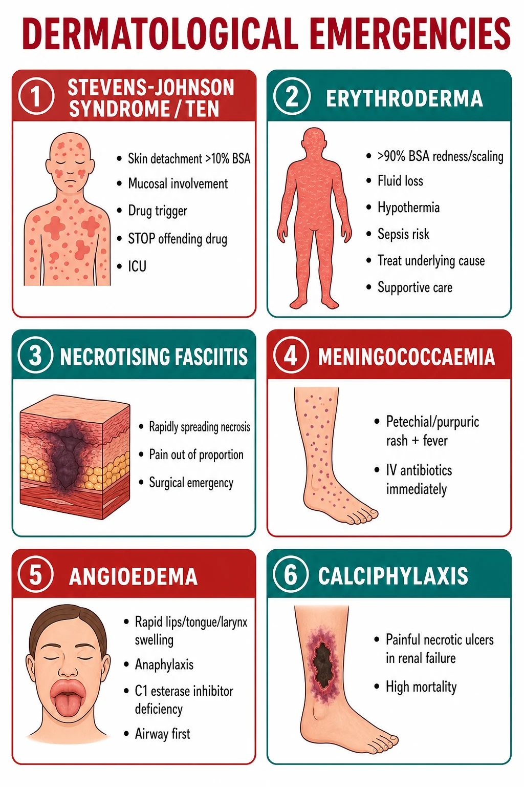
The conditions group naturally into four mechanisms. Severe cutaneous adverse drug reactions (SJS/TEN, DRESS, AGEP) are T-cell-mediated drug hypersensitivity syndromes. Skin-failure states (erythroderma, staphylococcal scalded skin syndrome, generalised pustular psoriasis) involve loss of epidermal integrity across a large surface area. Infective and necrotising disorders (necrotising fasciitis, purpura fulminans/meningococcaemia) involve rapidly progressive tissue destruction and systemic sepsis. Immediate hypersensitivity and vascular disorders (angioedema/anaphylaxis, calciphylaxis) involve mediator-driven oedema or small-vessel occlusion. A fifth thread — immunobullous and autoimmune emergencies such as pemphigus vulgaris and dermatomyositis — is covered in dedicated topics. [1]
Two principles govern the whole topic and should frame every answer. First, a febrile patient with a rash and systemic upset is assumed to have a dermatological emergency until proven otherwise — the cost of missing meningococcaemia or TEN is death, while the cost of over-calling a viral exanthem is a brief admission. Second, recognition and resuscitation precede diagnosis: the immediate actions (ABCDE, stop new drugs, fluid resuscitation, IV antibiotics for purpura with fever, surgical referral for necrosis, airway protection for angioedema) are taken on the clinical pattern alone, before biopsy, cultures, or specialist review confirm the specific entity. The dermatologist is called early but does not delay the first hour of care.[1][2]
Classification
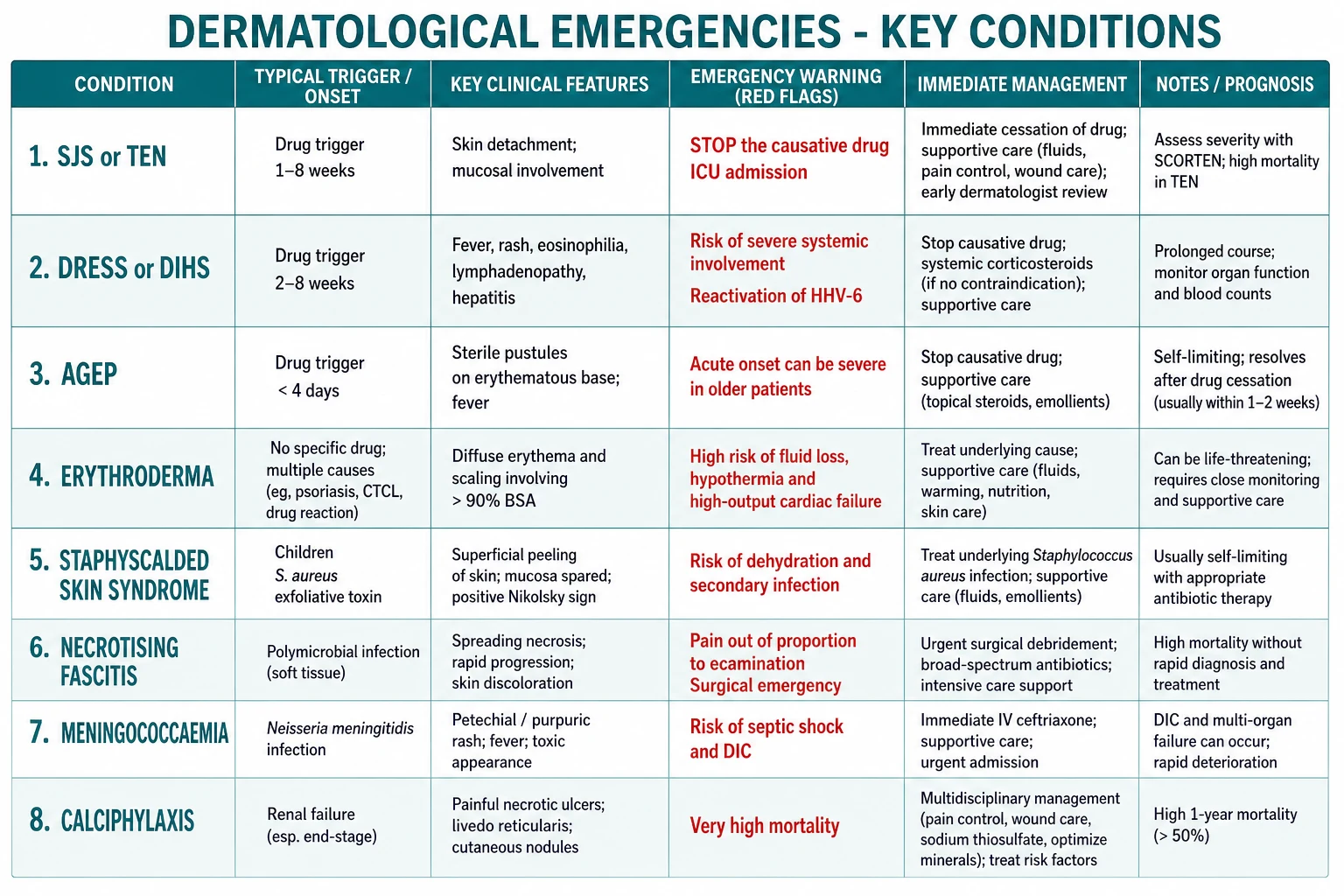
Severe cutaneous adverse reactions (SCAR)
- Drug-triggered T-cell reactions
- SJS/TEN, DRESS, AGEP
- Onset days to weeks after drug
- STOP the culprit drug is treatment step one
Acute skin failure
- Loss of barrier across large BSA
- Erythroderma, SSSS, von Zumbusch
- Fluid, protein, heat loss
- Manage like a burn: ICU, fluids, temperature
Infective / necrotising
- Rapid tissue destruction + sepsis
- Necrotising fasciitis, purpura fulminans
- Time-critical antibiotics and surgery
- Mortality 20-40% or higher
Immediate hypersensitivity / vascular
- Mediator-driven oedema or vessel occlusion
- Angioedema, anaphylaxis, calciphylaxis
- Adrenaline (histamine) or C1-INH/icatibant (bradykinin)
- Airway is the immediate threat
Epidemiology & Risk Factors
The epidemiology matters because the high-risk drugs, host factors, and mortality figures are exactly what examiners probe. SJS and TEN are rare but feared: combined incidence is roughly 1 to 6 per million person-years, with drugs implicated in over 80% of cases and mortality rising steeply with the percentage of detached epidermis.[10] DRESS is an order of magnitude more common, with an estimated incidence of 1 per 1000 to 10000 drug exposures and a case-fatality of 5 to 10%, most often from fulminant hepatitis.[7]
Staphylococcal scalded skin syndrome overwhelmingly affects neonates and children under 5 years, where mortality is around 4%; in adults — typically those with renal failure or immunocompromise — the mortality climbs to over 50% because the toxin cannot be renally cleared.[4] Necrotising fasciitis incidence is rising, driven by diabetes, obesity, immunosuppression, intravenous drug use, and ageing populations; mortality ranges from 20 to 40% and is halved by surgical debridement within 24 hours.[11] Erythroderma shows a male preponderance with a mean onset age over 40 years, and an underlying cause is eventually identified in roughly 70% — a pre-existing dermatosis (psoriasis, eczema), a drug, or a malignancy such as cutaneous T-cell lymphoma (Sézary syndrome) — leaving up to 30% idiopathic.
The principal risk factors for a severe drug reaction are: a personal or family history of drug hypersensitivity; specific HLA alleles (HLA-B*15:02 with carbamazepine in Han Chinese and South-East Asians; HLA-B*58:01 with allopurinol); active viral infection (HIV, EBV, CMV) which lowers the reaction threshold; renal or hepatic impairment which prolongs drug exposure; and high cumulative drug dose.[6]
Pathophysiology
Each emergency has a distinct mechanism, and the examiner rewards the candidate who can name the molecular target rather than say "it is an allergic reaction." [1]
Stevens-Johnson syndrome and toxic epidermal necrolysis are drug-induced, full-thickness keratinocyte apoptosis. The dominant effector is granulysin, a cytotoxic molecule released by drug-specific CD8+ T cells and natural killer cells; Fas-Fas ligand interactions, perforin/granzyme, and tumour necrosis factor also contribute. The result is confluent epidermal necrosis with a subepidermal split, leaving a denuded, bleeding dermis analogous to a thermal burn.[10]
Staphylococcal scalded skin syndrome (SSSS) is toxin-mediated, not T-cell-mediated. Phage group II Staphylococcus aureus at a distant focus (often conjunctivitis, otitis, or a wound) produces exfoliative toxin A or B, which circulates haematogenously and specifically cleaves the extracellular domain of desmoglein-1 in the stratum granulosum. The split is therefore high and superficial — only the uppermost epidermis separates. Because desmoglein-3 compensates in mucosal epithelium, the mucosae are spared, which is the cardinal distinction from SJS/TEN.[4][5]
DRESS/DIHS is a systemic drug hypersensitivity with a characteristic 2 to 8 week latency. The mechanism combines drug-specific T-cell activation, a defect in detoxification of the drug's reactive metabolites (relevant for sulphonamides and anticonvulsants), and reactivation of human herpesviruses — most consistently HHV-6, but also CMV and EBV — which drives the biphasic, relapsing course and the eosinophilia.[7]
AGEP is the fastest-onset severe cutaneous drug reaction. Drug-specific CD4+ T cells home to the epidermis and release the cytokine IL-8 (CXCL8), a potent neutrophil chemoattractant; IL-8 and granulocyte-macrophage colony-stimulating factor recruit neutrophils that fill the subcorneal space as sterile pustules. The cleavage is subcorneal and superficial, the pustules are non-follicular and culture-negative, and the lack of deep keratinocyte necrosis explains the low mortality and rapid resolution once the drug is withdrawn.[2]
Erythroderma is a failure of epidermal turnover and barrier function. The skin vasodilates massively over a large surface area, producing a continuous high insensible loss of water, electrolytes, and protein; heat loss causes shivering and hypothermia; and the persistent cutaneous vasodilation generates a hyperdynamic circulation that can precipitate high-output cardiac failure, especially in the elderly and those with underlying cardiac disease.[8]
Necrotising fasciitis is a spreading infection of the subcutaneous tissue and deep fascia that spares (initially) the overlying muscle and skin. Three microbiological types are recognised: Type I is polymicrobial (anaerobes plus facultative Gram-negatives), typically after surgery or in diabetics; Type II is monomicrobial Group A streptococcal (± staphylococcal) infection, classically after minor trauma; and Type III involves marine organisms such as Vibrio vulnificus or Aeromonas after seawater exposure. The tissue destruction is driven both by direct bacterial invasion and by bacterial exotoxins (streptococcal pyrogenic exotoxins) that produce a toxic-shock phenotype.[11]
Purpura fulminans is disseminated intravascular coagulation localised to the dermal microvasculature. Microthrombi occlude dermal capillaries and venules, producing haemorrhagic infarction of the skin with a characteristic retiform (net-like or stellate) pattern that progresses to full-thickness necrosis. The three settings are neonatal homozygous protein C deficiency, acute infectious (overwhelming meningococcaemia, pneumococcaemia, or varicella), and idiopathic post-infectious with acquired protein S deficiency.[13]
Hereditary angioedema is a deficiency (Type I, 85%) or dysfunction (Type II, 15%) of C1 esterase inhibitor. Unopposed activation of the contact (kallikrein-kinin) pathway generates excess bradykinin, which is the dominant mediator of the oedema. Because the swelling is bradykinin-mediated rather than histamine-mediated, conventional anaphylaxis therapy — adrenaline, antihistamines, and corticosteroids — is largely ineffective; the specific antidotes target the bradykinin pathway.[15][16]
Clinical Presentation
Erythroderma (exfoliative dermatitis)
The definition is erythema and scaling involving over 90% of the body surface area, with an abrupt or insidious onset. Patients complain of shivering, feeling cold, and tightness of the skin; on examination the skin is diffusely red, warm, and scaling, with varying degrees of oedema, fissuring, and palmoplantar keratoderma. Lymphadenopathy is common (dermopathic lymphadenopathy), and ectropion and alopecia may accompany chronic disease. The acute danger signs are tachycardia, hypotension, oliguria (hypovolaemia), confusion (hypothermia or sepsis), and respiratory distress (high-output cardiac failure).[8]
Stevens-Johnson syndrome / toxic epidermal necrolysis
A flu-like prodrome (fever, malaise, upper respiratory symptoms) is followed by a macular, targetoid, or purpuric rash that coalesces and then detaches. The classification is by percentage of detached epidermis: SJS under 10% BSA, SJS-TEN overlap 10 to 30%, and TEN over 30%. Mucosal involvement — oral, conjunctival, tracheobronchial, genital, and urethral — is present in over 90% and is the single most reliable discriminator from SSSS. Positive Nikolsky and Asboe-Hansen signs indicate the plane of cleavage has extended. Oedema, fever, skin pain, and skin tenderness are prominent; the patient looks systemically unwell.[10]
DRESS / DIHS
The presentation is a triad of fever, rash, and internal organ involvement beginning 2 to 8 weeks after drug exposure. The rash is typically a morbilliform or exanthematous eruption that becomes infiltrated and oedematous, with a characteristic involvement of the face (facial oedema is a clue). Generalised lymphadenopathy is common, and visceral involvement drives the severity: hepatitis (the leading cause of death), interstitial nephritis, pneumonitis, myocarditis, and colitis. Eosinophilia (often over 1.0 x 10^9/L) and atypical lymphocytes support the diagnosis. The course is often biphasic, with relapse as the corticosteroid is tapered, reflecting ongoing viral reactivation.[7]
AGEP (Acute Generalised Exanthematous Pustulosis)
Hundreds of small, sterile, non-follicular pustules erupt on an erythematous base within under 4 days of drug exposure (penicillins, macrolides). Fever and leucocytosis accompany the rash; mucosal involvement is minimal or absent; and systemic organ involvement is uncommon. The condition is usually self-limiting once the drug is withdrawn, resolving with desquamation over 1 to 2 weeks.[2] The EuroSCAR/AGEP validation score helps distinguish AGEP from pustular psoriasis: rapid onset (under 4 days), a typical culprit drug, fever, hundreds of small non-follicular pustules on an erythematous base, and a neutrophilic rather than lymphocytic infiltrate on biopsy all score positively; a higher score supports AGEP over pustular psoriasis. Management is withdrawal of the culprit drug (penicillins, macrolides, pristinamycin, terbinafine, diltiazem, hydroxychloroquine) and supportive care — antipyretics, fluids, and topical corticosteroids for symptomatic relief; systemic corticosteroids are reserved for extensive or refractory disease. Resolution with desquamation over 5 to 14 days is the rule, and mortality is under 5%, far lower than SJS/TEN or DRESS.[2]
Staphylococcal scalded skin syndrome
In an infant or young child (classically under 5 years), a focal staphylococcal infection (conjunctivitis, otitis media, umbilical or wound infection) is followed by the systemic spread of exfoliative toxin. The sequence is: diffuse tender erythema, then large flaccid bullae that rupture easily, then sheet-like peeling leaving a moist, glistening, scalded-appearing surface. The Nikolsky sign is positive. The mucosae are spared — oral, conjunctival, and genital mucosae are unaffected — because desmoglein-3 maintains mucosal integrity.[4][5]
Necrotising fasciitis
The red flag is pain that is grossly disproportionate to the visible skin signs. Early, the skin may show only mild erythema and oedema indistinguishable from cellulitis; over hours the erythema spreads rapidly, the skin becomes dusky and indurated, bullae form (haemorrhagic bullae are ominous), and cutaneous anaesthesia develops (infarction of cutaneous nerves). Crepitus may be present in gas-forming infection. Systemically, the patient develops high fever, tachycardia, hypotension, altered mental state, and multi-organ failure — a toxic-shock picture out of proportion to the visible skin lesion.[11]
Purpura fulminans / meningococcaemia
The rash begins as small petechiae and rapidly coalesces into retiform (net-like, geographic) purpura with sharply demarcated, dusky edges that progress to frank cutaneous necrosis, especially on the extremities. This is accompanied by fever, shock, and signs of DIC. In meningococcal disease there may be meningism (neck stiffness, photophobia, altered consciousness) but meningococcaemia can occur without meningitis. The rash with fever is the trigger to treat now, not to investigate.[13]
Generalised pustular psoriasis (von Zumbusch)
An explosive eruption of widespread, sterile, pinpoint pustules on an intensely erythematous skin, accompanied by high fever, rigors, malaise, and marked leukocytosis. The patient is erythrodermic and unwell; the pustules coalesce into lakes of pus and then desquamate. There may be a history of chronic plaque psoriasis, and precipitants include systemic corticosteroid withdrawal, infections, pregnancy (impetigo herpetiformis), hypocalcaemia, and irritating topical therapy.[14]
Angioedema / anaphylaxis
Rapidly progressive, non-pitting swelling of the deep dermis and subcutis, most dangerous when it involves the lips, tongue, floor of mouth, and larynx (stridor, drooling, respiratory distress). Histamine-mediated angioedema (allergic/anaphylaxis) is accompanied by urticaria, itching, and other features of systemic allergy (bronchospasm, hypotension, gastrointestinal symptoms). Bradykinin-mediated angioedema (hereditary, acquired, ACE-inhibitor) causes episodic swelling without urticaria, pruritus, or hypotension, and tends to be slower in onset but more refractory to standard therapy — the recurrent, unexplained, non-urticarial swellings are the clue.[15][16]
Toxic shock syndrome and staphylococcal streptococcal toxic illness
Toxic shock syndrome (TSS) is a toxin-mediated superantigen emergency, not a direct skin infection. Menstrual TSS (tampon-associated, Staphylococcus aureus toxic shock syndrome toxin-1) and non-menstrual TSS (post-surgical, post-burn, skin and soft tissue foci) present with the abrupt onset of high fever, hypotension, a diffuse sunburn-like macular erythroderma, conjunctival and mucosal hyperaemia, strawberry tongue, and multi-organ failure (gastrointestinal, hepatic, renal, muscular, central nervous system). Desquamation of the palms and soles follows in 1 to 2 weeks. Streptococcal toxic shock syndrome (Group A streptococcus, often with necrotising fasciitis) is clinically similar but bacteremic and rapidly progressive. Management is aggressive fluid resuscitation, source control (remove tampon, drain focus), IV clindamycin (to suppress toxin synthesis) plus a beta-lactam, IV immunoglobulin for severe disease, and critical care support.[1][2]
Differential Diagnosis
The differentials cluster around a small number of high-yield discriminating questions: is the mucosa involved? What is the cleavage plane? What is the latency from drug to rash? Is there systemic toxicity? [1]
SSSS
- Children under 5
- Superficial split (stratum granulosum)
- Mucosa SPARED
- Staphylococcal toxin; Nikolsky positive
- IV flucloxacillin
SJS/TEN
- Any age, drug exposure
- Full-thickness epidermal necrosis
- Mucosa INVOLVED
- Drug-specific T cells; Nikolsky positive
- STOP drug; ICU/burns
Bullous impetigo
- Localised, not generalised
- Same toxin, local effect
- Mucosa spared
- Culture positive for S. aureus
- Topical/oral antibiotic
Pemphigus foliaceus
- Chronic, not acute
- Superficial split (subcorneal)
- Mucosa spared
- Autoantibodies to desmoglein-1
- Corticosteroids
DRESS
- Onset 2-8 weeks after drug
- Fever + rash + eosinophilia + lymphadenopathy
- Hepatitis, nephritis
- HHV-6 reactivation
- Systemic corticosteroids; slow taper
AGEP
- Onset under 4 days after drug
- Sterile pustules on erythema
- Minimal systemic involvement
- Self-limiting after drug cessation
- Supportive care
Viral exanthem
- No eosinophilia, no internal organ involvement
- Self-limiting
- Often paediatric
- Symmetrical, acral or centripetal
- Symptomatic
Serum sickness
- Onset 1-2 weeks after exposure
- Urticaria + arthralgia + fever
- Immune-complex mediated
- Low complement
- Self-limiting or corticosteroids
For a rapidly spreading, painful red leg, the differential is cellulitis (slow, well-demarcated, painful but not systemically toxic), erysipelas (sharply demarcated, raised), gas gangrene (clostridial myonecrosis, severe systemic toxicity, crepitus, bronze discoloration), and calciphylaxis (renal failure, painful necrotic ulcers with livedoid edges, not infectious). The discriminator for necrotising fasciitis is the combination of severe pain out of proportion, rapid progression, and systemic toxicity.[11]
For a pustular eruption, distinguish AGEP (drug, under 4 days, sterile, self-limiting), von Zumbusch pustular psoriasis (background psoriasis, fever, erythroderma, systemic retinoid), subcorneal pustular dermatosis (Sneddon-Wilkinson, annular/polycyclic, flexural, older women), and IgA pemphigus (autoimmune, chronic). For erythroderma, the cause categories — in roughly equal thirds — are pre-existing dermatosis (psoriasis, eczema, pityriasis rubra pilaris), drug reaction, and underlying disease (CTCL/Sézary, pemphigus foliaceus, ichthyosiform disorders), with idiopathic the remainder. [1]
Clinical & Bedside Assessment
Every suspected dermatological emergency is assessed first by ABCDE, then by a focused skin examination. The airway is the immediate threat in angioedema with laryngeal involvement and in SJS/TEN with tracheobronchial sloughing. Breathing may be compromised by bronchospasm (anaphylaxis) or by the hyperdynamic/pulmonary oedema of erythroderma. Circulation is threatened by hypovolaemia (erythroderma, SSSS, SJS/TEN all lose fluid through the denuded skin) and by distributive shock (necrotising fasciitis, purpura fulminans). Disability reflects hypothermia or septic encephalopathy. Exposure is the dermatological examination itself. [1]
[1]The Nikolsky sign is performed by sliding a firm finger or cotton-tipped applicator laterally on normal-appearing skin; superficial shearing or sloughing is positive, indicating a cleavage plane high in the epidermis (SSSS) or full-thickness necrosis (SJS/TEN). In necrotising fasciitis, examine for the "hardwood" induration of the skin, loss of the normal subcutaneous softness, and the "woody hard" subcutaneous tissue that distinguishes it from cellulitis. Look for portal of entry (trauma, surgical wound, IV cannula, fissure). Palpate for crepitus (gas). [1]
In suspected DRESS, examine the face for oedema, palpate all lymph node groups, and examine the abdomen for hepatomegaly. In suspected HAE, ask about a family history, recurrent unexplained abdominal pain (bowel wall oedema), and previous episodes triggered by dental work or trauma; measure the swelling velocity. [1]
Investigations
Investigations support, but never delay, treatment in the time-critical conditions. The named scores below are reproduced verbatim because they are the examinable material. [1]
SCORTEN (severity of illness for TEN)
Calculated at 24 hours and again at 72 hours after admission. Each criterion scores one point; the sum predicts mortality.[9]
[1]RegiSCAR criteria (DRESS/DIHS diagnosis)
A scoring system that classifies a case as definite, probable, possible, or no DRESS based on the pattern of features. The components are: fever (over 38.5 degrees C); rash (extent over 50% BSA, oedema, infiltration, purpura, desquamation); eosinophilia (over 0.7 x 10^9/L, or over 10% if leucopenic) or atypical lymphocytes; enlarged lymph nodes (at least two sites, over 1 cm); internal organ involvement (hepatitis, interstitial nephritis, interstitial pneumonitis, myocarditis); resolution of the suspected drug within 15 days (or longer if prolonged); and exclusion of infectious and autoimmune causes.[6]
LRINEC score (laboratory risk indicator for necrotising fasciitis)
A score to raise or lower the suspicion of necrotising soft tissue infection when the clinical picture is ambiguous. A score of 6 or more suggests necrotising fasciitis; a score of 5 or less does not exclude it — clinical judgement overrides the score, and a low score must never delay surgery in a toxic patient.[11][12]
Biopsy and laboratory
In SSSS, a skin biopsy with frozen section shows a superficial intraepidermal split at the stratum granulosum with no necrosis, no inflammation, and no organisms — distinguishing it from SJS/TEN (full-thickness necrosis) and bullous impetigo (subcorneal pustules with organisms). In SJS/TEN, biopsy shows full-thickness epidermal necrosis with a sparse inflammatory infiltrate; a shave or punch biopsy is performed but should not delay treatment. [1]
In DRESS, the laboratory phenotype drives both diagnosis and monitoring. Send a full blood count with differential (eosinophilia over 0.7 x 10^9/L, or over 10% if leucopenic; atypical lymphocytes), liver function tests (ALT and AST over 2 to 5 times the upper limit of normal support the diagnosis; a rise in bilirubin and INR signals fulminant hepatitis), renal function and urinalysis (interstitial nephritis with sterile pyuria and eosinophiluria), and creatine kinase and troponin if myocarditis is suspected. Where available, quantitative HHV-6, CMV, and EBV PCR on whole blood should be sent at baseline and weekly: rising titres confirm the diagnosis, predict a biphasic relapsing course, and warn of imminent flare as corticosteroids are tapered. Recheck the full blood count and liver function every 2 to 3 days during the acute phase, then weekly during the long corticosteroid taper.[7]
In necrotising fasciitis, the operative finding is the investigation: a foul-smelling "dishwater" fluid, grey necrotic fascia that separates easily from underlying tissue (positive "finger test"), thrombosis of perforating vessels, and lack of bleeding from the fascia. Tissue and deep wound cultures (not superficial swabs) guide directed antibiotic therapy. Blood cultures are positive in up to 25% of Type II streptococcal disease. Imaging (plain X-ray, CT, or MRI for subcutaneous gas, fascial thickening, or fluid) is reserved for the haemodynamically stable patient with an ambiguous presentation — in a classic toxic patient, theatre is the investigation.[11]
In purpura fulminans, send coagulation (PT, APTT, fibrinogen, D-dimer), a full blood count with blood film (schistocytes suggest microangiopathy), blood cultures (meningococcus, pneumococcus), and protein C, protein S, and antithrombin levels (drawn before transfusion if possible, since they guide replacement therapy). A skin biopsy shows dermal microvascular thrombosis with full-thickness cutaneous infarction and scant inflammation — the paucity of inflammation distinguishes purpura fulminans from leukocytoclastic vasculitis.[13]
Rapid Assessment Algorithm
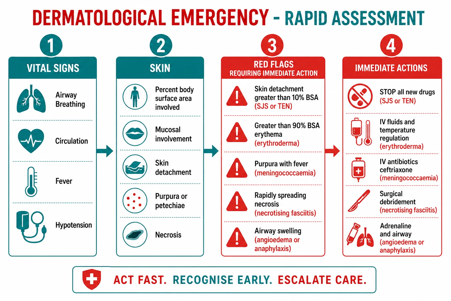
1. ABCDE
Secure airway (angioedema, SJS); oxygen; two large-bore cannulae; fluids if shocked.
2. Vitals
Temperature, heart rate, blood pressure, oxygen saturation, glucose; sepsis screen.
3. Skin
Estimate BSA, detachment percent, mucosal involvement, morphology (purpura, pustules, bullae, necrosis); Nikolsky sign.
4. STOP drugs
Stop all drugs started within the last 8 weeks — the single most useful intervention in SCAR.
5. Named score
SCORTEN (TEN), RegiSCAR (DRESS), LRINEC (necrotising fasciitis) — calculate and act on the result.
6. Definitive action
IV ceftriaxone for purpura with fever; surgical debridement for necrotising fasciitis; ICU/burns for TEN; dermatology referral for all.
Management — Resuscitation
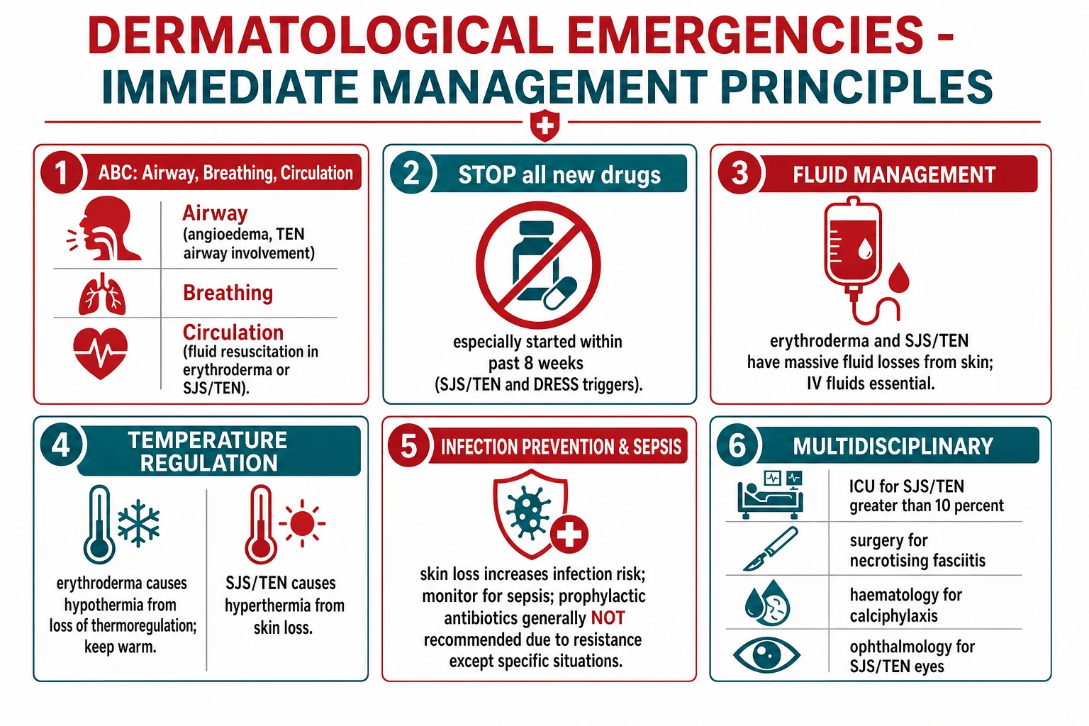
The resuscitation phase is identical in principle across all dermatological emergencies and is what the patient is dying of while you make the diagnosis. [1]
A — Airway: angioedema with stridor, drooling, or tongue swelling needs early anaesthetic and ENT involvement; prepare for awake fibre-optic intubation or surgical airway. In SJS/TEN with tracheobronchial involvement, anticipate a difficult, bleeding airway. [1]
B — Breathing: high-flow oxygen; bronchodilators for bronchospasm (anaphylaxis); monitor for pulmonary oedema in erythroderma. [1]
C — Circulation: two large-bore IV cannulae. In erythroderma, SSSS, and SJS/TEN, fluid losses through denuded skin are enormous and equivalent to a burn of the same surface area — resuscitate with warmed crystalloid guided by urine output (over 1 mL/kg/h in adults; over 1 mL/kg/h in children) and haemodynamics. In necrotising fasciitis and purpura fulminans, treat distributive and septic shock with crystalloid boluses, vasopressors (noradrenaline), and early IV antibiotics. In anaphylaxis, give IM adrenaline 0.5 mg (500 micrograms) into the anterolateral thigh, repeated at 5 minute intervals if no response, with IV fluid support.[2]
D — Disability: correct hypothermia (warmed fluids, forced-air warming, ambient temperature up); glucose; treat seizures. [1]
E — Exposure and Environment: fully expose the skin (then cover to preserve heat), examine, and document BSA and detachment. Stop all new drugs started within 8 weeks. Decubitus care, eye care (ophthalmology within hours for SJS/TEN), and catheterisation (SJS/TEN with urethral involvement) are part of resuscitation, not an afterthought. [1]
Management — Definitive & Stepwise
SJS / TEN
SJS/TEN bundle
The Cochrane review of systemic interventions for SJS/TEN found no agent — corticosteroids, IVIG, ciclosporin, plasmapheresis, thalidomide — with proven mortality benefit, and randomised trial evidence is sparse; treatment remains supportive with centre-specific immunomodulation.[10] Prophylactic antibiotics are not given (they do not prevent sepsis and increase resistance); treat documented infection.
DRESS / DIHS
Stop the drug. Start systemic corticosteroids — oral prednisolone 1 mg/kg/day (or equivalent IV methylprednisolone 0.5-1 mg/kg/day if severe) — and plan a slow taper over weeks to months because relapse is common as HHV-6 reactivates. Treat organ failure supportively (hepatitis, renal failure, pneumonitis). In corticosteroid-refractory or corticosteroid-dependent disease, ciclosporin, mycophenolate, or IVIG have been used. Re-challenge is absolutely contraindicated. The taper should be guided by eosinophil count, liver function, and clinical signs; consider a switch to a steroid-sparing agent at 4 to 6 weeks.[7]
Staphylococcal scalded skin syndrome
SSSS bundle
Skin generally heals within 1 to 2 weeks without scarring because the split is superficial. Adults with SSSS have a much worse prognosis and need ICU-level care and renal-dose-adjusted antibiotics to clear the toxin.[4]
Erythroderma
Admit, ideally to a warm side-room or ICU depending on severity. Treat the underlying cause. Fluid and electrolyte management is central: IV crystalloid to replace ongoing losses, with daily weighing and urine output monitoring. Temperature regulation: keep the ambient temperature high (over 28 degrees C), use warmed fluids and forced-air warming, and avoid overheating (which worsens the hyperdynamic circulation). Apply emollients frequently and mild to mid-potency topical corticosteroids (avoid potent corticosteroids over large areas due to systemic absorption). Monitor for and treat sepsis (blood cultures, antibiotics for documented infection); thromboprophylaxis with low-molecular-weight heparin is indicated because erythroderma is a prothrombotic state. High-output cardiac failure is treated by fluid balance and diuretics.[8]
Necrotising fasciitis
This is a surgical emergency. The definitive treatment is emergency surgical debridement of all necrotic tissue down to bleeding, healthy muscle and fascia, with a low threshold for re-look debridement at 24 to 48 hours. Antibiotics are adjunctive and started immediately: empiric broad-spectrum cover such as piperacillin-tazobactam 4.5 g IV eight-hourly plus clindamycin 600 mg IV eight-hourly (to suppress toxin production) for polymicrobial disease, or benzylpenicillin plus clindamycin for Group A streptococcal disease; add vancomycin if MRSA is possible. Intravenous immunoglobulin and hyperbaric oxygen are adjuncts used in some centres without strong evidence. Vasopressor and ventilatory support as needed for septic shock.[11]
Purpura fulminans
Treat the underlying cause aggressively. For meningococcal disease give IV ceftriaxone 2 g (adult) or 50-100 mg/kg (child) immediately — do not wait for investigations. Supportive care for DIC and shock: fresh frozen plasma to replace consumed clotting factors and protein C/S, protein C concentrate or activated protein C where available, and antithrombin in some protocols. Heparin infusion is used in some centres. The necrotic skin requires surgical assessment for fasciotomy, escharotomy, and eventual debridement and grafting; amputation rates are high in survivors.[13]
Generalised pustular psoriasis (von Zumbusch)
Admit; this is a dermatological emergency with mortality from sepsis, hypocalcaemia, and high-output cardiac failure. Systemic retinoid — acitretin 0.5-1 mg/kg/day — is first-line and acts rapidly. Alternatives where rapid control is needed are ciclosporin 3-5 mg/kg/day (fast onset, useful in pregnancy where retinoids are contraindicated) or infliximab 5 mg/kg at weeks 0, 2, and 6 (very rapid, used for refractory disease). Avoid topical irritants, coal tar, and systemic corticosteroids (withdrawal precipitates pustular flares). Treat precipitants (infection, withdraw offending drugs), correct hypocalcaemia, monitor fluid balance, and give thromboprophylaxis.[14]
Angioedema / anaphylaxis
Angioedema by mechanism
The critical decision is whether the swelling is histamine- or bradykinin-mediated, because the treatments are entirely different. Adrenaline works for histamine-mediated anaphylaxis; it does not work for hereditary angioedema, which requires a bradykinin-pathway antagonist. Giving escalating adrenaline to refractory angioedema without urticaria or hypotension is a classic error — think HAE and reach for icatibant or C1-INH concentrate.[15][16]
Calciphylaxis (calcific uraemic arteriolopathy)
Included here as a vascular dermatological emergency in the dialysis patient: painful, stellate-livedoid, necrotic skin ulcers over areas of subcutaneous fat (abdomen, thighs, breasts) with calcification of small subcutaneous vessels on biopsy. Mortality is 50 to 80% at one year, usually from sepsis. Management is wound care, optimisation of calcium-phosphate metabolism (lower calcium-based phosphate binders, use a non-calcium binder, adequate dialysis), sodium thiosulphate IV during dialysis, and aggressive treatment of any infection. There is no proven cure.[2]
Specific Subtypes & Scenarios
Erythroderma by aetiology. The dermatosis group (psoriasis, eczema/atopic dermatitis, pityriasis rubra pilaris) is treated by intensifying or adjusting the underlying therapy (methotrexate, ciclosporin, or a biologic for psoriasis; emollients and topical corticosteroids for eczema). The drug group withdraws the culprit (sulphonamides, antiepileptics, gold, penicillin, allopurinol). The malignancy group — above all cutaneous T-cell lymphoma (Sézary syndrome, the leukaemic variant of mycosis fungoides) — requires systemic oncology-directed therapy. The idiopathic group requires long-term surveillance because a lymphoma may declare itself months to years later; skin biopsy with T-cell receptor gene rearrangement and peripheral blood flow cytometry for Sézary cells are indicated.[8]
Necrotising fasciitis by type. Type I polymicrobial (anaerobes plus Gram-negative facultatives) occurs after surgery, in diabetics, and in the perineum (Fournier gangrene). Type II monomicrobial Group A streptococcal follows minor trauma, varicella, or IV drug use, and is often accompanied by streptococcal toxic shock. Type III marine (Vibrio vulnificus, Aeromonas) follows seawater or raw seafood exposure, especially in liver disease, and runs a fulminant course. [1]
Angioedema by mechanism. Hereditary angioedema Type I (low C1-INH antigen and function, 85%) and Type II (normal antigen, low function, 15%) are autosomal dominant; a Type III (normal C1-INH, oestrogen-related) is recognised. Acquired angioedema is associated with B-cell lymphoproliferative disease or autoantibodies to C1-INH. ACE-inhibitor angioedema is increasingly common, occurs months to years after starting the drug, is bradykinin-mediated, and responds to drug cessation plus icatibant in severe cases.[16]
Purpura fulminans by setting. Neonatal purpura fulminans is homozygous protein C deficiency presenting with DIC and skin necrosis in the first days of life; treat with protein C concentrate and FFP. Acute infectious purpura fulminans complicates overwhelming meningococcaemia, pneumococcaemia, or varicella. Idiopathic post-infectious purpura fulminans follows a viral illness with autoantibodies to protein S.[13]
Pemphigus vulgaris as a dermatological emergency. Although typically subacute, pemphigus vulgaris can present as a crisis with widespread flaccid bullae, painful oral erosions that prevent eating, and extensive skin denudation — the positive Nikolsky sign and suprabasal acantholysis on biopsy (with intercellular IgG/C3 on direct immunofluorescence) distinguish it from SJS/TEN. The emergency is fluid and electrolyte loss, sepsis, and severe oral disease causing malnutrition; treatment is high-dose systemic corticosteroids (prednisolone 1-1.5 mg/kg/day) plus a steroid-sparing agent (rituximab 375 mg/m^2 weekly for 4 weeks is now first-line for moderate-severe disease), with wound care and analgesia.[2]
Kawasaki disease (mucocutaneous lymph node syndrome). A paediatric vasculitis emergency because untreated coronary artery aneurysms develop in up to 25%. The criteria are fever for at least 5 days plus four of five: bilateral non-purulent conjunctivitis, oral changes (strawberry tongue, cracked lips, pharyngeal erythema), polymorphous rash, extremity changes (erythema of palms/soles, periungual desquamation later), and cervical lymphadenopathy. Treatment is IVIG 2 g/kg as a single infusion plus aspirin (high-dose then low-dose), ideally within the first 10 days of fever to prevent coronary aneurysms. A red strawberry tongue with a peeling rash and fever in a child is Kawasaki until proven otherwise.[1]
Complications & Pitfalls
Erythroderma causes hypothermia (heat loss), hypovolaemia (fluid loss), hypoalbuminaemia (protein loss with skin shedding and a catabolic state), high-output cardiac failure (chronic cutaneous vasodilation), hypothermia-induced coagulopathy, sepsis from skin organisms, and venous thromboembolism (a recognised hypercoagulable state).[8]
SJS/TEN causes corneal adhesions, symblepharon, and blindness (the leading cause of long-term disability); oesophageal, urethral, vaginal, and bronchial strictures; cutaneous scarring and dyspigmentation; nail dystrophy and loss; chronic ocular surface disease; and death from sepsis or multi-organ failure. Early and daily ophthalmology is non-negotiable. [1]
SSSS causes dehydration, electrolyte disturbance, hypothermia, and secondary skin infection. The classic errors are (1) confusing it with SJS/TEN and treating with corticosteroids rather than flucloxacillin, and (2) debriding the skin (the split is superficial and will re-epithelialise).[4]
Necrotising fasciitis causes limb loss, multi-organ failure, and death. The classic errors are (1) sending the patient for CT/MRI and waiting for the report before operating, (2) attributing severe pain to a "bad cellulitis" and treating with oral antibiotics, and (3) inadequate initial debridement necessitating multiple returns to theatre.[11]
DRESS causes fulminant hepatitis (the leading cause of death), renal failure, pneumonitis, and myocarditis; the relapsing course on corticosteroid taper, autoimmune sequelae (thyroiditis, type 1 diabetes), and a long convalescence are often under-appreciated.[7]
Exam application bank (NEET-PG / INICET)
One-line answer
Dermatological emergencies are acute skin eruptions in which delayed recognition causes death or permanent disability. The catalogue spans (1) severe cutaneous adverse drug reactions — SJS/TEN (skin detachment with mucosal involvement; drug trigger 1-8 weeks; STOP drug; ICU/burns; SCORTEN), DRESS/DIHS (drug 2-8 weeks; fever, rash, eosinophilia, lymphadenopathy, hepatitis; HHV-6 reactivation; RegiSCAR; systemic corticosteroids), AGEP (drug under 4 days; sterile pustules; self-limiting); (2) erythroderma/exfoliative dermatitis (over 90% BSA erythema and scaling; fluid, protein and heat loss; high-output cardiac failure); (3) staphylococcal scalded skin syndrome (children under 5; staphylococcal exfoliative toxin; superficial split; mucosa spared); (4) necrotising fasciitis (pain out of proportion; surgical emergency; LRINEC); (5) purpura fulminans (DIC with skin necrosis; meningococcaemia;
Worked stems (answer without another resource)
Stem 1 — Classic presentation. Map symptoms to mechanism; name the first investigation and first treatment step with dose/route if drug therapy is standard. [1]
Stem 2 — Unstable / complicated. List red flags that force immediate resuscitation, theatre, ICU, antidote, or reperfusion — and what you do in the first 15 minutes. [1]
Stem 3 — Atypical group. Elderly, pregnancy, child, or immunocompromised: how presentation and thresholds change. [1]
Stem 4 — Differential trap. Name the three closest mimics and one discriminator for each. [1]
Stem 5 — Disposition. Who goes home with safety-netting, who is admitted, who needs HDU/ICU/theatre, and what follow-up is mandatory. [1]
Rapid viva checklist
- Definition + classification
- Pathophysiology chain
- Bedside signs / criteria
- Score with exact components (if any)
- Emergency bundle
- Definitive therapy with doses
- Complications of disease and of treatment
- Special populations
- Guideline/trial name if classic
- Three exam traps
Coverage self-check
If you cannot answer any stem above from this page alone, re-read the matching section — the page is intended to be self-sufficient for final-prof and NEET-PG/INICET questions on Dermatological emergencies.
[1]Prognosis & Disposition
Prognosis is captured by the named scores. SCORTEN at 72 hours is the most accurate mortality predictor for TEN (a score of 0-1 carries roughly 3% mortality; a score of 5 or more over 90%).[9] SSSS mortality is about 4% in children but over 50% in adults with renal failure or immunocompromise.[4] Necrotising fasciitis mortality is 20 to 40%, halved by surgical debridement within 24 hours; amputation rates reach 20 to 30% in survivors.[11] DRESS mortality is 5 to 10%, driven by hepatitis.[7] Purpura fulminans mortality is high (up to 40% for meningococcal, higher with multi-organ failure), and survivors have a significant amputation rate.[13] Calciphylaxis carries a one-year mortality of 50 to 80%.
Disposition thresholds: admit to ICU or a burns unit any SJS/TEN with epidermal detachment over 10% BSA, SCORTEN over 2, or comorbidity; admit erythroderma for fluid and temperature management if unstable; admit all necrotising fasciitis and purpura fulminans to a surgical or critical care environment; admit DRESS with hepatitis or renal failure. All patients need dermatology referral, and SJS/TEN needs ophthalmology within hours. [1]
Special Populations
Paediatric. The dermatological emergencies of childhood are distinct from those of adults. SSSS (staphylococcal toxin, mucosa spared, flucloxacillin), Kawasaki disease (coronary vasculitis, IVIG plus aspirin), staphylococcal and streptococcal toxic shock, neonatal pustular melanosis and neonatal emergencies, and varicella complicated by Group A streptococcal necrotising fasciitis dominate the list. Doses are weight-based: flucloxacillin 25-50 mg/kg/day in four divided doses, ceftriaxone 50-100 mg/kg (avoid in neonates under 28 days — use cefotaxime), IVIG for Kawasaki 2 g/kg as a single infusion over 10 to 12 hours, adrenaline 10 micrograms/kg (0.01 mL/kg of 1:1000 IM), aciclovir 20 mg/kg IV for suspected neonatal herpes. The airway is smaller and the surface-area-to-mass ratio is higher, so fluid and heat losses are proportionally greater and hypothermia develops faster. Drug doses must be calculated for every child and checked against a formulary; paediatric SJS/TEN behaves like adult disease but SCORTEN was not validated in children, so apply it cautiously. The most common error is under-dosing analgesia and fluids in a child with extensive skin loss.[1]
Pregnancy. Systemic retinoids (acitretin, isotretinoin) are absolutely contraindicated (teratogenic); use ciclosporin or infliximab for von Zumbusch pustular psoriasis in pregnancy (impetigo herpetiformis). Penicillins and cephalosporins are safe. IVIG crosses the placenta and is safe. Avoid tetracyclines, sulphonamides in the third trimester (kernicterus), and chloramphenicol. [1]
Elderly and immunocompromised. The inflammatory response is blunted, so the skin signs may be subtle (less fever, less erythema) while the systemic insult is severe — a high index of suspicion is essential. Drug reactions accumulate with polypharmacy; re-check every drug started in the last 8 weeks. Renal impairment prolongs flucloxacillin and ciclosporin exposure; dose-adjust. [1]
Renal failure and dialysis. Calciphylaxis is the dermatological emergency of this group. SSSS in adults occurs here because the exfoliative toxin is not cleared. Icatibant and C1-INH are used without dose adjustment; flucloxacillin and piperacillin-tazobactam need renal dose adjustment. [1]
Anticoagulated patients. Warfarin and direct oral anticoagulants complicate the bleeding of skin failure and the surgery of necrotising fasciitis; reverse per protocol before emergency debridement. Warfarin is itself a risk factor for calciphylaxis-like calcific arteriolopathy. [1]
Evidence, Guidelines & Regional Differences
The Cochrane review of systemic interventions for SJS/TEN (2022, updated) found no agent with proven mortality benefit — corticosteroids, IVIG, ciclosporin, plasmapheresis, anti-TNF, and thalidomide were all of uncertain benefit — and called for adequately powered randomised trials.[10] Despite this, IVIG (1-3 g/kg total) and ciclosporin are widely used in specialist centres; corticosteroid use has declined because of concerns about sepsis and re-epithelialisation delay.
Cochrane 2022 — systemic interventions for SJS/TEN
Systematic review of randomised and controlled studies of corticosteroids, IVIG, ciclosporin, plasmapheresis, anti-TNF, and thalidomide in SJS/TEN
Key finding
No systemic intervention showed a clear mortality benefit. Evidence quality is low and trial sizes small. The reviewers conclude that treatment remains largely supportive and that adequately powered randomised trials are urgently needed.
Practice change
IVIG and ciclosporin are used in specialist centres without proven benefit; supportive care (STOP drug, fluids, wound care, ophthalmology) is the evidence-based core.
SCORTEN — Bastuji-Garin et al., 2000
Prospective cohort of 52 TEN patients; derived and validated a 7-item severity score
Key finding
Seven independent predictors (age over 40, malignancy, heart rate over 120, detachment over 10%, urea over 10 mmol/L, bicarbonate under 20, glucose over 14 mmol/L) stratified mortality from 3% (0-1 points) to over 90% (5 or more). SCORTEN at 72 hours is more accurate than at 24 hours.
Practice change
SCORTEN is the standard triage and prognostic tool for SJS/TEN and guides ICU versus burns-unit admission.
The RegiSCAR consortium defined the high-risk drugs for SCAR across European registries: allopurinol (now the leading cause of SJS/TEN and DRESS in Europe and Asia, especially with HLA-B*58:01), anticonvulsants (carbamazepine with HLA-B*15:02 in Han Chinese; lamotrigine, phenytoin), antibacterial sulphonamides (co-trimoxazole), nevirapine, and oxicam NSAIDs.[6] Genetic screening for these HLA alleles before prescribing is now guideline-mandated in several East Asian health systems.
[1] [1]Australasian practice (RACP, ASCIA) emphasises IM adrenaline first for anaphylaxis and self-injector provision; HAE home therapy with C1-INH concentrate or icatibant is standard for known patients; meningococcal disease gets IV ceftriaxone 2 g pre-hospital by protocol.
In South Asia (ICMR/IMA practice, NEET-PG context), early ceftriaxone for meningococcaemia, anti-staphylococcal therapy for SSSS, and systemic retinoids for von Zumbusch are emphasised; access to IVIG and icatibant is variable, so corticosteroids and supportive care remain common for SJS/TEN. Genetic HLA screening is not routine.
Controversies. Whether corticosteroids help or harm in SJS/TEN remains debated; the role of prophylactic antibiotics is settled (no); LRINEC has poor sensitivity in early disease and must not reassure the clinician; sodium thiosulphate for calciphylaxis has weak evidence; and the optimal taper of corticosteroids in DRESS to prevent relapse is empirical.[7][12]
Exam Pearls
DRUGS
Quick self-test: a 3-year-old presents with diffuse erythema, skin tenderness, and sheet-like peeling; the mouth is normal. What is the diagnosis and first treatment?
Answer: Staphylococcal scalded skin syndrome (SSSS) — the spared mucosa, superficial split, and age make it toxin-mediated, not SJS/TEN. First treatment is IV flucloxacillin (anti-staphylococcal), fluids, gentle wound care, and analgesia; do NOT debride and do NOT use corticosteroids.[4]
References
- [1]Gruver JR, Kirkorian AY. Pediatric dermatologic emergencies Curr Opin Pediatr, 2024.PMID 39400084
- [2]Usatine RP, Sandy N. Dermatologic emergencies Am Fam Physician, 2010.PMID 20879700
- [3]Wortsman X. Sonography of Dermatologic Emergencies J Ultrasound Med, 2017.PMID 28470978
- [4]Pediatric Staphylococcal Scalded Skin Syndrome investigators. Pediatric Staphylococcal Scalded Skin Syndrome: A Systematic Review of the Literature to Inform Work-Up and Management Pediatr Dermatol, 2025.PMID 40650480
- [5]Liu X, Wang X, Du Y, et al. Staphylococcal-scalded skin syndrome: evaluation, diagnosis, and management World J Pediatr, 2018.PMID 29508362
- [6]Kardaun SH, Sidoroff A, Valeyrie-Allanore L, et al. Drug reaction with eosinophilia and systemic symptoms (DRESS): an original multisystem adverse drug reaction. Results from the prospective RegiSCAR study Br J Dermatol, 2013.PMID 23855313
- [7]Husain Z, Reddy BY, Schwartz RA. Drug Reaction With Eosinophilia and Systemic Symptoms: A Systematic Review J Allergy Clin Immunol Pract, 2023.PMID 36893848
- [8]Erythrodermic Psoriasis Systemic Therapies investigators. Management of Erythrodermic Psoriasis with Systemic Therapies: A Systematic Review Am J Clin Dermatol, 2025.PMID 40856907
- [9]Bastuji-Garin S, Fouchard N, Bertocchi M, et al. SCORTEN: a severity-of-illness score for toxic epidermal necrolysis J Invest Dermatol, 2000.PMID 10951229
- [10]Zimmermann S, Sekula P, Venhoff M, et al. Systemic interventions for treatment of Stevens-Johnson syndrome (SJS), toxic epidermal necrolysis (TEN), and SJS/TEN overlap syndrome Cochrane Database Syst Rev, 2022.PMID 35274741
- [11]Fernando SM, Tran A, Cheng W, et al. Necrotizing Soft Tissue Infection: Diagnostic Accuracy of Physical Examination, Imaging, and LRINEC Score: A Systematic Review and Meta-Analysis Ann Surg, 2019.PMID 29672405
- [12]Bechar J, Sepehrvand N, Mertz D, et al. A Systematic Review and Meta-Analysis of the Effectiveness of LRINEC Score for Predicting Upper and Lower Extremity Necrotizing Fasciitis J Foot Ankle Surg, 2022.PMID 34657810
- [13]Davis MD, Wright AM, Sue GJ, et al. Surgical outcomes in adults with purpura fulminans: a systematic review and patient-level meta-synthesis Burns Trauma, 2019.PMID 31641673
- [14]Generalized Pustular Psoriasis Pathophysiology investigators. Pathophysiology of Generalized Pustular Psoriasis Am J Clin Dermatol, 2022.PMID 35061228
- [15]Busse PJ, Christiansen SC, Riedl MA, et al. Hereditary Angioedema: A Review of the Current and Evolving Treatment Landscape J Allergy Clin Immunol Pract, 2023.PMID 37116793
- [16]Farkas H, Martinez-Saguer I, Bork K, et al. Hereditary Angioedema: Diagnosis, Clinical Implications, and Pathophysiology Adv Ther, 2023.PMID 36609679