Dermatology · Medicine
Dermatology in HIV
Also known as Dermatology in HIV · HIV-associated skin disease · Cutaneous manifestations of HIV/AIDS · AIDS-defining skin conditions
Cutaneous disease affects more than 90 percent of people living with HIV and is often the first clinical clue to infection. The skin manifestations are organised by CD4 count. CD4 above 500 (early): acute seroconversion morbilliform rash, severe seborrhoeic dermatitis, oral candidiasis, recurrent HSV, multidermatomal zoster. CD4 200 to 500 (moderate): oral hairy leukoplakia (EBV; lateral tongue; non-scrapable), Kaposi sarcoma (HHV-8; AIDS-defining), eosinophilic folliculitis (intensely pruritic facial papules), molluscum, psoriasis flare, bacillary angiomatosis. CD4 below 200 (severe): chronic ulcerative HSV/VZV (over 1 month = AIDS-defining), disseminated fungal (cryptococcus, histoplasmosis, coccidioidomycosis), crusted scabies, giant molluscum, bacillary angiomatosis. CD4 below 50: disseminated MAC, CMV, deep mycobacterial ulcers. Drug eruptions are 10 to 100 times more common (cotrimoxazole morbilliform; abacavir HLA-B*5701; nevirapine SJS). Key oral distinction: candidiasis is scrapable vs hairy leukoplakia is NOT scrapable. IRIS is paradoxical worsening after ART. ART (HAART) is the single most effective treatment for all HIV skin disease.
On this page & tools
Your progress
Saved locally on this device.
Exam tags
Red flags
Overview & Definition
The mucocutaneous manifestations of HIV infection constitute one of the richest and most examined areas of dermatology, and for good reason: the skin is frequently the first organ system to declare the diagnosis, and the character of the eruption is a near-direct readout of the degree of immune compromise. Over 90 percent of untreated people living with HIV develop at least one dermatological condition during the course of their illness, and in many a severe, atypical, or treatment-resistant eruption is the event that triggers HIV testing.[1][2]
HIV-associated skin disease should be understood not as a list of unrelated conditions but as a CD4-stratified continuum. As the CD4 count falls, common dermatoses become more severe and atypical, opportunistic infections emerge, oncogenic viruses find permissive tissue, and inflammatory pathways become dysregulated. The introduction of combination antiretroviral therapy (ART, formerly HAART) has transformed the landscape — many classic HIV dermatoses are now seen only in late presenters, in those with untreated or resistant virus, or as paradoxical immune phenomena (the immune reconstitution inflammatory syndrome, IRIS) in the first weeks of treatment.[1][5]
The spectrum of HIV skin disease is profoundly shaped by geography and resources. In high-income settings with universal ART access, late-stage dermatoses (crusted scabies, giant molluscum, bacillary angiomatosis, disseminated mycoses) are increasingly rare and the dermatologist more often manages drug eruptions and IRIS. In sub-Saharan Africa, South and South-East Asia, and Latin America, late presentation remains common and the full spectrum — including disseminated histoplasmosis, emerggomycosis, and tropical dermatoses — is seen regularly. Regional syphilis and HSV-2 seroprevalence further colour the clinical picture.
Classification
HIV-associated skin disease is classified along two intersecting axes: aetiology (the category of disease) and immune stage (the CD4 count at which each condition characteristically appears). Both axes must be mastered, because an examiner may ask either "what are the cutaneous neoplasms of HIV?" or "what skin diseases occur at CD4 of 300?". [1]
By aetiology
- Viral: HSV, VZV, molluscum, CMV, HPV (warts)
- Fungal: candidiasis, dermatophytosis, deep mycoses (cryptococcus, histoplasmosis, coccidioidomycosis, penicilliosis/Talaromyces)
- Bacterial: bacillary angiomatosis (Bartonella), syphilis, staphylococcal infections, mycobacteria (TB, MAC)
- Parasitic: crusted scabies
- Kaposi sarcoma (HHV-8) — AIDS-defining
- Non-melanoma skin cancer: SCC (increased, HPV-driven), BCC
- Melanoma (more aggressive course)
- Lymphoma (cutaneous T- and B-cell)
- Anal intraepithelial neoplasia (HPV, HPV-16)
- Seborrhoeic dermatitis (severe, early)
- Psoriasis (severe, paradoxical flare)
- Eosinophilic folliculitis (HIV-specific)
- Pruritic papular eruption (PPE)
- Acne/folliculitis, atopic dermatitis, xerosis
- Cotrimoxazole: morbilliform (most common)
- Abacavir: systemic hypersensitivity (HLA-B*5701)
- Nevirapine: SJS/TEN
- Efavirenz: rash, neuropsychiatric
- Tenofovir: rarely
By immune stage (the clinical organising principle)
Epidemiology & Risk Factors
Several epidemiological truths make HIV skin disease exam-worthy and clinically central. [1]
Skin disease is the rule, not the exception. Before ART, more than 90 percent of HIV-infected individuals developed cutaneous manifestations, and in roughly a third a skin complaint was the presenting symptom. Multiple simultaneous dermatoses are typical at low CD4 counts — a patient may have oral candidiasis, seborrhoeic dermatitis, molluscum and KS at the same examination.[1]
ART has changed everything. Effective antiretroviral therapy dramatically reduces the incidence and severity of most HIV-associated dermatoses by restoring CD4 counts. Conditions that were once AIDS-defining and near-universal in advanced disease — eosinophilic folliculitis, oral hairy leukoplakia, disseminated molluscum, chronic herpetic ulcers — are now comparatively rare in well-treated populations.[2] Two countervailing trends have emerged: an increase in drug eruptions (because more patients are on ART, cotrimoxazole prophylaxis, and antibiotics) and the appearance of IRIS as a new clinical entity.[5]
The strongest risk factor for any given HIV dermatosis is the CD4 count. Other risk factors include untreated high viral load, non-adherence or virological failure of ART, concomitant STIs (HSV-2, syphilis), chronic immunosuppression, and tropical residence (which exposes the patient to a wider fungal and mycobacterial pool).[1]
AIDS-defining cutaneous conditions. By CDC criteria, the following skin presentations in an HIV-positive patient are themselves AIDS-defining illnesses: Kaposi sarcoma, chronic (over 1 month) or disseminated HSV or VZV ulcers, and certain disseminated fungal infections (extrapulmonary histoplasmosis, coccidioidomycosis, disseminated cryptococcosis).[3][14]
Pathophysiology
HIV produces skin disease through three convergent mechanisms, and understanding each predicts which condition will appear at which stage. [1]
1. Quantitative immune deficiency (CD4 depletion). HIV binds CD4 and the CCR5 or CXCR4 co-receptor on T-helper cells and on mucocutaneous Langerhans cells, ultimately destroying the CD4 pool through direct cytopathic effects, syncytium formation, apoptosis, pyroptosis and cytotoxic-T-cell killing. As the absolute CD4 count falls, the patient progressively loses cell-mediated immunity, which is the principal defence against intracellular pathogens (mycobacteria, fungi, herpesviruses) and the surveillance mechanism that holds oncogenic viruses in check. The lower the CD4, the more unusual and severe the pathogen and the more atypical its morphology.[1][2]
2. Qualitative immune dysregulation (the Th1/Th2 shift). Even before profound CD4 depletion, HIV produces a skew from Th1 (cell-mediated) toward Th2 (humoral/eosinophilic) immunity. This shift, combined with polyclonal B-cell activation and dysregulated cytokine signalling, is the engine behind several "idiopathic" HIV dermatoses that are not driven by an identifiable pathogen — most notably eosinophilic folliculitis and the pruritic papular eruption of HIV (PPE), both of which are intensely pruritic and driven by eosinophilic infiltration of hair follicles and dermis.[12]
3. Epidermal barrier dysfunction. HIV-infected skin is constitutionally dry (xerosis) and shows altered lipid composition, microbial dysbiosis with increased Staphylococcus aureus colonisation, and impaired antimicrobial peptide production. This barrier failure explains the severity of seborrhoeic dermatitis, the frequency of staphylococcal folliculitis and impetiginisation, and the intractable pruritus that complicates late-stage disease.[1]
A fourth mechanism — oncogenesis — operates through the permissive effect of immunosuppression on oncogenic viruses: HHV-8 drives Kaposi sarcoma; HPV drives squamous cell carcinoma and anal intraepithelial neoplasia; EBV drives oral hairy leukoplakia and certain lymphomas.[3][13]
Clinical Presentation
Acute HIV seroconversion illness
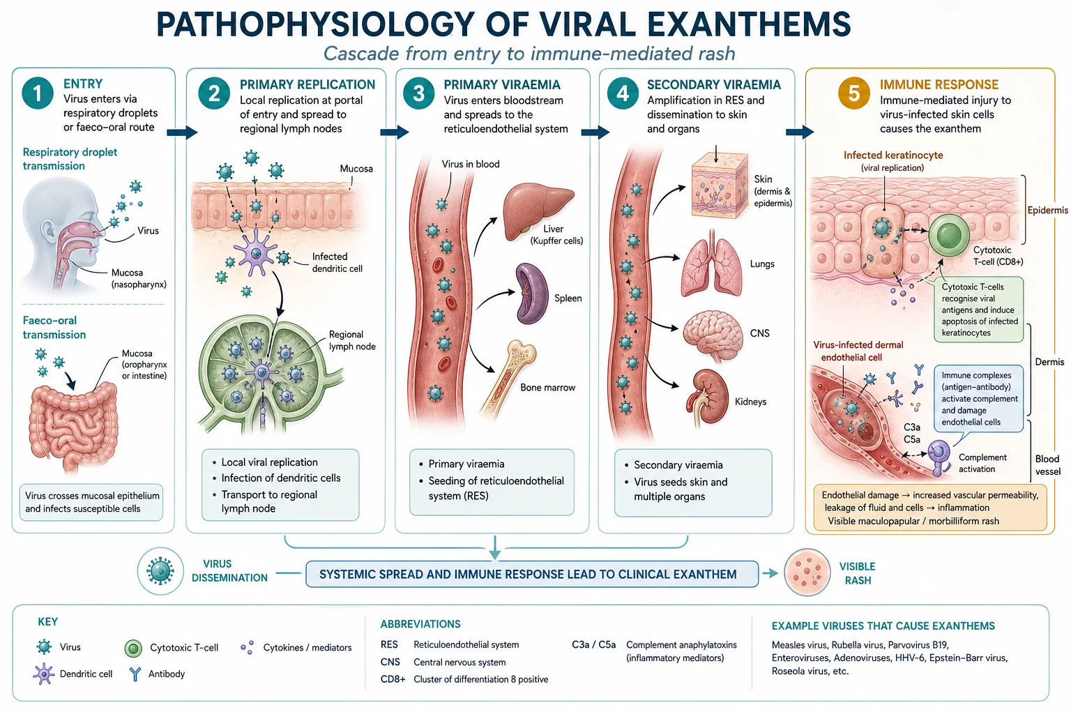
Acute (primary) HIV infection produces a mononucleosis-like illness two to four weeks after acquisition, lasting one to two weeks. The cutaneous hallmark is a morbilliform (measles-like) rash on the trunk, accompanied by fever, lymphadenopathy, pharyngitis, arthralgia and mucosal ulcers (aphthous or herpetiform).[1] The rash is easily missed or misattributed — to glandular fever (EBV), a drug reaction, rubella, or secondary syphilis — and is one of the highest-yield exam stems because recognising it allows diagnosis at the most infectious stage. Diagnosis at seroconversion requires an HIV RNA (viral load) PCR (positive) combined with a negative or indeterminate antibody test (the "window period").
Severe or atypical common dermatoses (CD4 above 500 to early decline)
The earliest cutaneous markers of HIV are common dermatoses behaving badly. Seborrhoeic dermatitis is the single most common HIV-related dermatosis and may be severe, extensive, treatment-resistant and atypical (involving the chest, axillae and groin rather than just the scalp and nasolabial folds).[1] Recurrent orolabial and anogenital HSV, multidermatomal or early-onset herpes zoster (shingles in a young person is an HIV red flag), recurrent oral candidiasis, extensive or treatment-resistant warts, and florid acne or folliculitis should each prompt HIV testing.[2]
Moderate immunosuppression (CD4 200 to 500)
As the CD4 count crosses below 500 and toward 200, the pathognomonic HIV dermatoses emerge.[1]
- Oral hairy leukoplakia (OHL): white, corrugated, non-scrapable plaques on the lateral border of the tongue; caused by EBV; pathognomonic for HIV (though occasionally seen in other immunosuppression).[13]
- Kaposi sarcoma (KS): purple-brown to dark red papules, nodules and plaques; AIDS-defining; see dedicated section below.[3]
- Eosinophilic folliculitis: intensely pruritic, urticarial, follicular papules on the face, neck and upper trunk; HIV-specific; eosinophilia common.[12]
- Molluscum contagiosum: numerous, large (over 1 cm), atypical lesions; may be giant or facial and refractory to standard treatment.
- Psoriasis: may be severe, erythrodermic, or first appear with HIV; paradoxically may flare on ART (IRIS).[1]
- Bacillary angiomatosis: vascular red-purple papules and nodules caused by Bartonella; mimics KS.[9]
- Immune thrombocytopenia (ITP): petechiae and ecchymoses from anti-platelet antibodies.
Severe immunosuppression (CD4 below 200)
Below 200, all moderate-stage conditions persist and worsen, and new life-threatening entities appear.[1]
- Chronic ulcerative HSV or VZV: herpetic ulcers that are chronic, deep, non-healing and often perianal, penile or orofacial; ulceration persisting over 1 month is AIDS-defining.[14]
- Disseminated fungal infections: cryptococcosis (umbilicated papules mimicking molluscum on the face, often with meningitis); histoplasmosis (ulcerated nodules, oral ulcers, petechiae); coccidioidomycosis (nodules, verrucous plaques); Talaromyces (Penicillium) marneffei (umbilicated papules in South-East Asia).[10][15]
- Crusted (Norwegian) scabies: hyperkeratotic, scaly, heavily crusted plaques teeming with millions of mites; highly contagious; an institutional outbreak risk.[11]
- Giant molluscum contagiosum: lesions over 1 cm, confluent, facial; refractory.
- Bacillary angiomatosis: may be disseminated with bacteraemia and visceral (hepatic, splenic) peliosis.[9]
Very severe immunosuppression (CD4 below 50)
At the lowest CD4 counts, the patient is prey to disseminated mycobacteria and CMV. [1]
- Disseminated Mycobacterium avium complex (MAC): skin lesions are rare but the systemic illness dominates with fever, weight loss, night sweats, diarrhoea and anaemia; cutaneous nodules, abscesses or ulcerations are a clue to disseminated disease that usually presents at CD4 below 50.
- Major aphthous ulcers: large (over 1 cm), deep, painful oral and oropharyngeal ulcers that can impair nutrition; distinguished from HSV by negative viral testing; severe or refractory disease responds to topical or intralesional corticosteroids, thalidomide 50 to 200 mg daily, or dapsone.[2]
- Cytomegalovirus (CMV): cutaneous ulcers (perianal, peristomal, digital) usually with retinitis or colitis.
- Disseminated mycobacteria and aggressive deep fungi: non-tuberculous mycobacteria may produce scattered nodules, abscesses and ulcerations.[1] At this stage multiple simultaneous dermatoses are the rule, not the exception, and a single patient may carry candidiasis, molluscum, KS and a chronic herpetic ulcer at one examination.
Differential Diagnosis
The four highest-yield dermatological distinctions in HIV are tabulated below; each is a classic exam stem. [1]
- White curd-like pseudomembranous plaques OR red atrophic patches
- SCRAPABLE — wipes off with gauze, leaving erythematous base
- KOH positive (pseudohyphae/budding yeast)
- CD4 below 500
- Candida albicans
- White corrugated vertical striations on lateral tongue
- NOT SCRAPABLE — adherent to mucosa
- EBV (in situ hybridisation); benign squamous hyperplasia
- CD4 below 200 to 300
- Pathognomonic for HIV
- Purple-brown papules, nodules, plaques; hard palate, lower legs, genitalia
- HHV-8 positive; spindle cells + slit-like vascular spaces on H and E
- Extravasated RBCs; positive CD31/CD34
- AIDS-defining
- Treat with ART +/- chemo/radiotherapy
- Red-purple friable vascular papules/nodules (mimics KS)
- Bartonella henselae/quintana; neutrophilic lobular panniculitis
- Warthin-Starry stain positive (black bacilli)
- CD4 below 100 to 200
- Treat with erythromycin or doxycycline
SCRAPES
A key mimic to always consider is disseminated cryptococcosis, whose cutaneous lesions (umbilicated, translucent papules on the forehead and face) can be indistinguishable from molluscum contagiosum; biopsy with PAS/GMS stain and serum cryptococcal antigen resolve it. The stakes are high because cutaneous cryptococcosis usually signals disseminated disease with cryptococcal meningitis.[10]
Specific Subtypes & Scenarios — Infections
Viral infections
Herpes simplex virus (HSV). In HIV, HSV runs an atypical course — chronic, deep, ulcerative, often perianal or penile, sometimes verrucous or tumid rather than the classic grouped vesicles. The cardinal exam point is that mucocutaneous HSV ulceration persisting over 1 month is an AIDS-defining illness.[14] Diagnosis is clinical with confirmation by PCR or viral culture of lesion swab. Treatment is oral valaciclovir 500 mg to 1 g twice daily or aciclovir 400 mg five times daily for 7 to 10 days; severe, extensive or resistant disease (common in low CD4 and with aciclovir-resistant thymidine-kinase-negative strains) needs IV aciclovir 5 to 10 mg/kg every 8 hours, and resistant strains require foscarnet.[2]
Varicella zoster virus (VZV). Herpes zoster in a young adult is an HIV red flag. In advanced HIV, zoster may be multidermatomal, disseminated, haemorrhagic or chronic, and may leave intractable post-herpetic neuralgia. Treat with oral valaciclovir 1 g three times daily for 7 days for uncomplicated disease and IV aciclovir 10 mg/kg every 8 hours for disseminated, visceral or severe disease.[1]
Molluscum contagiosum. A poxvirus producing umbilicated pearly papules. In HIV the lesions are numerous, large (giant, over 1 cm), confluent and facial, and may be refractory to standard destructive therapy. Differential includes cryptococcosis and KS. Treatment: curettage, cryotherapy, topical podophyllotoxin, imiquimod, or cantharidin — but the definitive treatment is ART, with which lesions usually resolve as immunity recovers. Refractory disease has responded to cidofovir (topical or intravenous).[2]
Cytomegalovirus (CMV). Cutaneous CMV is a disease of very low CD4 (below 50), producing painful ulcers (perianal, peristomal, digital) usually in a patient with CMV retinitis or colitis. Diagnosis needs biopsy with characteristic owl-eye intranuclear inclusions; treatment is IV ganciclovir or foscarnet.[1]
Human papillomavirus (HPV). Extensive, refractory and atypical warts, condylomata, and HPV-driven intraepithelial neoplasia are common. High-risk HPV-16 and 18 drive anal intraepithelial neoplasia (AIN) and cervical intraepithelial neoplasia, which progress to invasive squamous carcinoma at increased rates in HIV — particularly in MSM. This is the rationale for anal Pap screening and high-resolution anoscopy in at-risk HIV patients.[1]
Fungal infections
Oral and oesophageal candidiasis. Candidiasis (usually Candida albicans) is the most common oral manifestation of HIV. Pseudomembranous (thrush — white scrapable plaques), erythematous (red patches), angular cheilitis and atrophic forms occur. Oesophageal candidiasis is AIDS-defining. Treatment: mild disease with oral nystatin suspension or clotrimazole troches; moderate disease with oral fluconazole 100 to 200 mg daily for 7 to 14 days; oesophageal or refractory disease with fluconazole 200 to 400 mg daily or IV echinocandin.[2]
Dermatophytosis and onychomycosis. More extensive, atypical (proximal subungual onychomycosis is suggestive of HIV) and treatment-resistant.[1]
Disseminated deep mycoses. These are AIDS-defining and are among the most dangerous cutaneous events in HIV. Cryptococcus neoformans produces umbilicated, translucent, gelatinous papules on the forehead, face and neck that mimic molluscum; cutaneous cryptococcosis implies dissemination and mandates a lumbar puncture to exclude cryptococcal meningitis and serum cryptococcal antigen testing; treatment is IV liposomal amphotericin B plus flucytosine for induction then fluconazole consolidation.[10] Histoplasma capsulatum produces ulcerated nodules, oral ulcers, a papular or acneiform eruption, petechiae and molluscum-like lesions, particularly in the Americas; cutaneous disease implies disseminated histoplasmosis and is treated with amphotericin B then itraconazole.[15] Coccidioides produces nodules, plaques and verrucous lesions. Talaromyces (Penicillium) marneffei produces umbilicated papules in South-East Asia and China and is an AIDS-defining illness in endemic regions.[2]
Bacterial infections
Bacillary angiomatosis (Bartonella henselae or B. quintana). A vascular proliferative disorder unique to immunocompromised hosts (chiefly advanced HIV) that produces friable red to purple papules, nodules and angiomatous plaques that closely mimic Kaposi sarcoma. Disseminated disease produces hepatic and splenic peliosis with bacteraemia. The distinction from KS rests on biopsy: bacillary angiomatosis shows lobular proliferations of capillaries with neutrophils and neutrophilic debris, and a Warthin-Starry stain reveals the black-staining bacilli; KS shows spindle cells and HHV-8 positivity. Treatment is oral erythromycin 500 mg four times daily or doxycycline 100 mg twice daily for at least 3 months.[9]
Syphilis. HIV alters the natural history of syphilis: the rash of secondary syphilis may be atypical or florid, serology may be unreliable (the prozone phenomenon giving false-negatives), and there is accelerated progression to neurosyphilis and ocular syphilis. Ulcerative lesions mimicking chancroid or HSV occur. Because syphilis and HIV are co-transmitted, all patients with syphilis should be HIV-tested and vice versa. Treatment is standard penicillin-based therapy but with a low threshold for a lumbar puncture to exclude neurosyphilis.[16]
Staphylococcal infections (folliculitis, impetigo, ecthyma, botryomycosis) are common and severe due to high S. aureus colonisation. Mycobacteria — both TB (scrofuloderma, disseminated) and non-tuberculous mycobacteria — produce ulcers, nodules and scattered lesions, especially at very low CD4.[1]
Parasitic infestations
Crusted (Norwegian) scabies. Hyperinfestation with Sarcoptes scabiei producing thick, hyperkeratotic, scaly, grey crusted plaques over the hands, feet, scalp and extensor surfaces. The patient harbours millions of mites, is extraordinarily contagious, and may not complain of intense itch (the immune response that drives pruritus is blunted). It is a public-health emergency requiring isolation, topical permethrin 5 percent applied head-to-toe repeated weekly, oral ivermectin 200 micrograms/kg on days 1, 2, 8, 9, 15, and 22, and treatment of all contacts and healthcare workers.[11]
Specific Subtypes & Scenarios — Neoplasms
Kaposi sarcoma (KS)
Kaposi sarcoma is an AIDS-defining, HHV-8-driven vascular tumour and the single most examined neoplasm in HIV dermatology.[3]
- Mediterranean, Eastern European or Jewish elderly men
- Indolent violet plaques on the lower legs
- Best prognosis
- Equatorial Africa, independent of HIV
- More aggressive, may be lymphadenopathic in children
- Transplant and chronic immunosuppression patients
- May regress on reducing immunosuppression
- Most aggressive; widely disseminated; visceral (GI, pulmonary)
- AIDS-defining; sexually co-acquired with HIV
- Strongly ART-responsive
Clinical features. Purple, brown, dark red or violaceous macules, papules, plaques and nodules, which may be oedematous (reflecting lymphatic involvement) and may coalesce into confluent plaques. Characteristic sites are the lower extremities (classic), the hard palate (highly suggestive in HIV), the face, the genitalia and the trunk. A palatal lesion is almost pathognomonic for epidemic KS.[3]
Diagnosis. Skin biopsy is diagnostic: proliferating spindle cells forming slit-like vascular spaces, with extravasated erythrocytes, haemosiderin and a variable inflammatory infiltrate. HHV-8 immunohistochemistry is positive. Staging assesses visceral involvement (GI endoscopy, chest imaging for pulmonary nodules and pleural effusions).[3]
Treatment. For limited cutaneous disease in a treatment-naive patient, ART alone is often sufficient, as immune restoration leads to regression of KS. For localised symptomatic lesions, intralesional chemotherapy, cryotherapy or local radiotherapy. For extensive cutaneous, progressive or visceral disease, systemic chemotherapy with pegylated liposomal doxorubicin 20 mg/m2 every 3 weeks, or paclitaxel is first-line.[4] The key management principle: optimise ART first, since untreated HIV drives KS and IRIS may flare it.
Non-melanoma skin cancer, melanoma and lymphoma
Immunosuppression increases the risk of squamous cell carcinoma (SCC), which in HIV is more often HPV-driven, arises in anogenital and sun-exposed skin, and runs a more aggressive course with higher metastatic potential. Basal cell carcinoma shows a modest increase. Melanoma in HIV behaves more aggressively with reduced survival. Cutaneous lymphomas (both T-cell, including mycosis fungoides, and B-cell) occur at increased rates, as does CD30+ anaplastic large cell lymphoma.[1] Anal intraepithelial neoplasia (AIN) and invasive anal SCC, driven by HPV-16, are markedly increased in HIV-positive MSM and are screened for with anal cytology.[1]
Specific Subtypes & Scenarios — Inflammatory Dermatoses
Seborrhoeic dermatitis. The commonest HIV dermatosis. Presents with greasy yellow scale and erythema on the scalp, nasolabial folds, eyebrows, ears, chest and groin — but in HIV it is severe, extensive, treatment-resistant and atypical. Treat with topical ketoconazole 2 percent shampoo/cream and a mild topical corticosteroid; recalcitrant disease responds to oral itraconazole or fluconazole and to ART.[1]
Psoriasis. May be severe, pustular or erythrodermic, and may first appear in the setting of HIV or paradoxically flare on ART (IRIS). Treatment is challenging because traditional systemic immunosuppressants (methotrexate, ciclosporin) carry added infection risk in the immunocompromised; phototherapy (NBUVB) is effective and safe, and biologics (anti-TNF, anti-IL-17, anti-IL-23) are increasingly used with ART optimisation.[1]
Eosinophilic folliculitis. An HIV-specific, intensely pruritic eruption of urticarial follicular papules and pustules on the face, neck, upper trunk and extensor arms. Histology shows a perifollicular infiltrate rich in eosinophils; peripheral eosinophilia is common. It typically appears at CD4 of 200 to 300. It is exquisitely pruritic (a key exam clue) and resistant to standard anti-acne therapy. Treatment: topical corticosteroids, antihistamines, phototherapy (NBUVB), topical permethrin, and in resistant cases oral isotretinoin or itraconazole; ART is the most effective long-term therapy.[12]
Pruritic papular eruption of HIV (PPE). Chronic, intensely pruritic, symmetric papules and excoriated nodules on the extremities and trunk, driven by an eosinophilic response. Distinct from scabies (no burrows, negative scrapings) and eosinophilic folliculitis (not folliculocentric). Treat with antihistamines, topical corticosteroids, phototherapy and ART.[12]
Acne and folliculitis are often severe; atopic dermatitis may flare; xerosis (dry skin) and acquired ichthyosis are common and contribute to intractable pruritus in late disease.[1]
Drug Eruptions
Drug eruptions are dramatically more common in HIV — a 10 to 100-fold increased risk — reflecting immune dysregulation, polypharmacy, slow drug metabolism, and the specific immune-activating effects of several antiretrovirals and cotrimoxazole.[2] The spectrum runs from trivial morbilliform exanthems through to fatal Stevens-Johnson syndrome/toxic epidermal necrolysis (SJS/TEN) and drug reaction with eosinophilia and systemic symptoms (DRESS).
Cotrimoxazole (trimethoprim-sulfamethoxazole, TMP-SMX). The single most common culprit, used for Pneumocystis prophylaxis. Morbilliform maculopapular rash with fever occurs in roughly 40 to 60 percent of HIV patients (versus around 3 percent of the general population). Most cases are mild and resolve or can be managed with desensitisation, but SJS/TEN can occur. Alternatives include dapsone, atovaquone or aerosolised pentamidine for prophylaxis.[2]
Abacavir (an NRTI). Causes a characteristic systemic hypersensitivity reaction (fever, rash, GI symptoms, malaise, respiratory symptoms) appearing within the first 6 weeks. It is HLA-B*5701-restricted, and all patients must be screened for HLA-B*5701 before starting abacavir — if positive, abacavir is contraindicated and never rechallenged (re-exposure can cause fatal anaphylaxis).[7]
Nevirapine (an NNRTI). A high-risk drug for SJS/TEN, with the risk greatest in women with CD4 above 250 cells/microL (and men above 400) — for this reason nevirapine is avoided in these groups and a lead-in dose escalation is used. A cohort from South Africa documented nevirapine-induced SJS in children living with HIV.[8]
Efavirenz (an NNRTI). Causes a mild maculopapular rash and characteristic neuropsychiatric symptoms (vivid dreams, insomnia, anxiety, depression) that usually settle within weeks. Tenofovir, zidovudine, lamivudine, dolutegravir and raltegravir are comparatively benign cutaneously.[2]
Immune Reconstitution Inflammatory Syndrome (IRIS)
IRIS is the paradoxical worsening of an existing condition, or the unmasking of a subclinical infection, within weeks to months of starting ART, as recovering immunity mounts an exaggerated inflammatory response to antigens that were previously present but immunologically "quiet". A systematic review and meta-analysis established its incidence and risk factors.[5]
Risk factors for IRIS: a low baseline CD4 count (the single strongest predictor), a high CD4 rise or viral load fall after ART, a short interval between treatment of an opportunistic infection and starting ART, and the presence of an unrecognised opportunistic infection at ART initiation.[5]
Cutaneous IRIS produces a flare of pre-existing dermatoses or the unmasking of occult infection:[5]
- KS-IRIS: rapid enlargement, oedema and visceral progression of Kaposi sarcoma in the first weeks of ART — can be life-threatening if visceral or with significant lymphoedema.
- Acne or folliculitis flare (sometimes severe cystic acne).
- Worsening psoriasis or first presentation of severe psoriasis.
- Unmasking of previously subclinical infection: cryptococcosis (with meningitis), MAC, mycobacterial disease, HSV/VZV reactivation.
- Severe or refractory molluscum during immune recovery. [1]
Management of IRIS: do NOT stop ART (the temptation, and the wrong move). Continue ART, treat the unmasked or flared condition specifically, and use systemic corticosteroids (for example oral prednisolone 1 to 2 mg/kg/day tapering) for severe or organ-threatening IRIS — for example severe KS-IRIS, granulomatous reactions, or major inflammatory compromise of organ function. In severe paradoxical reactions to mycobacterial disease, anti-TNF agents such as infliximab and thalidomide have a role.[6]
IRIS versus treatment failure. The critical distinction: both may produce worsening disease after starting ART. IRIS shows a falling viral load and rising CD4 (the immune system is recovering), while treatment failure shows a rising or unsuppressed viral load. A viral load is mandatory before attributing worsening to IRIS.[5]
Clinical & Bedside Assessment
A focused dermatological examination in suspected or known HIV is systematic and exposes the patient fully. [1]
Examine the whole skin, the mucosae and the anogenital area. Inspect the scalp and face (seborrhoeic dermatitis, molluscum, eosinophilic folliculitis, KS, acne), the oral cavity (candidiasis, hairy leukoplakia, aphthae, oral KS, gingivitis, oral hairy leukoplakia — lift the tongue to see the lateral border), the trunk (molluscum, KS, drug rash, syphilis), the flexures and anogenital area (HSV ulcers, condyloma, warts, AIN, crusted scabies in web-spaces and genitals), the nails (proximal subungual onychomycosis, periungual warts), and the palms and soles (syphilis, scabies). Palpate the lymph nodes (generalised lymphadenopathy of HIV, lymphoma) and examine for lymphoedema (KS).[1]
Bedside tests: a KOH preparation distinguishes candidiasis (pseudohyphae) from hairy leukoplakia (negative); a Tzanck smear of a vesicle shows multinucleated giant cells in HSV/VZV; dermoscopy of a suspicious lesion distinguishes a melanocytic from a vascular lesion (KS shows a violaceous homogenous area with rainbow pattern); skin scrapings in mineral oil identify scabies mites; and the simple gauze-wipe test settles the candidiasis-versus-leukoplakia question at the bedside (candidiasis scrapes off, leukoplakia does not).[1]
Investigations
Skin biopsy is central to confirming KS, bacillary angiomatosis, deep fungal infection, mycobacterial disease, lymphoma and drug eruption. The choice of stain matters: H and E for routine histology; Warthin-Starry stain for Bartonella; PAS and GMS (methenamine silver) for fungi (cryptococcus shows narrow-based budding yeasts with a mucin-positive capsule on mucicarmine); Fite-Faraco for mycobacteria; HHV-8 immunohistochemistry for KS. Culture and PCR of lesions, swabs and blood confirm HSV/VZV, Bartonella, mycobacteria and fungi. Serology (RPR/VDRL plus TPPA for syphilis; cryptococcal antigen) and flow cytometry for lymphoma complete the workup.[1][9]
Baseline HIV staging bloods in any newly-diagnosed patient: CD4 count and percentage, HIV viral load (RNA PCR), full blood count, liver and renal function, HBV and HCV serology, syphilis serology, Toxoplasma IgG, and tuberculosis screen (IGRA or symptom screen plus chest X-ray). The CD4 count dictates the differential diagnosis and the urgency of ART.[2]
Management — Resuscitation
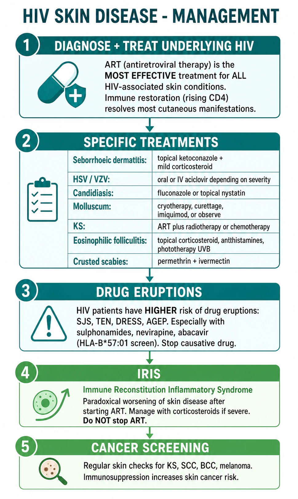
Most HIV skin disease is not an emergency, but four scenarios are. [1]
SJS/TEN. The defining emergency. STOP the offending drug immediately (nevirapine, cotrimoxazole, abacavir, or any newly-introduced agent), admit to an ICU or burns unit, provide intravenous fluids and electrolyte support, wound care, nutritional support, and urgent ophthalmology (ocular adhesions are the leading cause of long-term morbidity). Intravenous immunoglobulin or ciclosporin may be considered; systemic corticosteroids are controversial.[8]
Disseminated fungal infection with sepsis. Disseminated histoplasmosis, coccidioidomycosis and cryptococcosis may present with cutaneous lesions and septic shock or meningitis — start IV amphotericin B and manage the airway, circulation and meningitis.[10][15]
Crusted scabies outbreak. Isolate the patient, treat with permethrin plus ivermectin, and prophylactically treat all healthcare worker and household contacts, because a missed case can seed a ward-wide outbreak.[11]
Severe IRIS. KS-IRIS with airway compromise or extensive visceral disease, or IRIS with organ failure, needs systemic corticosteroids and specialist input; do not stop ART.[5][6]
Management — Definitive & Stepwise
The management of HIV skin disease rests on five pillars.[1][2]
1. Antiretroviral therapy (ART) — the cornerstone. Effective ART, by restoring CD4 counts and suppressing viral load, resolves the majority of HIV-associated dermatoses and prevents recurrence. The landmark START trial demonstrated that ART should be initiated in all patients regardless of CD4 count — the modern recommendation is "test and treat", with ART started on diagnosis. For a patient presenting with an AIDS-defining cutaneous illness, ART is urgent.[2]
2. Condition-specific dermatological therapy. Each dermatosis is treated in its own right, but ART is added (or optimised) in parallel. The high-yield specifics are tabulated below. [1]
Phototherapy and physical modalities. Narrowband UVB (NBUVB) phototherapy is a workhorse in HIV dermatology and deserves its own mention because it elegantly sidesteps the systemic immunosuppression that complicates HIV. It is first-line or adjunctive for eosinophilic folliculitis, pruritic papular eruption, severe psoriasis, and severe atopic dermatitis in HIV, where it reduces pruritus and clears lesions over several weeks of treatment. Importantly, UVB is not contraindicated in HIV (older concerns about UV-driven immune suppression have not translated into clinical harm in modern practice) and it avoids the additive infection risk of methotrexate or ciclosporin. PUVA is reserved for refractory disease. Cryotherapy, curettage, electrodessication and laser (CO2 or pulsed-dye) are the physical modalities for molluscum, warts, condylomata and small KS lesions; intralesional vincristine or interferon has a role in refractory KS.[1]
Symptomatic and supportive therapy. Intractable pruritus — a defining misfeature of late HIV skin disease — is managed with emollients and soap avoidance for xerosis, oral antihistamines (sedating at night such as hydroxyzine 25 to 50 mg), and topical menthol/camphor or capsaicin for neuropathic itch. Thalidomide (50 to 100 mg at night) is highly effective for severe refractory aphthous ulceration of the mouth and oropharynx in advanced HIV, but mandates strict contraception and monitoring for peripheral neuropathy. Topical and systemic analgesia manage the pain of chronic herpetic ulcers, KS lymphoedema and post-herpetic neuralgia.[2]
Follow-up and monitoring. A patient with HIV skin disease is followed jointly with the HIV physician. The CD4 count and viral load are the master variables — a dermatosis that fails to resolve despite specific therapy, or a new eruption in a treated patient, should prompt a check of virological suppression and ART adherence. Every visit is an opportunity for total-body skin examination (skin cancer surveillance), STI screening, and mental-health and adherence support, because untreated depression and stigma are major drivers of poor outcomes.[1]
3. Drug-eruption management. Stop the offending drug (and document it as an allergy). Screen for HLA-B*5701 before abacavir; avoid nevirapine in women with CD4 above 250; consider cotrimoxazole desensitisation for mild reactions where prophylaxis is essential; switch to an alternative agent within the same class (for example efavirenz to rilpivirine, or dolutegravir) guided by a HIV physician.[7][8]
4. IRIS management. Continue ART; treat the unmasked or flared condition; add systemic corticosteroids for severe or organ-threatening IRIS.[5][6]
5. Cancer screening and prevention. Regular total-body skin examination for KS, SCC, BCC and melanoma; anal Pap smear and high-resolution anoscopy for at-risk patients (AIN screening); cervical screening; sun-protection counselling; and HPV vaccination.[1]
Complications & Pitfalls
Pitfalls in diagnosis. The commonest error is to mistake disseminated cryptococcosis (umbilicated papules) for molluscum contagiosum, delaying lumbar puncture and antifungal therapy with potentially fatal consequences. A second classic error is to confuse bacillary angiomatosis with KS and treat with chemotherapy rather than antibiotics — biopsy with Warthin-Starry stain resolves it.[9][10]
Pitfalls in management. Stopping ART during IRIS is a recurrent and dangerous error; paradoxical worsening is a sign of successful immune recovery and should be managed with corticosteroids, not drug withdrawal. Re-challenging abacavir after a hypersensitivity reaction is potentially fatal and must never be done. Failing to isolate crusted scabies seeds institutional outbreaks.[5][7][11]
Complications of the diseases themselves. Untreated KS produces lymphoedema, pain, visceral (GI bleeding, pulmonary) disease and disfigurement. Chronic herpetic ulcers may become secondarily infected and act as a portal for bacteraemia. Long-standing pruritic papular eruption and eosinophilic folliculitis produce disabling pruritus, sleep deprivation, stigmatisation and depression. Drug eruptions cause the SJS/TEN spectrum with its high mortality and ocular morbidity.[1]
Prognosis & Disposition
The prognosis of HIV skin disease is transformed by effective ART. The majority of inflammatory and infectious dermatoses — seborrhoeic dermatitis, candidiasis, molluscum, eosinophilic folliculitis, oral hairy leukoplakia, chronic herpetic ulcers — resolve or markedly improve as CD4 counts recover. Epidemic KS frequently regresses with ART alone. Late-stage dermatoses (crusted scabies, giant molluscum, disseminated mycoses) are largely preventable with timely ART and, when they occur, indicate late presentation or treatment failure.[2][4]
Disposition. A patient with a new AIDS-defining cutaneous illness (KS, chronic herpetic ulcer, disseminated mycosis) needs urgent HIV physician input, immediate ART initiation, and management of the specific condition. Patients on ART with mild dermatoses are managed as outpatients. Any suspicion of SJS/TEN, disseminated fungal sepsis, or crusted scabies mandates admission. A rising CD4 with paradoxical worsening is IRIS until proven otherwise and is managed in collaboration with the HIV team.[5]
Special Populations
Children with perinatally-acquired HIV frequently present with severe, recurrent candidiasis, extensive warts, refractory molluscum, chronic paronychia and failure to thrive; drug dosing is weight-based and nevirapine-induced SJS is documented in paediatric cohorts.[8]
Pregnant women living with HIV require ART for both maternal health and prevention of mother-to-child transmission; efavirenz is now considered safe in the first trimester, but drug choice must be coordinated with the obstetric and HIV teams. Drug eruptions and IRIS may complicate pregnancy, and untreated syphilis co-infection is catastrophic for the fetus.[2]
Late presenters (CD4 below 200 at diagnosis) bear the full classic dermatological spectrum and are at highest risk of IRIS when ART is started; a period of treatment of active opportunistic infection before ART initiation may reduce IRIS risk in selected cases (for example, in TB or cryptococcal meningitis), reflecting a regionally debated timing decision.[5]
Evidence, Guidelines & Regional Differences
The START trial (2015) established that immediate ART (at CD4 above 500) reduces serious AIDS and non-AIDS events and death compared with deferred ART, cementing the "test and treat" strategy and confirming ART as the foundation of management for every HIV dermatosis.[2]
HLA-B*5701 screening before abacavir was validated in the PREDICT-1 randomised study and is now standard of care worldwide; a negative screen essentially abolishes immunologically-mediated abacavir hypersensitivity.[7]
Nevirapine CD4-based prescribing restrictions (avoid in women with CD4 above 250 and men above 400) derive from pivotal cohort data and are embedded in WHO and national guidelines.[8]
Regional differences are substantial. WHO guidelines support universal ART and co-trimoxazole prophylaxis for adults and children in resource-limited settings, where late presentation drives the epidemiology. In tropical regions, disseminated histoplasmosis, Talaromyces (Penicillium) marneffei infection, chromoblastomycosis, and tropical dermatophytosis are prominent AIDS-defining cutaneous illnesses that are rare in temperate climates. Disseminated histoplasmosis is such a common AIDS-defining illness in Latin America that Histoplasma antigen testing is recommended in any febrile HIV patient there. Access to ART, to liposomal amphotericin B, and to dermatopathology strongly influences outcome.
Exam Pearls
Red Flags
Exam application bank (NEET-PG / INICET)
One-line answer
Cutaneous disease affects more than 90 percent of people living with HIV and is often the first clinical clue to infection. The skin manifestations are organised by CD4 count. CD4 above 500 (early): acute seroconversion morbilliform rash, severe seborrhoeic dermatitis, oral candidiasis, recurrent HSV, multidermatomal zoster. CD4 200 to 500 (moderate): oral hairy leukoplakia (EBV; lateral tongue; non-scrapable), Kaposi sarcoma (HHV-8; AIDS-defining), eosinophilic folliculitis (intensely pruritic facial papules), molluscum, psoriasis flare, bacillary angiomatosis. CD4 below 200 (severe): chronic ulcerative HSV/VZV (over 1 month = AIDS-defining), disseminated fungal (cryptococcus, histoplasmosis, coccidioidomycosis), crusted scabies, giant molluscum, bacillary angiomatosis. CD4 below 50: disseminated MAC, CMV, deep mycobacterial ulcers. Drug eruptions are 10 to 100 times more common (cotrim
Worked stems (answer without another resource)
Stem 1 — Classic presentation. Map symptoms to mechanism; name the first investigation and first treatment step with dose/route if drug therapy is standard. [1]
Stem 2 — Unstable / complicated. List red flags that force immediate resuscitation, theatre, ICU, antidote, or reperfusion — and what you do in the first 15 minutes. [1]
Stem 3 — Atypical group. Elderly, pregnancy, child, or immunocompromised: how presentation and thresholds change. [1]
Stem 4 — Differential trap. Name the three closest mimics and one discriminator for each. [1]
Stem 5 — Disposition. Who goes home with safety-netting, who is admitted, who needs HDU/ICU/theatre, and what follow-up is mandatory. [1]
Rapid viva checklist
- Definition + classification
- Pathophysiology chain
- Bedside signs / criteria
- Score with exact components (if any)
- Emergency bundle
- Definitive therapy with doses
- Complications of disease and of treatment
- Special populations
- Guideline/trial name if classic
- Three exam traps
Coverage self-check
If you cannot answer any stem above from this page alone, re-read the matching section — the page is intended to be self-sufficient for final-prof and NEET-PG/INICET questions on Dermatology in HIV.
References
- [1]Karadag AS, Elmas ÖF, Altunay İK, et al. Cutaneous manifestations associated with HIV infections: A great imitator Clin Dermatol, 2020.PMID 32513397
- [2]Mohseni Afshar Z, Goodarzi A, Emadi SN, et al. A Comprehensive Review on HIV-Associated Dermatologic Manifestations: From Epidemiology to Clinical Management Int J Microbiol, 2023.PMID 37496761
- [3]Cesarman E, Damania B, Krown SE, et al. Kaposi sarcoma Nat Rev Dis Primers, 2019.PMID 30705286
- [4]Patel R, Lurain K, Yarchoan R, et al. Clinical management of Kaposi sarcoma herpesvirus-associated diseases: an update on disease manifestations and treatment strategies Expert Rev Anti Infect Ther, 2023.PMID 37578202
- [5]Müller M, Wandel S, Colebunders R, et al. Immune reconstitution inflammatory syndrome in patients starting antiretroviral therapy for HIV infection: a systematic review and meta-analysis Lancet Infect Dis, 2010.PMID 20334848
- [6]Armange L, Lacroix A, Petitgas P, et al. The use of TNF-α antagonists in tuberculosis to control severe paradoxical reaction or immune reconstitution inflammatory syndrome: a case series and literature review Eur J Clin Microbiol Infect Dis, 2023.PMID 36795280
- [7]Waters LJ, Mandalia S, Gazzard B, et al. Prospective HLA-B*5701 screening and abacavir hypersensitivity: a single centre experience AIDS, 2007.PMID 18025891
- [8]du Toit JD, Kotze K, van der Westhuizen HM, et al. Nevirapine-induced Stevens-Johnson syndrome in children living with HIV in South Africa South Afr J HIV Med, 2021.PMID 33824730
- [9]Akram SM, Anwar MY, Thandra KC, et al. Bacillary Angiomatosis 2026.PMID 28846267
- [10]Noguchi H, Matsumoto T, Kimura U, et al. Cutaneous Cryptococcosis Med Mycol J, 2019.PMID 31787730
- [11]Niode NJ, Adji A, Gazpers S, et al. Crusted Scabies, a Neglected Tropical Disease: Case Series and Literature Review Infect Dis Rep, 2022.PMID 35735761
- [12]Long H, Zhang G, Wang L, et al. Eosinophilic Skin Diseases: A Comprehensive Review Clin Rev Allergy Immunol, 2016.PMID 25876839
- [13]Rathee M, Jain P. Hairy Leukoplakia 2026.PMID 32119478
- [14]Sirka CS, Panda M, Jena A, et al. Clinical features of a large chronic ulcer on the genital and perianal region in HIV-infected patients can be a strong clinical clue for the diagnosis of herpes simplex infection Indian J Sex Transm Dis AIDS, 2020.PMID 33817593
- [15]Lin MJ, Mazzoni D, Gin D. Disseminated cutaneous-only histoplasmosis in a patient with AIDS Australas J Dermatol, 2019.PMID 31250916
- [16]Stefani A, Riello M, Rossini F, et al. Neurosyphilis manifesting with rapidly progressive dementia: report of three cases Neurol Sci, 2013.PMID 24062212