Dermatology · Medicine
Dermatomyositis
Also known as Dermatomyositis · DM · Amyopathic dermatomyositis · ADM · Clinically amyopathic dermatomyositis · CADM · Idiopathic inflammatory myopathy · IIM
Dermatomyositis (DM) is an autoimmune idiopathic inflammatory myopathy (IIM) characterised by pathognomonic cutaneous signs (Gottron papules, heliotrope rash, Gottron sign, shawl sign, V-sign, holster sign, mechanic's hands, nailfold capillary changes) and a spectrum of muscle involvement ranging from fulminant proximal weakness to clinically amyopathic disease (ADM/CADM). Pathogenesis centres on two interlocking pathways — a type I interferon signature (plasmacytoid dendritic cell release of IFN-alpha/beta, MHC class I upregulation, sustained innate/adaptive activation) and complement-mediated microangiopathy (C1q → C3 → C5b-9 membrane attack complex on endomysial capillaries → capillary dropout → perifascicular atrophy, the histological hallmark). Myositis-specific autoantibodies (anti-Mi-2, anti-MDA5/CADM-140, anti-TIF1-gamma/p155-p140, anti-NXP2, anti-SAE, anti-Jo-1/anti-synthetase, anti-SRP, anti-CN1A, anti-Mi-2-alpha/beta) define discrete clinical phenotypes; anti-MDA5 carries the highest 6-month mortality because of rapidly progressive interstitial lung disease (RP-ILD). Approximately 15 to 30 percent of adult DM is paraneoplastic, mandating age-appropriate cancer screening (CT chest/abdomen/pelvis, mammography, colonoscopy, ovarian CA-125 + transvaginal ultrasound, PSA, nasopharyngoscopy in Asian populations) at diagnosis and annually for 3 to 5 years. Management combines high-dose corticosteroids (prednisolone 1 mg/kg/day, or IV methylprednisolone 500–1000 mg/day × 3–5 days for severe disease) with steroid-sparing immunosuppression (methotrexate 7.5–25 mg/week, azathioprine 2 mg/kg/day, mycophenolate mofetil 2–3 g/day, ciclosporin or tacrolimus), IVIG (2 g/kg/cycle over 2–5 days, every 4 weeks) for refractory skin and severe oesophageal/diaphragmatic involvement, rituximab 1 g on days 0 + 14 for refractory myositis, JAK inhibitors (tofacitinib 5 mg BD) in selected cases, hydroxychloroquine 200–400 mg/day for cutaneous disease (with annual OCT maculopathy surveillance), strict photoprotection (SPF 50+ daily), and aggressive combination immunosuppression for anti-MDA5 RP-ILD (high-dose IV methylprednisolone + calcineurin inhibitor + cyclophosphamide +/− rituximab, with early lung transplant evaluation for refractory disease). Fellowship candidates must master the pathognomonic cutaneous signs, autoantibody stratification, the paraneoplastic work-up, the EULAR/ACR 2017 classification criteria, the juvenile DM complication set (calcinosis cutis, GI and CNS vasculopathy), and the operative distinction from polymyositis (PM), inclusion body myositis (IBM), immune-mediated necrotising myopathy (IMNM), and cutaneous lupus erythematosus (CLE).
On this page & tools
Your progress
Saved locally on this device.
Exam tags
Red flags
1. Definition & Classification
Dermatomyositis (DM) is an autoimmune idiopathic inflammatory myopathy (IIM) defined by pathognomonic cutaneous signs combined with a variable spectrum of muscle inflammation. It is one of the three classic IIMs — alongside polymyositis (PM) and inclusion body myositis (IBM) — and is uniquely characterised by both skin and muscle involvement, by complement-mediated microangiopathy (rather than the T-cell-mediated cytotoxicity that dominates PM), and by a strong association with autoantibodies, interstitial lung disease, and malignancy.[1][2][7]
Operational classification (Bohan & Peter 1975; EULAR/ACR 2017):[5][5]
| Subtype | Defining features |
|---|---|
| Classic DM | Pathognomonic skin signs (Gottron papules OR heliotrope rash) + proximal muscle weakness + elevated muscle enzymes + myopathic EMG and/or muscle biopsy |
| Amyopathic DM (ADM, "clinically amyopathic DM", CADM) | Cutaneous signs of DM for ≥ 6 months without clinical muscle weakness and with normal CK and aldolase; ± subclinical muscle involvement on MRI/biopsy |
| Hypomyopathic DM | Cutaneous signs + subclinical muscle involvement detected by EMG, MRI or biopsy but no clinical weakness |
| Juvenile DM (JDM) | Onset < 18 years; same cutaneous + muscle features; calcinosis cutis and vasculopathy (GI ulceration, CNS) more frequent |
| Cancer-associated DM (paraneoplastic) | DM with a concurrent or temporally-associated malignancy (~ 15–30 percent of adults); enriched for anti-TIF1-gamma and anti-NXP2 |
| Anti-synthetase syndrome (anti-Jo-1 and related) | Myositis + ILD + arthritis + Raynaud's + mechanic's hands + fever; ± DM skin signs |
| Clinically amyopathic DM with anti-MDA5 (CADM-140) | Amyopathic/hypomyopathic DM with anti-MDA5 antibody; rapidly progressive ILD (RP-ILD) is the lethal phenotype |
The EULAR/ACR 2017 classification criteria score variables across muscle weakness, skin signs (heliotrope, Gottron, Gottron papules, V-sign, shawl), muscle enzymes, EMG, MRI and biopsy to produce an aggregate probability of IIM (no biopsy cut-off = 55 percent probability; with biopsy = ≥ 90 percent probability) and a sub-classification into DM, PM, IBM or amyopathic DM.[1][5]
2. Epidemiology & Risk Factors
DM is a rare disease. The two peaks of incidence are childhood (5 to 15 years) for juvenile DM and adulthood (40 to 60 years) for classic and paraneoplastic DM, with a smaller third peak in the elderly overlapping with IBM and cancer.[1][5][7]
Risk factors include female sex, age (bimodal — children and mid-adult), family or personal history of autoimmune disease, HLA-DRB1*03:01 (adult DM), HLA-DRB1*03 and HLA-DQA1*0301 (JDM), seasonality (JDM clusters in spring/early summer, implicating an environmental trigger), preceding viral or bacterial infection (parvovirus B19, Coxsackie B, group A streptococcus, hepatitis B/C, HIV), UV light exposure (worsens cutaneous disease), and underlying malignancy (15 to 30 percent of adult cases).[1][2][5]
3. Pathophysiology
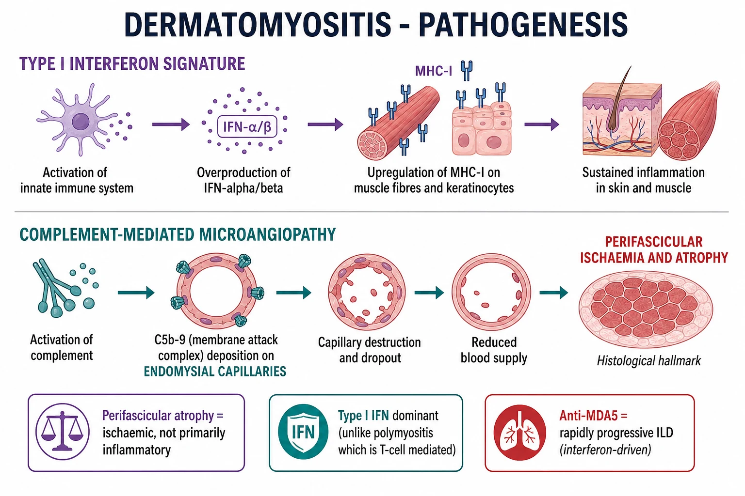
DM is the prototypical humoral + complement-mediated microvasculopathy of muscle and skin. Although T cells and autoantibodies participate, the dominant injury pathway is vascular: complement activation on endomysial capillaries → capillary dropout → ischaemic injury to the perifascicular myofibres → atrophy. A type I interferon signature in skin and muscle generates a self-amplifying inflammatory loop that maintains disease.[1][2][7]
3.1 Type I interferon signature
- Trigger: unknown (viral PAMP, tumour antigen, UV light) activates plasmacytoid dendritic cells (pDCs) in skin and muscle.
- Effector: pDCs release IFN-alpha and IFN-beta → upregulation of interferon-stimulated genes (ISGs): MX1, IFIT1, ISG15, IFI44L.
- Downstream: MHC-I upregulation on myofibres (which are normally MHC-I negative) → visible to CD8+ T cells; keratinocyte MHC-I upregulation → interface dermatitis.
- Clinical correlate: The IFN signature is most intense in anti-MDA5 DM (explaining the aggressive skin/lung phenotype) and anti-TIF1-gamma DM (paraneoplastic, in which the tumour drives the IFN signature).[2][3][7]
3.2 Complement-mediated microangiopathy
- Trigger: autoantibody (or immune complex) binds an endomysial capillary antigen → C1q activation → classical complement pathway → C3 cleavage → assembly of the C5b-9 membrane attack complex (MAC) on the capillary endothelial surface.
- Effector: MAC punches pores in endothelial cells → microvascular necrosis, capillary dropout, luminal narrowing → reduced perfusion of the perifascicular rim of myofibres.
- Outcome: perifascicular atrophy — small, angulated, MHC-I-positive myofibres at the edge of the fascicle. This is the histological hallmark that distinguishes DM from PM.[1][7]
- Skin parallel: identical interface dermatitis with C5b-9 deposition on dermal vessels → basal vacuolar change and the visible violaceous rash.
3.3 Adaptive immunity
- CD4+ T cells dominate perimysial and perivascular infiltrates (in contrast to PM, which is endomysial CD8+ mediated).
- B cells and plasma cells drive autoantibody production (anti-Mi-2, anti-MDA5, anti-TIF1-gamma, anti-NXP2, anti-SAE, anti-Jo-1, anti-SRP, anti-CN1A).
- Anti-MDA5 is a gain-of-function autoantibody that binds MDA5 (a cytosolic viral RNA sensor), activates its signalling, and amplifies type I IFN production — explaining the most aggressive DM phenotype.[3][6]
- Anti-TIF1-gamma recognises transcriptional intermediary factor 1-gamma, a tumour-suppressor; its presence suggests a paraneoplastic IFN drive from an underlying malignancy.[2][3]
3.4 Why the skin? Why the muscle?
Both skin and muscle share the same microvascular architecture (small vessels with fenestrated endothelium overlying a basal lamina) and the same vulnerability to type I IFN and complement injury. The perifascicular rim of muscle fibres (the watershed) is the most distal territory of the capillary bed and is therefore the first to become ischaemic. The skin's basal keratinocyte layer (also watershed) shows interface change for the same reason.[1][7]
4. Clinical Presentation
DM characteristically presents with skin signs preceding or coincident with muscle weakness; in amyopathic/hypomyopathic DM, the skin signs dominate and the muscle disease is minimal or subclinical. The tempo is subacute (weeks to a few months) in classic DM, acute in juvenile DM, and may be indolently chronic in amyopathic DM.[1][2][4]
4.1 The cutaneous signs (the dermatologist's gateway)[2][4]
| Sign | Description | Pathology |
|---|---|---|
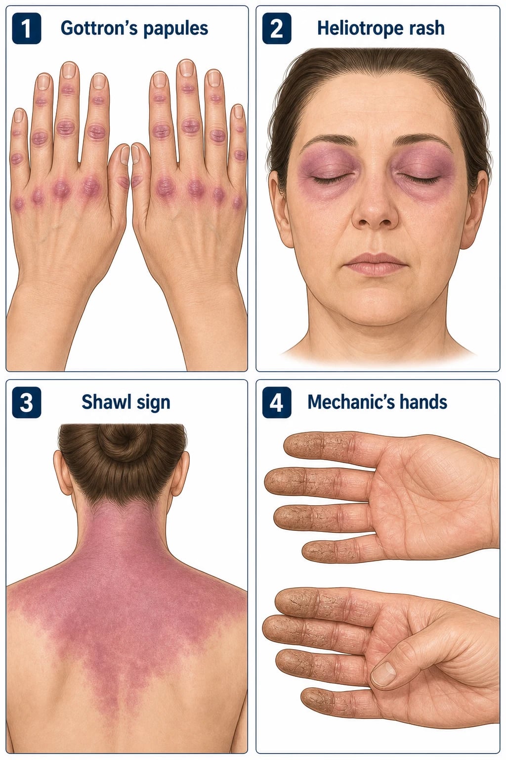
| Gottron papules | Violaceous (purple-red) flat-topped papules over the MCP and IP joints; ± overlying scale | Pathognomonic for DM; interface dermatitis |
| Gottron sign | Violaceous erythema over extensor surfaces (elbows, knees, medial malleoli), without papules | Highly characteristic; photoaggravated |
| Heliotrope rash | Violaceous erythema of the upper eyelids ± periorbital oedema; may involve the entire periorbital region | Pathognomonic for DM |
| Shawl sign | Violaceous erythema on the posterior neck, shoulders and upper back (photosensitive distribution) | Interface dermatitis |
| V-sign | Violaceous erythema on the anterior neck and upper chest (V of the chest) | Same pathogenesis |
| Holster sign | Violaceous erythema on the lateral thighs (where a holster would sit) | Highly specific; often missed |
| Mechanic's hands | Hyperkeratotic, fissured, "dirty-appearing" skin on the radial/palmar fingers | Suggests anti-synthetase overlap |
| Cuticular overgrowth | Ragged, hypertrophic cuticles with periungual telangiectasia | Nailfold capillary change at the cuticle |
| Nailfold capillary changes | Dilated capillary loops, dropout, haemorrhage; visible to the naked eye or by capillaroscopy | Same finding as in systemic sclerosis |
| Calcinosis cutis | Subcutaneous calcium deposits, often over bony prominences (elbows, knees, buttocks, fingertips) | Common in juvenile DM |
| Lipodystrophy | Acquired loss of subcutaneous fat (regional or generalised); associated with insulin resistance and hypertriglyceridaemia | More common in JDM and partial lipodystrophy variants |
| Ulceration | Necrotic ulceration over Gottron papules, digital tips or the chest wall | Suggests anti-MDA5 DM, severe vasculopathy |
4.2 The muscle component (when present)
- Distribution: symmetric proximal weakness — shoulder girdle (deltoid, supraspinatus/infraspinatus, scapular stabilisers) and hip girdle (iliopsoas, gluteus, quadriceps).
- Symptoms: difficulty rising from a low chair, climbing stairs, lifting arms overhead, combing hair; facial and extraocular muscles are usually spared; dyspnoea (diaphragm/intercostals), dysphagia (pharyngeal/upper oesophageal), dysphonia (laryngeal).
- Examination: reduced MRC power (typically 3+ to 4−/5), preserved reflexes and sensation, no fasciculations.
- Tempo: subacute progression over weeks to a few months; juvenile DM may progress over days.[1][4][7]
4.3 Other organ involvement
- Interstitial lung disease (ILD): ~ 20 to 40 percent overall; up to 90 percent in anti-MDA5 and 70 percent in anti-Jo-1. May be chronic progressive (antisynthetase) or rapidly progressive (anti-MDA5 RP-ILD, 6-month mortality 40 to 60 percent).[3][6]
- Cardiac: arrhythmias (conduction block), myocarditis, pericarditis; subclinical cardiac involvement is common and contributes to mortality.
- Gastrointestinal: dysphagia (cricopharyngeal, upper oesophageal striated muscle), reflux, dysmotility, constipation; in JDM — visceral vasculopathy with GI ulceration/perforation/pneumatosis intestinalis.
- Arthritis: non-erosive, small and large joints, especially in antisynthetase syndrome.
- Constitutional: fever (notably in antisynthetase), weight loss, fatigue.
4.4 Atypical and special-population presentations
- Amyopathic/hypomyopathic DM (ADM/CADM): cutaneous signs only, often subtle, with normal CK. Diagnosis missed for years; risk of anti-MDA5 RP-ILD remains.
- Elderly: DM in the > 70s is often paraneoplastic — must be screened.
- Juvenile DM: calcinosis cutis and vasculopathy dominate; malignancy is rare.
- Pregnancy: DM may flare postpartum or during pregnancy; some women first present in pregnancy.
- Drug-induced DM-like disease: hydroxyurea, statins (although the dominant statin myopathy is IMNM), TNF inhibitors, immune checkpoint inhibitors (nivolumab, pembrolizumab, ipilimumab).[1][4]
5. Differential Diagnosis[1][2][7]
6. Clinical & Bedside Assessment
The focused examination in suspected DM must cover skin, muscle, lungs, joints, and the systemic screen for malignancy and ILD.[1][4][7]
6.1 The skin examination
- Inspect face for heliotrope rash (upper eyelids, ± oedema), periorbital erythema, malar erythema (distinguish from SLE).
- Inspect hands for Gottron papules (over MCP/IP joints), Gottron sign (over knuckles without papules), mechanic's hands (radial fingers), nailfold capillary change (use a dermatoscope or a handheld ophthalmoscope at +20 D).
- Inspect trunk for shawl sign, V-sign, holster sign.
- Inspect extensor surfaces (elbows, knees, medial malleoli) for erythema, papules, ulceration.
- Inspect for calcinosis — palpate for subcutaneous nodules, especially in juvenile DM.
- Dermoscopy of nailfold: dilated loops, haemorrhage, dropout, avascular areas — the same changes seen in systemic sclerosis. [1]
6.2 The muscle examination
- Inspect for muscle wasting (deltoid, quadriceps).
- Test power at shoulder abduction, elbow flexion/extension, hip flexion, knee extension, neck flexion. Use MRC grade.
- Special tests:
- Gower sign — patient uses hands to "climb up the legs" when rising from the floor (proximal weakness).
- Standing-from-chair test — inability to rise without using arms.
- Neck flexor weakness — sensitive for DM (often weakened early).
- Respiratory reserve — forced vital capacity (FVC) and sniff nasal inspiratory pressure (SNIP); cough effectiveness. [1]
6.3 The systemic and lung screen
- Auscultate for bibasal "Velcro" crackles (ILD).
- Palpate lymph nodes (especially in juvenile DM and paraneoplastic screening).
- Cardiovascular exam — pulse rate and rhythm (conduction disease), blood pressure lying and standing (autonomic involvement), JVP (cardiac failure).
- Joint exam — small and large joint arthritis (suggests antisynthetase).
- Abdominal exam — visceromegaly (paraneoplastic), lipodystrophy pattern.
- Raynaud's phenomenon — colour change in cold; suggests antisynthetase or scleroderma overlap. [1]
7. Investigations
DM is a clinicopathological diagnosis. The work-up defines the extent of muscle disease, the antibody profile (which predicts organ risk and prognosis), the lung screen, the malignancy screen, and (when needed) the histological proof.[1][4][7][5]
7.1 Muscle enzymes
- CK (creatine kinase): the most sensitive enzyme; up to 50 × ULN in classic DM, normal in ADM and sometimes in JDM; monitor every 4 to 12 weeks to assess response.
- Aldolase: elevated when CK can be normal (alternative marker); particularly useful in juvenile DM and ADM.
- AST/ALT/LDH: elevated in active myositis (but confounded by hepatotoxic drugs — methotrexate, azathioprine — interpret in context).
- Troponin-I (cardiac isoform) and troponin-T (skeletal re-expressed in chronic myositis): interpret with caution; troponin-T may be falsely elevated in chronic myositis. Use troponin-I or cardiac MRI for true cardiac involvement.[1][4]
7.2 Myositis-specific antibodies (MSA) and myositis-associated antibodies (MAA)
| Antibody | Phenotype | Key clinical implication |
|---|---|---|
| Anti-Mi-2-alpha/beta (anti-Mi-2) | Classic DM with Gottron papules, heliotrope, V-sign; responds well to steroids | Good prognosis; low malignancy rate |
| Anti-MDA5 (CADM-140) | Clinically amyopathic/hypomyopathic DM; cutaneous ulceration; palmar papules | Rapidly progressive ILD; mortality 40 to 60 percent within 6 months untreated |
| Anti-TIF1-gamma (p155/p140) | Extensive cutaneous disease; severe rash; low muscle CK | Strongly associated with malignancy in adults over 40 |
| Anti-NXP2 (p140, MJ) | Myositis + oedema; calcinosis cutis in juvenile DM | Adult: malignancy-associated; juvenile: severe muscle disease and atrophy |
| Anti-SAE | Skin-dominant onset → myositis + dysphagia; usually mild muscle disease | Adult DM; can develop severe dysphagia and systemic features |
| Anti-Jo-1 (anti-synthetase, anti-histidyl-tRNA synthetase) | Myositis + ILD (chronic) + mechanic's hands + arthritis + Raynaud's + fever | Antisynthetase syndrome |
| Anti-PL-7, anti-PL-12, anti-EJ, anti-OJ, anti-Zo, anti-KS | Other anti-synthetase antibodies; ILD may dominate | Antisynthetase syndrome variants |
| Anti-SRP | Necrotising myopathy; very high CK; severe weakness; poor steroid response | IMNM (NOT classic DM) |
| Anti-HMGCR | Statin (or rarely statin-naive) necrotising myopathy | IMNM (NOT classic DM) |
| Anti-CN1A | IBM and DM/IBM overlap; severe dysphagia | More common in IBM |
| Anti-Ro52 | MAA; associated with antisynthetase and ILD | Worsens the prognosis of anti-Jo-1 DM |
7.3 Other serology
- ANA: positive in 60 to 80 percent of DM (often speckled or nucleolar pattern); anti-dsDNA and anti-Smith usually negative (distinguishes from SLE).
- Inflammatory markers: ESR/CRP usually modestly elevated; can be very high in anti-MDA5 DM. [1]
7.4 Electromyography (EMG)[1][7]
- Myopathic pattern: short-duration, small-amplitude, polyphasic motor unit potentials with early recruitment; fibrillation potentials and positive sharp waves (membrane irritability).
- Distribution: proximal muscles symmetrically; deltoid, biceps, quadriceps, paraspinals most often abnormal.
- Use: most useful when MRI/biopsy is not available, and to confirm the myopathic nature of weakness. [1]
7.5 Muscle MRI
- T1-weighted: chronic atrophy and fatty replacement of muscle.
- T2-weighted / STIR: muscle oedema in active disease (hyperintensity in affected muscles, especially proximal).
- Fat-suppressed gadolinium: active inflammation with hyperaemia.
- Use: guides biopsy site (highest-yield area of oedema); distinguishes active from burnt-out disease; detects subclinical involvement in ADM/hypomyopathic DM.[1][7]
7.6 Muscle biopsy (the diagnostic gold standard)[1][7]
- Site: the side of previously identified MRI oedema OR a less-affected muscle (avoid the most-affected/end-stage muscle where fatty replacement obscures findings).
- H&E findings in DM:
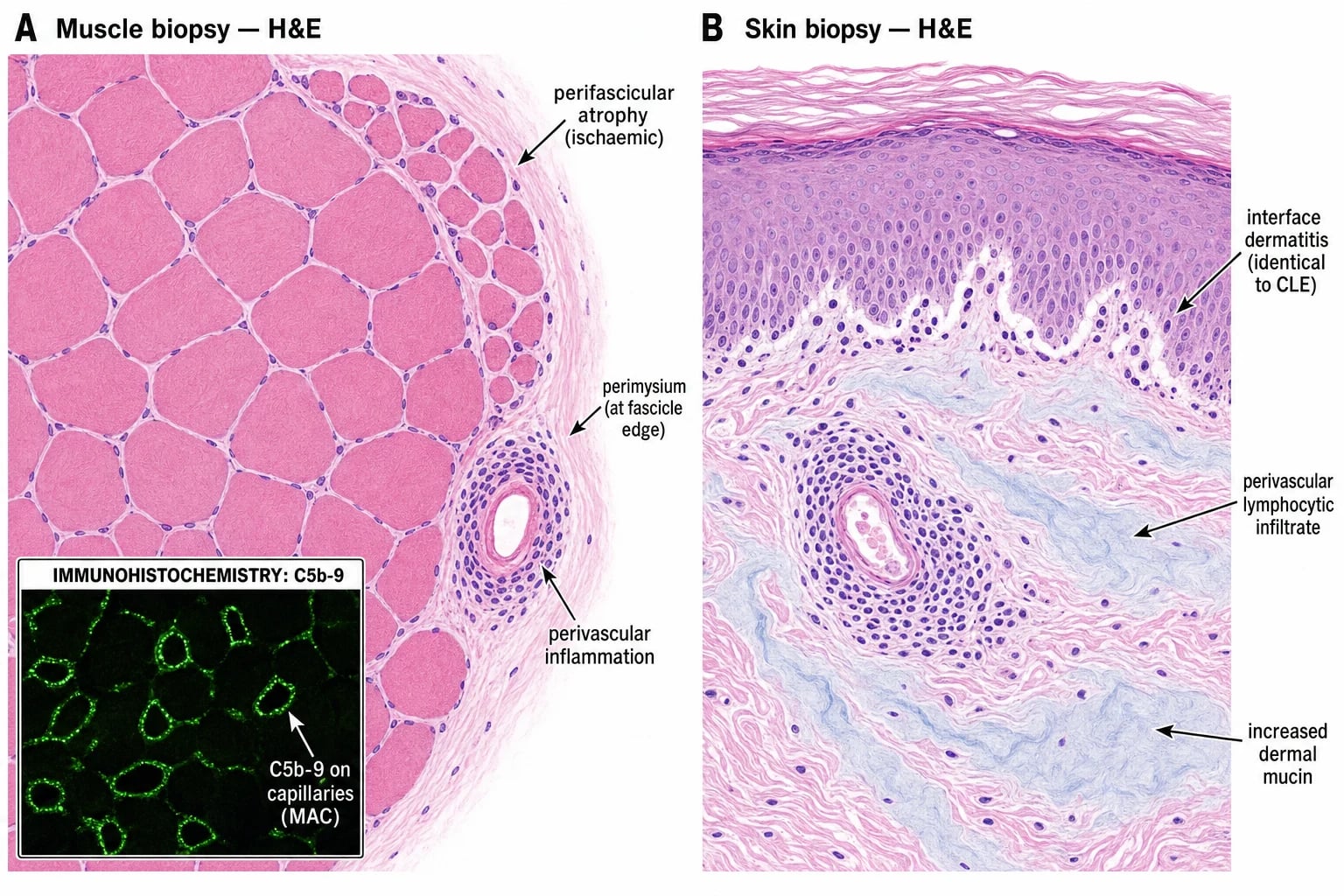
- Perifascicular atrophy — small, angulated, basophilic myofibres at the periphery of the fascicle; the histological hallmark.
- Perivascular, perimysial inflammation with CD4+ T cells and CD20+ B cells.
- Scattered necrotic and regenerating fibres.
- Immunohistochemistry:
- C5b-9 (MAC) deposition on endomysial capillaries — diagnostic of complement-mediated microangiopathy.
- MHC class I upregulation — diffuse, sarcolemmal, on perifascicular fibres especially.
- MxA (an IFN-inducible protein) — positive in the perifascicular rim and capillaries (IFN signature).
- Skin biopsy (H&E): identical to cutaneous lupus erythematosus — interface dermatitis (basal vacuolar change, perivascular lymphocytic infiltrate, increased dermal mucin).[2]
7.7 Interstitial lung disease (ILD) screen
- PFTs: FVC, TLC, DLCO. A falling DLCO and falling FVC in a DM patient over weeks → screen aggressively for ILD.
- HRCT chest: bilateral, basal, subpleural reticular opacities, traction bronchiectasis, ground-glass (and in anti-MDA5 RP-ILD, rapidly progressive consolidation with or without pneumomediastinum).
- 6-minute walk test with pulse oximetry — exercise desaturation supports ILD.
- Echocardiogram if pulmonary hypertension suspected.[3][6]
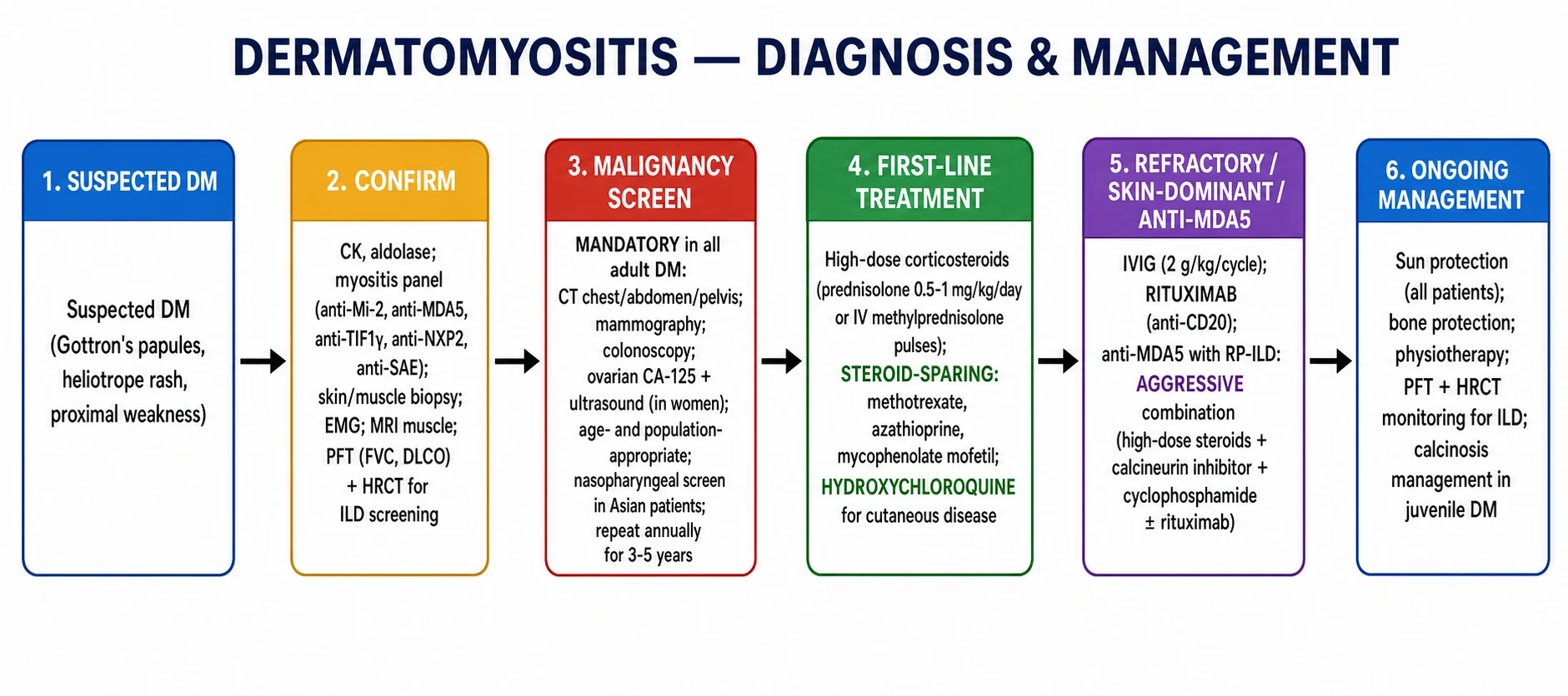
7.8 Malignancy screen (MANDATORY in adults)[1][2][4]
Because ~ 15 to 30 percent of adult DM is paraneoplastic, age- and sex-appropriate cancer screening is mandatory at diagnosis and annually for 3 to 5 years: [1]
- All adults: CT chest/abdomen/pelvis with contrast; colonoscopy (age-appropriate, often ≥ 45); FBC, ESR, CRP, lactate dehydrogenase, transaminases; PSA (men > 50); CA-125 + transvaginal ultrasound (women, especially anti-TIF1-gamma and anti-NXP2 positive); mammography (women ≥ 40, ≥ 50 per region).
- Asian populations (especially East/Southeast Asian): nasopharyngoscopy ± MRI nasopharynx; EBV serology — nasopharyngeal carcinoma is the leading paraneoplastic malignancy in this cohort.
- Repeat annually for 3 to 5 years after diagnosis; the window of highest risk is the first 12 months.
- Anti-TIF1-gamma and anti-NXP2 are the strongest individual predictors of an associated malignancy.[1][2][3]
8. Management — Resuscitation & Acute Care
DM is rarely a true resuscitation emergency, but three scenarios are: [1]
- Anti-MDA5 rapidly progressive ILD (RP-ILD): admit, often to HDU/ICU, for IV methylprednisolone pulses (500 to 1000 mg/day × 3 to 5 days), then combination immunosuppression.
- Bulbar/diaphragmatic weakness with respiratory failure: ICU admission, non-invasive or invasive ventilation, NG/PEG feeding, IVIG 2 g/kg over 2 to 5 days.
- Visceral vasculopathy in juvenile DM (GI perforation, CNS): resuscitation, surgical co-management, active immunosuppression.[3][4][4]
9. Management — Definitive Treatment
DM management is staged and antibody-informed.[4][4]
9.1 Muscle disease — induction
- Corticosteroids (first-line):
- Oral prednisolone 1 mg/kg/day (typical max 60 to 80 mg/day) for 4 to 6 weeks, then taper slowly over 9 to 12 months.
- IV methylprednisolone 500 to 1000 mg/day × 3 to 5 days for severe disease (dysphagia, respiratory weakness, marked weakness, ILD), followed by oral prednisolone.
- Steroid-sparing immunosuppression (start concurrently to allow steroid taper):
- Methotrexate 7.5 to 25 mg orally or SC weekly + folic acid 5 mg on non-MTX days. Most effective steroid-sparing for DM.[4][4]
- Azathioprine 2 mg/kg/day (after TPMT activity checked); alternative first-line.
- Mycophenolate mofetil 2 to 3 g/day — preferred in ILD-positive DM for steroid-sparing lung benefit.
- Ciclosporin or tacrolimus — preferred in anti-MDA5 DM (lung benefit).
- IVIG (2 g/kg/cycle over 2 to 5 days, every 4 weeks for 3 to 6 months, then taper): effective for refractory disease and for severe oesophageal/diaphragmatic involvement; can be added to steroid + immunosuppression.
- Refractory disease:
- Rituximab 1 g IV on days 0 and 14 (anti-CD20) — the RIM trial missed its primary endpoint but a secondary analysis showed steroid-sparing benefit, especially in anti-synthetase and anti-Mi-2 subgroups.
- JAK inhibitors (tofacitinib 5 mg BD; baricitinib 2 to 4 mg/day) — emerging evidence for refractory disease including anti-MDA5 DM.
- Cyclophosphamide (IV, monthly for 6 to 9 months) — used for RP-ILD and severe systemic disease.
- Anti-TNF agents (infliximab, adalimumab) — paradoxical use; reserved for refractory cutaneous disease.
- Apremilast — emerging for refractory cutaneous disease.[4][4]
9.2 Cutaneous disease
- Photoprotection: SPF 50+ broad-spectrum sunscreen daily; physical barrier (long sleeves, hat) — the single most important intervention for cutaneous disease.[2]
- Topical corticosteroids (mid-to-high potency for the body; lower potency for face/folds).
- Topical calcineurin inhibitors (tacrolimus 0.1 percent ointment or pimecrolimus) for face/folds and steroid-sparing.
- Hydroxychloroquine 200 to 400 mg/day (5 to 6.5 mg/kg lean body weight) for cutaneous disease; annual OCT macula + visual fields because of retinopathy risk. Add quinacrine if monotherapy fails.[2][4]
- IVIG for refractory cutaneous disease (highly effective).
- Methotrexate, mycophenolate, JAK inhibitors for refractory cutaneous disease.
9.3 Anti-MDA5 RP-ILD (a separate, urgent pathway)
- IV methylprednisolone 500 to 1000 mg/day × 3 days followed by 1 mg/kg/day prednisolone.
- Plus a calcineurin inhibitor (tacrolimus trough 5 to 10 ng/mL or ciclosporin 3 to 5 mg/kg/day).
- Plus IV cyclophosphamide (500 to 750 mg/m² every 4 weeks × 6).
- Plus / or rituximab (1 g on days 0 + 14) for refractory or as part of induction.
- Consider tofacitinib (5 mg BD) and plasma exchange in refractory disease; early referral for lung transplantation for irreversible disease.[3][6]
9.4 Malignancy-associated DM
- Treat the malignancy first — this often improves the DM. Continue DM treatment concurrently.
- Repeat malignancy screening annually for 3 to 5 years, especially in the first 12 months.[1][4]
9.5 Calcinosis (juvenile DM)
- No proven medical therapy. Surgical excision if symptomatic (pain, ulceration, infection).
- Investigational: bisphosphonates (e.g., pamidronate), probenecid, sodium thiosulphate, TNF inhibitors.
- Prevention: early aggressive treatment of DM reduces calcinosis risk.[1][5]
10. Subtypes & Special Scenarios
10.1 Classic DM[1][4]
The prototypical adult with Gottron papules, heliotrope rash and subacute proximal weakness, CK elevated, responds well to steroids + methotrexate, and the prognosis is favourable if anti-Mi-2 positive. [1]
10.2 Amyopathic/Hypomyopathic DM (ADM/CADM)[2][4][3][6]
- 5 to 20 percent of DM cases.
- Cutaneous signs only (amyopathic) or with subclinical muscle involvement (hypomyopathic), with normal CK for ≥ 6 months.
- Anti-MDA5 DM is enriched in the amyopathic cohort — never assume ADM is "safe": actively screen for ILD with HRCT and PFTs (baseline and serial).
- In amyopathic anti-Mi-2 DM, prognosis for skin is often favourable; in amyopathic anti-MDA5 DM, prognosis is guarded because of RP-ILD. [1]
10.3 Anti-MDA5 DM[3][6]
- Demographics: more common in East Asian adults, but recognised globally.
- Skin: Gottron papules, often with ulceration; palmar papules (painful erythematous papules over the interphalangeal joints and palmar creases); mechanic's hands; panniculitis; oral ulceration; hair loss.
- Joint: often severe arthritis.
- Lung: RP-ILD with bilateral consolidation, ground-glass and (in severe cases) pneumomediastinum.
- Prognosis: without aggressive combination immunosuppression, 6-month mortality 40 to 60 percent. With early aggressive treatment, mortality falls toward 20 to 30 percent.
- Markers of poor prognosis: elevated ferritin, KL-6, IL-18, rapid fall in DLCO, anti-MDA5 titre trend. [1]
10.4 Anti-synthetase syndrome[1][4]
- Antibodies: anti-Jo-1 (anti-histidyl-tRNA synthetase), anti-PL-7, anti-PL-12, anti-EJ, anti-OJ, anti-KS, anti-Zo.
- Clinical pentad (often incomplete): myositis, ILD, mechanic's hands, arthritis, Raynaud's, fever.
- ILD: tends to be chronic progressive rather than rapidly progressive (contrast with anti-MDA5), but can still be severe and is the main driver of mortality.
- Treatment: steroids + methotrexate or mycophenolate; rituximab for steroid-sparing effect.[4][4]
10.5 Paraneoplastic DM[1][2][3]
- Antibody signature: anti-TIF1-gamma (strongest predictor, ~ 50 percent have cancer), anti-NXP2.
- Common cancers: ovary, breast, lung, gastric, colorectal, pancreatic, prostate, bladder, nasopharyngeal (especially East/Southeast Asian populations), non-Hodgkin lymphoma.
- Management: cancer treatment first; DM often improves with cancer cure.
- Tempo: the malignancy may precede, be concurrent with, or follow the DM diagnosis; the malignancy risk window is greatest in the first 12 to 24 months. [1]
10.6 Juvenile DM (JDM)[5][5]
- Demographics: peak 5 to 15 years; female predominance 2 to 3:1.
- Features: same skin and muscle signs as adult; calcinosis cutis more common; vasculopathy (GI, CNS) more common.
- Course: generally more responsive to treatment than adult DM.
- Malignancy: rare; not a routine screen.
- Treatment: steroids + methotrexate (front-line), IVIG for refractory disease; biologics (rituximab, tofacitinib) for refractory disease.
- Outcome criteria: ACR/EULAR 2016 response criteria for JDM define minimal, moderate and major improvement.[5]
10.7 Immune checkpoint inhibitor-induced DM[1]
- Triggers: anti-PD-1 (nivolumab, pembrolizumab), anti-PD-L1, anti-CTLA-4 (ipilimumab).
- Onset: variable; can be weeks to months after first infusion; some cases after drug cessation.
- Phenotype: classic DM, hypomyopathic DM, anti-MDA5 DM, anti-TIF1-gamma DM.
- Management: immunotherapy pause; systemic corticosteroids; severe disease may require IVIG or other immunosuppression; oncological input on balance of cancer treatment vs DM control. [1]
11. Complications & Pitfalls[1][2][3][4]
11.1 Disease complications
- Interstitial lung disease: chronic progressive (antisynthetase) or rapidly progressive (anti-MDA5); respiratory failure is the leading cause of DM mortality.
- Cardiac: conduction abnormalities, myocarditis, pericarditis, heart failure.
- Dysphagia, dysphonia, aspiration pneumonia: cricopharyngeal and upper oesophageal striated muscle weakness; in JDM, may be associated with visceral vasculopathy.
- Calcinosis cutis (JDM): chronic painful ulceration, secondary infection.
- Vasculopathy (JDM): GI ulceration/perforation, CNS vasculopathy with stroke-like events.
- Secondary infection: from chronic immunosuppression + skin breakdown.
- Malignancy: paraneoplastic in 15 to 30 percent of adult DM; malignancy drives prognosis. [1]
11.2 Treatment complications
- Corticosteroids: Cushingoid, weight gain, hyperglycaemia, hypertension, osteoporosis, cataract, infection, adrenal suppression on taper.
- Methotrexate: hepatotoxicity, marrow suppression, pneumonitis, mucositis, teratogenicity; strict contraception in women of childbearing age.
- Azathioprine: marrow suppression (especially in TPMT-deficient patients — test activity before use), hepatotoxicity, increased infection risk, rare lymphoma/SCC.
- Mycophenolate: GI upset, leucopenia, hepatotoxicity; teratogenic (avoid in pregnancy).
- Ciclosporin/tacrolimus: nephrotoxicity, hypertension, tremor, hyperkalaemia, gum hyperplasia (ciclosporin).
- Cyclophosphamide: haemorrhagic cystitis, bladder cancer, infertility, marrow suppression.
- Rituximab: infusion reactions, hepatitis B reactivation (screen), progressive multifocal leukoencephalopathy (PML, rare), hypogammaglobinaemia, late-onset neutropenia.
- IVIG: aseptic meningitis, thrombosis, acute kidney injury (sucrose formulations), anaphylaxis in IgA deficiency (screen). [1]
11.3 Diagnostic and management pitfalls
- Missing amyopathic DM: attributing Gottron papules and heliotrope rash to "eczema" or "rosacea" for years.
- Missing anti-MDA5 RP-ILD: failing to order anti-MDA5 (and PFTs + HRCT) in any ADM patient.
- Missing paraneoplastic DM: omitting cancer screening in any adult DM.
- Misdiagnosis as SLE: CLE and DM share an interface biopsy; check for Gottron papules, heliotrope rash, and muscle weakness.
- Misdiagnosis as IBM: assuming any older adult with weakness has IBM — IBM is distal and asymmetric; missing DM is a treatable disease.
- Undertreating calcinosis: no proven medical therapy; surgery reserved for symptomatic lesions; prevention via early DM control.
- Treating PM as DM: the two have different mechanisms (DM: humoral/complement microvascular; PM: cellular CD8+ endomysial); in PM, the steroid-sparing ladder and IVIG use are the same, but the malignancy screen is less aggressive (paraneoplastic PM is less common).
- Methotrexate + hydroxychloroquine GI upset: often intolerable; consider topical, switching to mycophenolate, or IVIG.
- Sun exposure worsening: all DM patients need SPF 50+ education (the rash is photoaggravated).
- Pregnancy planning: methotrexate and mycophenolate are teratogenic; switch to azathioprine or IVIG before conception.[4]
12. Prognosis & Disposition[1][3][4]
DM prognosis is antibody-driven: [1]
| Antibody | Prognosis |
|---|---|
| Anti-Mi-2 | Best — treatment-responsive, low malignancy, low ILD |
| Anti-NXP2 (adult) | Intermediate; malignancy drives prognosis |
| Anti-SAE | Intermediate; severe dysphagia common |
| Anti-Jo-1 | Intermediate; chronic progressive ILD |
| Anti-TIF1-gamma | Intermediate; malignancy drives prognosis (~ 50 percent have cancer) |
| Anti-MDA5 | Worst — RP-ILD mortality 40 to 60 percent at 6 months untreated |
| JDM | Generally good prognosis; calcinosis and vasculopathy drive morbidity |
Disposition: [1]
- Classic DM without ILD: managed as outpatient; urgent rheumatology + dermatology review.
- DM with mild ILD: outpatient management with PFT/HRCT surveillance every 3 to 6 months; immunosuppression initiated.
- DM with progressive ILD (falling FVC, falling DLCO, progressive dyspnoea): hospital admission; aggressive combination therapy.
- DM with RP-ILD or anti-MDA5: ICU/HDU; IV methylprednisolone pulses; combination immunosuppression.
- DM with dysphagia/aspiration: speech and language therapy, NG/PEG feeding, supervised oral intake.
- JDM with visceral vasculopathy: emergency admission, surgical co-management. [1]
13. Special Populations
13.1 Pregnancy[4]
- Pre-conception: switch methotrexate and mycophenolate to azathioprine (safer in pregnancy) and plan quiescent disease for at least 6 months before conception.
- During pregnancy: prednisolone (avoid fluorinated steroids like dexamethasone/betamethasone in the first two trimesters unless fetal lung maturation), azathioprine (continued), hydroxychloroquine (continue), IVIG (safe).
- Continue: PFT monitoring; foetal monitoring; tight glucose control; consider aspirin prophylaxis.
- Post-partum flare is common — anticipate and treat aggressively.
- Multidisciplinary management with obstetrics, rheumatology, dermatology, neonatology. [1]
13.2 Elderly ([2])[1][2]
- High paraneoplastic risk: malignancy screening mandatory and high-yield.
- Differential with IBM is important: DM is subacute, proximal, symmetric, steroid-responsive; IBM is chronic, distal, asymmetric, steroid-resistant.
- Comorbidity burden — cardiac, renal, diabetes — limits immunosuppression choices; hydroxychloroquine and IVIG are safer than cyclophosphamide.
- Falls risk from proximal weakness + visual/vestibular issues — falls prevention program. [1]
13.3 Children (JDM)[5][5]
- Distinct from adult DM: calcinosis and vasculopathy dominate; malignancy is rare.
- Treat actively and early to prevent calcinosis and chronic disability.
- Steroid + methotrexate is first-line; IVIG for refractory; rituximab and JAK inhibitors for further refractory disease.
- Multidisciplinary team: paediatric rheumatology, dermatology, physiotherapy, psychology, school liaison.
- ACR/EULAR 2016 response criteria are used to define improvement.[5]
13.4 Immunocompromised and HIV[1]
- DM can occur in HIV but is uncommon; differential includes HIV myopathy (a separate entity with proximal weakness but no skin signs), drug-induced myopathy (AZT), and opportunistic infection.
- Treatment of DM in HIV requires caution with immunosuppression; rituximab is a relatively safe option.
- Hepatitis B and C screening before rituximab; HCV-associated DM may respond to direct-acting antiviral therapy. [1]
14. Evidence, Guidelines & Controversies[1][4][5][4]
14.1 Major landmark trials and sources
- EULAR/ACR 2017 classification criteria for adult and juvenile IIM (Bottai, Lundberg et al.) — the modern diagnostic framework.[1][5]
- EULAR 2019 recommendations for treatment of IIM.
- RIM trial (Oddis 2013): rituximab in refractory myositis — missed primary endpoint at 44 weeks (placebo crossover design), but secondary analysis showed benefit; now widely used.
- PROFILE trial and biomarker studies — building the evidence base for new biologics and JAK inhibitors.
- Lundberg & Fujimoto 2021 Nature Reviews Disease Primers (the most comprehensive review).[1]
- Lu & Peng 2024 Nat Rev Rheumatol review of anti-MDA5 DM.[3]
14.2 Regional guideline differences
EULAR/ACR 2017 classification criteria are the international diagnostic framework. Treat with high-dose corticosteroids + methotrexate/azathioprine/mycophenolate, IVIG for refractory disease, rituximab for refractory myositis. Anti-MDA5 RP-ILD: aggressive combination immunosuppression.
Australian and New Zealand consensus aligns with EULAR. Mycophenolate often first-line for ILD-positive DM. PBS subsidised IVIG is available for refractory DM via the Australian National Blood Authority criteria.
14.3 Current controversies
- Is a muscle biopsy always necessary? EULAR/ACR 2017 allows a > 55 percent probability without biopsy and > 90 percent with biopsy; in classic DM with typical skin and EMG, biopsy may not change management. In atypical or suspected IBM/IMNM, biopsy is mandatory.
- Rituximab positioning: the RIM trial missed its primary endpoint; many clinicians still use it for refractory disease, but its optimal place (after one or two steroid-sparing agents) remains debated.
- JAK inhibitors: growing evidence, especially for anti-MDA5 and refractory cutaneous disease, but licensing is off-label and long-term safety data are emerging.
- Duration of immunosuppression: most clinicians continue treatment for 2 to 3 years minimum after remission; some adult patients require long-term low-dose therapy. [1]
15. Exam Pearls & High-Yield Minutiae[1][2][4][7]
DM cutaneous signs — mnemonic
HELP-GOT-SHAV
Pathognomonic — violaceous periorbital (upper eyelid)
Often accompanies heliotrope
Acquired; insulin resistance; juvenile DM
Anti-MDA5 — painful over palmar creases
Pathognomonic — over MCP and IP joints
Erythema over knuckles (not papules)
Nailfold capillary change
Posterior neck/shoulders photosensitive
Lateral thighs; very specific
Anterior neck/chest photosensitive
Anterior neck/chest photosensitive distribution
DM autoantibody prognosis ladder — mnemonic
Mi-Mi-N-M-T-J-M
Classic DM, treatment-responsive, low malignancy
Adult malignancy; juvenile calcinosis
Strongest cancer predictor
Chronic ILD + mechanic's hands + arthritis + Raynaud's
RP-ILD; mortality 40–60% at 6 months untreated
Red Flags
Exam application bank (NEET-PG / INICET)
One-line answer
Dermatomyositis (DM) is an autoimmune idiopathic inflammatory myopathy (IIM) characterised by pathognomonic cutaneous signs (Gottron papules, heliotrope rash, Gottron sign, shawl sign, V-sign, holster sign, mechanic's hands, nailfold capillary changes) and a spectrum of muscle involvement ranging from fulminant proximal weakness to clinically amyopathic disease (ADM/CADM). Pathogenesis centres on two interlocking pathways — a type I interferon signature (plasmacytoid dendritic cell release of IFN-alpha/beta, MHC class I upregulation, sustained innate/adaptive activation) and complement-mediated microangiopathy (C1q → C3 → C5b-9 membrane attack complex on endomysial capillaries → capillary dropout → perifascicular atrophy, the histological hallmark). Myositis-specific autoantibodies (anti-Mi-2, anti-MDA5/CADM-140, anti-TIF1-gamma/p155-p140, anti-NXP2, anti-SAE, anti-Jo-1/anti-synthetase,
Worked stems (answer without another resource)
Stem 1 — Classic presentation. Map symptoms to mechanism; name the first investigation and first treatment step with dose/route if drug therapy is standard. [1]
Stem 2 — Unstable / complicated. List red flags that force immediate resuscitation, theatre, ICU, antidote, or reperfusion — and what you do in the first 15 minutes. [1]
Stem 3 — Atypical group. Elderly, pregnancy, child, or immunocompromised: how presentation and thresholds change. [1]
Stem 4 — Differential trap. Name the three closest mimics and one discriminator for each. [1]
Stem 5 — Disposition. Who goes home with safety-netting, who is admitted, who needs HDU/ICU/theatre, and what follow-up is mandatory. [1]
Rapid viva checklist
- Definition + classification
- Pathophysiology chain
- Bedside signs / criteria
- Score with exact components (if any)
- Emergency bundle
- Definitive therapy with doses
- Complications of disease and of treatment
- Special populations
- Guideline/trial name if classic
- Three exam traps
Coverage self-check
If you cannot answer any stem above from this page alone, re-read the matching section — the page is intended to be self-sufficient for final-prof and NEET-PG/INICET questions on Dermatomyositis.
[1]References
- [1]Lundberg IE, Fujimoto M, Vencovsky J, et al. Idiopathic inflammatory myopathies Nat Rev Dis Primers, 2021.PMID 34857798
- [2]DeWane ME, Waldman R, Lu J. Dermatomyositis: Clinical features and pathogenesis J Am Acad Dermatol, 2020.PMID 31279808
- [3]Lu X, Peng Q, Wang G. Anti-MDA5 antibody-positive dermatomyositis: pathogenesis and clinical progress Nat Rev Rheumatol, 2024.PMID 38057474
- [4]Waldman R, DeWane ME, Lu J. Dermatomyositis: Diagnosis and treatment J Am Acad Dermatol, 2020.PMID 31279813
- [5]Ashton C, Paramalingam S, Stevenson B, et al. Idiopathic inflammatory myopathies: a review Intern Med J, 2021.PMID 34155760
- [6]Castro-Molina SA, Méndez-Flores S. [Anti-MDA5 dermatomyositis. Literature review] Rev Med Inst Mex Seguro Soc, 2023.PMID 36542793
- [7]Tanboon J, Nishino I. Update on dermatomyositis Curr Opin Neurol, 2022.PMID 35942671