Dermatology · Medicine
Dermoscopy and image-based diagnosis
Also known as Dermatoscopy · Epiluminescence microscopy · Image-based skin diagnosis · Digital dermoscopy monitoring · Chaos and clues · Teledermoscopy · Dermoscopic pattern analysis
Board-level application module for dermoscopy as image-based diagnosis: two-step algorithm, chaos-and-clues pattern analysis, vascular morphology dictionary, non-melanocytic tumour patterns, special-site rules (face, acral, nail), dermoscopy–histology correlation, inflammoscopy and trichoscopy snapshots, digital monitoring thresholds, teledermoscopy quality, AI-assisted classification limits, and a red-flag biopsy algorithm. Complements the principles leaf with decision-focused exam content.
On this page & tools
Your progress
Saved locally on this device.
Exam tags
Red flags
Scope of this leaf
This module is the application / image-diagnosis companion to dermoscopy-principles. Focus: decision algorithms, pattern dictionaries, special sites, monitoring vs excision thresholds, and AI/teledermoscopy limits for board exams (FRCDerm, ABD, FACD, MRCP SCE, NEET-PG/IADVL). [1]
Instrumentation quick recap
- Non-polarised immersion: liquid interface required; excellent for superficial pigment and keratin plugs.[1]
- Polarised: no fluid; better for vessels and shiny white lines (chrysalis).[1]
- Hybrid devices toggle modes — use both when a lesion is equivocal.
The diagnostic algorithm
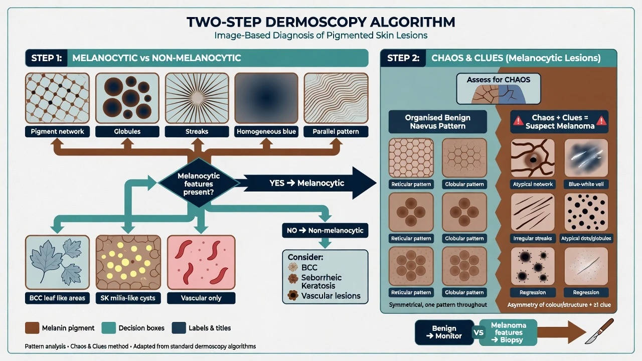
Step 1 — Melanocytic or not?
Melanocytic if any classic criterion is present: pigment network, aggregated globules, streaks/pseudopods, homogeneous blue pigmentation, or parallel pattern (acral/mucosa).[1][2] If none → non-melanocytic pathway (BCC, SK, vascular, keratinocyte neoplasia, dermatofibroma, etc.).
Step 2 — Within melanocytic lesions
Use pattern analysis (asymmetry of colours/structures) and/or checklists: [1]
| Tool | Core idea | Exam hook |
|---|---|---|
| Chaos & clues | Disorder + melanoma clues → biopsy | Practical primary-care/derm workflow[3][4] |
| Menzies | 2 negative features exclude; ≥1 of 9 positives suspicious | High sensitivity screening |
| Argenziano 7-point | Major 2 pts / minor 1 pt; ≥3 suspicious | Semi-quantitative |
| Stolz ABCD | Weighted A/B/C/D score bands | Historic prospective validation[5] |
| 3-point checklist | Asymmetry, atypical network, blue-white | Rule-out tool for non-experts |
IDS web-based work supports that trained criteria can be applied with measurable validity and reliability — training matters.[2]
Chaos and clues (routine practice)
- Is there chaos (asymmetric colour or structure)?
- If yes, seek clues (e.g. eccentric structureless areas, thick reticular lines, grey/blue structures, peripheral black dots/globules, pseudopods, white lines, polymorphous vessels).
- Chaos + clue(s) → biopsy/excise rather than long-term observation.[3][4]
Organised lesions without clues may be monitored or left — unless history (change, symptoms) or special-site rules intervene. [1]
Vascular and structure dictionary
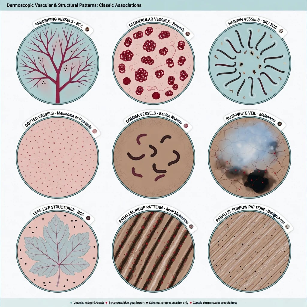
| Pattern | Classic association |
|---|---|
| Arborising vessels | BCC |
| Glomerular vessels | Bowen disease (SCC in situ) |
| Hairpin vessels | SK / some SCC |
| Dotted vessels | Melanoma, Spitz, psoriasis (context!) |
| Comma vessels | Benign dermal naevus |
| Linear-irregular / polymorphous | Melanoma red flag |
| Crown vessels | Sebaceous hyperplasia |
| Blue-white veil | Melanoma (with other chaos) |
| Leaf-like / spoke-wheel | BCC |
| Milia-like cysts, comedo openings | SK |
| Parallel ridge | Acral melanoma |
| Parallel furrow / lattice / fibrillar | Often benign acral naevus |
Vascular decoding and keratinocyte progression features (AK → SCC) are high-yield for image stations.[1][9]
Special sites
Face (lentigo maligna pathway)
Sun-damaged facial skin uses pseudonetwork architecture. Concerning: asymmetric pigmented follicular openings, rhomboidal structures, annular-granular pattern, progressive obliteration of follicular openings.[7] Partial mapping biopsies may be guided by the most dermoscopically atypical sectors of a large patch.
Acral
Parallel ridge pattern (pigment on ridges that bear eccrine openings) is a melanoma red flag; parallel furrow, lattice, and fibrillar patterns favour acral naevi when classic.[6][7] Do not apply truncal network rules naively to palms and soles.
Nail
Assess band width, colour homogeneity, border regularity, and periungual pigment (micro-Hutchinson). Single-digit progressive melanonychia in adults needs a low threshold for specialist matrix assessment.[7]
Dermoscopy–histology correlation
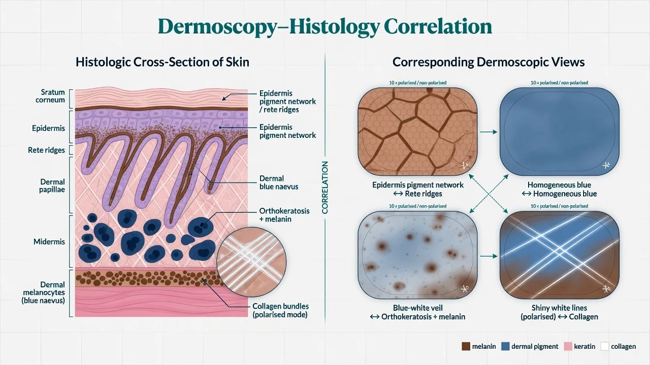
Understanding correlates prevents magical thinking: [1]
- Pigment network ↔ melanin along elongated rete ridges.[1]
- Globules ↔ melanocytic nests.
- Blue structures ↔ deeper dermal melanin (Tyndall).
- Blue-white veil ↔ compact orthokeratosis over melanin-rich dermis.
- Shiny white lines ↔ collagen remodel under polarised light.
Correlation also explains why dermoscopy improves biopsy targeting and can reduce excision of stereotypical benign SK/BCC patterns in expert hands — without replacing histology for suspicious lesions.[1]
Beyond tumours: inflammoscopy, trichoscopy, entomodermoscopy
- Inflammoscopy: regularly distributed dotted vessels on a red background suggest psoriasis; Wickham striae favour lichen planus; patterns refine differentials among common inflammatory dermatoses.[8]
- Trichoscopy: yellow dots, black dots, broken hairs and exclamation-mark hairs support alopecia areata; white dots / loss of follicular openings favour scarring alopecias — the dermatoscope is now standard in hair clinics.[12]
- Entomodermoscopy: classic scabies “delta wing / jet with contrail” sign speeds bedside confirmation (details also in scabies atlas leaf).
Digital monitoring, teledermoscopy, and AI
Short-term digital dermoscopy monitoring
Appropriate for flat, feature-poor, low-suspicion melanocytic lesions when immediate excision is not mandatory. Combine static morphology with dynamic change calculators/features; clear growth, new colours, or new melanoma clues end monitoring.[11] Exclude nodular, ulcerated, amelanotic red-flag, and high-anxiety non-compliant contexts.
Teledermoscopy
Requires sharp focus, correct mode (polarised/immersion), macroscopic context photos, and clinical history. Garbage-in images produce garbage-out triage. [1]
AI
Convolutional networks have reached dermatologist-level classification performance on curated image tasks (landmark Esteva 2017 Nature work), but real-world deployment faces dataset shift, skin-of-colour under-representation, rare entities, and medicolegal accountability.[10] Exam-safe stance: AI may assist; clinicopathologic correlation decides.
Image-based diagnosis — exam anchors
Decision algorithm: observe, monitor, or biopsy
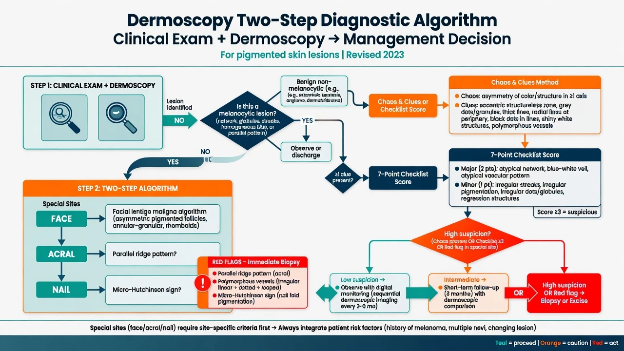
- Clinical context (history of change, immunosuppression, prior melanoma).
- Macroscopic ABCDE / EFG for nodules.
- Dermoscopy two-step + site rules.
- Excise/biopsy now if melanoma-specific features, parallel ridge, LM facial clues, micro-Hutchinson pathway, or polymorphous vessels in a concerning lesion.
- Short-term digital monitor only if flat, low-suspicion, and follow-up assured.[11]
- Histology remains gold standard for definitive diagnosis of excised tissue.
Preferred biopsy when melanoma is realistic: full-thickness excisional biopsy with narrow margins (or carefully planned partial sampling of large LM) — do not rely on superficial shave that understages Breslow. [1]
Biopsy now (image red flags)
RIDGE
Parallel ridge until proven otherwise
Polymorphous / linear-irregular
Chaos and melanoma clues
With other atypical features
Micro-Hutchinson; rhomboidal face
Pitfalls
- SK-like verrucous melanoma and collision tumours.
- Recurrent naevus (pseudomelanoma) after incomplete shave — history is everything.
- Over-reliance on a single criterion (e.g. any dotted vessels).
- Monitoring a nodular pink lesion because “dermoscopy was inconclusive.”
- Trainee overconfidence without structured pattern training.[2]
Regional notes
Core pattern language is global (IDS terminology). Resource-limited settings still gain from handheld dermoscopy for triage; AI smartphone tools need the same red-flag discipline as clinic devices.[10]
Clinical pearl
Red flags
Exam application bank (NEET-PG / INICET)
One-line answer
Board-level application module for dermoscopy as image-based diagnosis: two-step algorithm, chaos-and-clues pattern analysis, vascular morphology dictionary, non-melanocytic tumour patterns, special-site rules (face, acral, nail), dermoscopy–histology correlation, inflammoscopy and trichoscopy snapshots, digital monitoring thresholds, teledermoscopy quality, AI-assisted classification limits, and a red-flag biopsy algorithm. Complements the principles leaf with decision-focused exam content.
Worked stems (answer without another resource)
Stem 1 — Classic presentation. Map symptoms to mechanism; name the first investigation and first treatment step with dose/route if drug therapy is standard. [1]
Stem 2 — Unstable / complicated. List red flags that force immediate resuscitation, theatre, ICU, antidote, or reperfusion — and what you do in the first 15 minutes. [1]
Stem 3 — Atypical group. Elderly, pregnancy, child, or immunocompromised: how presentation and thresholds change. [1]
Stem 4 — Differential trap. Name the three closest mimics and one discriminator for each. [1]
Stem 5 — Disposition. Who goes home with safety-netting, who is admitted, who needs HDU/ICU/theatre, and what follow-up is mandatory. [1]
Rapid viva checklist
- Definition + classification
- Pathophysiology chain
- Bedside signs / criteria
- Score with exact components (if any)
- Emergency bundle
- Definitive therapy with doses
- Complications of disease and of treatment
- Special populations
- Guideline/trial name if classic
- Three exam traps
Coverage self-check
If you cannot answer any stem above from this page alone, re-read the matching section — the page is intended to be self-sufficient for final-prof and NEET-PG/INICET questions on Dermoscopy and image-based diagnosis.
Expanded exam teaching (depth pass)
Clinical reasoning
For Dermoscopy and image-based diagnosis, examiners test whether you can prioritise life threats, choose the right first test, and give specific therapy (agent, dose, route, timing). Generic phrases without numbers score poorly.
Mechanism → feature map
Build a short chain: cause → pathophysiologic intermediate → clinical feature → complication. Every major symptom in the classic vignette should sit on that chain.
Investigation strategy
- Bedside/first-line tests that change immediate management
- Confirmatory or staging tests
- What a normal result does not exclude
- When not to delay treatment for imaging (unstable patient)
Management ladder
- Resuscitation / ABC / sepsis or haemorrhage bundle as relevant
- Specific antidote / procedure / antimicrobial / reperfusion / surgery
- Supportive care and monitoring targets
- Definitive long-term therapy and secondary prevention
- Disposition and safety-net advice
Special populations
Always prepare one line each for children, pregnancy, elderly, renal/hepatic impairment, and immunocompromised patients when the topic allows.
Pitfalls that fail candidates
- Treating the number not the patient
- Missing pregnancy status when relevant
- Imaging before stabilisation
- Wrong empiric cover or wrong antidote timing
- Incomplete counselling on recurrence, adherence, or red-flag return
Board-level application module for dermoscopy as image-based diagnosis: two-step algorithm, chaos-and-clues pattern analysis, vascular morphology dictionary, non-melanocytic tumour patterns, special-site rules (face, acral, nail), dermoscopy–histology correlation, inflammoscopy and trichoscopy snapshots, digital monitoring thresholds, teledermoscopy quality, AI-assisted classification limits, and a red-flag biopsy algorithm. Complements the principles leaf with decision-focused exam content. [1]
Structured revision sheet
Must-know numbers and names
List every score, size threshold, dose, and time window from this topic on a blank page from memory, then check against the sections above.
Three classic MCQ angles
- Most likely diagnosis given a vignette
- Next best step in management
- Most appropriate investigation
Three classic SAQ angles
- Pathophysiology in five steps
- Management algorithm with doses
- Complications and prevention
Clinical station flow
Greet → focused history → targeted exam → investigations → explain diagnosis → emergency care → definitive plan → safety-net / follow-up → answer examiner questions on mechanism and pitfalls.
References
- [1]Yélamos O, Braun RP, Liopyris K, Wolner ZJ, et al. Dermoscopy and dermatopathology correlates of cutaneous neoplasms J Am Acad Dermatol, 2019.PMID 30321581
- [2]Carrera C, Marchetti MA, Dusza SW, Argenziano G, et al. Validity and Reliability of Dermoscopic Criteria Used to Differentiate Nevi From Melanoma: A Web-Based International Dermoscopy Society Study JAMA Dermatol, 2016.PMID 27074267
- [3]Rosendahl C, Cameron A, McColl I, Wilkinson D. Dermatoscopy in routine practice - 'chaos and clues' Aust Fam Physician, 2012.PMID 22762066
- [4]Ramji R, Valdes-Gonzalez G, Oakley A, Rademaker M. Dermoscopic 'Chaos and Clues' in the diagnosis of melanoma in situ Australas J Dermatol, 2018.PMID 29094749
- [5]Nachbar F, Stolz W, Merkle T, Cognetta AB, et al. The ABCD rule of dermatoscopy. High prospective value in the diagnosis of doubtful melanocytic skin lesions J Am Acad Dermatol, 1994.PMID 8157780
- [6]Saida T, Koga H, Uhara H. Key points in dermoscopic differentiation between early acral melanoma and acral nevus J Dermatol, 2011.PMID 21175752
- [7]Thomas L, Phan A, Pralong P, Poulalhon N, et al. Special locations dermoscopy: facial, acral, and nail Dermatol Clin, 2013.PMID 24075549
- [8]Sgouros D, Apalla Z, Ioannides D, Katoulis A, et al. Dermoscopy of Common Inflammatory Disorders Dermatol Clin, 2018.PMID 30201145
- [9]Álvarez-Salafranca M, Zaballos P. [Translated article] Dermoscopy of Squamous Cell Carcinoma: From Actinic Keratosis to Invasive Forms Actas Dermosifiliogr, 2024.PMID 39102978
- [10]Esteva A, Kuprel B, Novoa RA, Ko J, et al. Dermatologist-level classification of skin cancer with deep neural networks Nature, 2017.PMID 28117445
- [11]Zenone M, Zocchi L, Moccia C, Passerini SG, et al. Digital dermoscopy monitoring of melanocytic lesions: Two novel calculators combining static and dynamic features to identify melanoma J Eur Acad Dermatol Venereol, 2022.PMID 34862986
- [12]Pirmez R. The dermatoscope in the hair clinic: Trichoscopy of scarring and nonscarring alopecia J Am Acad Dermatol, 2023.PMID 37591567