Dermatology · Medicine
Dermoscopy: principles and patterns
Also known as Dermoscopy · Dermatoscopy · Epiluminescence microscopy · Skin surface microscopy · Dermatoscopy of skin tumours · Trichoscopy · Onychoscopy
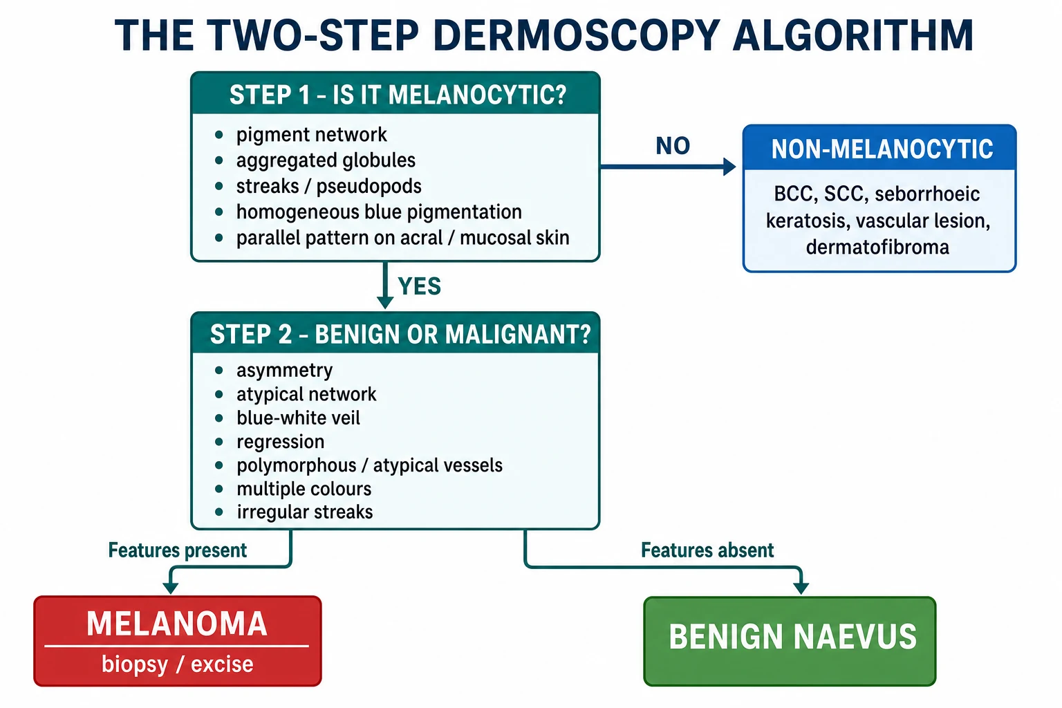
Dermoscopy (dermatoscopy, epiluminescence microscopy) is in-vivo 10x magnified skin-surface microscopy using a handheld contact dermatoscope that has transformed the accuracy of skin-cancer, pigmented-lesion, hair, nail and inflammatory-skin diagnosis. The foundational framework is the two-step algorithm (Argenziano): Step 1 — melanocytic vs non-melanocytic using pigment network, aggregated globules, streaks, homogeneous blue pigmentation or parallel pattern; Step 2 — within melanocytic lesions, apply melanoma-specific criteria (asymmetry, atypical network, blue-white veil, regression, polymorphous vessels, irregular streaks) through pattern analysis (Pehamberger) or checklist tools (Menzies 11-point; Argenziano 7-point; Stolz ABCD rule; Zalaudek 3-point; Lallas CASH algorithm for keratinising tumours). Equipment: contact polarised vs non-polarised immersion dermatoscope; the immersion fluid (alcohol/gel) is required with non-polarised instruments to abolish surface reflection. Vascular structures decoded: arborising=BCC, glomerular=Bowen's, hairpin=SK/SCC, dotted=melanoma/Spitz/psoriasis, comma=benign naevus, linear-irregular=melanoma, polymorphous=melanoma, crown=sebaceous hyperplasia, milky-red=melanoma. Special sites need bespoke criteria: face (pseudonetwork, rhomboidal structures=lentigo maligna), acral (parallel ridge=melanoma vs parallel furrow/lattice/fibrillar=benign), nail (micro-Hutchinson, longitudinal melanonychia bands), mucosa (parallel pattern, regression). Trichoscopy (hair) and entomodermoscopy (scabies delta-wing, Demodex follicular openings, tungiasis) and inflammoscopy (psoriasis glomerular/dotted, lichen planus Wickham striae) extend the technique. AI-assisted dermoscopy (Esteva 2017 Nature CNN) approaches dermatologist-level melanoma sensitivity. Examination-relevant pitfalls: small amelanotic/hypomelanotic and nodular melanomas, verrucous/papillomatous melanomas that mimic SK, and digital-monitoring false reassurance.
On this page & tools
Your progress
Saved locally on this device.
Exam tags
Red flags
Definition, Instrumentation and Physical Basis
Dermoscopy (dermatoscopy, epiluminescence microscopy, skin-surface microscopy) is the in-vivo examination of skin lesions through a hand-held magnification device — the dermatoscope — that couples a 10× optical system with either polarised light or a liquid interface to abolish surface reflection and render the stratum corneum translucent.[4] The technique raises diagnostic accuracy for melanoma from roughly 55 per cent with the naked eye to >85 per cent with dermoscopy in trained hands, with even greater gains for pigmented basal cell carcinoma and seborrhoeic keratosis.[4][8] It is now a required competency in dermatology training programmes worldwide.[4]
Both designs operate at 10–20× magnification; cross-polarised light penetrates to the superficial dermis and reveals vascular and collagen features invisible with immersion instruments, while non-polarised immersion instruments are excellent for superficial pigment pattern and keratin-filled structures (milia-like cysts, comedo-like openings). Modern hybrid instruments allow toggling between modes. [1]
Polarised vs non-polarised immersion
PIG
Crossed filters; NO fluid needed; reveals shiny white lines and deep vessels
Non-polarised REQUIRES alcohol/gel interface; surface becomes translucent
Ultrasound/chlorhexidine gel is non-flammable; alcohol is convenient but flammable
The Two-Step Algorithm
The foundational dermoscopic framework — every pigmented lesion is approached in two steps:[4][8]
Step 1: Melanocytic vs non-melanocytic
Decide whether the lesion is melanocytic (contains melanocytes producing melanin in a recognisable pattern). A lesion is melanocytic if ANY of the following are present: [1]
- Pigment network (reticular network) — brown lines forming a mesh with hypopigmented holes; represents melanin in basal keratinocytes along rete ridges.
- Aggregated globules — brown/black/blue dots = nests of melanocytes at the dermo-epidermal junction or papillary dermis.
- Streaks/pseudopods — radial projections at the periphery = spindle melanocytes.
- Homogeneous blue pigmentation — dermal melanin (e.g., blue naevus).
- Parallel pattern — on acral (palms/soles) or mucosal surfaces.
- If none of the above → classify as non-melanocytic (BCC, SCC, SK, vascular, dermatofibroma, etc.).[4]
Step 2: Benign vs malignant (melanocytic lesions only)
If melanocytic, assess for melanoma-specific features using pattern analysis, the Menzies 11-point checklist, the Argenziano 7-point checklist, the Stolz ABCD rule, the 3-point (Zalaudek) checklist or the BLINK algorithm. Each is examined.[4][7][8]
Algorithmic Approaches to Step 2
No single method is uniformly best; examiners frequently contrast pattern analysis (the gold standard for the experienced) with checklist methods that allow reproducible, semi-quantitative scoring.[4][8]
Pattern analysis (Pehamberger / consensus)
Method: assess each lesion for ten or more local features grouped under asymmetry, colours and dermoscopic structures, integrate them and reach a gestalt. The 2003 International Dermoscopy Society consensus meeting identified the original consensus terminology and is the foundation of the language of dermoscopy.[4][8] Validity and reliability were confirmed in the 2016 International Dermoscopy Society web-based study of more than 200 dermoscopists evaluating >1,000 lesions.[8]
Menzies 11-point checklist
Designed as a bedside algorithm. Two negative features (symmetry of pattern, presence of a single colour) must be present for a lesion to be considered benign. Then one or more of nine positive features flags melanoma: blue-white veil, multiple brown dots, pseudopods/radial streaming, scarlike depigmentation, peripheral black dots/globules, multiple blue-grey dots, atypical network, five to six colours, and broadened network. Sensitivity ~95 per cent, specificity ~75 per cent for experienced users. [1]
Argenziano 7-point checklist
Three major criteria (2 points each): atypical pigment network, blue-white veil, atypical vascular pattern. Four minor criteria (1 point each): irregular pigmentation, irregular streaks/pseudopods, regression structures, irregular dots/globules. A score of ≥3 is suspicious and triggers excision.[4]
Stolz ABCD rule of dermoscopy
A semi-quantitative, computer-adaptable score.[7] Each lesion is scored A — Asymmetry (0-2), B — Border (sharp 0 / cutoff 1 / multiple cutoffs 2 segments of eight), C — Colour (1-6 colours), D — Dermoscopic structures (0-5 types). Dermoscopy score = A × 1.3 + B × 0.1 + C × 0.5 + D × 0.5. Score under 4.75 benign; 4.75-5.45 suspicious; above 5.45 melanoma.[7] The rule had prospective validation in 1994.[7] Modern computer-vision extensions of the ABCD rule converge with CNN approaches.[9]
3-point / Zalaudek checklist
A screening tool for non-experts. Score 1 for each: (1) asymmetry of pigmentation pattern; (2) atypical network; (3) blue-white structures. Any positive mandates biopsy; sensitivity ~95 per cent, specificity poor — useful only to rule out melanoma, never to confirm. [1]
CASH algorithm (keratinising tumours)
Lallas' algorithm for squamous cell carcinoma vs actinic keratosis vs Bowen's vs SK: C – colour (red, hyperkeratotic, keratin), A – architecture (growth pattern: nodular, hyperkeratotic, flat), S – structures (glomerular vessels, white circles, scaling), H – halo around vessels (keratin asymmetry). Lower score = AK; higher score = SCC. [1]
Step-2 algorithm performance at a glance
Non-Melanocytic Lesion Features
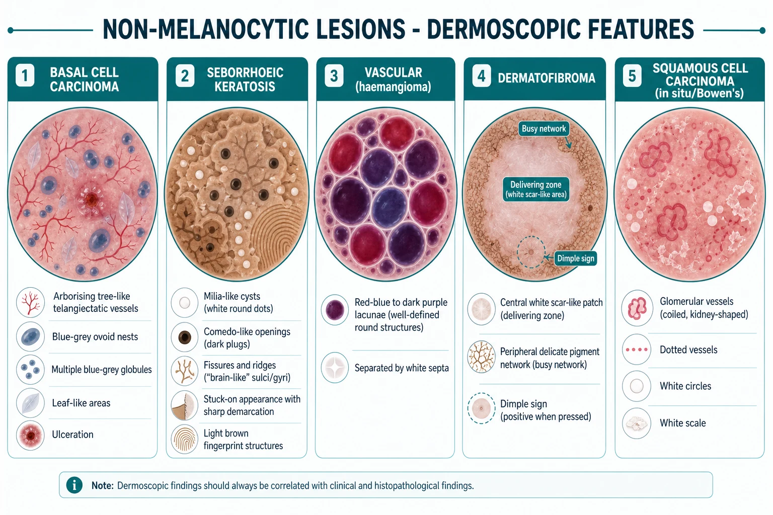
Basal cell carcinoma (BCC)
- Arborising (tree-like) telangiectatic vessels — sharply in focus; branch like a tree.
- Blue-grey ovoid nests — large confluent pigmented oval structures.
- Multiple blue-grey globules — smaller, separate.
- Leaf-like (maple-leaf-like) areas — bulbous brown extensions at the periphery.
- Ulceration — single small or multiple erosions.
- Shiny white-red uniform areas; spoke-wheel structures (radial projections).
- No pigment network (key: BCC is non-melanocytic).[5]
Seborrhoeic keratosis (SK)
- Milia-like cysts — small white round structures = intraepidermal horn cysts.
- Comedo-like openings — dark brown/black plugs of keratin (yellowish-brown).
- Fissures and ridges — "brain-like" / sulci-gyri pattern.
- Fingerprint-like structures — light brown linear parallel ridges.
- Sharp demarcation ("stuck-on" appearance); typically no arborising vessels.[4]
Vascular lesions (haemangioma, angiokeratoma, pyogenic granuloma)
- Red-blue to dark purple lacunae — well-defined round/oval structures = dilated vascular spaces in the dermis; separated by whitish fibrous septa.
- Multiple lacunae in a grouped arrangement.
- Pyogenic granuloma: reddish homogeneous area with white collarette of scale (may mimic melanoma — always excise for histology).[4]
Dermatofibroma
- Central white scar-like patch (the "delivering zone") + peripheral delicate pigment network ("busy network").
- Clinical: dimple sign (central dimpling on lateral compression — pathognomonic).[4]
Squamous cell carcinoma (SCC) / Bowen's disease (SCC in situ)
- Glomerular vessels — coiled, kidney-shaped vessels (like renal glomeruli).
- Dotted vessels (small pinpoint red dots).
- White circles / white halo around follicular openings.
- White scale; irregular surface; in pigmented Bowen's: brown/grey dots arranged linearly along edges ("globs").[4][16]
Clear cell acanthoma
- "String of pearls" — dotted vessels arranged linearly at the periphery (pathognomonic).[4]
Vessel Morphology Lexicon
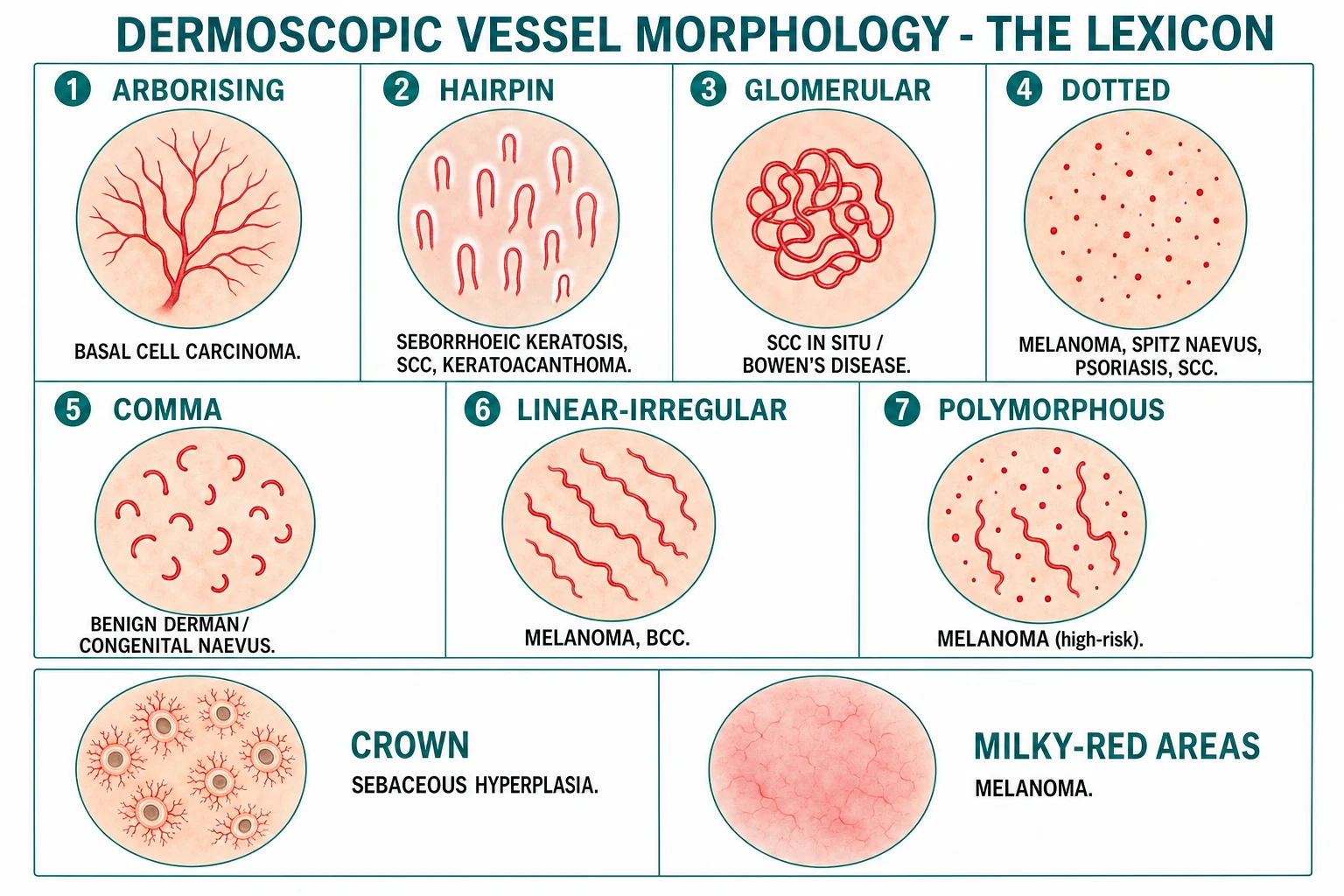
| Vessel morphology | Appearance | Points to |
|---|---|---|
| Arborising | Tree-like branching telangiectasia | BCC |
| Glomerular | Coiled, kidney-shaped (like a glomerulus) | SCC in situ / Bowen's |
| Hairpin | U-shaped elongated loop (± whitish halo) | SK, SCC, keratoacanthoma |
| Dotted | Tiny pinpoint red dots | Melanoma, Spitz naevus, psoriasis, SCC |
| Comma | Curved/comma-shaped | Benign dermal/congenital naevus |
| Linear-irregular | Irregular linear red lines | Melanoma, BCC |
| Polymorphous | Multiple vessel types (dotted + linear) | Melanoma (high-risk) |
| Crown | Wreath-like arrangement around follicles | Sebaceous gland hyperplasia |
| Strawberry / hairpin-white | Dense glomerular + dotted + white halos, pink background | Pink actinic keratosis / facial Bowen's |
| Corkscrew | Tightly twisted loops | Tungiasis, amelanotic melanoma |
| Milky-red areas | Pinkish-red structureless areas | Melanoma (invasive component) |
Vessel-pattern pearls
ABCD-V
BCC
Glomerular
Benign naevus
Melanoma, Spitz, psoriasis
Hairpin = SK/SCC; corkscrew = tungiasis, amelanotic melanoma
Melanoma-Specific Features
For melanocytic lesions in Step 2, the following are high-risk features: [1]
- Asymmetry — of pigmentation and structural components (axes: colour, pattern, vessels).
- Atypical pigment network — irregular thickening/thinning, abrupt peripheral cutoff.
- Blue-white veil — blue colour overlaid with a whitish ground-glass "veil" (due to orthokeratosis overlying heavily pigmented dermis / mass of melanoma cells). Strong predictor of melanoma.
- Multiple colours — ≥5-6 colours (light brown, dark brown, black, red, white, blue-grey) with chaotic distribution.
- Regression structures — scar-like depigmentation (white, structureless) + peppering (blue-grey dots = melanophages in papillary dermis).
- Atypical/polymorphous vessels — dotted + linear-irregular; milky-red areas.
- Streaks/pseudopods — irregular distribution (asymmetric) = melanoma; regular/radial = benign Spitz/Reed naevus.
- Peripheral tan/grey structureless areas.
- Shiny white lines / chrysalis structures — visible only with polarised dermoscopy; collagen remodelling; seen in melanoma, BCC, Spitz, dermatofibroma.[4]
Simplified tools
- 3-point checklist (screening; ≥1 positive → excise/biopsy): (1) asymmetry; (2) atypical network; (3) blue-white structures. Sensitivity ~95%.
- 7-point checklist (more detailed; ≥3 points → suspicious): major (atypical network, blue-white veil, atypical vessels = 2 points each); minor (irregular pigmentation, irregular streaks/pseudopods, regression, irregular globules = 1 point each). [1]
Special Site Dermoscopy
Special-site dermoscopy has site-specific features because skin architecture varies: the face lacks rete ridges, acral skin is ridged, nails are specialised epithelium, and mucosa is non-keratinised.[12]
Acral (palms and soles)
The parallel pattern — pigment distribution relative to surface furrows and ridges:[4][11][12]
| Pattern | Feature | Significance |
|---|---|---|
| Parallel furrow | Linear pigment on the furrows (sulci superficiales) | Benign acral naevus |
| Parallel ridge | Linear pigment on the surface ridges (cristae superficiales) | Acral melanoma |
| Lattice | Cross-linking pigment along furrows | Benign acral naevus (arch) |
| Fibrillar | Fine fibrillar pigment across friction lines | Benign acral naevus |
| Globular / reticular | Diffuse dots/network on non-acral skin | Benign naevus |
| Homogeneous / structureless | Diffuse pigment, no spatial pattern | Melanoma if irregular |
Parallel ridge pattern = melanoma — the single most examinable acral dermoscopic sign. The pigment is on the ridges (not furrows) because melanoma cells are in the crista intermedia (which underlies the surface ridge).[4][11] Koga and Saida's seminal study clarified the early distinction and is the basis of all subsequent diagnostic pathways.[11]
Facial (lentigo maligna)
- Pseudonetwork — facial skin has few rete ridges; follicular openings create a network-like pattern rather than a true pigment network.
- Asymmetric pigmented follicular openings — irregular pigment around hair follicles.
- Rhomboidal structures — rhomboid-shaped grey-brown areas around follicles.
- Annular-granular pattern / slate-grey targets — dots/granules around follicles in long-standing lentigo maligna.
- Black blotches / obliterated follicular openings — late LM.[4][12]
Nail (onychoscopy)
- Longitudinal melanonychia — single-digit pigmented band in an adult is melanoma until proven otherwise; biopsy the proximal nail matrix.[12]
- Micro-Hutchinson sign — pigmentation on the proximal nail fold (visible only with dermoscopy) = subungual melanoma; macro-Hutchinson (naked-eye pigmentation) is even more specific.
- Triangular band — band wider at proximal than distal edge suggests melanoma.
- Grey/black colour + irregular pigment + periungual spread — high-risk.
Mucosa
- Parallel pattern (analogous to acral): pigment along mucosal ridges is suspicious for mucosal melanoma; benign mucosal melanosis tends to be uniform and parallel to the surface.
- Multifocal irregular pigmentation + blue-grey dots + regression = mucosal melanoma. [1]
Nailfold capillaroscopy
Used in connective tissue disease assessment:[2]
- Scleroderma pattern — dilated/giant capillaries, avascular areas (dropout), bushy capillaries; pericapillary haemorrhages. Specificity high for SSc.
- SLE pattern — non-specific tortuous and dilated capillaries without giant loops.
- Dermatomyositis pattern — dystrophia, branching "bushy" capillaries, periungual haemorrhages. [1]
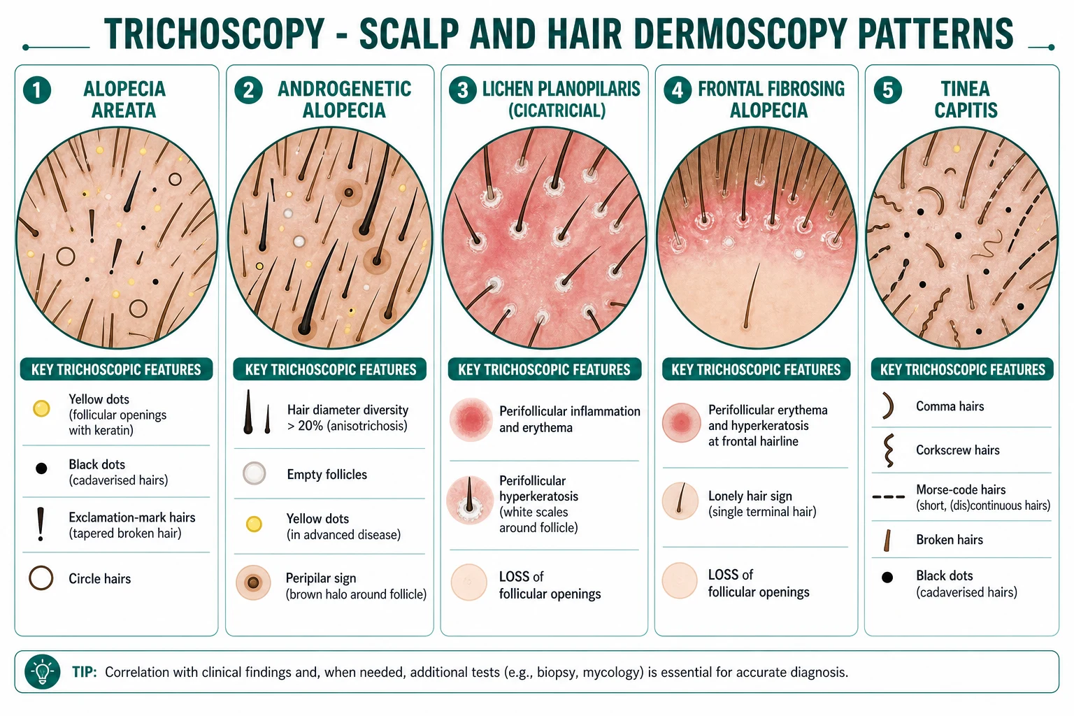
Trichoscopy (Hair and Scalp Dermoscopy)
| Diagnosis | Key trichoscopic features |
|---|---|
| Alopecia areata | Yellow dots (follicular openings with keratin plugs), exclamation-mark hairs (tapered broken hairs), black dots (cadaverised hairs), circle hairs, broken hairs[6] |
| Androgenetic alopecia | Hair diameter diversity more than 20% (anisotrichosis; thick + thin hairs), yellow dots in advanced disease, empty follicles, peripilar sign (brown halo around emerging hair shaft)[1][6] |
| Lichen planopilaris (cicatricial) | Perifollicular inflammation/erythema, perifollicular hyperkeratosis (white scales around follicle), loss of follicular openings (scarred skin)[6] |
| Frontal fibrosing alopecia (FFA) | Perifollicular erythema/hyperkeratosis at frontal/temporal hairline, lonely hair sign (single terminal hair), loss of follicular openings; often with eyebrow/body hair loss[1][6] |
| Discoid lupus | Large radial/branching vessels, white scales, hyperkeratotic follicular plugging, "carpet-tack" sign (keratotic spike on undersurface when scale removed) |
| Tinea capitis | Comma hairs, corkscrew hairs, Morse-code hairs, broken hairs, "black dot" pattern |
| Trichotillomania | Broken hairs of varying lengths, coiled hairs, black dots, empty follicles; no inflammation |
| Scabies | Burrows (delta-wing / "jet with contrail" / "butterfly" sign — triangular structure = mite with linear burrow); dotted vessels on hands/wrists[3] |
Trichoscopy distinguishes scarring (loss of follicular openings) from non-scarring alopecia (follicular openings preserved) and identifies specific diagnoses. The loss of follicular openings is the hallmark of cicatricial alopecia — its presence mandates a biopsy to confirm.[6]
Inflammmoscopy (Inflammatory Dermoscopy)
Dermoscopy of inflammatory skin disease reveals patterns invisible to the naked eye and allows non-invasive diagnostics.[10]
- Psoriasis — uniformly arranged dotted vessels in a linear/curvilinear arrangement; light-red background; diffuse white scale; **histioid" glomerular vessels in chronic plaques.
- Lichen planus — Wickham striae (white reticular lines) on the surface; dotted/linear vessels at the edge; violet/brown background; dot pattern around Wickham striae.
- Discoid lupus — follicular plugging (keratotic plugs in follicular openings); peripheral pigment network interruption; arborising/branching radial vessels; white structureless areas.
- Rosacea — polygonal vessels; follicular plugs; orange-yellow areas (Demodex folliculorum).
- Dermatitis (eczema) — scattered dotted vessels; yellow serocrusts; patchy distribution of vessels (vs uniform psoriasis).
- Lichen sclerosus — bright white structureless areas with comedo-like openings; multiple yellowish plugs. [1]
Entomodermoscopy (Infestations & Parasites)
Dermoscopy of ectoparasitoses is highly specific and increasingly first-line.[3][13][15]
- Scabies — delta-wing sign (Sarcoptes scabiei adult mite: a delta-shaped brown structure at the start of a serpiginous burrow), sometimes called "jet with contrail" or "butterfly"; the terminal part is the burrow, the delta is the mite itself. Eggs and faecal pellets are also visible as small white and brown dots/globules.[3]
- Demodicosis — Demodex follicular tails (plugged follicular openings with whitish cylindrical content protruding); Demodex follicular openings (round, conical, whitish, 0.1–0.2 mm follicular "spikes"). Pigmented demodicidosis presents as reticular pigmented networks on the face.[13]
- Tungiasis (Tunga penetrans) — black central pore surrounded by a chitinous ring in a pathognomonic pattern, sometimes with bluish-grey halo; dorsal subungual location common.[15]
- Ticks — the engorged tick is visible as a round, blue-grey structure with retained mouthparts — useful for removal guidance.
- Pediculosis / Phthirus pubis — lice and nits visible attached to hair shafts.
AI-Assisted Dermoscopy and Deep Learning
The Esteva 2017 Nature paper demonstrated that a convolutional neural network (CNN) trained on 129,450 images of 2,032 diseases classified skin lesions at dermatologist-level accuracy, marking the inflection point for computer-vision in dermoscopy.[9] Subsequent systems (e.g. Model Derm, SkinVision, proprietary CNNs used by the International Skin Imaging Collaboration) have matched or exceeded average dermatologist performance in binary melanoma vs benign discrimination while remaining inferior to expert-level dermatologists for rare lesions.[9]
Key concepts the examination expects: [1]
- CNN architecture (Inception, ResNet, EfficientNet) trained on large labelled dermoscopic image datasets (ISIC archive).
- Sensitivity vs specificity trade-off: computer-vision tools can be tuned to extremely high sensitivity (low miss rate, but more excisions) — the clinical role is therefore often as a "second reader" after a human clinician.
- Limitations: image quality, skin phototype under-representation, performance on acral/nail/facial sites still inferior, regulatory approval varies by jurisdiction.
- Total body photography and sequential digital dermoscopy (SDDI): whole-body imaging + serial dermoscopic comparison for high-risk patients, improving detection of new/changing lesions while reducing unnecessary excisions.[4]
AI-assisted dermoscopy at a glance
Training and Competence
Dermoscopy is operator-dependent, and training improves accuracy. Key evidence: [1]
- Magnitude of benefit: meta-analyses show dermoscopy reduces the benign-to-malignant excision ratio roughly five-fold in trained hands, and sensitivity rises from ~55% to >85% with formal training.
- Trainee patterns: in novices, the 3-point checklist is preferred for triage; pattern analysis with checklist cross-check is preferred for residents; expert gestalt for consultant-level practice.
- Maintenance: weekly 5–10 minute digital review of personal cases, peer review of difficult lesions, attendance at international meetings (IDS, AAD, EADV).
- Teledermoscopy and AI-assisted decision support are increasingly part of training — validating AI logic on known teaching cases is a common exercise.
- Documentation: digital dermoscopic archive + annotation of every lesion seen, correlated with histology where possible — the foundation of continuous improvement.[4]
Pitfalls and Limitations
Exam application bank (NEET-PG / INICET)
One-line answer
Dermoscopy (dermatoscopy, epiluminescence microscopy) is in-vivo 10x magnified skin-surface microscopy using a handheld contact dermatoscope that has transformed the accuracy of skin-cancer, pigmented-lesion, hair, nail and inflammatory-skin diagnosis. The foundational framework is the two-step algorithm (Argenziano): Step 1 — melanocytic vs non-melanocytic using pigment network, aggregated globules, streaks, homogeneous blue pigmentation or parallel pattern; Step 2 — within melanocytic lesions, apply melanoma-specific criteria (asymmetry, atypical network, blue-white veil, regression, polymorphous vessels, irregular streaks) through pattern analysis (Pehamberger) or checklist tools (Menzies 11-point; Argenziano 7-point; Stolz ABCD rule; Zalaudek 3-point; Lallas CASH algorithm for keratinising tumours). Equipment: contact polarised vs non-polarised immersion dermatoscope; the immersion f
Worked stems (answer without another resource)
Stem 1 — Classic presentation. Map symptoms to mechanism; name the first investigation and first treatment step with dose/route if drug therapy is standard. [1]
Stem 2 — Unstable / complicated. List red flags that force immediate resuscitation, theatre, ICU, antidote, or reperfusion — and what you do in the first 15 minutes. [1]
Stem 3 — Atypical group. Elderly, pregnancy, child, or immunocompromised: how presentation and thresholds change. [1]
Stem 4 — Differential trap. Name the three closest mimics and one discriminator for each. [1]
Stem 5 — Disposition. Who goes home with safety-netting, who is admitted, who needs HDU/ICU/theatre, and what follow-up is mandatory. [1]
Rapid viva checklist
- Definition + classification
- Pathophysiology chain
- Bedside signs / criteria
- Score with exact components (if any)
- Emergency bundle
- Definitive therapy with doses
- Complications of disease and of treatment
- Special populations
- Guideline/trial name if classic
- Three exam traps
Coverage self-check
If you cannot answer any stem above from this page alone, re-read the matching section — the page is intended to be self-sufficient for final-prof and NEET-PG/INICET questions on Dermoscopy: principles and patterns.
Expanded exam teaching (depth pass)
Clinical reasoning
For Dermoscopy: principles and patterns, examiners test whether you can prioritise life threats, choose the right first test, and give specific therapy (agent, dose, route, timing). Generic phrases without numbers score poorly.
Mechanism → feature map
Build a short chain: cause → pathophysiologic intermediate → clinical feature → complication. Every major symptom in the classic vignette should sit on that chain.
Investigation strategy
- Bedside/first-line tests that change immediate management
- Confirmatory or staging tests
- What a normal result does not exclude
- When not to delay treatment for imaging (unstable patient)
Management ladder
- Resuscitation / ABC / sepsis or haemorrhage bundle as relevant
- Specific antidote / procedure / antimicrobial / reperfusion / surgery
- Supportive care and monitoring targets
- Definitive long-term therapy and secondary prevention
- Disposition and safety-net advice
Special populations
Always prepare one line each for children, pregnancy, elderly, renal/hepatic impairment, and immunocompromised patients when the topic allows.
Pitfalls that fail candidates
- Treating the number not the patient
- Missing pregnancy status when relevant
- Imaging before stabilisation
- Wrong empiric cover or wrong antidote timing
- Incomplete counselling on recurrence, adherence, or red-flag return
Dermoscopy (dermatoscopy, epiluminescence microscopy) is in-vivo 10x magnified skin-surface microscopy using a handheld contact dermatoscope that has transformed the accuracy of skin-cancer, pigmented-lesion, hair, nail and inflammatory-skin diagnosis. The foundational framework is the two-step algorithm (Argenziano): Step 1 — melanocytic vs non-melanocytic using pigment network, aggregated globules, streaks, homogeneous blue pigmentation or parallel pattern; Step 2 — within melanocytic lesions, [1]
Structured revision sheet
Must-know numbers and names
List every score, size threshold, dose, and time window from this topic on a blank page from memory, then check against the sections above.
Three classic MCQ angles
- Most likely diagnosis given a vignette
- Next best step in management
- Most appropriate investigation
Three classic SAQ angles
- Pathophysiology in five steps
- Management algorithm with doses
- Complications and prevention
Clinical station flow
Greet → focused history → targeted exam → investigations → explain diagnosis → emergency care → definitive plan → safety-net / follow-up → answer examiner questions on mechanism and pitfalls.
Clinical Pearls
[1]Self-test: A 60-year-old gardener with Fitzpatrick II skin and a history of NMSC presents with a 7-mm pigmented lesion on the left heel; dermoscopy shows linear brown pigment along the surface ridges. What is your next step?
Excisional biopsy. The parallel ridge pattern — pigment along the surface ridges rather than along the furrows — is the single most reliable dermoscopic sign of acral melanoma. Koga & Saida showed that pigmentation on the ridges corresponds to melanoma cells within the crista intermedia. Parallel furrow / lattice / fibrillar patterns all signify a benign acral naevus; a parallel-ridge pattern does not. Margin-guided wide local excision follows histological confirmation with Breslow thickness. Delayed diagnosis of acral melanoma is the most common cause of the worst-prognosis melanomas — do not reassure on this sign.
References
This topic synthesises evidence from the International Dermoscopy Society consensus terminology (2003 consensus and 2016 validation), the Seventh American Joint Committee on Cancer staging guidelines, the AAD/ACD/IDC melanoma guidelines, and a structured review of recent systematic reviews of dermoscopic accuracy.[4][8] Future updates will track revisions from the International Dermoscopy Society and the AI-assisted dermoscopy (IAD) working group, including ongoing large-scale validation trials of deep-learning–augmented clinical pathways.[9]
References
- [1]Griggs J, Trüeb RM, Gavazzoni Dias MFR, et al. Fibrosing alopecia in a pattern distribution J Am Acad Dermatol, 2021.PMID 31926219
- [2]Smith V, Ickinger C, Hysa E, et al. Nailfold capillaroscopy Best Pract Res Clin Rheumatol, 2023.PMID 37419757
- [3]Uzun S, Durdu M, Yürekli A, et al. Clinical practice guidelines for the diagnosis and treatment of scabies Int J Dermatol, 2024.PMID 38922701
- [4]Yélamos O, Braun RP, Liopyris K, et al. Dermoscopy and dermatopathology correlates of cutaneous neoplasms J Am Acad Dermatol, 2019.PMID 30321581
- [5]Heath MS, Bar A. Basal Cell Carcinoma Dermatol Clin, 2023.PMID 36410973
- [6]Pirmez R. The dermatoscope in the hair clinic: Trichoscopy of scarring and nonscarring alopecia J Am Acad Dermatol, 2023.PMID 37591567
- [7]Stolz W, Riemann R, Cognetta AB, et al. The ABCD rule of dermatoscopy. High prospective value in the diagnosis of doubtful melanocytic skin lesions J Am Acad Dermatol, 1994.PMID 8157780
- [8]Carrera C, Marchetti MA, Dusza SW, et al. Validity and Reliability of Dermoscopic Criteria Used to Differentiate Nevi From Melanoma: A Web-Based International Dermoscopy Society Study JAMA Dermatol, 2016.PMID 27074267
- [9]Esteva A, Kuprel B, Novoa RA, et al. Dermatologist-level classification of skin cancer with deep neural networks Nature, 2017.PMID 28117445
- [10]Lallas A, Errichetti E, Apalla Z, et al. Dermoscopy of Common Inflammatory Disorders Dermatol Clin, 2018.PMID 30201145
- [11]Koga H, Saida T. Key points in dermoscopic differentiation between early acral melanoma and acral nevus J Dermatol, 2011.PMID 21175752
- [12]Zalaudek I, Kreusch J, Giacomel J, et al. Special locations dermoscopy: facial, acral, and nail Dermatol Clin, 2013.PMID 24075549
- [13]Pinnock C, Sarri N, Krowchuk DP. Pigmented demodicidosis - an under-recognized cause of facial hyperpigmentation Int J Dermatol, 2022.PMID 34897670
- [14]Yélamos O, Braun RP, Liopyris K, et al. Dermoscopy and dermatopathology correlates of cutaneous neoplasms J Am Acad Dermatol, 2019.PMID 30321581
- [15]Bezerra RJS, Di Chiacchio N, et al. Potential use of dermoscopy in atypical tungiasis J Travel Med, 2021.PMID 33969417
- [16]Ardigo M, Papoutsaki M, Falconieri G, et al. [Translated article] Dermoscopy of Squamous Cell Carcinoma: From Actinic Keratosis to Invasive Forms Actas Dermosifiliogr, 2024.PMID 39102978