Dermatology · Medicine
Dissecting cellulitis of the scalp (perifolliculitis capitis)
Also known as Dissecting cellulitis of the scalp (DCS) · Perifolliculitis capitis abscedens et suffodiens · Hoffmann's disease · Dissecting folliculitis · Folliculitis abscedens et suffodiens
Dissecting cellulitis of the scalp (DCS) is a chronic, progressive, suppurative follicular disorder producing painful boggy nodules, abscesses, and interconnected sinus tracts on the scalp vertex and occiput, ultimately causing scarring alopecia. It is part of the follicular occlusion tetrad (with hidradenitis suppurativa, acne conglobata, and pilonidal sinus) and predominantly affects young Black men aged 20 to 40. First-line therapy is oral tetracyclines; escalation is with rifampicin plus clindamycin, oral isotretinoin, anti-TNF biologics (adalimumab), or surgical excision with grafting for refractory disease.
On this page & tools
Your progress
Saved locally on this device.
Exam tags
Red flags
Overview and definition
Dissecting cellulitis of the scalp (DCS) — also called perifolliculitis capitis abscedens et suffodiens and Hoffmann disease — is a rare, chronic, progressive, suppurative follicular disorder of the scalp in which occluded hair follicles rupture into the dermis, generating an intense neutrophilic and granulomatous inflammatory reaction that organises into abscesses and interconnected subcutaneous sinus tracts. The clinical result is a boggy, oedematous, boggy-nodular scalp discharging purulent or serosanguineous fluid through multiple surface openings, with progressive destruction of follicles and permanent scarring alopecia.[1][3][5]
DCS does not exist in isolation for the examination candidate. It is the scalp member of the follicular occlusion tetrad, sharing a common pathophysiology with hidradenitis suppurativa (axillae and groins), acne conglobata (chest and back), and pilonidal sinus (natal cleft). Recognising this unifying concept is the single highest-yield fact on this topic, because the same triad of occlusion, rupture, and neutrophilic inflammation — and largely the same drugs — appear across all four conditions.[1][3]
Synonyms and naming
The eponymous and descriptive names are deliberately evocative, and a viva examiner may ask you to translate them: [1]
- Perifolliculitis capitis abscedens et suffodiens — Latin for "perifollicular inflammation of the head, with abscesses and tunneling (undermining)." The word suffodiens ("undermining") captures the interconnected sinus tracts beneath the scalp surface that are the morphological hallmark.
- Hoffmann disease — after the German dermatologist Hoffmann, who described it in the early 20th century (distinct from Hoffman-Purdy syndrome).
- Dissecting folliculitis — emphasises that the inflammation dissects (tunnels) through the subcutaneous tissue. [1]
Classification and the follicular occlusion tetrad
DCS has no universally adopted formal staging system of its own; in practice it is classified by severity and anatomical extent, and clinical decision-making borrows from the Hurley staging used in hidradenitis suppurativa (the sister disease in the tetrad). The most important conceptual classification for the exam is the follicular occlusion tetrad itself. [1]
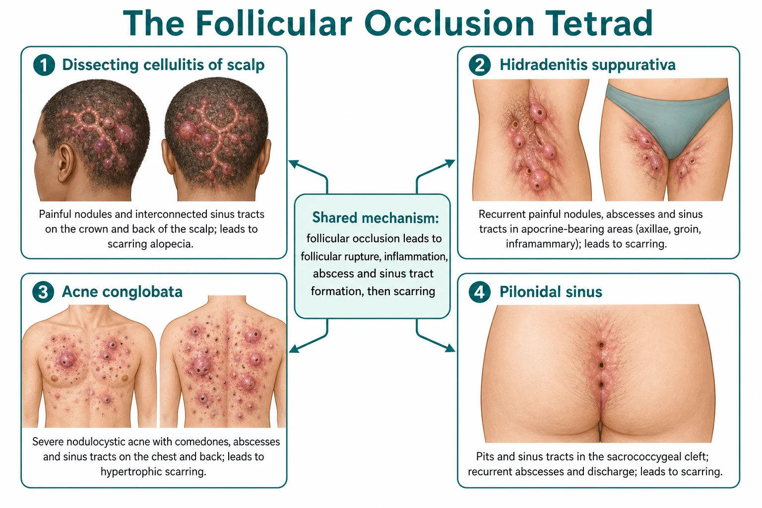
The tetrad shares a common pathomechanistic sequence, and a candidate who can articulate it can answer almost any question on any of the four conditions: [1]
Dissecting cellulitis of scalp
Hidradenitis suppurativa
Acne conglobata
Pilonidal sinus
Severity framework (Hurley-adapted)
Because DCS lacks a dedicated grading scale, clinicians adapt the Hurley stages used for hidradenitis suppurativa: [1]
Severity
Severe (Hurley III)
3 — Diffuse coalescing boggy plaques, multiple interconnected sinus tracts discharging purulent material, extensive bridging scars and scarring alopecia.
Epidemiology and risk factors
DCS is uncommon, and true population prevalence figures are not established because the disease is rare, under-recognised, and under-reported. What is consistent across series is a striking demographic signature. [1]
The disease overwhelmingly affects young Black men in the second to fourth decade, although it is described in women, in children, in Caucasians, and in older adults — atypical demographics should prompt confirmation with biopsy and exclusion of mimics, especially tinea capitis (kerion) in children.[3][5]
Risk factors and associations: [1]
- Male sex and African ancestry — strong, reproducible associations; likely relate to hair follicle density, hair curvature, and sebum characteristics, though the precise mechanism is unproven.
- Family history / genetics — familial clusters are reported; rare families harbour loss-of-function mutations in gamma-secretase pathway genes (PSENEN, NCSTN, PSEN1), the same pathway implicated in familial hidradenitis suppurativa. This is a key molecular link explaining why the tetrad co-segregates in some kindreds.
- Follicular occlusion tetrad — coexistence with hidradenitis suppurativa, acne conglobata, or pilonidal sinus should be actively sought; one condition of the tetrad increases the odds of another.
- Obesity, smoking, and metabolic syndrome — established accelerants in hidradenitis suppurativa and reported in DCS case series; weight and smoking status should be addressed as part of management.
- Immunosuppression and HIV — DCS is reported with unusual severity in HIV-positive patients, and a new severe scalp process in an at-risk individual warrants HIV testing. [1]
Pathophysiology
The pathophysiology of DCS is the follicular occlusion sequence, identical in principle to hidradenitis suppurativa and acne conglobata. The candidate who can walk through this cascade at viva can defend the entire management ladder, because every drug class targets a step in it. [1]
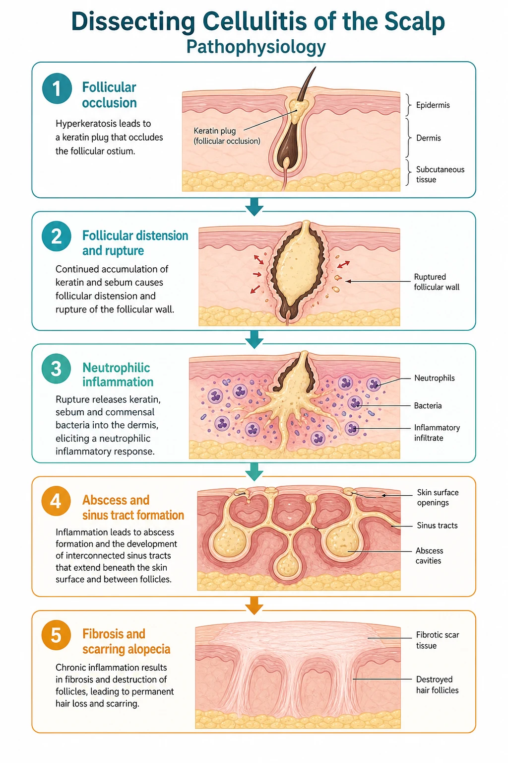
The cascade, step by step
Follicular occlusion
Follicular distension and rupture
Neutrophilic then granulomatous inflammation
Abscess and sinus tract formation
Fibrosis and scarring alopecia
The hallmark morphological consequence is interconnected sinus tract formation — channels that dissect (hence "dissecting") through the subcutaneous tissue and connect multiple follicular units. Pressing on one nodule may express pus from a distant opening, a useful bedside confirmation of the diagnosis. Sinus tracts become lined by squamous epithelium, which is the substrate for later malignant change (see Complications).[3][6]
Molecular and genetic basis
In a minority of patients, DCS is part of a familial follicular occlusion syndrome driven by loss-of-function mutations in the gamma-secretase complex — most often NCSTN (nicastrin), PSENEN (presenilin enhancer 2), and PSEN1 (presenilin 1). Gamma-secretase is a transmembrane protease that cleaves Notch and other substrates; impaired Notch signalling in the hair follicle is hypothesised to alter follicular keratinisation and immune homeostasis. The same gene family underlies familial hidradenitis suppurativa, which is why DCS and HS co-segregate in some families. This molecular link is high-yield for fellowship vivas and explains why a family history should be sought.[2]
The role of bacteria
Bacterial infection is secondary, not primary: cultures are frequently sterile or yield skin commensals and opportunists (Staphylococcus aureus, coagulase-negative staphylococci, Proteus species, anaerobes, and occasionally Klebsiella or Pseudomonas). Antibiotics work predominantly through anti-inflammatory and immunomodulatory mechanisms rather than by eradicating a causative organism — a fact that explains why tetracyclines help even when cultures are negative, and why the disease relapses when antibiotics are stopped.[3][4]
Clinical presentation
DCS runs a chronic, relapsing, and progressive course over years to decades. The presentation is usually unmistakable once seen, but the candidate must be able to describe it precisely, distinguish it from mimics, and recognise atypical scenarios. [1]
Distribution
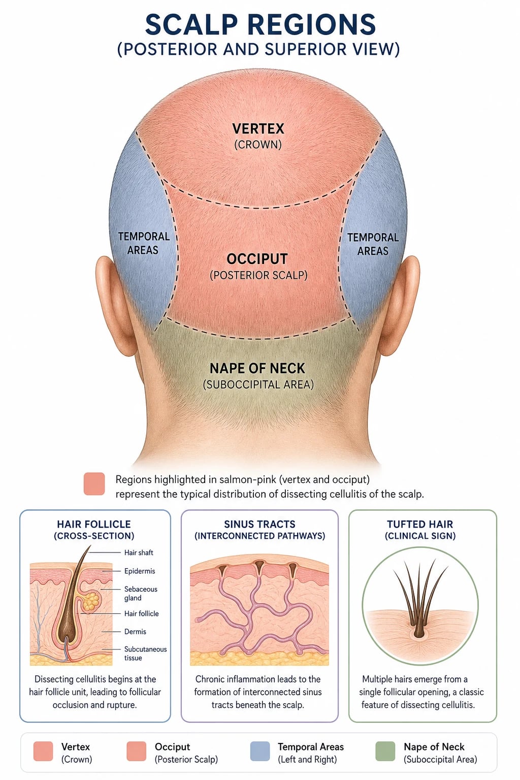
- Scalp vertex (crown) — the most common and earliest site.
- Occiput (posterior scalp) and nape of neck — frequent extension.
- Rarely the eyebrows, beard, axillae, groin, or chest (when these are involved, suspect overlap with the rest of the follicular occlusion tetrad).[3]
Morphology and evolution
The disease evolves through characteristic stages: [1]
- Early — multiple tender follicular papules and pustules on the vertex and occiput, easily mistaken for ordinary folliculitis.
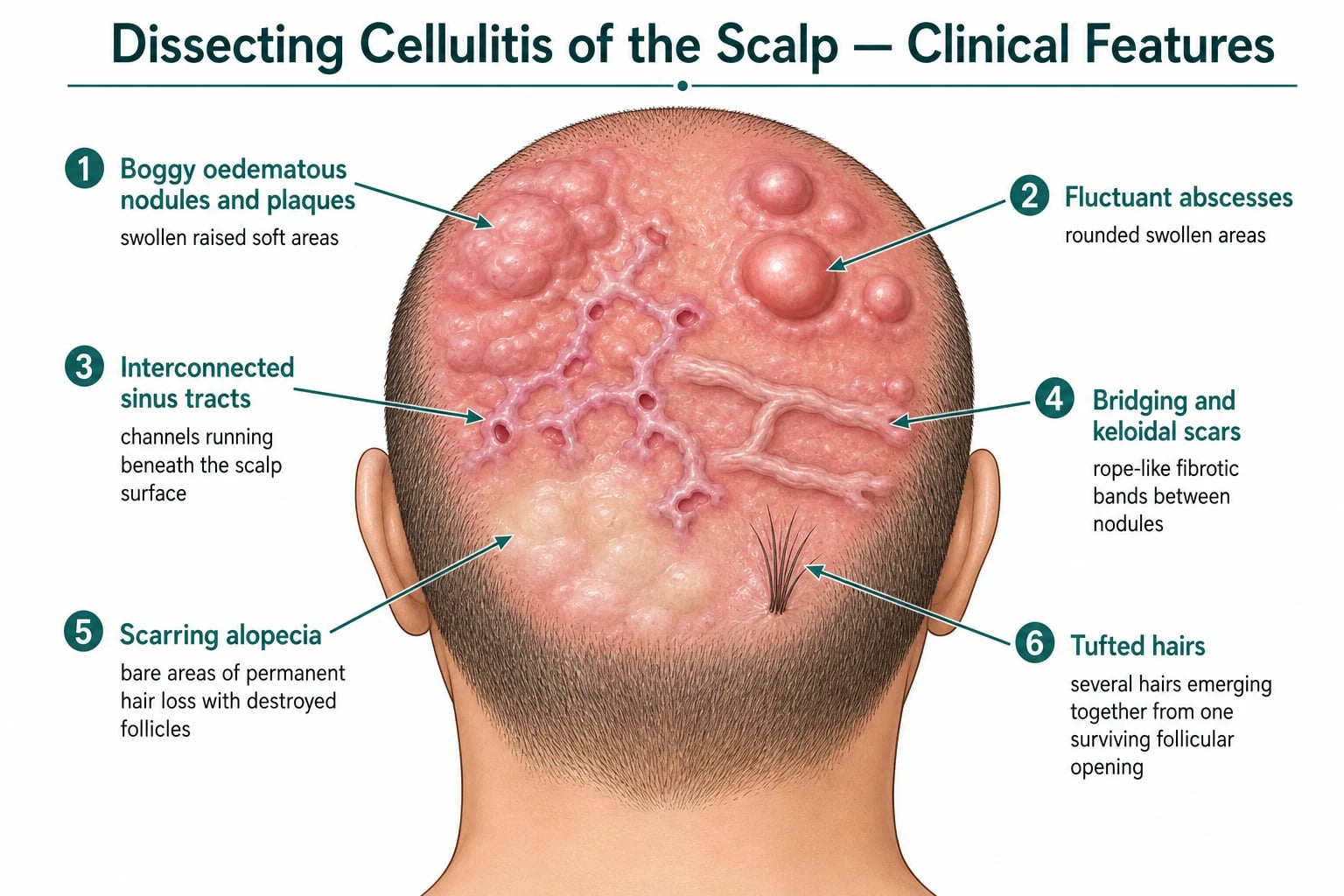
- Established — boggy, oedematous, doughy nodules and plaques that become fluctuant, forming abscesses. The scalp feels soft and depressible ("boggy") on palpation.
- Sinus tract / discharge — interconnected subcutaneous channels discharge purulent, serosanguineous, or serous fluid from multiple surface openings; pressing one nodule may express pus from another (the "dissecting" hallmark). The appearance can be kerion-like and is often malodorous.
- Fibrosis — bridging (keloidal) scars, tubular fibrotic bands, and permanent scarring alopecia; surviving follicles may show tufted hairs (multiple hairs emerging from a single opening).[1][3]
Symptoms
- Pain and tenderness — often severe, the chief reason patients present.
- Purulent discharge and odour — socially distressing; patients may report soiled pillowcases and clothing.
- Pruritus and a sensation of scalp pressure.
- Psychological distress — depression, anxiety, social withdrawal, and reduced quality of life from pain, odour, discharge, and visible hair loss. [1]
Systemic features
Fever, malaise, and regional lymphadenopathy (occipital, posterior cervical) may accompany acute suppurative flares, but DCS is usually a localised disease and systemic upset should prompt a search for secondary infection or an alternative diagnosis. [1]
Examination of the tetrad
Because DCS is one quarter of the follicular occlusion tetrad, the focused examination always looks for the other members: inspect the axillae, groins, inframammary folds, buttocks, and natal cleft (hidradenitis suppurativa, pilonidal sinus), and the chest, back, and face (acne conglobata). Detecting an unsuspected tetrad partner changes the therapeutic ladder and supports the diagnosis. [1]
Differential diagnosis
The differential of a boggy, discharging scalp with hair loss is broad, and the high-yield discriminator is whether the process is follicular and suppurative (DCS), fungal (kerion), staphylococcal (furunculosis), or scarring alopecic. The examiner will probe the distinguishing features. [1]
Dissecting cellulitis
Tinea capitis / kerion
Bacterial furunculosis
Folliculitis decalvans
Acne keloidalis nuchae
Squamous cell carcinoma
Pseudopelade of Brocq
Key discriminating points the examiner rewards: [1]
- DCS versus kerion — kerion is most often in children, is acute and unilateral, has broken hairs and lymphadenopathy, and is KOH/fungal-culture positive. DCS is in young adults, is chronic and bilateral, has sinus tracts discharging pus, and is culture-negative or commensal. Failing to exclude tinea before starting steroids or isotretinoin is a classic and serious error.
- DCS versus acne keloidalis nuchae (AKN) — AKN sits on the nape of the neck and low occipital hairline as firm keloidal papules; DCS dominates the vertex and occiput as boggy nodules and sinus tracts. They can coexist.
- DCS versus folliculitis decalvans (FD) — FD is a pustular folliculitis with tufted hair folliculitis but lacks the large boggy abscesses and sinus tracts of DCS.[3]
Clinical and bedside assessment
Diagnosis is clinical and is made at the bedside on the characteristic morphology and demographic. A focused scalp examination documents: [1]
- Distribution and extent — map the involved scalp regions (vertex, occiput, nape); photograph for serial comparison.
- Number and type of lesions — papules, pustules, boggy nodules, abscesses, sinus openings.
- Sinus tract integrity — gentle pressure on one nodule to see if pus appears at a distant opening confirms subcutaneous interconnection.
- Scarring alopecia — extent of permanent hair loss; presence of tufted hairs.
- Lymphadenopathy — occipital and cervical nodes; tender nodes suggest secondary infection.
- Tetrad screen — axillae, groins, inframammary folds, natal cleft, chest, back. [1]
There is no pathognomonic blood test or imaging finding. Dermoscopy/trichoscopy may show perifollicular pustules, tufted hairs, keratin plugs, yellow crusts, and absence of follicular openings in scarred areas, supporting a cicatricial process, but it is adjunctive rather than diagnostic.[3]
Investigations
DCS is a clinical diagnosis, and investigations are performed chiefly to exclude mimics, characterise secondary infection, and screen for systemic disease and complications before starting systemic therapy. [1]
Test
Bacterial culture and sensitivity of pus
KOH mount and fungal culture (skin scrapings, hair pluckings)
Skin biopsy (4 mm punch)
HIV serology
Baseline bloods before systemic therapy
Scalp swab
Histopathology
The microscopic picture mirrors the pathophysiological cascade and is supportive rather than pathognomonic: follicular hyperkeratosis and occlusion, follicular rupture, a dense neutrophilic and granulomatous (foreign-body) infiltrate with giant cells around keratin and hair fragments, abscess formation, sinus tracts lined by stratified squamous epithelium, and progressive dermal fibrosis with follicular dropout. Biopsy is reserved for atypical presentations, refractory disease, or suspicion of malignant transformation.[3][6]
Management — resuscitation and acute flares
DCS has no resuscitation phase in the usual sense (it is not a time-critical emergency), but acute suppurative flares with painful abscesses require prompt symptom-relieving intervention while definitive therapy is planned: [1]
- Analgesia — paracetamol and NSAIDs for pain; severe flares may require stronger analgesia.
- Incision and drainage of a tense, fluctuant abscess for pain relief and decompression — a temporising measure, not curative, because the underlying sinus tracts remain.[1]
- Culture of pus at the time of drainage to guide antibiotic choice for secondary infection.
- Treat secondary infection with a short course of an anti-staphylococcal antibiotic (e.g. flucloxacillin, or vancomycin/linezolid if MRSA is suspected) guided by culture.[3]
- Address red flags — any non-healing ulcer within a long-standing lesion must be biopsied to exclude squamous cell carcinoma before immunosuppression is escalated.[6]
Management — definitive and stepwise
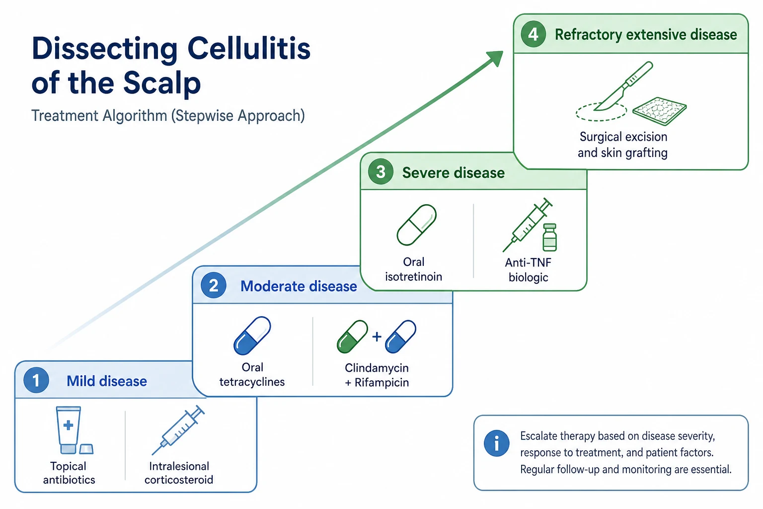
No therapy is curative, and the goal is disease control, reduction of flares, preservation of hair, and quality of life. Evidence is limited to case series and systematic reviews of uncontrolled data; there are no large randomised trials, and no agent is approved specifically for DCS.[1][4] The ladder escalates from anti-inflammatory antibiotics through retinoids to biologics and surgery.
Stepwise therapy ladder
Mild disease / first-line systemic
Moderate disease
Moderate–severe disease
Severe / refractory disease
Refractory extensive disease
Adjuncts and maintenance
First-line systemic therapy
Oral tetracyclines are the conventional first-line systemic therapy by virtue of anti-inflammatory (not antimicrobial) action via inhibition of neutrophil chemotaxis and matrix metalloproteinases. Doxycycline 100 mg twice daily (or lymecycline 40 mg daily, minocycline 100 mg twice daily) is given for a minimum of 3 months and continued as maintenance in responders. Tetracyclines are generally well tolerated; counsel about photosensitivity, gastrointestinal upset, and (for minocycline) vestibular side effects and drug-induced lupus. They are contraindicated in pregnancy and in young children.[1][4]
Combination antibiotic therapy
Rifampicin 300 mg twice daily plus clindamycin 300 mg twice daily for 10 to 12 weeks is the combination regimen borrowed from hidradenitis suppurativa and widely used in DCS, with documented efficacy in case series and systematic reviews. The pairing provides anti-inflammatory and antimicrobial synergy. Warn the patient that rifampicin stains body fluids orange/red and reduces the efficacy of the oral contraceptive pill. Monitor LFTs; review for Clostridioides difficile colitis with clindamycin. This is a reasonable second-line choice when a tetracycline has failed or is contraindicated.[4]
Oral isotretinoin
Oral isotretinoin 0.5 to 1 mg/kg/day for 6 to 12 months is regarded as the most effective single systemic agent in DCS, acting on the initiating event by suppressing sebum production and normalising follicular keratinisation. A lower starting dose (0.5 mg/kg/day) is sometimes preferred to avoid an initial inflammatory flare, escalating as tolerated. Mandatory precautions: highly teratogenic — exclude pregnancy, ensure two forms of reliable contraception, and enrol in a pregnancy-prevention programme; monitor lipids and LFTs at baseline and periodically; counsel about mucocutaneous dryness, photosensitivity, mood change, and musculoskeletal symptoms. Isotretinoin is often combined with systemic antibiotics in severe disease, though the traditional caution against co-prescribing with tetracyclines (pseudotumour cerebri risk) applies.[1][4]
Other systemic agents
- Dapsone 50 to 150 mg daily — an anti-neutrophilic sulphone; check G6PD before starting and monitor FBC and LFTs for haemolysis and methaemoglobinaemia. Useful in refractory or sulphone-responsive cases.
- Zinc sulphate (e.g. 135 mg three times daily) — adjunctive anti-inflammatory; limited evidence.
- Systemic corticosteroids — short courses (e.g. oral prednisolone) for severe acute flares; not suitable for long-term use.[4]
Biologic therapy for refractory disease
When antibiotics and isotretinoin fail, anti-TNF-alpha biologics are the most evidence-supported option. Adalimumab (the only biologic approved for hidradenitis suppurativa, the sister disease) is the best-studied agent in DCS, given as 40 mg subcutaneously every 2 weeks after a standard loading regimen. Infliximab (5 mg/kg IV at weeks 0, 2, 6, then 8-weekly) is an alternative supported by case reports and series. A 2025 systematic review confirmed that TNF-alpha blockers show the highest response rates among biologics in DCS, with anti-interleukin agents (ustekinumab anti-IL-12/23; secukinumab anti-IL-17) and small-molecule JAK inhibitors as emerging options for biologic-resistant disease.[2] Screen for latent tuberculosis, hepatitis B, and C before starting; counsel on infection risk and injection-site reactions.
Oral isotretinoin
Dose
0.5 to 1 mg/kg once daily, for 6 to 12 months; start at 0.5 mg/kg/day and escalate as tolerated to minimise an initial inflammatory flare.
Doxycycline
Dose
100 mg once or twice daily, continued for a minimum of 3 months and then maintained long term in responders.
Clindamycin plus rifampicin combination
Dose
Clindamycin 300 mg twice daily plus rifampicin 300 mg twice daily, together for 10 to 12 weeks.
Adalimumab (anti-TNF-alpha)
Dose
40 mg every 2 weeks (after a loading regimen per the hidradenitis suppurativa licence), continued for at least 3 to 6 months before assessing response.
Surgical and procedural therapy
- Incision and drainage — for acute abscess decompression; symptomatic, not curative.
- Wide surgical excision of all involved scalp down to the galea, with skin grafting or secondary intention healing — the most definitive treatment, reserved for localised, severe, treatment-resistant disease; extensive involvement makes the donor/defect size prohibitive, and recurrence at graft margins is possible if any sinus tract is left behind.[1]
- Laser epilation (long-pulsed Nd:YAG or diode) — reduces hair follicle-driven inflammation and is a useful adjunct, particularly for maintenance; works by destroying the follicular units that drive occlusion.
- Intralesional triamcinolone acetonide (5 to 10 mg/mL) — into individual inflammatory nodules to settle flares.
- Intralesional botulinum toxin and radiotherapy — reported anecdotally; not standard.[3][4]
Supportive and holistic care
Do not underestimate the burden: pain, odour, discharge, and visible alopecia cause depression, anxiety, and social isolation. Address obesity and smoking (both accelerate tetrad disease), provide psychological support, and consider patient advocacy groups. Serial photography and a patient diary help track flares and treatment response. [1]
Specific subtypes and scenarios
Paediatric dissecting cellulitis
DCS is rare in children but described, particularly in boys of African heritage. The critical differential is tinea capitis with kerion — every paediatric boggy scalp lesion needs KOH and fungal culture before any anti-inflammatory or retinoid therapy. Isotretinoid dosing is weight-based and requires careful counselling in adolescents (teratogenicity, mood, growth-plate considerations are debated). Tetracyclines are generally avoided in children under 8 years (dental staining) and under 12 in many guidelines.[3]
DCS in women
Women are far less commonly affected (sex ratio about 4 to 1 male). Female cases should prompt confirmation of the diagnosis and screening for the tetrad. Pregnancy planning is pivotal because isotretinoin, tetracyclines, and several biologics have restricted use in pregnancy; effective contraception is essential, and biologic choice should weigh pregnancy exposure (e.g. adalimumab has the most reproductive data among TNF inhibitors).[2]
DCS in the immunocompromised and HIV
DCS may be unusually severe or extensive in HIV-positive patients, and a new severe scalp process in an at-risk individual warrants HIV testing. Management parallels standard care, with attention to drug interactions, opportunistic infection screening, and the immune reconstitution inflammatory syndrome after antiretroviral therapy.[5]
Follicular occlusion tetrad overlap
When DCS coexists with hidradenitis suppurativa, acne conglobata, or pilonidal sinus, treat the most disabling component first and recognise that an agent effective in one (e.g. adalimumab for HS) may benefit another. A multidisciplinary approach (dermatology, general/plastic surgery, psychology) is often needed.[1]
Complications and pitfalls
Complication
Scarring alopecia (irreversible)
Squamous cell carcinoma (Marjolin)
Secondary bacterial infection
Keloid formation
Psychological distress
Treatment-related harm
Classic pitfalls
- Treating for DCS without excluding tinea capitis — a kerion looks boggy and discharging; starting steroids or retinoids without a KOH test can worsen a fungal infection and is a defensible complaint.
- Missing Marjolin transformation — a non-healing ulcer in a 20-year sinus tract is cancer until proven otherwise by biopsy.[6]
- Underestimating psychological burden — quality-of-life impairment in tetrad disease is comparable to other chronic skin diseases; screen and refer.
- Assuming antibiotics will cure — antibiotics are anti-inflammatory and suppressive, not curative; relapse on cessation is the rule, and an escalating plan should be set early.
- Neglecting the tetrad — missing a coexistent hidradenitis suppurativa or pilonidal sinus loses an opportunity for unified management.[1]
Prognosis and disposition
DCS is chronic and relapsing, with no cure; the realistic goal is disease control and quality of life, not eradication. Hair lost to scarring alopecia is permanent. Most patients are managed in the outpatient dermatology setting; surgical excision is reserved for localised refractory disease. Patients with long-standing active sinus tracts carry a small but real lifetime risk of squamous cell carcinoma and need long-term surveillance with a low threshold to biopsy change. Psychological morbidity is substantial and must be addressed in parallel.[1][3][6]
Special populations
Skin of colour
DCS predominantly affects people of African descent. Post-inflammatory hyperpigmentation, keloid formation, and pseudofolliculitis are more pronounced in skin of colour and influence treatment choice (favour early anti-inflammatory control, counsel on keloid risk before surgery). Laser epilation settings must be adjusted for higher Fitzpatrick skin types to avoid dyspigmentation and burns.[3]
Pregnancy
Isotretinoin is absolutely contraindicated (severe teratogenicity); tetracyclines are avoided after the first trimester (dental and skeletal effects). If treatment is essential in pregnancy, prefer intralesional corticosteroids and topical agents, and seek specialist advice; adalimumab has the most pregnancy safety data among TNF inhibitors but should be used only when the benefit justifies the risk.[2]
The anticoagulated and the elderly
Older patients with long-standing DCS are the group at risk of Marjolin transformation; age also raises the threshold for aggressive systemic retinoids and biologics, favouring surgery for localised lesions. Anticoagulation does not specifically modify DCS but increases bleeding risk from intralesional injection, biopsy, and surgery. [1]
Exam practice: SAQ, viva, and self-test
SAQ — Boggy scalp in a young Black man (10 marks, 10 minutes)
10 minutes · 10 marks
“”
Self-test: a 9-year-old boy has a unilateral, boggy, painful, hairless plaque on his scalp with occipital lymphadenopathy. KOH is positive. What is the diagnosis, what should you NOT give, and what is the correct treatment?
The diagnosis is tinea capitis with kerion, not dissecting cellulitis (DCS is in young adults, bilateral and chronic, and KOH-negative). Do NOT give systemic corticosteroids or isotretinoin first, and do not assume DCS. Confirm with fungal culture and treat with oral antifungals — griseofulvin (first-line for Microsporum) or terbinafine/itraconazole (for Trichophyton) — for 4 to 8 weeks, with adjunctive antifungal shampoo. A short course of oral corticosteroid is sometimes added to reduce kerion inflammation once antifungal therapy is underway, but only after the diagnosis is secured. The lesson: always KOH and fungal-culture a boggy scalp before labelling it DCS, especially in a child.
Evidence, guidelines, and regional differences
DCS is orphan in evidence terms: there are no large randomised controlled trials, and recommendations rest on case reports, case series, and systematic reviews of uncontrolled data. No therapy is approved specifically for DCS by the FDA, EMA, or MHRA; most agents are used off-label, borrowed from hidradenitis suppurativa (the same tetrad) where adalimumab is approved.[1][2][4]
Masson et al., 2023 — Systematic review and treatment algorithm (PMID 37740150)
Systematic review of treatments for DCS; proposed stepwise algorithm
Key finding
Tiered algorithm from topical/intralesional therapy through tetracyclines, clindamycin/rifampicin, isotretinoin, biologics, and surgery; evidence quality low but consistent.
Practice change
Provides the most widely cited framework for sequencing DCS therapy; no RCT backbone.
Heidari et al., 2025 — Systematic review of biologics in DCS (PMID 40383754)
Systematic review of TNF-alpha blockers, anti-interleukins, and small-molecule inhibitors
Key finding
Anti-TNF agents (adalimumab, infliximab) show the highest response rates; ustekinumab and secukinumab emerging; JAK inhibitors reported in refractory cases.
Practice change
Supports anti-TNF biologics as the preferred advanced therapy after conventional agents fail.
Thomas and Aguh, 2021 — Systematic review of refractory DCS (PMID 31348693)
Systematic review of treatment options in refractory disease
Key finding
Multiple modalities (antibiotics, retinoids, biologics, surgery, laser) with variable response; no single agent universally effective; combination approaches common.
Practice change
Reinforces that DCS requires individualised, multimodal, stepwise management.
Maranga et al., 2024 — Marjolin SCC in DCS (PMID 39319186)
Case report and review
Key finding
Squamous cell carcinoma arising within long-standing dissecting cellulitis sinus tracts (Marjolin-type); reinforces malignant potential.
Practice change
Any non-healing ulcer in chronic DCS mandates biopsy to exclude SCC.
Regional and guideline deltas
[1] [1]DCS is most common in populations of African descent and is frequently under-recognised where dermatology access is limited. In low-resource settings, tetracyclines and clindamycin/rifampicin remain the pragmatic backbone; biologics and surgery are reserved for centres that can deliver and fund them. WHO essential-medicine list availability shapes what is feasible.
Exam pearls
[1]OCCCLUSION
Red flags
Exam application bank (NEET-PG / INICET)
One-line answer
Dissecting cellulitis of the scalp (DCS) is a chronic, progressive, suppurative follicular disorder producing painful boggy nodules, abscesses, and interconnected sinus tracts on the scalp vertex and occiput, ultimately causing scarring alopecia. It is part of the follicular occlusion tetrad (with hidradenitis suppurativa, acne conglobata, and pilonidal sinus) and predominantly affects young Black men aged 20 to 40. First-line therapy is oral tetracyclines; escalation is with rifampicin plus clindamycin, oral isotretinoin, anti-TNF biologics (adalimumab), or surgical excision with grafting for refractory disease.
Worked stems (answer without another resource)
Stem 1 — Classic presentation. Map symptoms to mechanism; name the first investigation and first treatment step with dose/route if drug therapy is standard. [1]
Stem 2 — Unstable / complicated. List red flags that force immediate resuscitation, theatre, ICU, antidote, or reperfusion — and what you do in the first 15 minutes. [1]
Stem 3 — Atypical group. Elderly, pregnancy, child, or immunocompromised: how presentation and thresholds change. [1]
Stem 4 — Differential trap. Name the three closest mimics and one discriminator for each. [1]
Stem 5 — Disposition. Who goes home with safety-netting, who is admitted, who needs HDU/ICU/theatre, and what follow-up is mandatory. [1]
Rapid viva checklist
- Definition + classification
- Pathophysiology chain
- Bedside signs / criteria
- Score with exact components (if any)
- Emergency bundle
- Definitive therapy with doses
- Complications of disease and of treatment
- Special populations
- Guideline/trial name if classic
- Three exam traps
Coverage self-check
If you cannot answer any stem above from this page alone, re-read the matching section — the page is intended to be self-sufficient for final-prof and NEET-PG/INICET questions on Dissecting cellulitis of the scalp (perifolliculitis capitis).
References
- [1]Masson R, Jeong CY, Ma E, et al. Treatments for Dissecting Cellulitis of the Scalp: A Systematic Review and Treatment Algorithm Dermatol Ther (Heidelb), 2023.PMID 37740150
- [2]Heidari N, Ghannadzadeh Kermani Pour R, Farshbafnadi M, et al. A systematic review of tumor necrosis factor-α blockers, anti-interleukins, and small molecule inhibitors for dissecting cellulitis of the scalp treatment Orphanet J Rare Dis, 2025.PMID 40383754
- [3]Gamissans M, Romani J, Lopez-Llunell C, et al. Dissecting cellulitis of the scalp: A review on clinical characteristics and management options in a series of 14 patients Dermatol Ther, 2022.PMID 35674720
- [4]Thomas J, Aguh C Approach to treatment of refractory dissecting cellulitis of the scalp: a systematic review J Dermatolog Treat, 2021.PMID 31348693
- [5]Scheinfeld N Dissecting Cellulitis Skinmed, 2015.PMID 26380513
- [6]Maranga A, Ravipati A, Martinez-Escala ME, et al. Marjolin cutaneous squamous cell carcinoma arising in dissecting cellulitis of the scalp JAAD Case Rep, 2024.PMID 39319186