Dermatology · Medicine
Drug-induced skin disease
Also known as Drug-induced skin disease · Cutaneous adverse drug reactions · Drug eruptions · SCARs (severe cutaneous adverse reactions) · DRESS/DIHS
Cutaneous adverse drug reactions (CADRs) range from mild (exanthematous maculopapular rash, urticaria, fixed drug eruption) to life-threatening SCARs (SJS/TEN, DRESS/DIHS, AGEP). Onset timing is diagnostic: AGEP under 4 days, exanthem 7 to 14 days, SJS/TEN 1 to 8 weeks, DRESS 2 to 8 weeks. HLA-B15:02 (carbamazepine SJS in Han Chinese) and HLA-B58:01 (allopurinol SJS in Asian populations) mandate pre-prescription screening. Management of every suspected CADR begins with STOP the drug; severity dictates escalation: antihistamines and topical corticosteroids for mild reactions, systemic corticosteroids for DRESS, ICU/burns and ophthalmology for SJS/TEN, supportive care for AGEP.
On this page & tools
Your progress
Saved locally on this device.
Exam tags
Red flags
Drug-induced skin disease — collectively cutaneous adverse drug reactions (CADRs) — accounts for up to one-third of all adverse drug events and is, after gastrointestinal effects, the commonest reason a patient stops a prescribed medication.[1] The clinical range is striking: a transient pruritic morbilliform exanthem that resolves on stopping the drug sits at one end, while severe cutaneous adverse reactions (SCARs) — Stevens-Johnson syndrome/toxic epidermal necrolysis (SJS/TEN), drug reaction with eosinophilia and systemic symptoms (DRESS), and acute generalised exanthematous pustulosis (AGEP) — sit at the other, with mortality that can exceed 30%. The diagnosis rests on the drug history and timeline, not on a single laboratory test, and the single most important therapeutic act — for every CADR, mild or catastrophic — is to identify and stop the offending drug.
Overview & Definition
A cutaneous adverse drug reaction is any cutaneous or mucocutaneous change caused by a systemically or topically administered drug at standard doses, occurring through immunologic (allergic) or non-immunologic (pharmacologic, cumulative, idiosyncratic) mechanisms. It excludes reactions from overdose, drug-drug interactions producing toxicity, and the underlying disease being treated.[1]
The classical Rawlins and Thompson pharmacovigilance classification distinguishes Type A (augmented) reactions — predictable, dose-dependent extensions of the drug's known pharmacology (for example, alopecia from cytotoxic chemotherapy, mucositis) — from Type B (bizarre) reactions — unpredictable, dose-independent, and the category into which the vast majority of drug eruptions fall.[1] Type B reactions are then sub-categorised by mechanism (immunologic versus non-immunologic) and by temporal profile (immediate, accelerated, delayed), because both axes are diagnostic.
Across regions, SCARs (SJS/TEN, DRESS/DIHS, AGEP, and the generalized bullous variant of fixed drug eruption) are uniformly classified as serious adverse drug reactions requiring expedited pharmacovigilance reporting: the MHRA Yellow Card (UK), FDA MedWatch (US), and PvPI (India) systems all accept spontaneous reports. Japan maintains a separate diagnostic framework — DIHS (drug-induced hypersensitivity syndrome) — that requires HHV-6 reactivation, whereas the European RegiSCAR DRESS criteria do not.
Classification
CADRs are best classified simultaneously by clinical morphology, timing, and mechanism, because the three axes cross-reference one another at the bedside. The figure below shows the timing and clinical-pattern classification that drives the diagnostic checklist. [1]
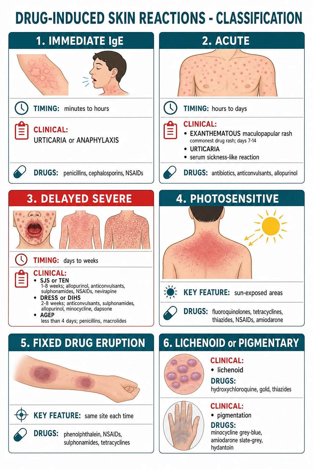
Four reactions — SJS/TEN, DRESS/DIHS, AGEP, and generalized bullous fixed drug eruption (GBFDE) — are grouped as the SCARs because they share a high mortality, characteristic drug triggers, and T-cell-mediated pathomechanisms.[1][2]
Epidemiology & Risk Factors
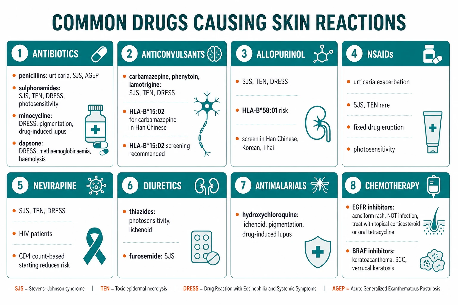
CADRs complicate roughly 2 to 3% of hospitalized patients and as many as 25% of ambulatory patients on chronic therapy; they account for around 1 in 100 emergency dermatology consultations.[1] Antibiotics, anticonvulsants, NSAIDs, allopurinol and, increasingly, targeted anticancer agents account for the bulk of cases — together responsible for more than 80% of all CADRs in the largest case series.[1]
Two classic host-pathogen-drug interactions are intensely examinable. Amoxicillin given during infectious mononucleosis produces a morbilliform rash in 90 to 100% of patients; this is not a true penicillin allergy — it reflects polyclonal T-cell activation by EBV — and patients tolerate penicillins subsequently.[3] Patients with HIV have a 40 to 60% rate of rash to trimethoprim-sulfamethoxazole, driven by glutathione deficiency, slow acetylation, immune dysregulation, and high CD8 T-cell activation.
Genetic susceptibility is now a major clinical theme. HLA-B*15:02 confers a 100-fold increased risk of carbamazepine-induced SJS/TEN in Han Chinese, Thai, and South-East Asian populations and underpins mandatory pre-prescription screening in those populations.[5] HLA-B*58:01 confers a similar risk for allopurinol-induced SJS/TEN in Asian populations.[6] HLA-A*31:01 is associated with carbamazepine-induced DRESS in European and Japanese patients. The lesson is direct: screen before prescribing carbamazepine in Han Chinese and before allopurinol in Han Chinese, Korean and Thai populations; reduction of allopurinol dose in chronic kidney disease independently lowers risk.[6]
Pathophysiology
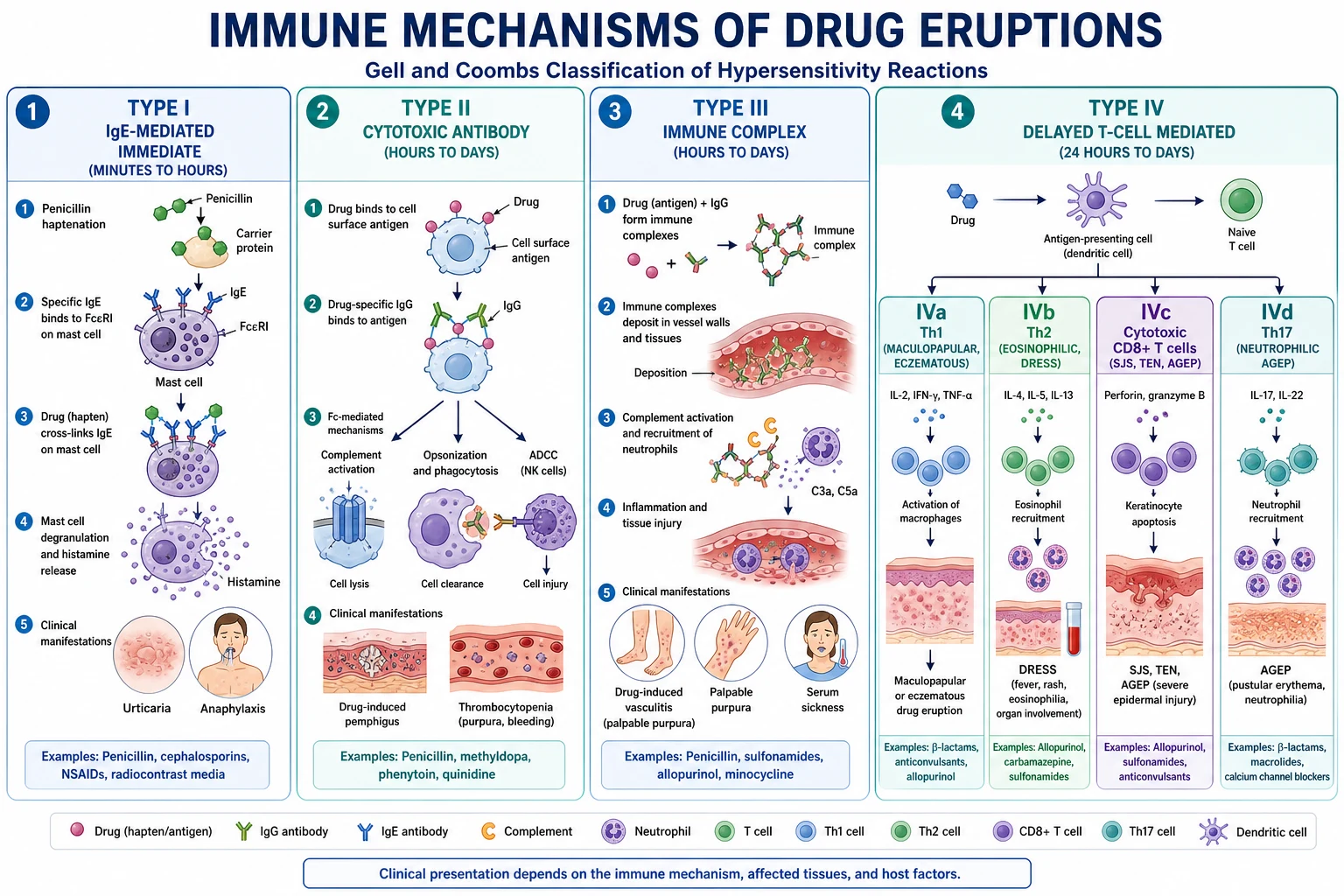
Drug eruptions arise by either non-immunologic mechanisms (predictable, dose-related, cumulative — for example warfarin-induced skin necrosis, minocycline pigment deposition, photo-toxicity) or immunologic (allergic) mechanisms, which conform to the Gell and Coombs Types I to IV, with Type IV (T-cell-mediated) subdivisions now expanded by Pichler to IVa through IVd. [1]
In SJS/TEN, the offending drug (or its reactive metabolite) is presented by HLA class I to clonal drug-specific CD8 cytotoxic T cells, which migrate into the epidermis and kill keratinocytes through the Fas-Fas ligand death-receptor pathway, perforin/granzyme B, and the cytotoxic molecule granulysin (the dominant mediator, present at concentrations 2 to 4 orders of magnitude higher than perforin or granzyme in SJS/TEN blister fluid).[2] The result is confluent full-thickness epidermal necrosis, positive Nikolsky sign, and sheet-like peeling.
In DRESS/DIHS, the pathogenesis has two integrated arms. First, an inherited or acquired defect in drug detoxification — most famously epoxide hydrolase deficiency for the aromatic anticonvulsants (carbamazepine, phenytoin, phenobarbital) — produces toxic arene oxide metabolites that damage keratinocytes and trigger a vigorous CD4 and CD8 T-cell response.[9] Second, between 2 and 3 weeks after onset, latent herpesviruses reactivate — HHV-6 most commonly, also CMV, EBV, HHV-7 — driving the characteristic biphasic flare, fever, and organ inflammation that distinguish true DRESS from a simple hypersensitivity reaction.[9][13]
AGEP is overwhelmingly T-cell mediated: drug-specific CD4 and CD8 T cells home to the epidermis, secrete IL-8 (CXCL8) and GM-CSF, and recruit neutrophils that fill a subcorneal cleft with sterile pustules; this is a Type IVd (Th17) pattern.[8]
Photosensitivity is mechanistically bipartite: phototoxic reactions are dose-dependent, non-immunologic, sunburn-like eruptions caused by UV-absorbing drug chromophores generating reactive oxygen species in the skin (any patient on sufficient drug plus sufficient sun is affected); photoallergic reactions are Type IV eczematous responses requiring prior sensitisation, can spread beyond sun-exposed skin, and persist after drug withdrawal. [1]
Clinical Presentation
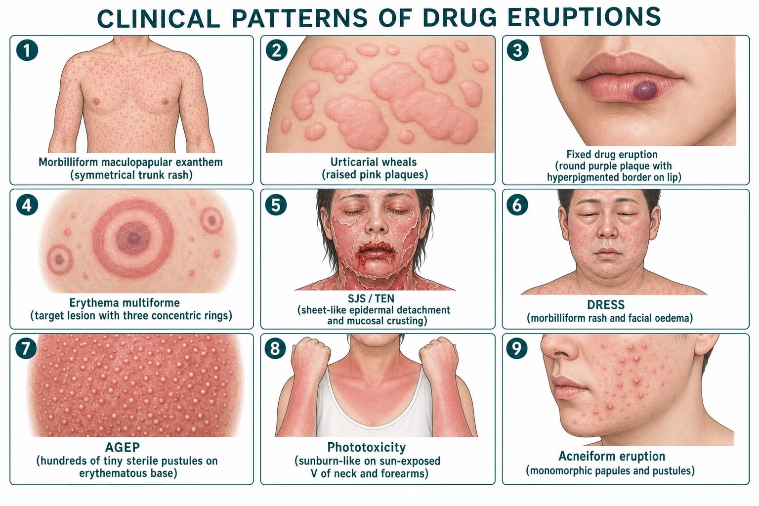
Because management hinges on pattern recognition, the catalogue of clinical morphologies — with their characteristic onset timing and high-yield culprit drugs — is the heart of this topic. The atlas below summarises the nine most examinable patterns. [1]
Exanthematous (morbilliform / maculopapular) — the commonest
The morbilliform (measles-like) maculopapular exanthem accounts for around 95% of all drug eruptions.[1] It begins as blanchable macules and papules that coalesce, starting on the trunk and proximal extremities and spreading symmetrically outwards, often sparing the face. Pruritus is mild to moderate; fever and eosinophilia may be present.
Day 7 to 14 after starting the new drug (1 to 2 days on re-exposure)
Symmetrical macules and papules, starting on trunk, spreading centrifugally
Confluence gives a morbilliform "measles-like" pattern
Mild pruritus, low-grade fever, possible eosinophilia
Resolves with desquamation 1 to 2 weeks after drug withdrawal
The classic offenders are penicillins (especially ampicillin/amoxicillin), sulfonamides, antiepileptics (phenytoin, carbamazepine, lamotrigine), allopurinol, and anti-TNF agents.[1] The amoxicillin-mono rash interaction deserves a separate mention because it is so common and so frequently mislabelled as penicillin allergy (see Special Populations).
Urticarial
Drug-induced urticaria and angioedema arise either by IgE-mediated (Type I) mechanisms — penicillins and other β-lactams are the classic cause — or by non-IgE (pseudoallergic) mechanisms, in which NSAID inhibition of cyclo-oxygenase-1 shunts arachidonic acid into the leukotriene pathway, producing urticaria and angioedema without specific IgE.[3] Opiates and radiocontrast media degranulate mast cells directly through MRGPRX2 receptors.
Distinguishing the two matters: an IgE-mediated reaction contraindicates the entire drug class and requires adrenaline on re-exposure; a pseudoallergic reaction can often be managed by switching to a COX-2 selective inhibitor (celecoxib) under cover. [1]
Fixed drug eruption (FDE)
Fixed drug eruption is the quintessentially recognisable pattern: a well-demarcated round purple-red plaque that recurs at the same anatomic site each time the drug is taken, resolving over 1 to 2 weeks to leave characteristic slate-grey-brown post-inflammatory hyperpigmentation.[1] Lesions are typically solitary but multiple and even generalised (generalised bullous fixed drug eruption, GBFDE — which can mimic TEN) variants exist.[14]
Favourite sites are mucocutaneous junctions: lips (most common), genitals (glans penis, vulva), oral mucosa, hands, feet, and perianal skin. Mucosal FDE may present as a painful erosion resembling aphthous ulceration. Common culprits are sulfonamides (including TMP-SMX), NSAIDs (especially piroxicam, mefenamic acid), tetracyclines, barbiturates, phenolphthalein (laxatives), metronidazole, and dapsone.[1]
[1]Erythema multiforme (EM)
Drug-induced erythema multiforme is less common than infection-driven EM (HSV, Mycoplasma) but is examinable. The classical target lesion has three concentric zones: a dark or bullous centre, a pale raised oedematous ring, and an erythematous outer ring. Lesions favour acral sites (palms, soles, dorsa of hands and feet) and extensor surfaces. EM minor is mucosal-sparing or has limited oral involvement; EM major has mucosal involvement at one or more sites. Drugs implicated include sulfonamides, antiepileptics (phenytoin, carbamazepine), NSAIDs, barbiturates, and penicillins.[2]
[1]SJS / TEN
Stevens-Johnson syndrome and toxic epidermal necrolysis form a spectrum defined by the percentage of body surface area (BSA) with epidermal detachment: SJS less than 10% BSA, SJS-TEN overlap 10 to 30% BSA, TEN greater than 30% BSA.[2] A flu-like prodrome (fever, malaise, upper-respiratory symptoms) precedes cutaneous signs by 1 to 3 days. The rash begins 1 to 8 weeks after starting the offending drug, with atypical target lesions — purpuric macules that evolve into flaccid bullae, then into sheets of necrotic epidermis that shear off with light lateral pressure (positive Nikolsky sign). Mucosal involvement at two or more sites is a defining feature: oral, conjunctival, tracheobronchial, genital, urethral, and gastrointestinal. The highest-yield drugs are allopurinol, anticonvulsants (carbamazepine, lamotrigine, phenytoin), sulfonamides (especially TMP-SMX), nevirapine, and oxicam NSAIDs.[2]
SJS/TEN is covered in depth in its dedicated topic; this catalogue focuses on recognition and immediate management (STOP drug, ICU/burns referral, ophthalmology within 24 hours). [1]
DRESS / DIHS
Drug reaction with eosinophilia and systemic symptoms (DRESS) — also called drug-induced hypersensitivity syndrome (DIHS) in the Japanese literature — begins 2 to 8 weeks after starting a new drug, far longer than most CADRs.[9][13] The presentation is dramatic:
Fever (often the first sign, 38 to 40 °C)
Facial oedema (characteristic, almost pathognomonic)
Morbilliform rash spreading caudally, may become exfoliative
Lymphadenopathy (cervical, generalised)
Eosinophilia (often greater than 1.5 × 10^9/L), atypical lymphocytes
Hepatitis (transaminases 2 to 5× upper limit of normal), sometimes acute liver failure
Multi-organ involvement: interstitial nephritis, pneumonitis, myocarditis, thyroiditis
Biphasic course: flare 2 to 3 weeks in as HHV-6 / CMV / EBV reactivate
The classic culprits form the aromatic anticonvulsant triad (carbamazepine, phenytoin, phenobarbital — cross-reactive via the arene oxide metabolite, so switch to a non-aromatic such as levetiracetam, valproate or lamotrigine), plus allopurinol, sulfonamides, minocycline, dapsone, vancomycin, and abacavir.[9][13]
DRESS is one of the great clinical mimics: it resembles infectious mononucleosis, viral hepatitis, lymphoma, serum sickness, Kawasaki disease, and idiopathic hypersensitivity. The clinical keys are (i) the long latency, (ii) the facial oedema, (iii) eosinophilia with hepatitis, and (iv) the characteristic drug list.[13]
AGEP
Acute generalised exanthematous pustulosis presents within 4 days (often within 24 to 48 hours) of starting a drug with hundreds of small (under 5 mm), sterile, non-follicular pustules lying on an erythematous base, with a flexural accentuation (inguinal, axillary, submammary).[8] Fever, leucocytosis (mainly neutrophilia), and mild systemic upset accompany the rash. Mucosal involvement is absent or minimal (distinguishing it from SJS). The rash is self-limiting: it resolves with superficial desquamation over 1 to 2 weeks after drug withdrawal.[8]
The two can be difficult to distinguish; the Sidoroff AGEP-validation score (EuroSCAR) helps (see Investigations). [1]
Photosensitivity
Drug-induced photosensitivity is divided into phototoxic (the common, predictable, sunburn-like reaction that any patient can get with sufficient drug plus sufficient UV) and photoallergic (the rarer, eczematous, Type IV, sensitisation-dependent reaction that spreads beyond sun-exposed skin).[1]
Pigmentary
Drug-induced pigmentation is usually mechanistically distinct (drug or drug-melanin complex deposition) rather than post-inflammatory. The patterns are exam classics. [1]
Drug-induced pigmentation may persist for months to years after the drug is stopped; minocycline type II can be helped by Q-switched Nd:YAG laser, but most types simply fade slowly.[10]
Acneiform
Drug-induced acneiform eruption differs from true acne vulgaris: lesions are monomorphic papules and pustules, without comedones, distributed on the upper trunk, shoulders, and face, and arise from follicular hyperkeratinisation and inflammation (not Cutibacterium acnes overgrowth).[1]
The EGFR inhibitor acneiform rash is so characteristic and so frequent (up to 90% of treated patients) that it is the textbook vignette: a follicular papulopustular eruption within 1 to 3 weeks of starting cetuximab or erlotinib, not an infection, and managed with topical corticosteroid plus oral tetracycline (doxycycline 100 mg BD) rather than antibiotics chosen for acne microbiology. The rash actually correlates with treatment response — patients who develop it tend to have better oncologic outcomes. [1]
Hair loss
Nail changes
Pseudoporphyria
Pseudoporphyria mimics porphyria cutanea tarda clinically — fragile sun-exposed skin, tense bullae on the dorsa of the hands, milia, scarring — but porphyrin studies are normal.[1] Culprits: NSAIDs (naproxen classically, also nabumetone, oxaprozin, diclofenac), tetracyclines, furosemide, dialysis-related. Management is withdrawal, photoprotection, and avoidance of the offending NSAID.
Lichenoid
Drug-induced lichenoid eruption mimics idiopathic lichen planus: flat-topped, polygonal, violaceous, pruritic papules with Wickham striae, but with features that betray a drug cause — the distribution is often photo-distributed, may be extensive or confluent, and resolves with prominent post-inflammatory hyperpigmentation. Histology differs: eosinophils and parakeratosis are present (unlike idiopathic LP). Culprits: hydrochlorothiazide, hydroxychloroquine, gold, penicillamine, beta-blockers (propranolol), ACE inhibitors, quinine, quinidine.[1]
Other examinable patterns
- Drug-induced lupus — anti-histone antibodies, ANA positive; culprits hydralazine, procainamide, minocycline, isoniazid, anti-TNF (paradoxically).
- Drug-induced vasculitis — palpable purpura on dependent sites (lower legs); culprits propylthiouracil, hydralazine, levetiracetam, anti-TNF, statins.
- Drug-induced pemphigoid / pemphigus — bullous pemphigoid-like tense bullae; culprits PD-1/PD-L1 inhibitors (pembrolizumab, nivolumab), loop diuretics, NSAIDs, penicillamine, captopril.[4]
- Warfarin-induced skin necrosis — days 3 to 10 of warfarin, in patients with protein C deficiency; painful purple-black plaques on breast, buttock, thigh; treat with vitamin K, heparin, and (if available) protein C concentrate.
- Drug-induced linear IgA bullous dermatosis — vancomycin is the classic cause; tense bullae in a "string of pearls" annular pattern.
- Erythroderma — generalized erythema and desquamation of greater than 90% BSA; culprits sulfonamides, antimalarials, phenytoin, dapsone, carbamazepine.
Differential Diagnosis
The differential diagnosis of a drug eruption depends entirely on the morphology and timeline, because the same morphology can be produced by both drugs and infections. The first task is to exclude non-drug causes for the pattern, then to ask whether a different SCAR is in play. [1]
For a morbilliform maculopapular eruption consider: (1) viral exanthem — measles, rubella, roseola (HHV-6), infectious mononucleosis (EBV), echovirus, parvovirus B19 — distinguished by prodrome, coryza, lymphadenopathy and a paucity of pruritus; (2) scarlet fever — sandpaper texture, Pastia lines, strawberry tongue, recent streptococcal pharyngitis; (3) Kawasaki disease in young children — fever for over 5 days, conjunctivitis, strawberry tongue, oedematous hands and feet, coronary artery dilation. [1]
For a febrile morbilliform eruption with organ involvement (the DRESS mimic) consider: (1) acute viral hepatitis — distinguished by high transaminases without eosinophilia and viral serology; (2) infectious mononucleosis — atypical lymphocytes, heterophile antibody, splenomegaly; (3) lymphoma — persistent lymphadenopathy, weight loss, night sweats, biopsy-confirmed; (4) systemic lupus erythematosus — ANA, anti-dsDNA, photosensitive rash without eosinophilia. [1]
For a pustular eruption (the AGEP mimic) consider: (1) generalised pustular psoriasis (von Zumbusch) — known psoriasis history, larger lakes of pus, nail and joint involvement, histological acantholysis; (2) subcorneal pustular dermatosis (Sneddon-Wilkinson) — older women, annular polycyclic lesions, chronic relapsing course; (3) IgA pemphigus — intra-epidermal neutrophils, direct immunofluorescence with intercellular IgA. [1]
For a bullous or desquamating eruption (the SJS/TEN mimic) consider: (1) staphylococcal scalded skin syndrome (SSSS) — superficial (intra-granular) split, no mucosal involvement, caused by exfoliative toxin, affects infants/young children; (2) toxic shock syndrome — hypotension, multi-organ failure, diffuse macular erythema with desquamation, no full-thickness necrosis; (3) acute graft-versus-host disease post stem-cell transplant; (4) pemphigus vulgaris — flaccid bullae, oral erosions, suprabasal acantholysis, positive Nikolsky; (5) bullous pemphigoid — tense bullae in the elderly, pruritic, subepidermal split with eosinophils. [1]
For palpable purpura consider infection-related vasculitis (meningococcaemia, Rocky Mountain spotted fever, infective endocarditis), Henoch-Schönlein purpura (paediatric, palpable purpura on lower legs plus abdominal pain, arthritis, haematuria), and cryoglobulinaemia. [1]
For drug-induced pigmentation consider post-inflammatory hyperpigmentation (resolves with time, follows known inflammation), melasma (facial, oestrogen-related, no drug history), Addison disease (generalised bronze pigmentation, mucosal involvement, hypocortisolism), and haemochromatosis (diabetes, hepatomegaly, iron overload). [1]
The two most common diagnostic errors are (i) calling a morbilliform drug eruption a viral exanthem (the difference is the drug history and timeline, not the morphology), and (ii) mislabelling DRESS as a viral illness — DRESS has eosinophilia and transaminitis that a viral exanthem does not. Equally important is distinguishing AGEP from pustular psoriasis: AGEP resolves spontaneously on drug withdrawal; pustular psoriasis requires systemic therapy and may recur. [1]
Clinical & Bedside Assessment
The single most informative investigation is a complete drug history and timeline. Record every drug, herbal, OTC preparation, and recreational substance, with start date, dose, route, duration, and any prior reaction.[1] Plot each against the morphology and timing to identify the most probable culprit.
DRUGS
Two formal causality tools are widely used. The Naranjo Adverse Drug Reaction Probability Scale scores 10 questions (yes +1 to +2, no −1 to 0, unknown 0): a score over 8 is definite, 5 to 8 probable, 1 to 4 possible, 0 or less doubtful.[1] The ALDEN (Algorithm of Drug Causality for Epidermal Necrolysis) is disease-specific for SJS/TEN, weighting timing, pre-exposure, drug half-life, de-challenge, re-challenge, and prior causality knowledge; a score over 6 is "very probable," 5 to 6 "probable," 2 to 4 "possible," 1 "unlikely," 0 "very unlikely."[2]
Bedside signs to elicit specifically: [1]
Investigations
Most CADRs are diagnosed clinically. Investigations serve to confirm a SCAR, to characterise organ involvement, and (in selected cases) to identify the culprit. [1]
Bloods
Skin biopsy
Histology is the tie-breaker for bullous or atypical eruptions. SJS/TEN: full-thickness confluent epidermal necrosis with a sparse dermal lymphocytic infiltrate. DRESS: lymphocytic interface dermatitis with eosinophils and atypical activated lymphocytes. AGEP: subcorneal aggregates of neutrophilic pustules with eosinophils, mild spongiosis, no necrosis. FDE: basal vacuolar change, apoptotic keratinocytes, melanophages in the dermis (the source of the residual pigmentation).[1]
In-vivo and ex-vivo tests for culprit identification
Delayed skin testing (patch, intradermal with delayed reading) is most useful for FDE, AGEP, and DRESS, with sensitivities of 30 to 80% depending on the drug and reaction.[11] The lymphocyte transformation test (LTT) measures drug-induced T-cell proliferation; it is particularly valuable for DRESS (sensitivity 60 to 80%) and increasingly used for SCARs generally.[12]
Named diagnostic and severity scores
SCORTEN should be calculated twice — within 24 h of admission and again at 72 h — as severity can evolve. The RegiSCAR study confirmed SCORTEN's discriminant performance in the largest European cohort.[15]
Genetic testing
For high-risk populations, HLA typing before prescription is now the standard of care: HLA-B*15:02 before carbamazepine, oxcarbazepine, and phenytoin in Han Chinese, Thai, Filipino, Vietnamese, Cambodian, and South-East Asian patients; HLA-B*58:01 before allopurinol in Han Chinese, Korean, Thai, and (by FDA guidance) all patients with chronic kidney disease regardless of ethnicity.[5][6]
Management — Resuscitation
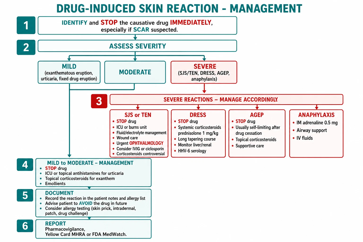
For most CADRs the immediate step is the same — stop the drug — but the resuscitative intensity scales steeply with severity. [1]
[1]The airway is at risk in SJS/TEN (tracheobronchial sloughing → stridor, hypoxia), anaphylaxis (laryngeal oedema), and severe DRESS with pneumonitis. Skin failure behaves like a burn: hypothermia, hypovolaemia, hypernatraemia, hypoalbuminaemia, and high sepsis risk. [1]
Management — Definitive & Stepwise
Identify and STOP the offending drug immediately (the single most important step)
Assess severity: mild (exanthem, urticaria, FDE, lichenoid, pigmentary, acneiform) vs severe (SJS/TEN, DRESS, AGEP, GBFDE, anaphylaxis, erythroderma)
Provide symptomatic relief: antihistamine (cetirizine 10 mg OD or loratadine 10 mg OD); topical corticosteroid (mometasone 0.1% OD); emollients
Stratify by severity — see the disease-specific bundles below
Document the allergy in the patient record, drug chart, and electronic health record; provide a patient-held allergy alert
Report to pharmacovigilance (MHRA Yellow Card, FDA MedWatch, PvPI India) for any serious or unusual reaction
Counsel the patient to avoid the drug and structurally related agents (cross-reactivity)
Consider allergy testing (skin prick, patch, LTT) 1 to 6 months after resolution
Mild reactions
Exanthem, urticaria (no anaphylaxis), FDE, lichenoid, pigmentary, acneiform: STOP drug; oral second-generation antihistamine for pruritus (cetirizine 10 mg OD, loratadine 10 mg OD, fexofenadine 180 mg OD); topical corticosteroid (mometasone furoate 0.1% OD for 1 to 2 weeks); emollients. Urticaria may need a short course of oral prednisolone 0.5 mg/kg/day for 3 to 5 days if extensive. Acneiform rash from EGFR inhibitors is managed with topical corticosteroid plus oral doxycycline 100 mg BD (anti-inflammatory, not antimicrobial) — do not stop the anticancer drug. [1]
Severe reactions
Specific situations
- Phototoxic reaction: STOP drug (or substitute if essential); strict photoprotection (broad-spectrum SPF 50+, protective clothing, avoidance); topical corticosteroid.
- Drug-induced pigmentation: STOP drug; may take months-years to fade; Q-switched Nd:YAG laser for minocycline type II; reassurance for most cases.
- Drug-induced vasculitis: STOP drug; NSAIDs for joint pain; systemic corticosteroid for extensive or visceral involvement; screen for internal organ involvement (renal, GI, CNS).
- Drug-induced lupus: STOP drug; NSAIDs; antimalarials for cutaneous disease; resolves over weeks-months.
- Warfarin-induced skin necrosis: STOP warfarin; vitamin K; heparin; protein C concentrate if available; surgical debridement if infected.
- Drug-induced pemphigoid/pemphigus: STOP drug; topical and systemic corticosteroid; doxycycline + nicotinamide as steroid-sparing. [1]
Documentation and pharmacovigilance
Every CADR must be documented in the patient's notes, drug chart, and electronic health record (allergy section), and the patient should be given a patient-held allergy alert (card or bracelet) and counselled on which drugs and drug classes to avoid. Pharmacovigilance reporting is mandatory for serious, unexpected, or new reactions — the MHRA Yellow Card (UK), FDA MedWatch (US), or PvPI (India) systems.[1]
Specific Subtypes & Scenarios
The catalogue below is the high-yield drug-by-reaction reference for viva and MCQ revision. [1]
Cross-reactivity pearls
- Aromatic anticonvulsants (carbamazepine, phenytoin, phenobarbital) share the arene oxide metabolite — 75% cross-reactivity for DRESS. Switch to valproate, levetiracetam, or lamotrigine.
- β-lactam cross-reactivity is now understood to be 2% or less between penicillins and third/fourth-generation cephalosporins; the historical 10% figure was over-estimated. Carbapenems and aztreonam are safe in penicillin allergy with negative skin testing.[3]
- NSAID class cross-reactivity for pseudoallergic urticaria is around 80% across non-selective NSAIDs; COX-2 selective inhibitors (celecoxib, etoricoxib) are usually tolerated and can be used as alternatives under challenge.
Complications & Pitfalls
Prognosis & Disposition
Prognosis is determined by reaction severity, comorbidities, and speed of drug withdrawal. [1]
Mild reactions resolve within days to weeks of drug withdrawal, though pigmentation may persist for months. DRESS typically requires months of corticosteroid taper, and patients need long-term follow-up for autoimmune sequelae (thyroiditis, type 1 diabetes, autoimmune enteropathy) that may emerge months after the acute episode.[9] SJS/TEN survivors need lifelong ophthalmology follow-up and may develop strictures requiring surgical reconstruction. Lifelong drug avoidance is mandatory for the offending agent and cross-reactive drugs; a medic-alert bracelet and patient-held allergy alert should be issued.
Special Populations
Paediatric
The most common paediatric CADRs are reactions to antibiotics (penicillins, cephalosporins, sulfonamides) and antiepileptics. Stevens-Johnson syndrome in children most often follows Mycoplasma pneumoniae infection or anticonvulsant use; the morphology and management are the same as in adults, but weight-based fluid resuscitation (weight-based 2 to 4 mL/kg/% BSA burn in the first 24 h using the Parkland formula) and paediatric dosing of corticosteroids and antihistamines apply. Kawasaki disease is an important mimic of morbilliform drug eruptions in young children. [1]
Pregnancy
Pregnancy limits some of the usual tools. Antihistamines — chlorphenamine and cetirizine are safe. Topical corticosteroids — mild to moderate potency safe; potent on small areas only. Systemic corticosteroids — prednisolone preferred over dexamethasone (less placental transfer). Avoid in pregnancy: thalidomide, isotretinoin and acitretin (teratogenic), minocycline (tooth discolouration), sulfonamides near term (kernicterus), NSAIDs after 20 weeks. Manage SCARs in pregnancy as in non-pregnant patients — the maternal mortality risk justifies the same aggressive therapy. [1]
Elderly
Polypharmacy makes the drug history complex and causality assessment challenging. Altered pharmacokinetics (reduced renal and hepatic clearance, reduced serum albumin) raise CADR risk; thiazides, anticoagulants, allopurinol (especially in CKD), and antibiotics dominate. Fever may be blunted, masking DRESS. Allopurinol dose should be reduced in CKD (start 100 mg/day or less in CKD stage 3 or worse) to lower DRESS/SJS risk; this is in the Beers Criteria and the ACR gout guideline.[6]
HIV and immunocompromised
HIV patients have markedly elevated CADR rates — 40 to 60% react to TMP-SMX, and high rates to dapsone, thalidomide, and sulfadiazine. Nevirapine causes SJS in up to 6% of patients (highest in women with CD4 over 250 and men with CD4 over 400 — these are relative contraindications). Abacavir causes a characteristic multi-system hypersensitivity reaction in HLA-B*57:01 positive patients — screen before prescribing. Stem-cell transplant patients develop graft-versus-host disease, which mimics drug eruptions and SJS/TEN histologically. Checkpoint inhibitor rash ranges from mild maculopapular to life-threatening bullous pemphigoid, SJS, or DRESS — multidisciplinary oncology-dermatology care is essential. [1]
Renal and hepatic impairment
Drug accumulation amplifies risk. Allopurinol in CKD converts to its long-half-life metabolite oxypurinol, increasing SJS/TEN risk; dose-reduce and screen HLA-B*58:01 in Asian populations.[6] Phenytoin is hepatically metabolised and protein-bound — hypoalbuminaemia raises free levels, precipitating DRESS.
Evidence, Guidelines & Regional Differences
- US FDA: Black-box warning on carbamazepine recommending HLA-B*15:02 testing in Asian ancestry patients before starting; recommended HLA-B*5801 testing before allopurinol in high-risk populations and dose reduction in CKD.
- EMA / European guidelines: same HLA recommendations; RegiSCAR-based DRESS criteria; EuroSCAR AGEP scoring standard.
- UK (NICE / MHRA): Yellow Card pharmacovigilance for all serious suspected CADRs; NICE gout guideline recommends allopurinol dose reduction in CKD.
- Japan (J-SCAR): separate DIHS criteria requiring HHV-6 reactivation; therapeutic approach emphasises slow corticosteroid taper and CMV monitoring.
- India (PvPI): Pharmacovigilance Programme of India; HLA-B*15:02 prevalence is lower than in Han Chinese but screening is increasingly practised in South-East Asian populations.
Controversies
The role of systemic corticosteroids in SJS/TEN remains debated: some observational data suggest benefit if given early (within 2 to 3 days), but delayed or prolonged steroids may increase infection and mortality. IVIG for SJS/TEN has mixed evidence — early observational studies suggested benefit, the largest retrospective studies showed none or harm; current European guidance reserves it for extensive disease with consultant multidisciplinary decision. Ciclosporin shows promising observational benefit and is increasingly used in Europe. DRESS management with corticosteroids is essentially universal; the controversies are the taper duration (most centres use 8 to 12 weeks, some longer) and the role of ganciclovir for CMV reactivation in visceral disease.[9]
Exam Pearls
[1]Exam application bank (NEET-PG / INICET)
One-line answer
Cutaneous adverse drug reactions (CADRs) range from mild (exanthematous maculopapular rash, urticaria, fixed drug eruption) to life-threatening SCARs (SJS/TEN, DRESS/DIHS, AGEP). Onset timing is diagnostic: AGEP under 4 days, exanthem 7 to 14 days, SJS/TEN 1 to 8 weeks, DRESS 2 to 8 weeks. HLA-B15:02 (carbamazepine SJS in Han Chinese) and HLA-B58:01 (allopurinol SJS in Asian populations) mandate pre-prescription screening. Management of every suspected CADR begins with STOP the drug; severity dictates escalation: antihistamines and topical corticosteroids for mild reactions, systemic corticosteroids for DRESS, ICU/burns and ophthalmology for SJS/TEN, supportive care for AGEP.
Worked stems (answer without another resource)
Stem 1 — Classic presentation. Map symptoms to mechanism; name the first investigation and first treatment step with dose/route if drug therapy is standard. [1]
Stem 2 — Unstable / complicated. List red flags that force immediate resuscitation, theatre, ICU, antidote, or reperfusion — and what you do in the first 15 minutes. [1]
Stem 3 — Atypical group. Elderly, pregnancy, child, or immunocompromised: how presentation and thresholds change. [1]
Stem 4 — Differential trap. Name the three closest mimics and one discriminator for each. [1]
Stem 5 — Disposition. Who goes home with safety-netting, who is admitted, who needs HDU/ICU/theatre, and what follow-up is mandatory. [1]
Rapid viva checklist
- Definition + classification
- Pathophysiology chain
- Bedside signs / criteria
- Score with exact components (if any)
- Emergency bundle
- Definitive therapy with doses
- Complications of disease and of treatment
- Special populations
- Guideline/trial name if classic
- Three exam traps
Coverage self-check
If you cannot answer any stem above from this page alone, re-read the matching section — the page is intended to be self-sufficient for final-prof and NEET-PG/INICET questions on Drug-induced skin disease.
[1]Quick check: which HLA allele and which drug?
- HLA-B*15:02 → carbamazepine (SJS, Han Chinese).
- HLA-B*58:01 → allopurinol (SJS, Asian populations).
- HLA-B*57:01 → abacavir (hypersensitivity, all populations — screen before starting).
- HLA-A*31:01 → carbamazepine (DRESS, European/Japanese).
- HLA-DQB1*06:02 / HLA-DRB1*15:01 → nevirapine (hepatotoxicity/SJS).
Quick check: timing of onset by reaction?
- Anaphylaxis: minutes to 1 hour.
- AGEP: under 4 days.
- Morbilliform exanthem: 7 to 14 days.
- SJS/TEN: 1 to 8 weeks.
- DRESS/DIHS: 2 to 8 weeks.
- Fixed drug eruption: hours to 2 days on re-exposure.
- Pseudoporphyria: months of chronic NSAID exposure.
Quick check: most common cause of SJS/TEN?
- In Europe and Israel: allopurinol is now the leading cause of SJS/TEN.
- In Asia: anticonvulsants (carbamazepine) and allopurinol predominate.
- In HIV: nevirapine and TMP-SMX.
- In children: anticonvulsants and Mycoplasma pneumoniae (the latter strictly a SJS-mycoplasma overlap rather than a drug cause).
References
- [1]Del Pozzo-Magaña BR, Liy-Wong C. Drugs and the skin: A concise review of cutaneous adverse drug reactions Br J Clin Pharmacol, 2024.PMID 35974692
- [2]Shah H, Parisi R, Mukherjee E, et al. Update on Stevens-Johnson Syndrome and Toxic Epidermal Necrolysis: Diagnosis and Management Am J Clin Dermatol, 2024.PMID 39278968
- [3]Zagursky RJ, Pichichero ME. Cross-reactivity in β-Lactam Allergy J Allergy Clin Immunol Pract, 2018.PMID 29017833
- [4]Verheyden MJ, Bilgic A, Murrell DF. A Systematic Review of Drug-Induced Pemphigoid Acta Derm Venereol, 2020.PMID 32176310
- [5]Chung WH, Hung SI, Hong HS, et al. Medical genetics: a marker for Stevens-Johnson syndrome Nature, 2004.PMID 15057820
- [6]Hung SI, Chung WH, Liou LB, et al. HLA-B*5801 allele as a genetic marker for severe cutaneous adverse reactions caused by allopurinol Proc Natl Acad Sci U S A, 2005.PMID 15743917
- [7]Kardaun SH, Sidoroff A, Valeyrie-Allanore L, et al. Variability in the clinical pattern of cutaneous side-effects of drugs with systemic symptoms: does a DRESS syndrome really exist? Br J Dermatol, 2007.PMID 17300272
- [8]Sidoroff A, Dunant A, Viboud C, et al. Risk factors for acute generalized exanthematous pustulosis (AGEP)-results of a multinational case-control study (EuroSCAR) Br J Dermatol, 2007.PMID 17854366
- [9]Mizukawa Y, Shiohara T. Recent advances in the diagnosis and treatment of DIHS/DRESS in 2025 Allergol Int, 2025.PMID 40251070
- [10]Alharithy R, Alhumaidi A, AlDukhiel F, et al. Drug induced hyperpigmentation: systematic review and meta-analysis Front Med (Lausanne), 2026.PMID 42023087
- [11]Barbaud A, Sterboul A, Waton J, et al. Delayed Skin Testing for Systemic Medications: Helpful or Not? J Allergy Clin Immunol Pract, 2024.PMID 38977212
- [12]Sittiwattanawong P, Kanchanapoomi T, Rerknimitr P, et al. In-vivo and ex-vivo tests for culprit drugs identification in severe cutaneous adverse drugs reactions J Dermatol, 2024.PMID 38605448
- [13]Cacoub P, Musette P, Descamps V, et al. The DRESS syndrome: a literature review Am J Med, 2011.PMID 21592453
- [14]Loo LY, Tan E, Pang R, et al. Generalized Bullous Fixed Drug Eruption: A Systematic Review J Allergy Clin Immunol Pract, 2026.PMID 41539429
- [15]Sekula P, Dunant A, Mockenhaupt M, et al. Evaluation of SCORTEN on a cohort of patients with Stevens-Johnson syndrome and toxic epidermal necrolysis included in the RegiSCAR study J Burn Care Res, 2011.PMID 21228709