Dermatology · Medicine
Ear and nose dermatoses
Also known as Chondrodermatitis nodularis helicis · Auricular dermatoses · Nasal dermatoses · Rhinophyma · Pinna chondritis spectrum
Multi-board ear and nose special-site dermatology: chondrodermatitis nodularis helicis as a pressure–ischaemia helix nodule, relapsing polychondritis with lobe-sparing chondritis, acute otitis externa co-management, seborrhoeic and contact dermatitis of the ear, rhinophyma within the rosacea spectrum, actinic keratosis and keratinocyte cancers of ear/nose, biopsy thresholds, and stepwise pressure-relief, topical, systemic, and procedural pathways.
On this page & tools
Your progress
Saved locally on this device.
Exam tags
Red flags
Overview & Definition
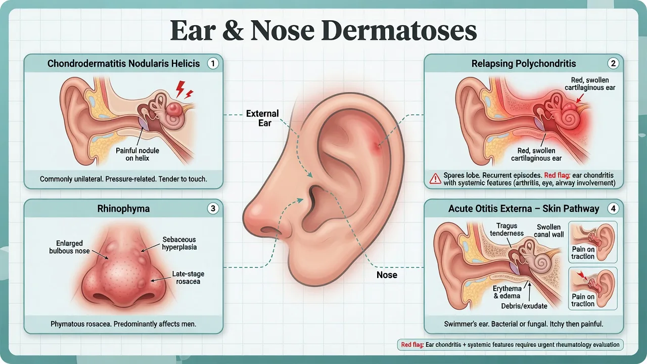
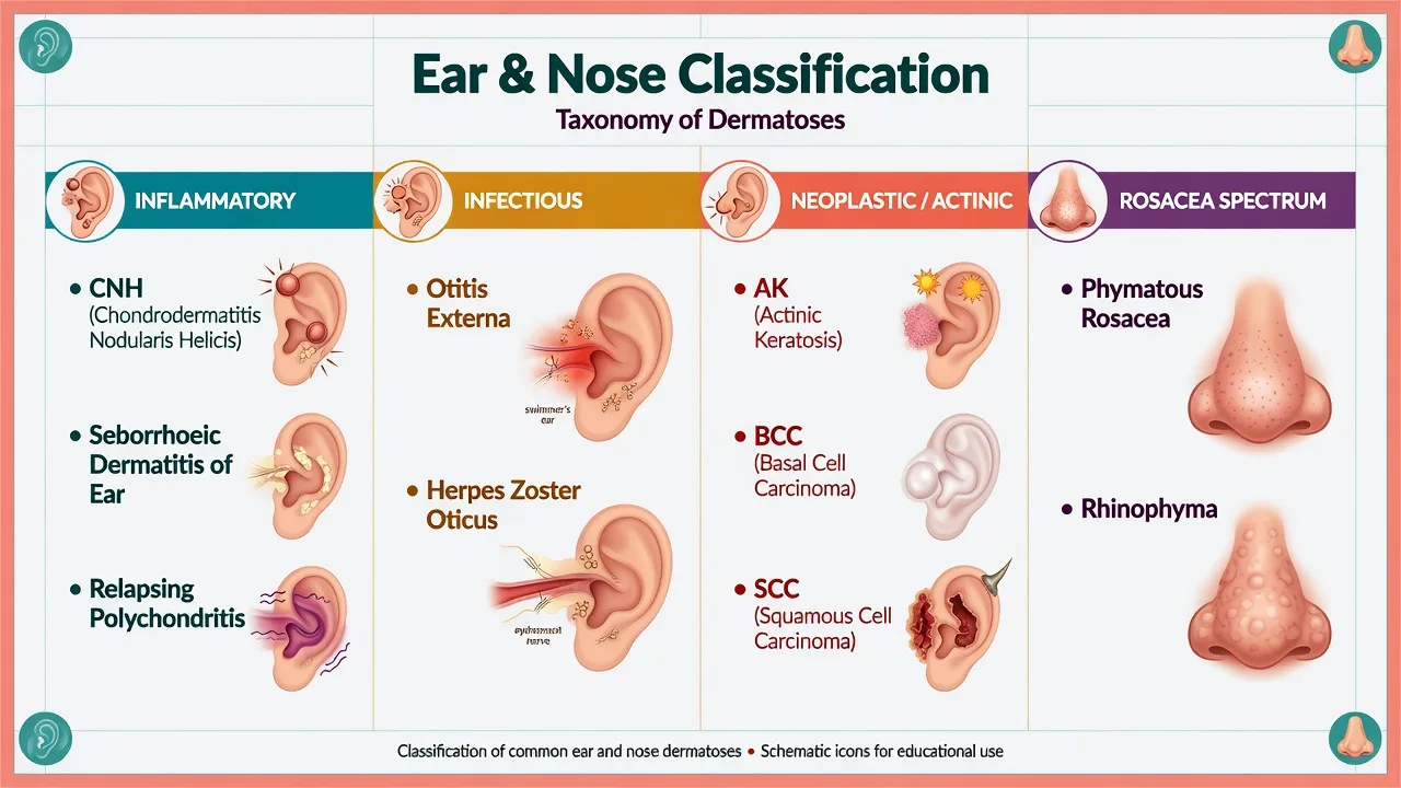
The external ear and nose are special sites: thin skin over cartilage, high UV dose, cosmetic visibility, and shared care with ENT. Examiners test whether you can separate local pressure disease (CNH), systemic cartilaginous disease (RP), canal infection (AOE), rosacea phymatous change, and actinic neoplasia.[1][4][9]
Classification
Inflammatory
Infectious
Actinic / neoplastic
Rosacea spectrum
Nasal analogue
Epidemiology & Risk Factors
CNH is most common in middle-aged and older adults who favour one sleep side; men are often over-represented in classic series.[1][3] AOE clusters with water exposure, trauma, hearing aids, and humid climates.[9][10] Rhinophyma is the phymatous end of rosacea and is more often clinically recognised in men.[8] Ear and nose keratinocyte cancers track cumulative UV and immunosuppression.[12]
Pathophysiology
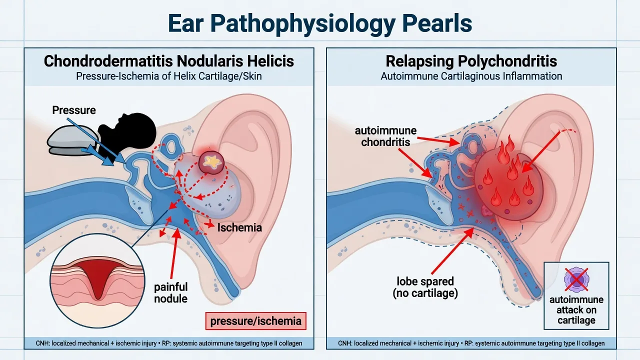
CNH is a pressure–ischaemia injury of skin tightly applied to cartilage; chronic microtrauma from sleep, phones, or headgear perpetuates a painful nodule, often with central crust or ulceration.[1][2][3]
Relapsing polychondritis targets cartilaginous structures; the earlobe is spared because it has no cartilage — a board-favourite distinguisher from diffuse pinna cellulitis.[4][5][6]
Rhinophyma reflects sebaceous and soft-tissue hyperplasia within phymatous rosacea, not “alcohol nose” folklore.[7][8]
Clinical Presentation
Chondrodermatitis nodularis helicis
Exquisitely tender small nodule on the helix (or antihelix), often unilateral on the preferred sleep side; patients wake when the ear is pressed. May show central crust.[1]
Relapsing polychondritis (auricular)
Acute or recurrent red, swollen, painful pinna with lobe sparing; may accompany nasal chondritis, airway symptoms, ocular inflammation, or seronegative arthritis.[4][5]
Acute otitis externa
Canal pain, discharge, tragal tenderness, pain on pinna traction; skin disease at the meatus may coexist.[9][10]
Rhinophyma
Bulbous, irregular nasal enlargement with prominent pores and thickened skin — end-stage phymatous change.[8]
Actinic / neoplastic
Scaly AK plaques; pearly BCC; indurated or ulcerated SCC on helix rim and nasal tip/ala — high-stakes special sites.[12]
Other high-yield
- Seborrhoeic dermatitis of concha and post-auricular crease.
- Nickel earring dermatitis of the lobe.
- Chondrodermatitis nodularis nasi as a nasal pressure analogue.[11]
- Herpes zoster oticus — vesicles in canal/concha ± facial palsy.
Differential Diagnosis
| Finding | Prefer | Key distinguisher |
|---|---|---|
| Tender sleep-side helix nodule | CNH | Pressure history; exquisite point tenderness |
| Red pinna, lobe spared, systemic clues | RP | Cartilage distribution; multi-organ review |
| Tragal tenderness + canal debris | AOE | Pain on traction; canal-centric |
| Bulbous nose, rosacea history | Rhinophyma | Phymatous subtype |
| Non-healing ulcer/crust | SCC/BCC | Biopsy |
| Vesicles + facial palsy | Zoster oticus | Dermatomal vesicles |
Clinical & Bedside Assessment
- Sleep side, headgear, hearing aids, UV occupation, prior skin cancers.
- Systemic screen for RP (joints, eyes, airway, nasal saddle change).
- Pull tragus/pinna; otoscopy when canal disease suspected.
- Full head-and-neck UV field exam.
- Document size and photograph nodules for response after offloading. [1]
Investigations
Most CNH and rosacea are clinical. Biopsy atypical, progressive, or non-healing lesions and any lesion where keratinocyte cancer cannot be excluded.[12] RP is a clinical multisystem diagnosis supported by inflammatory context — do not wait for a mythical single pathognomonic blood test before recognising the pattern.[4][6] Culture selected refractory AOE cases, especially immunocompromised hosts.[10]
Management — Resuscitation
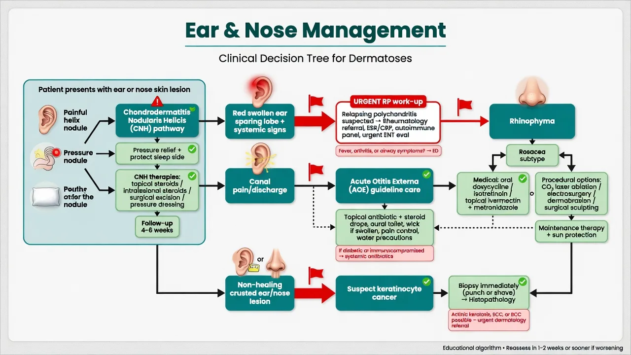
Airway-compromising RP or severe systemic chondritis is an emergency. Malignant (necrotising) OE concern in high-risk hosts with severe AOE needs urgent ENT, not another week of random drops alone.[4][9]
Management — Definitive & Stepwise
CNH: pressure relief is disease-modifying — change sleep side, protective doughnut pillow/padding, avoid local trauma. Adjuncts include topical or intralesional corticosteroids and, for refractory nodules, procedural options (excision, cartilage-sparing techniques) selected by experience and lesion morphology.[1][2][3]
AOE: aural toilet, appropriate topical therapy, pain control, and water precautions per AAO-HNS principles; add systemic therapy when extension or host risk demands it.[9][10]
RP: rheumatology-led systemic anti-inflammatory/immunosuppressive strategies once infection mimics are addressed; dermatology recognises the ear and protects cartilage long-term.[4][6]
Rhinophyma / rosacea: treat active inflammatory rosacea medically; established phymatous bulk may need electrosurgery, laser, or surgical recontouring after counselling.[7][8]
Keratinocyte neoplasia: biopsy-guided management along AK/SCC/BCC pathways; ear and nose often need specialist reconstructive planning.[12]
Specific Subtypes & Scenarios
Antihelix CNH and nasal chondrodermatitis follow the same pressure logic.[11]
Hearing-aid dermatitis mixes pressure, moisture, and contact allergy. [1]
Immunosuppressed patients: lower threshold to biopsy “CNH-like” lesions for SCC. [1]
Complications & Pitfalls
- Labelling RP as recurrent cellulitis.[5]
- Endless CNH creams without pressure offloading.[2]
- Missing helix SCC.[12]
- Stigmatising rhinophyma as alcohol use disorder.[8]
Prognosis & Disposition
CNH often improves when pressure is truly removed; residual nodules may need procedures.[2] RP is relapsing–remitting and needs long-term specialty follow-up.[4] Rhinophyma bulk does not fully reverse with cream alone once established.[8]
Special Populations
Diabetics and elderly: AOE complications. Transplant recipients: aggressive ear/nose SCC. Outdoor labourers: field cancerisation of helix and nasal tip. [1]
Evidence, Guidelines & Regional Differences
AAO-HNS AOE guidance underpins topical-first canal care with clear systemic exceptions.[9][10] CNH literature is therapy-review and case-series heavy — pressure modification remains the conceptual core.[2][3] Rosacea reviews sequence medical control before procedural rhinophyma work.[7]
Exam Pearls
EAR pearls
- Lobe sparing is the RP viva kill-shot.[5][6]
- CNH without pressure advice will fail management marks.[1]
- Non-healing crust = biopsy, not another antibiotic cream.[12]
- Tragal tenderness points to the canal.[9]
Exam application bank (NEET-PG / INICET)
One-line answer
Multi-board ear and nose special-site dermatology: chondrodermatitis nodularis helicis as a pressure–ischaemia helix nodule, relapsing polychondritis with lobe-sparing chondritis, acute otitis externa co-management, seborrhoeic and contact dermatitis of the ear, rhinophyma within the rosacea spectrum, actinic keratosis and keratinocyte cancers of ear/nose, biopsy thresholds, and stepwise pressure-relief, topical, systemic, and procedural pathways.
Worked stems (answer without another resource)
Stem 1 — Classic presentation. Map symptoms to mechanism; name the first investigation and first treatment step with dose/route if drug therapy is standard. [1]
Stem 2 — Unstable / complicated. List red flags that force immediate resuscitation, theatre, ICU, antidote, or reperfusion — and what you do in the first 15 minutes. [1]
Stem 3 — Atypical group. Elderly, pregnancy, child, or immunocompromised: how presentation and thresholds change. [1]
Stem 4 — Differential trap. Name the three closest mimics and one discriminator for each. [1]
Stem 5 — Disposition. Who goes home with safety-netting, who is admitted, who needs HDU/ICU/theatre, and what follow-up is mandatory. [1]
Rapid viva checklist
- Definition + classification
- Pathophysiology chain
- Bedside signs / criteria
- Score with exact components (if any)
- Emergency bundle
- Definitive therapy with doses
- Complications of disease and of treatment
- Special populations
- Guideline/trial name if classic
- Three exam traps
Coverage self-check
If you cannot answer any stem above from this page alone, re-read the matching section — the page is intended to be self-sufficient for final-prof and NEET-PG/INICET questions on Ear and nose dermatoses.
Expanded exam teaching (depth pass)
Clinical reasoning
For Ear and nose dermatoses, examiners test whether you can prioritise life threats, choose the right first test, and give specific therapy (agent, dose, route, timing). Generic phrases without numbers score poorly.
Mechanism → feature map
Build a short chain: cause → pathophysiologic intermediate → clinical feature → complication. Every major symptom in the classic vignette should sit on that chain.
Investigation strategy
- Bedside/first-line tests that change immediate management
- Confirmatory or staging tests
- What a normal result does not exclude
- When not to delay treatment for imaging (unstable patient)
Management ladder
- Resuscitation / ABC / sepsis or haemorrhage bundle as relevant
- Specific antidote / procedure / antimicrobial / reperfusion / surgery
- Supportive care and monitoring targets
- Definitive long-term therapy and secondary prevention
- Disposition and safety-net advice
Special populations
Always prepare one line each for children, pregnancy, elderly, renal/hepatic impairment, and immunocompromised patients when the topic allows.
Pitfalls that fail candidates
- Treating the number not the patient
- Missing pregnancy status when relevant
- Imaging before stabilisation
- Wrong empiric cover or wrong antidote timing
- Incomplete counselling on recurrence, adherence, or red-flag return
Multi-board ear and nose special-site dermatology: chondrodermatitis nodularis helicis as a pressure–ischaemia helix nodule, relapsing polychondritis with lobe-sparing chondritis, acute otitis externa co-management, seborrhoeic and contact dermatitis of the ear, rhinophyma within the rosacea spectrum, actinic keratosis and keratinocyte cancers of ear/nose, biopsy thresholds, and stepwise pressure-relief, topical, systemic, and procedural pathways. [1]
Structured revision sheet
Must-know numbers and names
List every score, size threshold, dose, and time window from this topic on a blank page from memory, then check against the sections above.
Three classic MCQ angles
- Most likely diagnosis given a vignette
- Next best step in management
- Most appropriate investigation
Three classic SAQ angles
- Pathophysiology in five steps
- Management algorithm with doses
- Complications and prevention
Clinical station flow
Greet → focused history → targeted exam → investigations → explain diagnosis → emergency care → definitive plan → safety-net / follow-up → answer examiner questions on mechanism and pitfalls.
Exam anchors
References
- [1]Zhang LW, Wu J, Chen T. Chondrodermatitis nodularis helicis Cleve Clin J Med, 2023.PMID 37263665
- [2]Shah S, Fiala KH. Chondrodermatitis nodularis helicis: A review of current therapies Dermatol Ther, 2017.PMID 27723195
- [3]Salah H, Urso B, Khachemoune A. Review of the Etiopathogenesis and Management Options of Chondrodermatitis Nodularis Chronica Helicis Cureus, 2018.PMID 29805936
- [4]Mertz P, Sparks J, Kobrin D, et al. Relapsing polychondritis: Best Practice & Clinical Rheumatology Best Pract Res Clin Rheumatol, 2023.PMID 37839908
- [5]Karp NC, Goglin SE. Ear Swelling of Relapsing Polychondritis J Gen Intern Med, 2023.PMID 36854868
- [6]Rapini RP, Warner NB. Relapsing polychondritis Clin Dermatol, 2006.PMID 17113965
- [7]Sharma A, Kroumpouzos G, Kassir M, et al. Rosacea management: A comprehensive review J Cosmet Dermatol, 2022.PMID 35104917
- [8]Tüzün Y, Wolf R, Kutlubay Z, et al. Rosacea and rhinophyma Clin Dermatol, 2014.PMID 24314376
- [9]Rosenfeld RM, Schwartz SR, Cannon CR, et al. Clinical practice guideline: acute otitis externa executive summary Otolaryngol Head Neck Surg, 2014.PMID 24492208
- [10]Rosenfeld RM, Brown L, Cannon CR, et al. Clinical practice guideline: acute otitis externa Otolaryngol Head Neck Surg, 2006.PMID 16638473
- [11]Kasitinon SY, Vandergriff T. Chondrodermatitis nodularis nasi J Cutan Pathol, 2020.PMID 32578245
- [12]Leiter U, Heppt MV, Steeb T, et al. S3 guideline actinic keratosis and cutaneous squamous cell carcinoma - update 2023, part 2: epidemiology and etiology, diagnostics, surgical and systemic treatment of cutaneous squamous cell carcinoma (cSCC), surveillance and prevention J Dtsch Dermatol Ges, 2023.PMID 37840404