Dermatology · Medicine
Eosinophilic folliculitis
Also known as Eosinophilic folliculitis (EF) · Eosinophilic pustular folliculitis (EPF) · Ofuji disease (classic variant)
Eosinophilic folliculitis is a chronic, intensely pruritic follicular disorder defined histologically by an eosinophil-rich infiltrate within and around hair follicles. Four variants exist: (1) classic Ofuji disease (annular, serpiginous plaques with peripheral extension and central clearing in non-HIV adults, classically Japanese), (2) HIV- or immunosuppression-associated (intensely pruritic urticarial follicular papules on the face and upper trunk, CD4 under 200-300), (3) infancy-associated, and (4) pregnancy-associated. Histology shows eosinophilic spongiosis and perifollicular eosinophilia; peripheral eosinophilia and elevated IgE are common. Optimisation of antiretroviral therapy is the most effective disease-modifying treatment for the HIV variant; narrowband UVB phototherapy is the most effective non-antiretroviral therapy.
On this page & tools
Your progress
Saved locally on this device.
Exam tags
Red flags
Overview and definition
Eosinophilic folliculitis (EF) — also called eosinophilic pustular folliculitis (EPF) — is a chronic, recurrent, intensely pruritic skin disorder in which eosinophils accumulate within and around hair follicles, producing follicular spongiosis, follicular damage, and a characteristic peripheral blood and tissue eosinophilia.[3] The disease is not an infection; it is an eosinophilic, immune-mediated folliculitis that sits within the broader family of eosinophilic dermatoses (which also includes Wells syndrome, eosinophilic cellulitis, and the papulonephritis of HIV).[2]
Although a single histological process unifies the variants, four clinically and demographically distinct forms are recognised, and the variant determines both the prognosis and the treatment. The two that dominate examination stems are the HIV- or immunosuppression-associated form — the one most often encountered in modern clinical practice in the post-HAART era — and classic Ofuji disease, the originally described annular, plaque-forming variant in non-HIV adults.[5]
The clinical problem EF poses is not cosmetic but symptomatic: the pruritus is characteristically exquisite and disproportionate to the visible lesion burden, driving a cycle of scratching, excoriation, crusting, secondary infection, and disfiguring post-inflammatory hyperpigmentation that is particularly marked in skin of colour.[4] Recognising EF matters because it may be the presenting feature of previously undiagnosed HIV, because it is frequently mislabelled as ordinary bacterial folliculitis or acne, and because its most effective disease-modifying therapy — optimisation of antiretroviral therapy — addresses the underlying immunodeficiency rather than the skin.[4] A brief historical perspective helps anchor the variants. The disease was first described by Iwao Ofuji in 1970 as a chronic, annular, eosinophilic folliculitis of otherwise-healthy Japanese adults — the form that still carries his name.[5] In the 1980s and early 1990s, as the HIV epidemic unfolded, an intensely pruritic, urticarial follicular variant emerged in patients with advanced AIDS and a CD4 count under 200 to 300, and this HIV-associated form came to dominate Western practice and the dermatology of immunosuppression. The introduction of combination antiretroviral therapy (HAART) in the mid-1990s transformed the prognosis: immune reconstitution resolved the eruption in most patients, and the population incidence fell sharply, turning EF from a common AIDS-defining skin problem into a marker of untreated or failing HIV.[4] The infancy-associated and pregnancy-associated variants complete the spectrum and are now well characterised.[3]
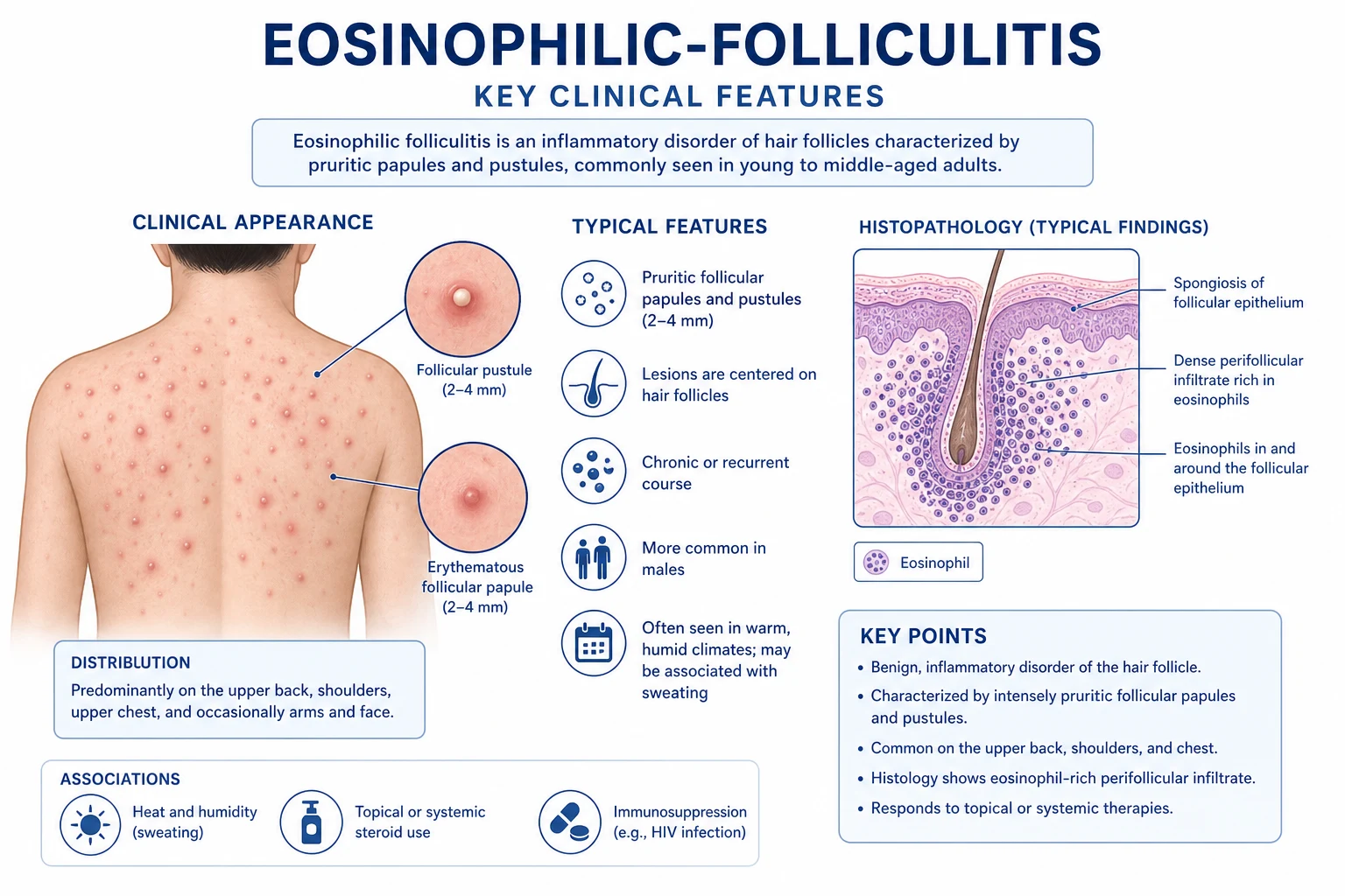
Classification
EF is unified by histology but divided by clinical context. The classification below is the one used in the major reviews and is the distinction examiners test.[2]
1. Classic Ofuji disease
- Originally described by Ofuji in 1970 in otherwise-healthy Japanese adults
- Annular, polycyclic or serpiginous plaques with peripheral extension and central clearing, leaving hyperpigmentation
- Distribution: seborrhoeic areas (face, trunk, extensor arms); palms and soles occasionally involved (unique to Ofuji)
- Histology: eosinophilic spongiosis and perifollicular eosinophilic infiltrate
- Responds variably to indomethacin, dapsone, NB-UVB, cyclosporin
- Chronic relapsing course over years
2. HIV / immunosuppression-associated
- The most common variant in Western clinical practice
- Intensely pruritic urticarial follicular papules and papulopustules on face, upper chest, shoulders, upper back
- Typical CD4 under 200 to 300 cells per microlitre; may be the presenting feature of HIV
- Peripheral eosinophilia and elevated serum IgE common
- Most effective disease-modifying therapy is optimisation of ART/HAART
- May flare transiently during immune reconstitution (IRIS)
3. Infancy / childhood-associated
- Clusters of pruritic papulopustules on the scalp of neonates and infants
- Usually self-limiting over weeks to months
- Exclude scabies and bacterial folliculitis
- Topical corticosteroid and antiseptic wash; avoid systemic agents
4. Pregnancy-associated
- Onset in the third trimester; intensely pruritic follicular papules
- Resolves post-partum; may recur in subsequent pregnancies
- Manage with pregnancy-safe topical corticosteroid and NB-UVB; avoid systemic retinoids and weigh dapsone
- Distinct from polymorphic eruption of pregnancy and PUPPP by follicular histology
The same histological signature — eosinophils within and around the follicular unit — underlies all four forms; what changes is the host context, and it is the host context that drives both prognosis and treatment.[3]
Epidemiology and risk factors
EF is uncommon, and most of the epidemiology comes from case series rather than population studies. The key fact an examiner rewards is the association with advanced HIV. [1]
Eosinophilic folliculitis — key numbers
Risk factors. The dominant risk factor for the HIV-associated variant is advanced, untreated or poorly controlled HIV infection, typically with a CD4 count under 200 to 300 cells per microlitre; the condition often appears at the point of profound immunodeficiency and may be the first clue to HIV in an undiagnosed patient.[4] In the pre-HAART era, EF was one of the most distressing cutaneous manifestations of AIDS; its incidence fell sharply once combination antiretroviral therapy became routine, and a new presentation should now prompt a careful look at ART adherence, virological failure, or a new HIV diagnosis.[4]
Beyond HIV, EF is described in association with haematological malignancy (chronic lymphocytic leukaemia, non-Hodgkin lymphoma, myelodysplasia), solid-organ and stem-cell transplantation, autoimmune connective-tissue disease, and a range of drug triggers including allopurinol, carbamazepine, minocycline, leukotriene-receptor antagonists and some monoclonal antibodies.[2] Classic Ofuji disease has a striking over-representation in Japanese adults, and a minority of cases are familial.[5] The pregnancy variant affects women in the third trimester and tends to recur in subsequent pregnancies.
Pathophysiology
EF is an immune-mediated eosinophilic folliculitis, not an infection. The precise trigger is unknown, but the dominant model is a skewed Th2-type immune response in which interleukin-4, interleukin-5, interleukin-13 and the eosinophil chemoattractant eotaxin recruit and activate eosinophils against the follicular unit.[3]
In the HIV-associated variant, progressive immunodeficiency creates a paradoxically dysregulated Th2 environment: as the CD4 count falls, the immune system loses its ability to mount balanced Th1/Th2 responses and tips toward an IgE-rich, eosinophil-promoting cytokine milieu. Two additional co-factors are invoked. First, altered sebum lipid composition and reduced sebaceous gland activity in advanced HIV change the follicular microenvironment, possibly unmasking antigens that are normally tolerated. Second, follicular organisms — Demodex folliculorum, Malassezia species, and skin bacteria — may act as antigenic triggers in a now-hypersensitive host, generating an eosinophilic reaction around the follicle.[2] The consistently elevated serum IgE supports an atopic, hypersensitivity flavour to the process.
The histological cascade mirrors this. Eosinophils first accumulate in the perifollicular dermis, then invade the follicular epithelium producing eosinophilic spongiosis; degranulation generates flame figures and damages the follicular wall, with secondary folliculitis and occasional impetiginisation. The result is the clinical papulopustule, and the intense pruritus — disproportionate to the visible lesion — reflects the heavy eosinophil-derived pruritogenic mediators (major basic protein, eosinophil-derived neurotoxin) released into the dermis.[3] The mediators that drive both the inflammation and the itch are worth naming because they explain the treatment. Interleukin-5 is the dominant eosinophilopoietin, supporting eosinophil production, survival and priming in the bone marrow and tissue; blocking it (with mepolizumab-style biologics) depletes tissue eosinophils and is the rationale for considering anti-IL-5 strategies in severe eosinophilic dermatoses. Eotaxin (CCL11) provides the chemotactic gradient that draws primed eosinophils into the perifollicular dermis, and interleukin-4 and interleukin-13 both skew the response toward Th2 and drive class-switching to IgE, which is why serum IgE is so often elevated. Once in the tissue, the eosinophil degranulates, releasing major basic protein, eosinophil cationic protein, eosinophil-derived neurotoxin and eosinophil peroxidase; these are directly toxic to the follicular epithelium (generating the spongiosis and flame figures) and are potent pruritogens that sensitise dermal C-fibres, which is why the itch is so disproportionate to the visible lesion count.[3]
Why HIV-associated EF resolves with effective ART fits the mechanism: restoring the CD4 count rebalances the Th1/Th2 axis, removes the dysregulated eosinophil-promoting milieu, and re-establishes tolerance to follicular antigens.[4] A second strand of the pathogenesis deserves emphasis because it explains two empirical treatments. The follicle in advanced HIV is antigenically altered: sebaceous gland atrophy and altered sebum lipid ratios change what the immune system "sees" at the follicle, and Demodex and Malassezia antigens are presented in a now-hypersensitive Th2 host. An autoreactive component is suspected in classic Ofuji disease, where circulating autoantibodies and an atopic background are common, which explains why immunomodulators (indomethacin, dapsone, cyclosporin) rather than antimicrobials dominate the Ofuji ladder. Finally, the leukotriene pathway is implicated, which is why the 5-lipoxygenase-inhibiting action of itraconazole produces benefit out of proportion to its antifungal effect and is a favourite examiner question.[2]
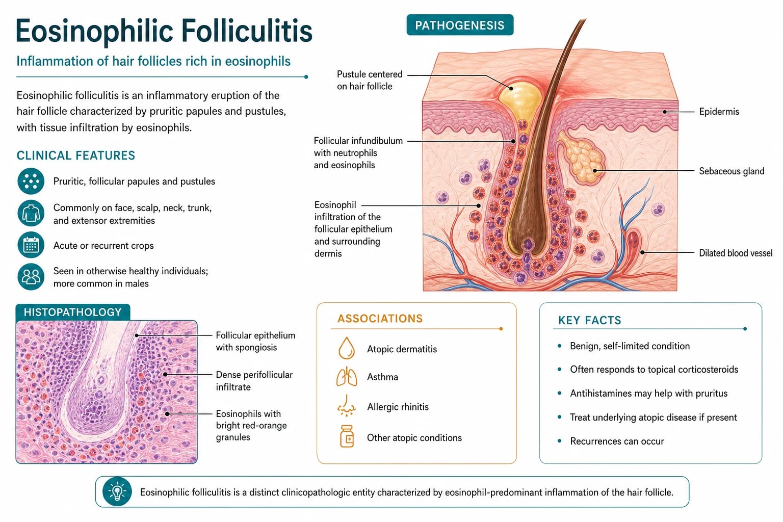
Clinical presentation
The clinical presentation is dominated by a single, decisive symptom — pruritus that is severe, persistent, and disproportionate to the number of visible lesions — together with follicular papules and papulopustules over the seborrhoeic areas. [1]
HIV-associated eosinophilic folliculitis
This is the variant most likely to be examined and the one most often seen in clinical practice. [1]
- Distribution. The face (especially cheeks and forehead), upper chest, shoulders and upper back bear the brunt; the extremities, particularly the extensor arms, are frequently involved, and the condition can extend more widely in advanced disease.[4]
- Morphology. Lesions begin as small (2 to 5 mm), erythematous, urticarial, follicular papules that evolve into papulopustules. Individual lesions may coalesce into plaques. The follicular origin is the key discriminator from acne and from non-follicular drug eruptions.
- Symptom. The pruritus is characteristically exquisite, frequently described as burning or stinging as well as itching, and is often severe enough to disrupt sleep. Excoriation is the rule rather than the exception, and crusts, erosions and even punctate scars follow.
- Secondary change. In skin of colour, the cycle of inflammation and excoration produces striking post-inflammatory hyperpigmentation, which can itself become a source of distress long after the active papules have settled.[4]
- Temporal pattern. The course is chronic and relapsing without treatment; in advanced HIV the lesions often persist until immune reconstitution is achieved.
A handful of nuances sharpen the clinical pattern and reward the candidate who names them. The papules are follicular and urticarial, so they often have a transient, evanescent quality early on before becoming fixed; this is why patients are sometimes dismissed as having "no rash" until the papules are looked for carefully in good tangential light. Dermoscopy is non-specific but typically shows follicular plugging, perifollicular erythema and a uniform network of small vessels around the hair follicle. The distribution is strikingly symmetrical and confined to the seborrhoeic and photo-exposed areas, which helps separate EF from a drug eruption (more generalised and acral) and from scabies (interdigital, flexural, genital). A Koebner-type isomorphic response is occasionally seen along lines of excoriation. Crucially, the pruritus is often the chief complaint and the lesions may be subtle, so a high index of suspicion is needed in any patient with advanced HIV and unexplained, severe, sleep-disturbing itch over the face and upper trunk.[4]
Classic Ofuji disease
The morphology here is distinctive and is the high-yield discriminator. [1]
- Annular, polycyclic or serpiginous plaques with peripheral extension: fresh papulopustules appear at the advancing edge while the centre clears, leaving hyperpigmentation.[5]
- Distribution favours the seborrhoeic areas — face, trunk and extensor surfaces; uniquely, the palms and soles may be involved in Ofuji disease, a feature not seen in the HIV variant.
- The typical patient is an adult without HIV, with an over-representation in Japanese subjects; a minority of cases are familial.
- Pruritus is variable and may be mild, in contrast to the HIV form.
Infantile and pregnancy-associated variants
The infantile variant presents as clusters of pruritic papulopustules on the scalp of neonates and infants, runs a self-limiting course over weeks to months, and must be distinguished from neonatal acne, scabies and bacterial folliculitis. The pregnancy-associated variant erupts as pruritic follicular papules in the third trimester, resolves post-partum, and may recur; it is clinically and histologically distinct from polymorphic eruption of pregnancy (PUPPP).[3]
Atypical presentations
Examiners deliberately test the corners. EF should be considered in an immunocompromised transplant recipient with new pruritic follicular papules, in a patient with newly diagnosed haematological malignancy, in an infant with scalp papulopustules refractory to standard antiseptic measures, and in a pregnant woman in the third trimester with intensely pruritic follicular lesions.[2]
Differential diagnosis
The differential is wide because pruritic follicular papules are common, and several mimics overlap with EF in distribution. The features that separate each are what the examiner rewards.[2]
Bacterial folliculitis (Staph. aureus)
- Discrete, tender pustules with surrounding erythema; spreads by autoinoculation
- Less intensely pruritic; more painful/tender
- S. aureus on bacterial culture
- Histology: neutrophilic, not eosinophilic, folliculitis
- Responds to antistaphylococcal therapy
Malassezia (Pityrosporum) folliculitis
- Monomorphic, uniform, erythematous papulopustules on chest/back
- KOH positive — budding yeast forms
- Often worse with sweating; common in young adults
- Histology: yeasts within follicle; mixed infiltrate
- Responds to oral itraconazole or fluconazole; topical ketoconazole
Demodex folliculitis
- Follicular papules and pustules on the face; rosacea-like
- High Demodex density on skin scraping
- Common in immunosuppression; overlaps with EF
- Responds to topical permethrin 5% or metronidazole 0.75% gel
- May coexist with EF and require dual treatment
Acne vulgaris
- Open and closed comedones plus papules, pustules, nodules
- Not intensely pruritic; chronic from adolescence
- Distribution: face, chest, upper back
- No peripheral eosinophilia
- Responds to retinoids, benzoyl peroxide, antibiotics
Scabies
- Intensely pruritic, nocturnal; burrows in web spaces, wrists, genitalia
- Family or household contacts affected
- Mite, eggs or faeces on microscopy of skin scrapings
- Symmetrical, acral, interdigital
- Responds to topical permethrin 5% or oral ivermectin
Dermatitis herpetiformis
- Intensely pruritic vesicles on extensor surfaces (knees, elbows, buttocks, scalp)
- Associated with coeliac disease/gluten enteropathy
- Direct immunofluorescence: granular IgA at dermal papillae
- Histology: subepidermal blister with neutrophils
- Responds to dapsone and a gluten-free diet
Drug eruption
- Temporal link to a new drug; morbilliform, acral or urticarial
- Often widespread; may have mucosal involvement in severe forms
- Eosinophilia possible but not follicular histology
- Resolves on withdrawal of the drug
- Review the drug chart and recent additions
The two most common diagnostic errors are labelling HIV-associated EF as ordinary bacterial folliculitis (and treating with repeated courses of antibiotics that fail) and missing the underlying HIV diagnosis by not testing. A skin biopsy and an HIV test resolve both.[4] A structured approach to the itchy follicular papule in an immunocompromised host prevents error. First, ask whether the lesions are truly follicular (centred on a hair) — this immediately narrows the list to the folliculitides and away from eczema, drug eruption and dermatitis herpetiformis. Second, ask whether the pruritus is disproportionate to the lesion count: intense itch with few lesions favours EF, scabies or dermatitis herpetiformis over ordinary bacterial folliculitis or acne. Third, ask about the tempo and distribution: a sudden crop of monomorphic papulopustules on the upper trunk in a hot, sweaty young adult points to Malassezia; chronic, relapsing, intensely pruritic follicular papules on the face and upper trunk in a patient with advanced HIV points to EF. Fourth, use the bedside and laboratory toolkit — KOH, skin scraping, bacterial culture and a biopsy with direct immunofluorescence — to resolve the remaining ambiguity rather than treating empirically.[2]
Clinical and bedside assessment
A focused assessment in suspected EF has three objectives: confirm the clinical pattern, identify the host context, and screen for the mimics and the systemic triggers. [1]
History. Establish the onset, distribution and tempo of the eruption; characterise the pruritus (severity, diurnal pattern, sleep impact using a visual analogue scale); ask explicitly about HIV risk factors (unprotected sex, men who have sex with men, injecting drug use, prior sexually transmitted infections, partner risk), known HIV and current ART, pregnancy, atopy, recent new drugs, and symptoms of lymphoproliferative disease (fever, night sweats, weight loss, lymphadenopathy). A family history is relevant for Ofuji disease.[2]
Examination. Examine the seborrhoeic areas in good light: face, upper chest, shoulders, upper back and extensor arms. Confirm the follicular origin of the papules, look for the annular configuration of Ofuji disease, and record the secondary changes — excoriation, crusting, erosions, prurigo nodularis, lichen simplex chronicus and post-inflammatory hyperpigmentation. Dermoscopy may show follicular plugging and perifollicular erythema but is non-specific. Perform a general examination: palpate for lymphadenopathy and hepatosplenomegaly (lymphoproliferative disease), and look for stigmata of HIV such as oral candidiasis, oral hairy leucoplakia and seborrhoeic dermatitis.[4]
Bedside decision. The single most important bedside decision is whether to offer an HIV test: EF is an indication for HIV testing in any adult with new-onset, intensely pruritic, follicular papules on the face and upper trunk.[4] Quantifying the itch. Because pruritus is the dominant symptom and the main treatment target, record its severity with a 0 to 10 numerical rating scale or visual analogue scale at baseline and at each visit, and ask specifically about sleep (time to onset, awakenings, daytime somnolence) and about impact on daily life using a short quality-of-life measure such as the Dermatology Life Quality Index (DLQI). A baseline DLQI also provides an objective endpoint against which the response to ART, phototherapy or systemic therapy can be judged, and it documents the burden that justifies escalation to second-line treatment. The itch of EF is characteristically worse at night and with heat, and patients often describe a burning or stinging quality in addition to itch; these features, together with the disproportionate severity, are what separate it from the milder itch of acne or bacterial folliculitis.[4]
Investigations
EF is a clinicopathological diagnosis: the clinical pattern plus the histological signature, set in the right host context. There is no single pathognomonic blood test, but several investigations confirm the diagnosis and exclude the mimics.[3]
[1]Skin biopsy is the diagnostic investigation. Histology shows eosinophilic spongiosis (eosinophils within the follicular epithelium), a perifollicular and intrafollicular eosinophilic infiltrate, occasional flame figures, and evidence of follicular damage; a neutrophilic component suggests superimposed bacterial infection.[3] The biopsy should be taken from a fresh, non-excoriated papule or papulopustule, because chronic excoriated lesions show only non-specific inflammation and the diagnostic perifollicular eosinophils may be obscured. Reading the histology with the question "where are the eosinophils?" is the key: in early EF they cluster in the perifollicular dermis and sebaceous gland, in established disease they invade the follicular epithelium (eosinophilic spongiosis), and in late disease flame figures (degranulated eosinophil protein coated by collagen) and follicular rupture mark tissue damage. A predominantly neutrophilic infiltrate redirects the diagnosis to bacterial folliculitis; yeast forms within the follicle point to Malassezia; Demodex mites in the follicular canal support Demodex folliculitis; and a subepidermal blister with neutrophils at the dermal papillae on routine histology, with granular IgA on direct immunofluorescence, redirects the diagnosis to dermatitis herpetiformis. The biopsy therefore both confirms EF and excludes its closest mimics in a single step.[3]
Bloods. A full blood count demonstrates peripheral eosinophilia in roughly half of patients and may show anaemia or lymphopenia pointing to HIV; serum IgE is frequently elevated; ESR/CRP may be mildly raised. HIV serology and a CD4 count are mandatory whenever the HIV variant is suspected — EF may be the presenting feature of HIV.[4]
Microbiology to exclude mimics. Bacterial culture of a pustule excludes Staphylococcal folliculitis; KOH examination and fungal culture exclude Malassezia folliculitis (budding yeast) and dermatophyte infection; a skin scraping excludes Demodex overgrowth and scabies (mite, eggs or faeces). Direct immunofluorescence of perilesional skin excludes dermatitis herpetiformis (granular IgA at the dermal papillae).[2]
Workup for an underlying systemic trigger. When EF appears in a non-HIV, non-pregnant adult without a drug trigger, screen for lymphoproliferative disease: a peripheral blood smear, lactate dehydrogenase, serum electrophoresis, and consideration of lymph-node biopsy or imaging if there is lymphadenopathy or hepatosplenomegaly.[2]
Management — resuscitation
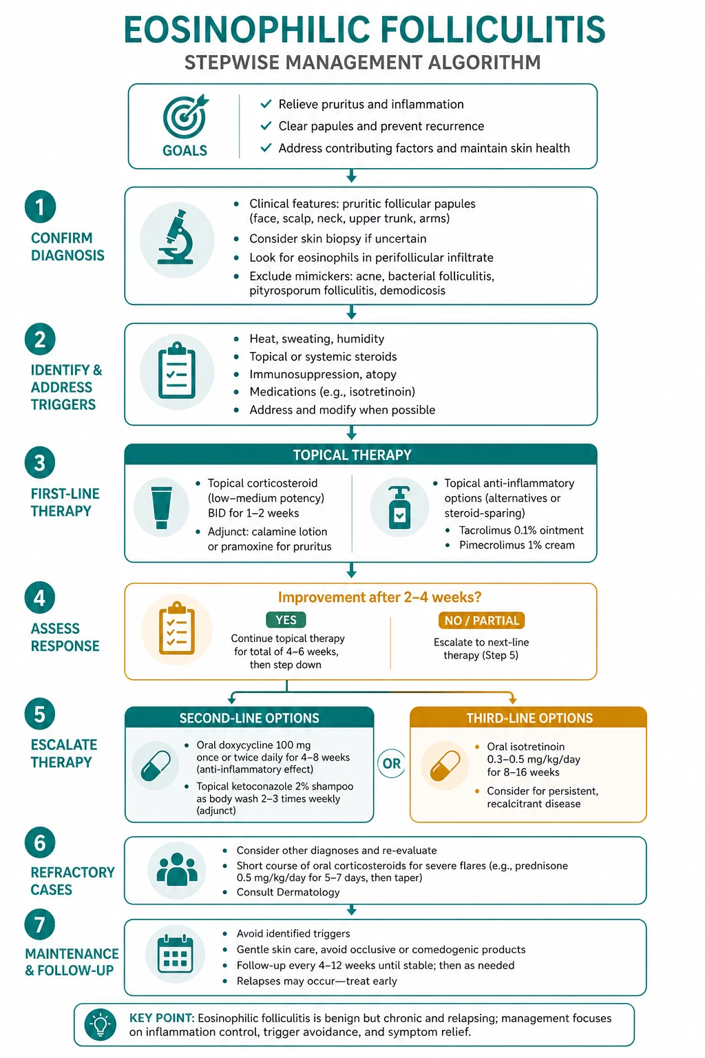
EF is rarely a dermatological emergency, but several situations demand prompt action. [1]
The most important urgent action is recognising EF as the possible presenting feature of HIV. A new diagnosis of HIV must trigger immediate linkage to HIV services for confirmation of the diagnosis, baseline CD4 and viral load, and initiation of combination antiretroviral therapy — the single most effective disease-modifying treatment for the HIV variant.[4]
The second urgent scenario is disabling, treatment-refractory pruritus with sleep deprivation. Immediate symptom control is humanitarian as much as medical: a sedating antihistamine at night (hydroxyzine 25 mg or promethazine) buys sleep while definitive therapy is arranged, and a short course of a potent topical corticosteroid (clobetasol 0.05% or betamethasone valerate 0.1%) dampens the inflammation. [1]
The third is secondary bacterial infection of excoriated lesions, which is treated with a topical or oral antistaphylococcal agent (flucloxacillin 500 mg four times daily orally for 7 days, or cephalexin in penicillin-allergic patients without anaphylaxis). Severe, widespread disease or suspected erythroderma warrants same-day dermatology referral.[2]
Management — definitive and stepwise
Definitive management is stratified by variant. In the HIV variant the principle is that optimisation of antiretroviral therapy is the most effective disease-modifying treatment, and all other measures are symptomatic or adjunctive.[4]
HIV-associated eosinophilic folliculitis
Optimise antiretroviral therapy (ART)
Confirm HIV, check CD4 and viral load, assess adherence, and start or optimise combination ART in collaboration with the HIV physician. Immune reconstitution (rising CD4) is the most effective disease-modifying therapy; EF typically resolves as the CD4 recovers above 200 to 300. Beware a transient flare in the first weeks of ART (IRIS).
Symptom control — first line
Potent topical corticosteroid (clobetasol 0.05% or betamethasone valerate 0.1% once to twice daily to active lesions for up to 2 weeks) plus an oral antihistamine (cetirizine 10 mg once daily, loratadine 10 mg once daily, or hydroxyzine 25 mg at night for sedation). Provide an emollient and a soap substitute.
Phototherapy — most effective non-ART therapy
Narrowband UVB (TL-01) three times weekly, gradually escalating dose, for a typical course of 15 to 30 treatments. NB-UVB is the most effective non-antiretroviral therapy for widespread EF; mechanism is immunomodulatory (altered eosinophil function and cytokine milieu). PUVA is an alternative.
Anti-inflammatory antifungal / antiparasitic adjuncts
Oral itraconazole 200 mg once daily (anti-inflammatory via 5-lipoxygenase inhibition and antifungal against Malassezia) for those with coexisting Malassezia; topical permethrin 5% (once weekly, 8 to 12 hours application) and metronidazole 0.75% gel twice daily where Demodex is implicated.
Second-line systemic therapy
For refractory disease: oral isotretinoin 0.5 to 1 mg/kg/day (sebocyte suppression; teratogenic — pregnancy prevention mandatory), dapsone 50 to 150 mg daily (screen G6PD first; monitor haemoglobin), or interferon-alpha. Reserve for severe, phototherapy-resistant cases under dermatology supervision.
Review and safety-net
Review response at 4 to 8 weeks; reinforce ART adherence; counsel about post-inflammatory hyperpigmentation and the expected timeline to improvement as the CD4 count recovers. Refer back to HIV services for any virological failure.
The systemic agents in step 5 carry specific monitoring that an examiner will probe. Dapsone (50 to 150 mg daily) requires G6PD screening before the first dose, a baseline full blood count, and repeat full blood counts weekly for the first month then monthly, watching for haemolytic anaemia and methaemoglobinaemia; warn the patient about the blue-grey discolouration of dapsone methaemoglobinaemia and about sulpha-drug cross-reactivity. Isotretinoin (0.5 to 1 mg/kg/day) is teratogenic and requires a pregnancy-prevention programme (reliable contraception, a negative pregnancy test before and during therapy, and a commitment to avoid pregnancy for at least one month after stopping), with baseline and serial lipids and liver enzymes; mucocutaneous dryness, mood change and the rare pseudotumour cerebri must be asked about. NB-UVB phototherapy requires cumulative-dose tracking, eye protection, and vigilance for the long-term skin-cancer risk that is amplified in immunosuppressed patients; a typical maintenance review of cumulative dose and skin surveillance is undertaken at each course. Coordinating all of this with the HIV physician is essential because several of these agents interact with antiretroviral drugs (itraconazole and macrolides inhibit CYP3A4 and raise levels of protease inhibitors and some integrase inhibitors).[1]
Classic Ofuji disease
Ofuji disease is managed with systemic anti-inflammatory and immunomodulatory agents, as it does not respond to ART. Oral indomethacin 25 to 75 mg daily is effective in a subset (implicating the prostaglandin pathway) and is a reasonable first choice. Dapsone 50 to 100 mg daily (after G6PD screening) and NB-UVB phototherapy are useful alternatives. Cyclosporin, colchicine and minocycline are described for refractory disease, and a short course of oral corticosteroid (prednisolone 0.5 mg/kg/day tapered over 2 to 3 weeks) can settle a severe flare.[1][5]
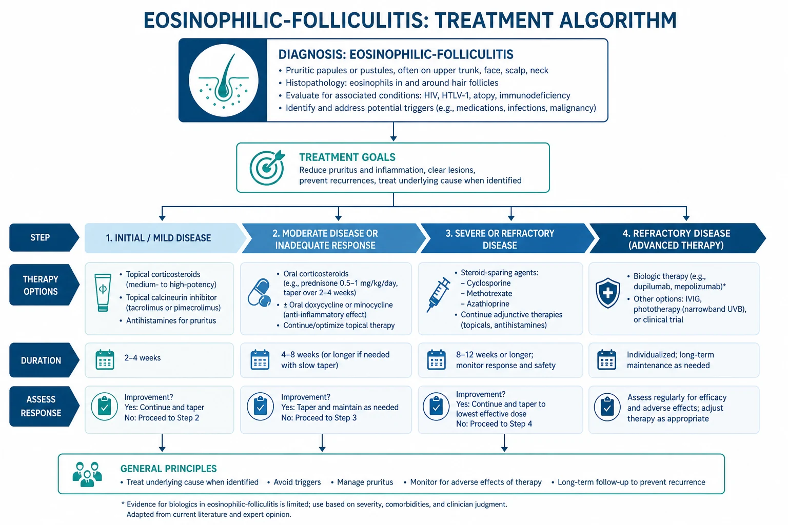
Pregnancy-associated and infantile variants
In pregnancy, manage with a pregnancy-safe topical corticosteroid (moderate potency, avoiding excessive amounts of potent agents) and NB-UVB, which is regarded as safe in pregnancy; avoid systemic retinoids (teratogenic) and weigh the risk-benefit of dapsone, which crosses the placenta. The condition resolves post-partum.[3]
In infants, the disease is usually self-limiting: a mild topical corticosteroid (such as hydrocortisone 1%) and an antiseptic wash (chlorhexidine or dilute potassium permanganate) suffice; avoid systemic agents and reserve them for the rare, severe, refractory case under specialist supervision. Exclude scabies (treat the whole household with permethrin 5% if confirmed) and neonatal acne (comedones, no itch), which are the two principal differential diagnoses in this age group. Reassure the parents that the eruption is benign and self-resolving, and review at two to four weekly intervals until clearance.[3]
Specific subtypes and scenarios
EF and immune reconstitution inflammatory syndrome (IRIS). When ART is started in a patient with subclinical EF — or in a patient whose EF had been suppressed — the recovering immune system can transiently amplify the eosinophilic response, producing a flare in the first weeks of therapy. This is IRIS, not treatment failure or drug allergy, and is managed with topical corticosteroids and antihistamines while ART is continued; the flare settles as immune reconstitution completes.[4]
EF in haematological malignancy and post-transplant. Here the principle is to treat the underlying disease (the lymphoma, the transplant immunosuppression) while providing topical corticosteroids and phototherapy for symptom control. Coordinating with haematology and transplant teams is essential because systemic agents may interact with chemotherapy or immunosuppressants.[2]
Drug-induced EF. Withdrawal of the suspect agent — commonly allopurinol, carbamazepine, minocycline or a leukotriene-receptor antagonist — combined with topical corticosteroids usually resolves the eruption; rechallenge should be avoided.[2]
EF in skin of colour. The dominant problem is post-inflammatory hyperpigmentation, which persists long after the active papules resolve and is itself a source of distress. Early, aggressive control of inflammation (phototherapy rather than prolonged potent topical steroids), sun protection, and reassurance about the slow resolution of pigmentation are central to management.[4]
Complications and pitfalls
The complications of untreated EF arise from the itch-scratch cycle and from the underlying immunodeficiency. Excoriation, crusting and erosion are followed by scarring, prurigo nodularis, lichen simplex chronicus and disfiguring post-inflammatory hyperpigmentation, particularly in skin of colour. Secondary bacterial infection with Staphylococcus aureus is common and may progress to cellulitis. The sleep deprivation, anxiety and depression driven by chronic, severe pruritus are under-recognised and should be asked about directly.[4]
The principal diagnostic pitfalls are four. First, missing the underlying HIV diagnosis by treating EF as ordinary folliculitis without offering an HIV test. Second, misdiagnosing EF as scabies or acne and treating empirically for months without a biopsy. Third, overlooking a coexisting Malassezia or Demodex component that requires additional targeted therapy. Fourth, confusing an IRIS flare with ART failure or drug allergy and inappropriately stopping effective antiretroviral therapy.[4]
The principal treatment pitfalls are equally important. Prolonged use of potent topical corticosteroids on the face causes atrophy, telangiectasia and steroid rosacea. Dapsone without G6PD screening can precipitate haemolytic anaemia and methaemoglobinaemia, particularly in patients of African, Mediterranean or South-East Asian descent. Isotretinoin in a woman of childbearing potential without contraception and pregnancy testing is teratogenic. Chronic phototherapy carries a cumulative skin-cancer and photoaging risk that must be weighed against the benefit, especially in immunosuppressed patients who already have a raised skin-cancer background risk.[1] The itch-scratch cycle deserves separate emphasis because it converts a symptomatic skin disease into a chronic, self-perpetuating one. Repeated scratching drives lichen simplex chronicus and prurigo nodularis, which themselves become intensely pruritic and lock the patient into scratching even when the underlying EF is settling; the result is scarring and indelible post-inflammatory hyperpigmentation that can persist for a year or more after disease control. The psychological burden is substantial and frequently overlooked: chronic, sleep-disrupting pruritus produces anxiety, low mood and social withdrawal, and these in turn lower the itch threshold and worsen the disease. Asking directly about sleep, mood and quality of life (a brief DLQI) and addressing them — with a sedating antihistamine at night, sleep hygiene, cognitive measures and, where appropriate, treatment of a mood disorder — is part of disease control, not an optional extra.[4]
Prognosis and disposition
The prognosis is determined by the variant and, in the HIV form, by the response to ART. [1]
HIV-associated EF improves and usually resolves as the CD4 count recovers above 200 to 300 cells per microlitre on effective ART; a transient IRIS flare may occur in the first weeks. Without immune reconstitution the course is chronic and relapsing, and the pruritus is debilitating.[4]
Classic Ofuji disease runs a chronic, relapsing course over years, responding variably to indomethacin, dapsone and phototherapy; sustained remission is achievable but relapse is common.[5]
Pregnancy-associated EF resolves post-partum and may recur in subsequent pregnancies; the infant is not affected. The infantile variant is self-limiting over weeks to months.[3]
What determines the outcome. Three factors drive the prognosis, and naming them shows understanding rather than rote recall. The first is immune reconstitution: in the HIV variant, recovery of the CD4 count above 200 to 300 cells per microlitre on effective ART is the single strongest predictor of resolution, and EF that persists despite a good CD4 response should prompt a search for an additional trigger (Malassezia, Demodex, a new drug, or an alternative diagnosis). The second is control of any underlying systemic disease: in EF associated with lymphoproliferative disease or transplant, the skin mirrors the systemic process, and effective treatment of the lymphoma or optimisation of immunosuppression is required for the skin to settle. The third is interruption of the itch-scratch cycle: even after the active folliculitis is controlled, residual prurigo nodularis, lichen simplex chronicus and post-inflammatory hyperpigmentation can perpetuate symptoms and require ongoing topical and behavioural management.[2]
Disposition. Most patients are managed jointly by dermatology and, where relevant, the HIV, haematology or obstetric team. The safety-net is clear: patients should return if the pruritus is uncontrolled despite first-line measures, if new lesions appear, if there are signs of secondary infection, or if ART side-effects develop. Routine follow-up is at 4 to 8 weeks to assess response and at intervals thereafter until the condition is controlled.[2]
Special populations
- Advanced HIV/AIDS. Coordinate all therapy with the HIV physician. Check for drug interactions between systemic agents and ART (avoid simvastatin with protease inhibitors; itraconazole and macrolides raise levels of many ART drugs via CYP3A4). Re-emphasise that ART optimisation is the disease-modifying step.
- Pregnancy. Use a moderate-potency topical corticosteroid in limited amounts and NB-UVB (regarded as safe in pregnancy). Avoid systemic retinoids (teratogenic). Weigh dapsone only if essential, and monitor the neonate for haemolysis and methaemoglobinaemia if used near term.
- Infants. Self-limiting; use a mild topical corticosteroid and antiseptic wash. Avoid systemic agents; exclude scabies.
- Haematological malignancy and transplant recipients. Treat the underlying disease; coordinate with haematology or the transplant unit; prefer phototherapy over prolonged systemic steroids.
- Skin of colour. Counsel early about post-inflammatory hyperpigmentation; favour phototherapy over prolonged potent topical steroids; use strict sun protection.
- India (NACO/IADVL). First-line ART is tenofovir-based; generic itraconazole, dapsone and isotretinoin are widely available. G6PD screening before dapsone is particularly relevant given regional G6PD deficiency prevalence. iPLEDGE-style isotretinoin pregnancy-prevention programmes are less formal than in the US; local counselling and contraception are emphasised.
- UK (BAD). NB-UVB is delivered in specialist phototherapy units with documented protocols and cumulative-dose tracking; HIV dermatoses are managed jointly with genitourinary medicine.
- US (AAD/DHHS). ART is initiated in all patients with HIV regardless of CD4 count; isotretinoin requires iPLEDGE registration; phototherapy skin-cancer surveillance is emphasised in immunosuppressed patients.
Evidence, guidelines and regional differences
The evidence base for EF is largely drawn from case series, case reports and expert review rather than randomised controlled trials; this is a recognised limitation that should be acknowledged to an examiner. The five landmark sources cited in this topic anchor the field.[1]
Ellis and Scheinfeld (2004, PMID 15186198) provide the comprehensive review of treatment options for eosinophilic pustular folliculitis that established the modern therapeutic ladder (topical corticosteroids, antihistamines, NB-UVB, itraconazole, indomethacin, dapsone, cyclosporin, isotretinoin).[1] Marzano and colleagues (2020, PMID 32394361) reframe EF within the broader family of eosinophilic dermatoses and update the recognition and management framework.[2] Long and colleagues (2016, PMID 25876839) describe the spectrum of eosinophilic skin diseases and the shared Th2 pathogenesis.[3] Parker and colleagues (2006, PMID 16734507) add the case series of EF in HIV-infected women and reinforce that EF may be the presenting feature of HIV and that ART is disease-modifying.[4] Katoh and colleagues (2013, PMID 23083212) review the Japanese literature on Ofuji disease, defining the annular morphology, the palmoplantar involvement and the demographic over-representation in Japanese patients.[5]
Regional differences in management are modest but real. The CD4 threshold at which ART is initiated has fallen over time: current US (DHHS) and WHO guidance is to start ART in all patients with HIV regardless of CD4 count, which has reduced the incidence of EF at the population level; India (NACO) has moved to the same treat-all policy.[4] The NB-UVB regimen (3 times weekly, 15 to 30 treatments) is consistent across the British Association of Dermatologists, the European Academy of Dermatology and Venereology, and the American Academy of Dermatology, but access to phototherapy and the formality of isotretinoin pregnancy-prevention programmes (iPLEDGE in the US, the PPP in the UK, more informal counselling in India) differ regionally. The G6PD screening that must precede dapsone is particularly important in populations with a high prevalence of G6PD deficiency, including much of South Asia, sub-Saharan Africa and the Mediterranean.[1]
The principal controversy is the relative place of itraconazole and indomethacin in the ladder: both have enthusiastic proponents and limited head-to-head evidence, and the choice is often pragmatic — itraconazole when Malassezia is suspected, indomethacin for classic Ofuji disease. Where the evidence is weakest is the long-term safety of repeated courses of phototherapy in immunosuppressed patients, in whom background skin-cancer risk is already elevated.[1] The biologics frontier is the most active area of new evidence. Because EF is driven by a Th2-eosinophil axis, anti-IL-5 (mepolizumab, benralizumab) and anti-IL-4-receptor-alpha (dupilumab) strategies have a sound mechanistic rationale and have been described in case reports and small series for refractory eosinophilic dermatoses, including EF. These remain off-label and evidence-poor, but they are the logical next step for severe, phototherapy-resistant, ART-optimised disease, and a candidate who names the cytokine target (IL-5 or the IL-4/13 receptor) demonstrates understanding of the pathogenesis rather than rote recall.[3] In practice, most EF is controlled by the cheaper, established ladder (topical corticosteroids, antihistamines, NB-UVB, itraconazole, indomethacin, dapsone, isotretinoin), and biologics are reserved for the small minority who fail all of these. The honest summary for an examiner is that EF is a disease where mechanism is well understood but the evidence base is weak, so treatment is built on expert opinion and small series rather than randomised trials, and individualisation — guided by the variant, the host context, drug interactions and the patient's tolerance — is central.[2]
Exam pearls
EOSINOPHILIC — high-yield hooks for EF
Exam application bank (NEET-PG / INICET)
One-line answer
Eosinophilic folliculitis is a chronic, intensely pruritic follicular disorder defined histologically by an eosinophil-rich infiltrate within and around hair follicles. Four variants exist: (1) classic Ofuji disease (annular, serpiginous plaques with peripheral extension and central clearing in non-HIV adults, classically Japanese), (2) HIV- or immunosuppression-associated (intensely pruritic urticarial follicular papules on the face and upper trunk, CD4 under 200-300), (3) infancy-associated, and (4) pregnancy-associated. Histology shows eosinophilic spongiosis and perifollicular eosinophilia; peripheral eosinophilia and elevated IgE are common. Optimisation of antiretroviral therapy is the most effective disease-modifying treatment for the HIV variant; narrowband UVB phototherapy is the most effective non-antiretroviral therapy.
Worked stems (answer without another resource)
Stem 1 — Classic presentation. Map symptoms to mechanism; name the first investigation and first treatment step with dose/route if drug therapy is standard. [1]
Stem 2 — Unstable / complicated. List red flags that force immediate resuscitation, theatre, ICU, antidote, or reperfusion — and what you do in the first 15 minutes. [1]
Stem 3 — Atypical group. Elderly, pregnancy, child, or immunocompromised: how presentation and thresholds change. [1]
Stem 4 — Differential trap. Name the three closest mimics and one discriminator for each. [1]
Stem 5 — Disposition. Who goes home with safety-netting, who is admitted, who needs HDU/ICU/theatre, and what follow-up is mandatory. [1]
Rapid viva checklist
- Definition + classification
- Pathophysiology chain
- Bedside signs / criteria
- Score with exact components (if any)
- Emergency bundle
- Definitive therapy with doses
- Complications of disease and of treatment
- Special populations
- Guideline/trial name if classic
- Three exam traps
Coverage self-check
If you cannot answer any stem above from this page alone, re-read the matching section — the page is intended to be self-sufficient for final-prof and NEET-PG/INICET questions on Eosinophilic folliculitis.
References
- [1]Ellis E, Scheinfeld N Eosinophilic pustular folliculitis: a comprehensive review of treatment options Am J Clin Dermatol, 2004.PMID 15186198
- [2]Marzano AV, Genovese G, et al. Eosinophilic Dermatoses: Recognition and Management Am J Clin Dermatol, 2020.PMID 32394361
- [3]Long H, Zhang G, Wang L, Lu Q Eosinophilic Skin Diseases: A Comprehensive Review Clin Rev Allergy Immunol, 2016.PMID 25876839
- [4]Parker SR, Parker DC, McCall CO Eosinophilic folliculitis in HIV-infected women: case series and review Am J Clin Dermatol, 2006.PMID 16734507
- [5]Katoh M, Nomura T, Miyachi Y, Kabashima K Eosinophilic pustular folliculitis: a review of the Japanese published works J Dermatol, 2013.PMID 23083212