Dermatology · Medicine
Epidermal naevus and naevus sebaceous
Also known as Epidermal naevus · Naevus sebaceous · Jadassohn naevus · Linear verrucous epidermal naevus · ILVEN · Epidermal naevus syndrome · Schimmelpenning syndrome · Becker naevus
Epidermal naevi are congenital hamartomas of keratinocytes or epidermal appendages that follow Blaschko lines, reflecting postzygotic somatic mosaicism. Verrucous epidermal naevus presents as linear warty papules; naevus sebaceous is a yellow-orange hairless scalp or facial plaque that thickens at puberty; ILVEN is a pruritic psoriasiform linear plaque. Epidermal naevus syndrome (Schimmelpenning) associates cutaneous lesions with neurological, skeletal, and ocular anomalies. Modern management of naevus sebaceous is conservative with biopsy of suspicious nodules, because the risk of basal cell carcinoma is low.
On this page & tools
Your progress
Saved locally on this device.
Exam tags
Red flags
Overview and Definition
An epidermal naevus is a benign, congenital, hamartomatous proliferation of epidermal or adnexal components that presents along the lines of embryonic ectodermal migration known as Blaschko's lines. The term is a clinical umbrella: it includes lesions derived predominantly from keratinocytes (keratinocytic epidermal naevi) and lesions derived from epidermal appendages such as sebaceous glands, hair follicles, or smooth muscle (organoid naevi).[6]
The unifying concept is somatic mosaicism. A postzygotic mutation occurring during early embryogenesis creates a clone of mutated keratinocytes or adnexal progenitor cells that expands and migrates along Blaschko's lines. Because the mutation is present in the embryo but usually not in the germline, most cases are sporadic and not inherited. The visible lesion is therefore a cutaneous map of the mutant clone.[6]
Clinically, epidermal naevi are present at birth or appear in early childhood. They grow in proportion to the child, often becoming more prominent during puberty under the influence of sex hormones and androgens. They are usually asymptomatic, although some variants—particularly inflammatory linear verrucous epidermal naevus (ILVEN)—can be intensely pruritic. The most important clinical implications are cosmetic disfigurement, the association with extracutaneous anomalies in epidermal naevus syndrome, and the risk of secondary neoplasia within naevus sebaceous. [1]
The terminology can be confusing. "Epidermal naevus" is sometimes used narrowly to mean keratinocytic epidermal naevus, and sometimes broadly to include naevus sebaceous and Becker naevus. In exam questions, the term usually refers to the Blaschkoid linear warty or velvety plaque, while naevus sebaceous and Becker naevus are usually named explicitly. Understanding the umbrella nature of the term helps avoid errors when a question describes a scalp lesion and asks for the diagnosis: a scalp lesion is more likely naevus sebaceous, whereas a trunk or limb lesion is more likely a keratinocytic epidermal naevus. [1]
[1]Classification
Epidermal naevi are classified first by the predominant tissue of origin and second by morphology, distribution, and histology. This classification is not merely academic; it drives the differential diagnosis, the risk of systemic associations, and the approach to treatment. [1]
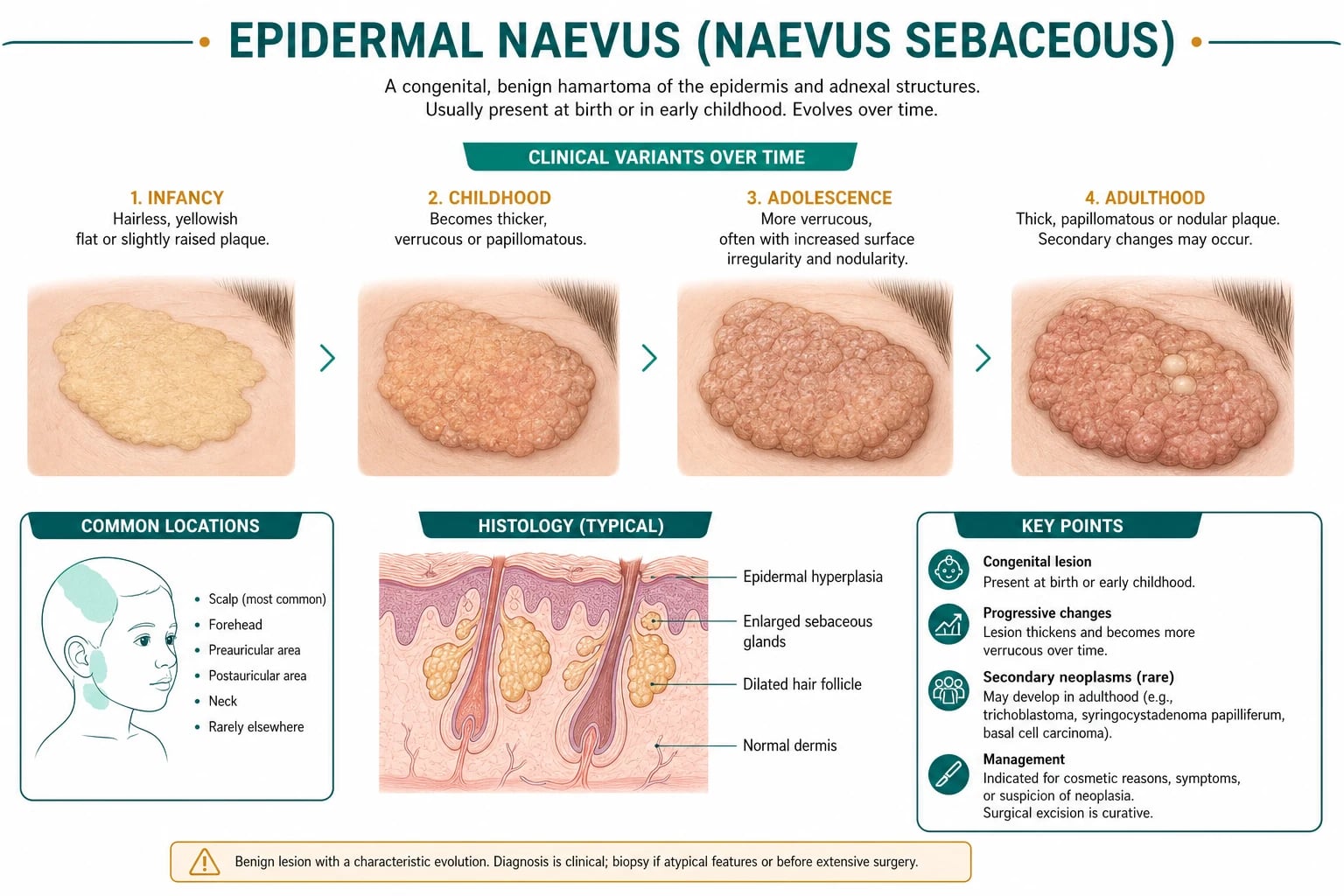
Keratinocytic epidermal naevi arise from keratinocytes. The common variants are: [1]
- Verrucous epidermal naevus (VEN): the classic linear warty plaque.
- Inflammatory linear verrucous epidermal naevus (ILVEN): a pruritic, psoriasiform, erythematous linear plaque.
- Epidermolytic epidermal naevus (EEN): a verrucous linear plaque with epidermolytic hyperkeratosis on histology.
- Naevus comedonicus: a cluster of dilated follicular openings with keratin plugs resembling comedones. [1]
Organoid epidermal naevi arise from adnexal structures: [1]
- Naevus sebaceous (Jadassohn naevus): a hamartoma of sebaceous glands, apocrine glands, and immature hair follicles, usually on the scalp or face.
- Becker naevus: a hamartoma presenting as a hyperpigmented, hypertrichotic plaque with increased smooth muscle bundles and androgen sensitivity. [1]
- Arises from keratinocytes
- Linear, warty or psoriasiform
- Follows Blaschko lines
- Risk of systemic disease lower than organoid syndromes, but ENS can occur
- Arises from adnexal structures
- Naevus sebaceous: yellow-orange, hairless, scalp/face
- Becker naevus: hyperpigmented, hypertrichotic, shoulder/upper trunk
- Higher risk of epidermal naevus syndrome (Schimmelpenning) with naevus sebaceous
Epidemiology and Risk Factors
Epidermal naevi are uncommon. Most estimates suggest a prevalence of roughly 1 in 1,000 live births, although many small lesions are never brought to medical attention. There is no consistent sex predilection for most keratinocytic epidermal naevi, but ILVEN shows a female predominance of approximately 4:1.[7]
Naevus sebaceous occurs in approximately 1 to 3 per 1,000 live births. It is usually sporadic. Unlike melanocytic naevi, it is not strongly associated with family history, because the causative mutations are typically postzygotic and not present in parental germ cells. Becker naevus is more common in males and usually presents in adolescence, reflecting its androgen dependence. [1]
The only "risk factor" is the occurrence of a postzygotic mutation during embryogenesis. The timing and tissue distribution of the mutation determine the extent and location of the naevus. If the mutation affects cells that also contribute to the central nervous system, skeleton, eye, or viscera, an epidermal naevus syndrome results. Large, extensive, or segmental lesions are more likely to be associated with systemic involvement, particularly when the naevus sebaceous is on the head and neck.[6]
Pathophysiology
The molecular basis of epidermal naevi is postzygotic somatic activating mutation in genes that regulate epidermal and adnexal growth. The mutation occurs after fertilisation, producing a mosaic individual in whom a subset of cells carries the mutation and a subset does not. Because the mutation is acquired during embryogenesis, the affected clone migrates along the paths of ectodermal cell movement and appears on the skin as a linear, swirled, or segmental pattern along Blaschko's lines.[6]
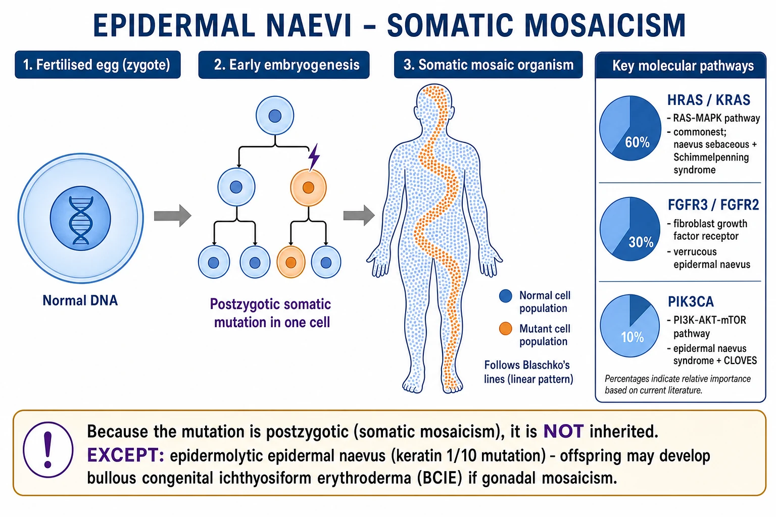
The key genes and their associated phenotypes are: [1]
- FGFR3 and PIK3CA: commonly implicated in keratinocytic epidermal naevi, particularly verrucous forms. Activating FGFR3 mutations drive epidermal proliferation; PIK3CA mutations activate the PI3K-AKT-mTOR pathway. These same pathways are implicated in other mosaic overgrowth disorders such as Proteus syndrome and megalencephaly-capillary malformation syndrome.[6]
- HRAS and KRAS: the characteristic mutations in naevus sebaceous and Schimmelpenning syndrome. The 2012 Nature Genetics study demonstrated that postzygotic activating mutations in HRAS or KRAS in the developing ectoderm cause both the cutaneous naevus sebaceous and the associated extracutaneous anomalies. These are mosaic RASopathies.[9]
- AKT1: the classic mutation in Proteus syndrome, causing asymmetric and progressive overgrowth of multiple tissues.
- KRT1 and KRT10: mutations in keratin 1 or keratin 10 produce epidermolytic epidermal naevus. These are the same genes that cause bullous congenital ichthyosiform erythroderma when the mutation is present in the germline. If the patient has gonadal mosaicism, offspring can inherit the full-blown genodermatosis.
The downstream signalling consequences are important for understanding associated syndromes. Activating HRAS or KRAS mutations drive the RAS-MAPK and PI3K-AKT pathways, which promote cell proliferation, survival, and migration. Because the mutation is present in the ectodermal cells that also give rise to parts of the brain, eye, and skeleton, the same mutant clone can produce both the cutaneous naevus and the extracutaneous malformations seen in Schimmelpenning syndrome. The severity of systemic disease correlates with the timing and tissue distribution of the mutation: an earlier postzygotic event affects more tissues and produces more extensive disease. FGFR3 mutations in keratinocytic epidermal naevi increase keratinocyte proliferation and inhibit apoptosis, producing the acanthosis and papillomatosis seen histologically. PIK3CA and AKT1 mutations cause overgrowth through the mTOR pathway and explain the overlap between extensive keratinocytic epidermal naevi and PIK3CA-related overgrowth spectrum disorders. KRT1 and KRT10 mutations disrupt the keratin cytoskeleton of suprabasal keratinocytes, producing cytolysis and epidermolytic hyperkeratosis. [1]
Blaschko's lines are not dermatomes, Langer's lines, or blood vessels. They reflect the direction of ectodermal cell migration during embryonic development. On the trunk they form S-shaped whorls; on the limbs they run longitudinally; on the scalp they form whorls. This pattern explains why epidermal naevi are often linear or segmental but not dermatomal. [1]
BLASCHKO
Clinical Presentation
The clinical presentation depends on the subtype. All epidermal naevi are present at birth or appear in early childhood, and most grow in proportion to the child. The key diagnostic clue is a congenital or early-childhood lesion in a Blaschkoid distribution. [1]
Keratinocytic epidermal naevus
Verrucous epidermal naevus (VEN) is the commonest keratinocytic form. It presents as a linear or serpiginous plaque of closely set, skin-coloured to brown, warty or velvety papules. The surface is often rough and hyperkeratotic. Sites of predilection are the trunk, neck, and limbs. The lesion is usually asymptomatic but can be mildly itchy. It is benign, stable after childhood, and does not resolve spontaneously.[6]
Inflammatory linear verrucous epidermal naevus (ILVEN) presents as a pruritic, erythematous, scaly, psoriasiform linear plaque. It typically appears in early childhood, often before age 5, and is more common in girls. The lower limbs, buttocks, and trunk are favoured sites. The intense pruritus and the refractory response to topical corticosteroids help distinguish it from linear psoriasis. Histologically, ILVEN shows psoriasiform epidermal hyperplasia with parakeratosis, which can be indistinguishable from psoriasis on a small biopsy; the clinical distribution and history are therefore decisive.[7]
Epidermolytic epidermal naevus (EEN) presents as a verrucous linear plaque. Histology shows epidermolytic hyperkeratosis: vacuolated cells of the granular layer with clumped keratin tonofilaments. This histological pattern is the same as that seen in bullous congenital ichthyosiform erythroderma. The finding of an epidermolytic naevus should prompt genetic counselling about gonadal mosaicism and the risk of transmitting the full genodermatosis. [1]
Naevus comedonicus presents as a cluster of dilated follicular openings plugged with dark keratin, resembling open comedones, arranged in a linear or Blaschkoid array. It is usually on the face, neck, upper trunk, or upper limb. It may be associated with skeletal, CNS, or ocular anomalies in naevus comedonicus syndrome. [1]
Organoid epidermal naevus
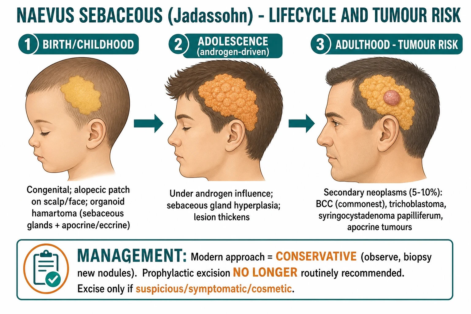
Naevus sebaceous (Jadassohn naevus) is the prototypical organoid epidermal naevus. At birth it appears as a well-circumscribed, yellow-orange, velvety, hairless plaque, most commonly on the scalp or face. Less often it occurs on the neck or trunk. During childhood the lesion is relatively flat and waxy. At puberty, under androgen stimulation, the sebaceous and apocrine components enlarge, and the lesion becomes thicker, more greasy, and more verrucous or nodular. Hair does not grow within the lesion because the hair follicles are immature.[6]
Becker naevus presents in adolescence, almost always in males, as a unilateral, brown, hyperpigmented patch that gradually enlarges and darkens and develops dark terminal hairs. It favours the shoulder, upper chest, and upper back. The lesion is androgen-dependent: androgen receptors are upregulated in lesional skin. It may be associated with ipsilateral breast hypoplasia, scoliosis, or limb shortening in Becker naevus syndrome. [1]
The size and shape of an epidermal naevus can be useful clinically. Small, localized lesions are usually isolated and have a low risk of systemic association. Extensive, segmental, or midline-crossing lesions are more likely to be associated with an epidermal naevus syndrome. Lesions that are plaque-like and follow broad bands of Blaschko lines are more concerning than small linear streaks. In naevus sebaceous, the size and location matter: large lesions on the scalp or face are more likely to be associated with Schimmelpenning syndrome than small lesions on the trunk. [1]
The evolution of symptoms over time is also informative. ILVEN often becomes more pruritic during childhood and may be exacerbated by heat, sweating, or friction. Verrucous epidermal naevus may become more hyperkeratotic and darker during puberty. Naevus sebaceous becomes more greasy and verrucous after puberty and may develop a characteristic orange peel-like surface. Any sudden change in a long-standing naevus should raise concern for a secondary process, such as infection, inflammation, or neoplasia. [1]
Differential Diagnosis
The differential diagnosis of epidermal naevus depends on the subtype and stage. The key discriminating features are the congenital or early-childhood onset, the Blaschkoid distribution, and the histology. [1]
- Psoriasiform linear plaque, often follows trauma (Koebner)
- History of psoriasis elsewhere, nail changes, arthritis
- Responds to topical corticosteroids and vitamin D analogues
- Histology identical to psoriasis; clinical context distinguishes
- Self-limited linear eruption in children
- Flat-topped violaceous or skin-coloured papules
- Resolves spontaneously over months to a few years
- Histology: band-like lymphocytic infiltrate at dermo-epidermal junction
- X-linked dominant; usually female
- Verrucous stage followed by swirled hyperpigmentation
- Systemic involvement: teeth, eyes, CNS
- Genetic: IKBKG/NEMO mutation
- Acquired in middle age or later
- Stuck-on waxy papule or plaque
- Does not follow Blaschko lines
- Histology: acanthosis, papillomatosis, horn cysts
- Viral, often on hands or feet
- Rough hyperkeratotic surface, thrombosed capillaries
- May be linear from autoinoculation (Koebner)
- HPV infection, not congenital
- Velvety hyperpigmented plaques in flexures
- Associated with insulin resistance, obesity, malignancy
- Not congenital, not linear in Blaschko distribution
- Histology: papillomatosis without significant hyperkeratosis
Naevus sebaceous must be distinguished from other yellow-orange scalp or facial plaques. Seborrhoeic dermatitis in infants can produce greasy, scaly plaques but is usually more diffuse and self-limited. Histiocytoses such as Langerhans cell histiocytosis can present as scalp papules or erosions but are accompanied by systemic signs and characteristic histology. Juvenile xanthogranuloma and mastocytoma can present as yellow nodules but are usually solitary, not plaque-like, and not congenital. [1]
ILVEN is most commonly mistaken for linear psoriasis. The distinction is clinical: ILVEN is intensely pruritic, refractory to topical steroids, and often more erythematous and scaly along its entire length. A therapeutic trial of potent topical corticosteroid or calcipotriol can help, because psoriasis usually improves whereas ILVEN does not. [1]
Becker naevus must be distinguished from congenital melanocytic naevus, café-au-lait macule, and smooth muscle hamartoma. The adolescent onset, hypertrichosis, shoulder distribution, and histology of acanthosis with increased smooth muscle fibres are diagnostic. [1]
Several additional conditions can mimic epidermal naevi and should be considered in the appropriate context. Porokeratosis may present as a linear plaque with a thread-like raised border (cornoid lamella) and is diagnosed histologically by the characteristic column of parakeratosis overlying an absent granular layer. Darier disease can produce greasy, scaly papules in a seborrhoeic distribution, but it is usually bilateral, associated with nail changes and palmar pits, and caused by ATP2A2 mutations. Epidermodysplasia verruciformis can produce widespread flat warts, but these are viral, acquired, and not arranged along Blaschko lines. Cutaneous T-cell lymphoma can present as a persistent scaly plaque, but it is usually acquired in adulthood and does not follow Blaschko lines. Nevus comedonicus can be mistaken for severe acne or comedonal acne, but the congenital onset and linear Blaschkoid arrangement distinguish it. [1]
Clinical and Bedside Assessment
The bedside assessment of a suspected epidermal naevus has three goals: confirm the diagnosis clinically, identify the subtype, and look for clues of an associated syndrome. [1]
First, map the distribution. Ask whether the lesion follows Blaschko's lines. Linear, S-shaped, or whorled patterns are characteristic. Dermatomal or segmental patterns suggest other diagnoses such as zoster or segmental vitiligo. Blaschko's lines are best appreciated on the trunk and limbs; on the scalp they appear as whorls. [1]
Second, examine the morphology. Note the colour, surface, scale, hair status, and presence of papules, nodules, or ulceration. A yellow-orange hairless plaque on the scalp or face is naevus sebaceous. A pruritic, erythematous, scaly linear plaque is ILVEN. A brown, velvety, warty linear plaque is VEN. A hyperpigmented, hypertrichotic patch on the shoulder is Becker naevus. [1]
Third, search for systemic associations. A full examination should include: [1]
- Neurological: head circumference, developmental milestones, seizures, hemiparesis.
- Skeletal: limb length discrepancy, scoliosis, genu varum or valgum, bony swellings.
- Ocular: visual acuity, strabismus, coloboma, choristoma.
- Cardiovascular and genitourinary: murmurs, blood pressure, abdominal masses, genital anomalies. [1]
Any infant with a large naevus sebaceous on the head or neck, or any child with a large keratinocytic epidermal naevus and neurological or skeletal signs, should be evaluated for an epidermal naevus syndrome. [1]
Bedside red flags that should trigger systemic work-up
- Naevus sebaceous on the face or scalp with seizures, developmental delay, or hemiparesis.
- Epidermal naevus with limb bowing, fractures, or short stature (hypophosphataemic rickets).
- Ipsilateral visual impairment, strabismus, or coloboma.
- Large or extensive lesions that cross the midline or involve multiple body segments.
Investigations
Most epidermal naevi are diagnosed clinically. Investigations are reserved for atypical presentations, suspected malignancy, or suspected systemic associations. [1]
Skin biopsy is the definitive diagnostic test when the diagnosis is uncertain. A shave or punch biopsy from the lesional skin is usually adequate. The key histological patterns are: [1]
- Verrucous epidermal naevus: regular acanthosis, papillomatosis, and hyperkeratosis, resembling a seborrhoeic keratosis but in a congenital/Blaschkoid clinical context.
- ILVEN: psoriasiform epidermal hyperplasia with parakeratosis, spongiosis, and a superficial perivascular lymphocytic infiltrate. This can look identical to psoriasis; the clinical history and distribution are essential.
- Epidermolytic epidermal naevus: epidermolytic hyperkeratosis with vacuolated granular keratinocytes and clumped eosinophilic keratin tonofilaments.
- Naevus sebaceous: large, mature sebaceous lobules that drain directly onto the skin surface rather than into hair follicles; immature hair follicles; apocrine glands; and often epidermal papillomatosis.
- Becker naevus: acanthosis, basal layer hyperpigmentation, and increased smooth muscle bundles in the dermis (smooth muscle hamartoma). [1]
Imaging and systemic work-up are indicated when an epidermal naevus syndrome is suspected: [1]
- MRI brain for seizures, developmental delay, hemiparesis, or hemimegalencephaly.
- Skeletal survey or plain radiographs for suspected rickets, scoliosis, limb length discrepancy, or bony overgrowth.
- Serum phosphate, calcium, alkaline phosphatase, parathyroid hormone, and FGF23 if hypophosphataemic rickets is suspected.
- Ophthalmology review for visual impairment, coloboma, or choristoma.
- Echocardiography and renal ultrasound for large facial naevus sebaceous or extensive syndromic lesions. [1]
Genetic testing is not routinely required for isolated epidermal naevi. It is considered when an underlying mosaic syndrome is suspected, when precise genetic counselling is needed—particularly for epidermolytic epidermal naevus because of the risk of gonadal mosaicism—or for research purposes. Next-generation sequencing of affected skin can identify somatic mutations in FGFR3, PIK3CA, HRAS, KRAS, or KRT1/KRT10. [1]
Dermoscopy is sometimes useful as a bedside adjunct. Verrucous epidermal naevus may show a cobblestone or fingerprint-like pattern with regular vessels. ILVEN may show dotted vessels on a red background, similar to psoriasis. Naevus sebaceous can show a characteristic pattern of yellowish lobules, arborising vessels, and comedo-like openings. However, dermoscopy is not a substitute for clinical assessment and biopsy when the diagnosis is uncertain. [1]
Photography is an important investigation in its own right. Baseline clinical photographs with a ruler and standard lighting allow objective comparison over time. This is particularly valuable for naevus sebaceous, where serial photography helps detect new nodules or surface changes that may indicate a secondary neoplasm. In resource-limited settings, good clinical photography may be the most useful surveillance tool. [1]
Laboratory investigations for suspected hypophosphataemic rickets include serum phosphate, calcium, alkaline phosphatase, parathyroid hormone, and FGF23. Hypophosphataemia with elevated alkaline phosphatase and elevated or inappropriately normal FGF23 is diagnostic. Urinary phosphate wasting may be demonstrated. These patients should also have wrist and knee radiographs to assess for rachitic changes and bone density. [1]
Management — Resuscitation
Epidermal naevi are not medical emergencies. There is no resuscitation or immediate life-saving intervention required for the lesion itself. However, two acute presentations require urgent assessment: [1]
- A new nodule, ulceration, or bleeding within a naevus sebaceous raises the possibility of basal cell carcinoma or another secondary neoplasm. The lesion should be biopsied, and the patient referred for definitive management.
- An infant with a large naevus sebaceous and new-onset seizures, lethargy, or focal neurological deficit may have Schimmelpenning syndrome with hemimegalencephaly or other structural brain abnormalities. Urgent neurological assessment and neuroimaging are indicated. [1]
For all other patients, management is elective and focused on surveillance, symptom control, cosmesis, and the detection of associated systemic disease. [1]
Management — Definitive and Stepwise
The management of epidermal naevi depends on the subtype, symptoms, location, size, and risk of complications. [1]
General principles
- Observation is appropriate for most small, asymptomatic epidermal naevi. These lesions are benign, stable after childhood, and do not require treatment unless they cause symptoms or significant cosmetic concern.
- Excision is indicated for symptomatic lesions, cosmetically unacceptable lesions, or lesions with suspicious change. Complete excision with appropriate margins is preferred for naevus sebaceous when malignancy is suspected.
- Medical and destructive therapies are used for symptomatic or cosmetically bothersome VEN, ILVEN, and limited naevus sebaceous, but recurrence is common and results are variable. [1]
Keratinocytic epidermal naevus
For verrucous epidermal naevus and ILVEN, first-line management depends on the patient's symptoms and the size of the lesion. Small, bothersome lesions can be excised. Larger lesions are challenging because excision may require extensive reconstruction or skin grafting. [1]
Medical options include: [1]
- Topical calcipotriol 0.005% ointment once or twice daily for ILVEN and some verrucous naevi; it may flatten the lesion and reduce scale, although response is variable.
- Topical corticosteroids (e.g., betamethasone dipropionate 0.05% ointment) for ILVEN; however, ILVEN is famously refractory, and failure of response is a diagnostic clue.
- Topical retinoids (e.g., tretinoin 0.05% cream or adapalene 0.1% gel) to reduce hyperkeratosis.
- Topical 5-fluorouracil 5% cream or imiquimod 5% cream have been reported anecdotally but are not standard. [1]
Procedural options include: [1]
- Full-thickness excision with primary closure, skin grafting, or flap reconstruction for small to medium lesions.
- Laser ablation with CO2 or erbium:YAG laser can flatten and lighten lesions, but recurrence is common and multiple sessions are usually required. A systematic review and a multicenter retrospective study suggest that laser can improve cosmesis in selected patients with verrucous epidermal naevi, but it is not curative.[5][8]
- Dermabrasion and curettage can reduce thickness but rarely remove the lesion completely.
Naevus sebaceous
Modern management of naevus sebaceous has shifted from routine prophylactic excision to a conservative, surveillance-based approach. Historical estimates of malignant transformation were as high as 10-30%, but contemporary studies show that the risk of basal cell carcinoma is much lower. An 18-year institutional review found that the risk of malignancy in naevus sebaceous is small, and many lesions previously labelled as basal cell carcinoma were actually benign trichoblastomas.[10]
Current recommendations are: [1]
- Observation for most asymptomatic naevus sebaceous lesions, with regular clinical photography and examination.
- Biopsy of any new nodule, papule, ulceration, or bleeding within the lesion to exclude basal cell carcinoma, trichoblastoma, or syringocystadenoma papilliferum.
- Excision if the lesion is symptomatic, cosmetically unacceptable, or shows suspicious change. Complete excision with histological examination is preferred. When malignancy is confirmed, standard basal cell carcinoma excision margins or Mohs micrographic surgery for facial lesions are appropriate.
- Avoid destructive treatments such as laser or curettage when malignancy is suspected, because they can destroy the architecture and make future diagnosis difficult. [1]
The timing of excision in children is controversial. Because the risk of malignancy before puberty is extremely low, observation through childhood is reasonable. If excision is planned for cosmetic reasons, many clinicians defer until adolescence or adulthood, when the lesion is fully developed and reconstruction is easier. [1]
Surgical technique depends on the size and site of the naevus sebaceous. Small scalp lesions can often be excised with primary closure under local anaesthesia using 1% lidocaine with 1:100,000 adrenaline. Larger lesions may require serial excision, tissue expansion, rotation flaps, or skin grafting. On the face, excision should be planned to respect cosmetic subunits and may require Mohs micrographic surgery if malignancy is suspected. Complete excision with histological examination of the entire specimen is preferred when secondary neoplasia is a concern, because punch biopsy may not be representative. [1]
Laser therapy for naevus sebaceous is generally reserved for selected cosmetic cases in which malignancy has been excluded. Ablative lasers such as CO2 or erbium:YAG can reduce thickness and improve surface texture, but they do not remove the lesion and may obscure future diagnosis of malignancy. They are not a substitute for surgical excision when tumour is suspected. For extensive lesions, treatment is usually palliative and requires multiple sessions with significant recurrence rates. [1]
Medical therapy for naevus sebaceous is limited. Topical retinoids or calcipotriol can sometimes reduce surface scale and hyperkeratosis, but they do not alter the underlying hamartoma. Antibiotic ointments may be used for secondary bacterial infection. For patients with secondary syringocystadenoma papilliferum or trichoblastoma, the treatment is surgical excision. [1]
Medical options for symptomatic epidermal naevi
Becker naevus
Becker naevus is usually managed conservatively. Laser hair removal or electrolysis can address hypertrichosis. Q-switched or pigment-specific lasers can lighten the hyperpigmentation, although results are variable. Excision is rarely needed. Associated breast hypoplasia or skeletal asymmetry may require reconstructive or orthopaedic referral. [1]
Specific Subtypes and Scenarios
Epidermal naevus syndrome (Schimmelpenning syndrome)
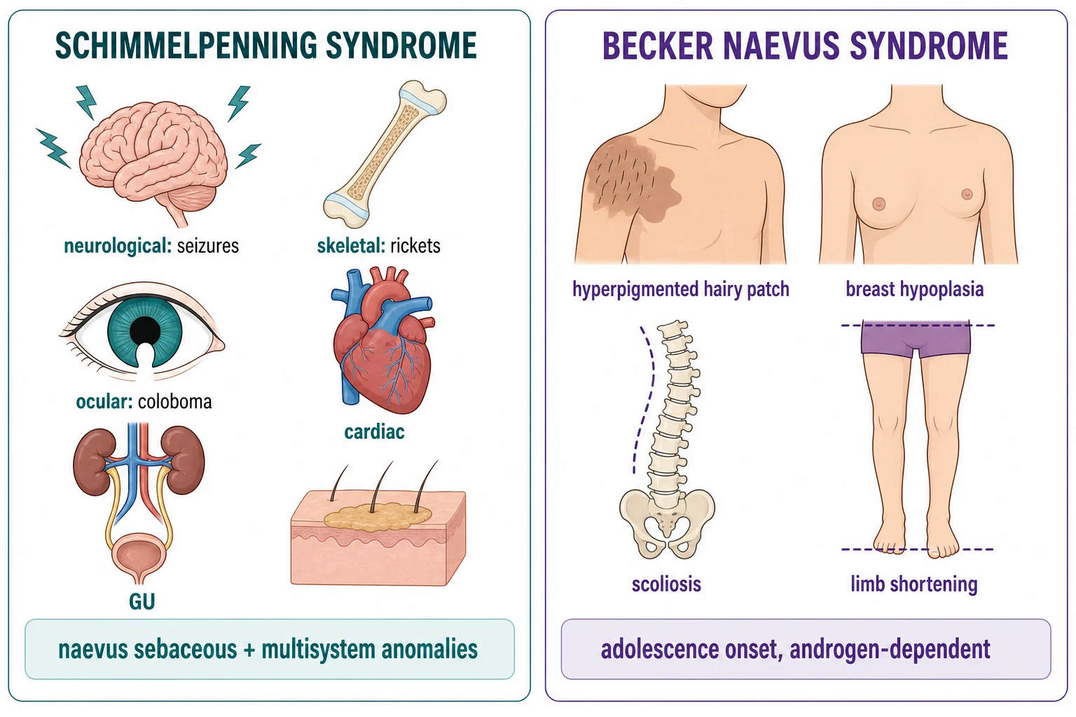
Epidermal naevus syndrome, also known as Schimmelpenning-Feuerstein-Mims syndrome, is a neurocutaneous mosaic disorder in which an epidermal naevus—most commonly naevus sebaceous on the face or scalp—is associated with ipsilateral extracutaneous anomalies. The underlying genetic basis is postzygotic activating mutations in HRAS or KRAS, which are present in the naevus and affected internal organs.[9]
The extracutaneous associations include: [1]
- Neurological: seizures, intellectual disability, hemimegalencephaly, cortical dysplasia, hydrocephalus, and intracranial lipomas or masses.
- Ocular: coloboma, choristoma, strabismus, visual impairment, and cataract.
- Skeletal: limb length discrepancy, scoliosis, and hypophosphataemic rickets. The rickets is driven by fibroblast growth factor 23 (FGF23) produced by the naevus tissue, leading to renal phosphate wasting. Treatment is with oral phosphate supplementation and calcitriol; some patients require anti-FGF23 therapy.[3]
- Cardiovascular and genitourinary: congenital heart defects, renal anomalies, and genital abnormalities.
Patients with suspected Schimmelpenning syndrome require multidisciplinary follow-up involving dermatology, paediatric neurology, orthopaedics, ophthalmology, and genetics. [1]
Nevus comedonicus syndrome
Nevus comedonicus syndrome is the association of naevus comedonicus with skeletal, CNS, or ocular abnormalities. The cutaneous lesions are clusters of dilated follicular openings with keratin plugs. Skeletal anomalies include scoliosis, fused vertebrae, and limb defects. CNS involvement can include seizures and intellectual disability. Ocular defects include cataract and corneal abnormalities. The syndrome is rare and the extracutaneous findings may be subtle, so a thorough examination is required. [1]
CHILD syndrome
CHILD syndrome is a rare X-linked dominant disorder caused by mutations in the NSDHL gene, which is involved in cholesterol biosynthesis. The name is an acronym: Congenital Hemidysplasia with Ichthyosiform naevus and Limb Defects. It presents with unilateral erythematous, scaly, ichthyosiform plaques following Blaschko's lines, ipsilateral limb undergrowth or other skeletal defects, and internal organ involvement. It is usually lethal in males; most affected patients are female because of X-inactivation mosaicism. The skin lesions are treated with topical agents; the ichthyosiform naevus is a key clinical marker for the underlying syndrome.[12]
Proteus syndrome
Proteus syndrome is a sporadic, progressive overgrowth disorder caused by postzygotic activating mutations in AKT1. It is characterised by asymmetric and disproportionate overgrowth of multiple tissues, including skin, bone, muscle, and adipose tissue. The cutaneous findings can include cerebriform connective tissue naevi, epidermal naevi, vascular malformations, and lipomas. The overlap with epidermal naevus syndrome is that some patients with extensive epidermal naevi may have features of Proteus syndrome or other PIK3CA-related overgrowth spectrum disorders. Diagnosis is clinical and genetic. [1]
Becker naevus syndrome
Becker naevus syndrome is the association of a Becker naevus with ipsilateral abnormalities, most commonly breast hypoplasia in females, scoliosis, and limb shortening. It is androgen-dependent and usually presents in adolescence. The histology shows a smooth muscle hamartoma in addition to epidermal acanthosis and basal hyperpigmentation. Management is supportive and cosmetic; significant skeletal or breast asymmetry may require surgical correction.[2]
Complications and Pitfalls
The complications of epidermal naevi depend on the subtype and any associated syndrome. [1]
Secondary neoplasms in naevus sebaceous
The most feared complication of naevus sebaceous is the development of a secondary tumour. The commonest benign tumours are trichoblastoma and syringocystadenoma papilliferum. The commonest malignant tumour is basal cell carcinoma. Historically, the risk of malignancy was overestimated; modern data suggest that the overall rate of secondary neoplasms is approximately 12.8% and the rate of malignant transformation is around 2.4%, with BCC-specific risk generally cited as 0% to 1.7%. Many lesions previously called BCC were actually trichoblastomas. The risk increases after puberty, and any new nodule or ulceration warrants biopsy.[10]
Epidermal naevus syndrome complications
The systemic complications of Schimmelpenning syndrome can be severe. Seizures may be refractory. Hypophosphataemic rickets can cause bowing, fractures, and short stature. Ocular anomalies can cause visual impairment. Cardiac and renal anomalies may require specialist intervention. Early recognition and multidisciplinary management improve outcomes. [1]
Diagnostic pitfalls
- Biopsy of ILVEN: the histology can be identical to psoriasis. The diagnosis rests on the clinical picture of a congenital or early-onset, intensely pruritic, linear, Blaschkoid plaque.
- Misdiagnosis of trichoblastoma as BCC: trichoblastoma is the commonest benign tumour in naevus sebaceous and can mimic basal cell carcinoma clinically and histologically. Expert pathological review is valuable.
- Destructive therapy obscuring malignancy: laser or curettage of a naevus sebaceous can destroy the architecture and make future diagnosis of malignancy difficult. Complete excision is preferred when malignancy is suspected.
- Missing systemic associations: a thorough examination for neurological, skeletal, and ocular abnormalities is essential in any infant with a large or extensive epidermal naevus, especially naevus sebaceous on the head or neck. [1]
Prognosis and Disposition
Most isolated epidermal naevi have an excellent prognosis. They are benign, stable after childhood, and do not affect life expectancy. Cosmetic concerns are the commonest indication for treatment. Recurrence after excision is uncommon if the lesion is completely removed; recurrence after laser or destructive therapy is common. [1]
The cosmetic and psychological impact of epidermal naevi can be significant, particularly when lesions are large, visible, or located on the face or scalp. Children and adolescents may experience bullying, anxiety, or reduced self-esteem. Early referral to a dermatologist, and where appropriate to a psychologist or support group, can help patients and families cope with the condition. Reassurance about the benign nature and the low malignancy risk is an important part of the consultation. [1]
Long-term follow-up is particularly important for naevus sebaceous. Although the risk of basal cell carcinoma is low, it is not zero, and malignancy can develop decades after puberty. Patients should be educated to self-examine the lesion and to report any new nodule, bleeding, ulceration, or rapid change. Annual dermatology review with clinical photography is reasonable for most patients, with more frequent review if the lesion is large or has previously changed. [1]
For patients with epidermal naevus syndrome, the prognosis is determined by the severity of neurological, skeletal, and ocular disease. Multidisciplinary follow-up should continue into adulthood, with particular attention to seizure control, skeletal health, visual function, and developmental and educational support. [1]
The prognosis of naevus sebaceous is also generally excellent, because the risk of malignancy is low. The modern approach is conservative surveillance with biopsy of suspicious change. Patients should be educated about warning signs: new nodules, ulceration, bleeding, or rapid change within the lesion. [1]
The prognosis of epidermal naevus syndrome is determined by the severity of the systemic associations. Seizures, developmental delay, hypophosphataemic rickets, and visual impairment can significantly affect quality of life. These patients require long-term multidisciplinary follow-up. [1]
Disposition: asymptomatic patients with isolated epidermal naevi can be discharged to primary care or self-surveillance with advice to return if the lesion changes. Patients with naevus sebaceous should have a baseline photograph and periodic review, particularly through puberty and into adulthood. Patients with suspected or confirmed epidermal naevus syndrome require referral to a multidisciplinary team. [1]
Special Populations
Neonates and infants
Epidermal naevi are usually noticed at birth or in the first months of life. In neonates, naevus sebaceous may appear as a flat, yellow-orange, hairless plaque without the verrucous surface seen later. A thorough examination for associated anomalies should be performed in infants with large or multiple naevi, especially on the head or neck. Imaging and specialist referral should be guided by clinical findings. [1]
Children
Most children with epidermal naevi do not require treatment. Observation is appropriate for naevus sebaceous until puberty, because the risk of malignancy before puberty is extremely low. Excision for cosmetic reasons can be deferred until the child is older and can participate in the decision. Topical therapies for ILVEN should be used cautiously in young children because of the risk of skin atrophy and systemic absorption. [1]
Adolescents and pregnancy
Puberty is a key transition for naevus sebaceous and Becker naevus. Androgen stimulation causes thickening and sebaceous hyperplasia in naevus sebaceous and darkening and hypertrichosis in Becker naevus. Adolescents should be counselled about the low risk of malignancy and the importance of surveillance. [1]
For women with epidermolytic epidermal naevus, genetic counselling is important before pregnancy. If the KRT1 or KRT10 mutation is present in germ cells, offspring can inherit bullous congenital ichthyosiform erythroderma. Prenatal counselling and, where available, genetic testing can inform reproductive decisions. [1]
Immunocompromised patients
Immunocompromised patients are not specifically at increased risk of epidermal naevi, but any rapidly growing or ulcerated lesion in an immunosuppressed patient should be biopsied promptly to exclude malignancy, because immunosuppression increases the risk of skin cancer generally. Solid organ transplant recipients, patients with chronic lymphocytic leukaemia, and those on long-term immunosuppressive therapy have a higher incidence of cutaneous squamous cell carcinoma and basal cell carcinoma. In a patient with naevus sebaceous, this lowered threshold for biopsy applies even though the baseline risk is low. For patients with HIV or other immunodeficiency states, unusual infections can also present as verrucous or ulcerated plaques, so biopsy with appropriate stains is prudent. [1]
Elderly patients
Elderly patients may present with changes in a long-standing naevus sebaceous. The cumulative effect of androgen stimulation over decades means that lesions are often markedly verrucous and greasy. Any new nodule, ulceration, or bleeding should be evaluated with a low threshold for biopsy, because the incidence of basal cell carcinoma increases with age. In patients with limited life expectancy or significant comorbidity, observation may be preferred over aggressive excision unless malignancy is confirmed or symptoms are troublesome. [1]
Patients with dark skin phototypes
Epidermal naevi occur across all skin phototypes. Post-inflammatory hyperpigmentation and hypertrophic scarring are more common after excision or laser in patients with darker skin. Therefore, conservative management and meticulous wound care are especially important. Laser therapy should be approached with caution, and any planned procedure should be discussed with realistic expectations about dyspigmentation and scarring. [1]
Evidence, Guidelines and Regional Differences
The evidence base for epidermal naevi is limited to case series, retrospective reviews, and expert consensus. There are no large randomised trials. [1]
The landmark 2012 Nature Genetics paper demonstrated that postzygotic HRAS and KRAS mutations cause naevus sebaceous and Schimmelpenning syndrome, establishing these conditions as mosaic RASopathies.[9] The 2009 review by Rosen and colleagues revised the understanding of malignancy risk in naevus sebaceous, contributing to the modern conservative approach.[10] Recent reviews have summarised the genetic landscape and therapeutic options, including emerging targeted therapies for refractory or extensive disease.[6]
Regional differences in practice exist. In North America and Europe, the trend has been toward observation and biopsy of suspicious change rather than routine prophylactic excision of naevus sebaceous. In some settings, particularly where follow-up is uncertain or cosmesis is a major concern, early excision remains common. There is no universal guideline; management should be individualised. [1]
Laser treatment for verrucous epidermal naevi has been studied in systematic reviews and multicenter retrospective series. Ablative lasers can improve appearance but are not curative and recurrence is common.[5][8]
Emerging targeted therapies are an area of active research. Because many epidermal naevi are driven by activating mutations in the RAS-MAPK or PI3K-AKT-mTOR pathways, inhibitors of these pathways are logical candidates for extensive or refractory disease. MEK inhibitors, PI3K inhibitors, and mTOR inhibitors have been used anecdotally in severe mosaic RASopathies and overgrowth disorders, but robust clinical trial data for isolated epidermal naevi are lacking. These agents are not standard of care and should only be considered in specialist centres with experience in mosaic disorders. [1]
Guidelines specifically for epidermal naevi are limited. Management is largely guided by expert consensus, retrospective studies, and the principle of balancing the low risk of malignancy against the morbidity of treatment. The British Association of Dermatologists and the American Academy of Dermatology have not issued standalone guidelines for naevus sebaceous, but general principles of skin cancer surveillance and biopsy of suspicious lesions apply. In the United Kingdom, the National Health Service approach favours conservative management with dermatology surveillance. In the United States, practice is more variable, and some centres still advocate prophylactic excision of naevus sebaceous in selected patients, particularly those with large or cosmetically significant lesions. [1]
Exam Pearls
[1]Exam application bank (NEET-PG / INICET)
One-line answer
Epidermal naevi are congenital hamartomas of keratinocytes or epidermal appendages that follow Blaschko lines, reflecting postzygotic somatic mosaicism. Verrucous epidermal naevus presents as linear warty papules; naevus sebaceous is a yellow-orange hairless scalp or facial plaque that thickens at puberty; ILVEN is a pruritic psoriasiform linear plaque. Epidermal naevus syndrome (Schimmelpenning) associates cutaneous lesions with neurological, skeletal, and ocular anomalies. Modern management of naevus sebaceous is conservative with biopsy of suspicious nodules, because the risk of basal cell carcinoma is low.
Worked stems (answer without another resource)
Stem 1 — Classic presentation. Map symptoms to mechanism; name the first investigation and first treatment step with dose/route if drug therapy is standard. [1]
Stem 2 — Unstable / complicated. List red flags that force immediate resuscitation, theatre, ICU, antidote, or reperfusion — and what you do in the first 15 minutes. [1]
Stem 3 — Atypical group. Elderly, pregnancy, child, or immunocompromised: how presentation and thresholds change. [1]
Stem 4 — Differential trap. Name the three closest mimics and one discriminator for each. [1]
Stem 5 — Disposition. Who goes home with safety-netting, who is admitted, who needs HDU/ICU/theatre, and what follow-up is mandatory. [1]
Rapid viva checklist
- Definition + classification
- Pathophysiology chain
- Bedside signs / criteria
- Score with exact components (if any)
- Emergency bundle
- Definitive therapy with doses
- Complications of disease and of treatment
- Special populations
- Guideline/trial name if classic
- Three exam traps
Coverage self-check
If you cannot answer any stem above from this page alone, re-read the matching section — the page is intended to be self-sufficient for final-prof and NEET-PG/INICET questions on Epidermal naevus and naevus sebaceous.
[1]References
- [1]Arjona-Aguilera C, Collantes-Rodríguez C, Villegas-Romero I, et al. Rounded and velvety epidermal naevus: Dermoscopic findings and literature review Australas J Dermatol, 2018.PMID 28736849
- [2]Schäfer K, Bauer B, Donhauser J, et al. Becker Naevus Syndrome of the Lower Body: One Case and Review of the Literature Acta Derm Venereol, 2017.PMID 27882383
- [3]Olivares JL, Ramos FJ, Carapeto FJ, et al. Epidermal naevus syndrome and hypophosphataemic rickets: description of a patient with central nervous system anomalies and review of the literature Eur J Pediatr, 1999.PMID 10048604
- [4]Zakrzewski JL, Luecke T, Bentele KH, et al. Epidermal naevus and segmental hypermelanosis associated with an intraspinal mass: overlap between different mosaic neuroectodermal syndromes Eur J Pediatr, 2001.PMID 11686504
- [5]Khan W, Ibrahim A, Alvaro A, et al. Laser Treatment of Verrucous Epidermal Naevi: A Systematic Review J Cutan Med Surg, 2022.PMID 35603930
- [6]Waldman AR, Hultman KA, Antaya RJ. Epidermal Nevi: What Is New Dermatol Clin, 2022.PMID 34799036
- [7]Atzmony L, Leventhal JS, Choi JN, et al. Inflammatory linear verrucous epidermal nevus (ILVEN) encompasses a spectrum of inflammatory mosaic disorders Pediatr Dermatol, 2022.PMID 35853659
- [8]Alkhalifah A, Al Onazi M, Al Aboud K, et al. Laser treatment of epidermal nevi: A multicenter retrospective study with long-term follow-up J Am Acad Dermatol, 2020.PMID 31202870
- [9]Groesser L, Peterhof E, Evert M, et al. Postzygotic HRAS and KRAS mutations cause nevus sebaceous and Schimmelpenning syndrome Nat Genet, 2012.PMID 22683711
- [10]Rosen H, Schmidt B, Lam HP, et al. Management of nevus sebaceous and the risk of Basal cell carcinoma: an 18-year review Pediatr Dermatol, 2009.PMID 19686305
- [11]Farschtschi S, Werchnik AE, Wolf P, et al. Keratinocytic epidermal nevus syndrome with Schwann cell proliferation, lipomatous tumour and mosaic KRAS mutation BMC Med Genet, 2015.PMID 25928347
- [12]Ramphul K, Mejias SG, Gundelly P. CHILD Syndrome 2026.PMID 29939590