Dermatology · Medicine
Epidermolysis Bullosa
Also known as Epidermolysis bullosa (EB) · EB simplex (EBS) · Junctional EB (JEB) · Dystrophic EB (DEB) · Hallopeau-Siemens disease
Epidermolysis bullosa (EB) is a group of inherited mechanobullous disorders caused by mutations in structural proteins of the skin and mucosa. Blistering follows minor mechanical trauma. Classification is by the level of cleavage: EB simplex (intraepidermal; KRT5/14), junctional EB (lamina lucida; laminin-332/COL17A1), dystrophic EB (sublamina densa; COL7A1/type VII collagen), and Kindler syndrome (mixed plane; FERMT1). Severe recessive dystrophic EB carries a cumulative cutaneous squamous cell carcinoma risk of around 90% by age 55 and remains the leading cause of early death. Diagnosis rests on immunofluorescence mapping and genetic testing; management is multidisciplinary supportive care, with topical gene therapy (beremagene geperpavec / Vyjuvek) now FDA-approved for dystrophic EB.
On this page & tools
Your progress
Saved locally on this device.
Exam tags
Red flags
Definition and Classification
Epidermolysis bullosa (EB) is a family of rare, inherited blistering disorders united by mechanical fragility of the skin and mucous membranes. Affected individuals develop blisters, erosions, and ulcers after trivial trauma that would not injure normal skin.[1] Historically described as a group of inherited mechanobullous disorders, EB is now recognised to comprise more than 30 subtypes, all arising from mutations in genes that encode structural proteins responsible for epidermal and dermo-epidermal integrity.[1]
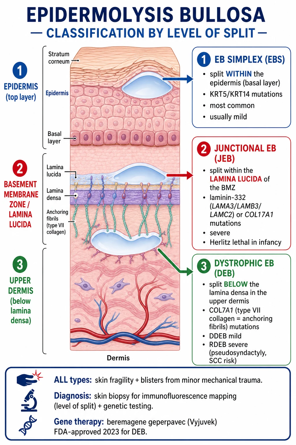
The level of cleavage within the skin is the fundamental classification axis. The four major groups are: [1]
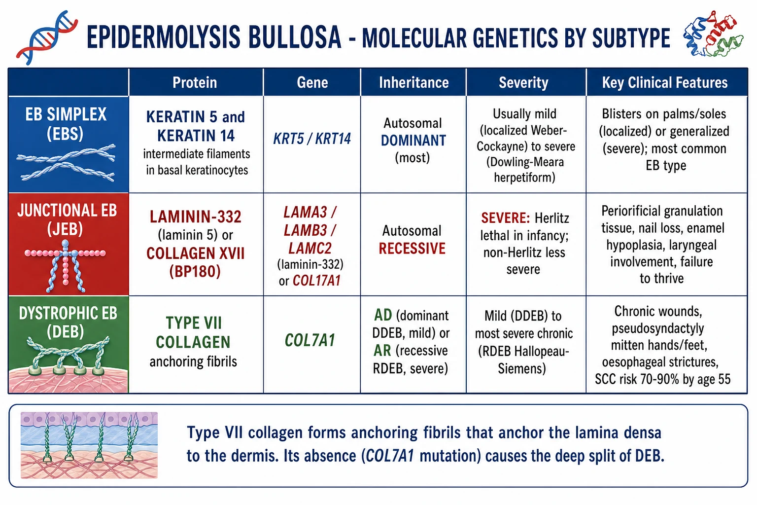
- Epidermolysis bullosa simplex (EBS) — cleavage is intraepidermal, within the basal keratinocytes. It is caused by mutations in KRT5 and KRT14, the genes encoding keratins 5 and 14, which form the intermediate-filament cytoskeleton of basal keratinocytes.[1]
- Junctional epidermolysis bullosa (JEB) — cleavage occurs at the lamina lucida of the basement membrane zone. It is caused by mutations in the genes encoding laminin-332 (LAMA3, LAMB3, LAMC2) or collagen XVII / BP180 (COL17A1).[1]
- Dystrophic epidermolysis bullosa (DEB) — cleavage is sublamina densa, below the basement membrane. It is caused by mutations in COL7A1, the gene encoding type VII collagen, which forms anchoring fibrils that tether the lamina densa to the upper dermis.[1]
- Kindler syndrome — cleavage occurs at multiple levels, giving a mixed-plane picture. It is caused by mutations in FERMT1, encoding kindlin-1, a protein involved in actin-cytoskeleton linking at keratinocyte focal adhesions and basement membrane adhesion.[1]
| Group | Level of split | Key proteins | Key genes | Inheritance | Severity |
|---|---|---|---|---|---|
| EBS | Intraepidermal (basal keratinocyte) | Keratin 5 / Keratin 14 | KRT5, KRT14 | Usually autosomal dominant | Usually mild; Dowling-Meara severe |
| JEB | Lamina lucida | Laminin-332, Collagen XVII | LAMA3, LAMB3, LAMC2, COL17A1 | Autosomal recessive | Severe; Herlitz often lethal |
| DEB | Sublamina densa | Type VII collagen | COL7A1 | Autosomal dominant or recessive | DDEB mild; RDEB severe |
| Kindler | Mixed plane | Kindlin-1 | FERMT1 | Autosomal recessive | Intermediate; photosensitivity, poikiloderma |
EB simplex subtypes
- EBS localised (Weber-Cockayne) — blistering is confined to the palms, soles, and other friction-prone sites. Onset may be in infancy or childhood, and disease activity often lessens with age. It is the classic presentation of EBS and is usually compatible with a normal life span.[1]
- EBS generalised intermediate (Koebner) — widespread blistering but of intermediate severity; patients usually survive to adulthood.
- EBS generalised severe (Dowling-Meara) — presents at birth with widespread herpetiform grouped blisters, severe palmoplantar keratoderma, and mucosal involvement. Neonatal disease can be life-threatening because of sepsis and fluid loss, but many patients improve with age and survival into adulthood is usual.[1]
- EBS with muscular dystrophy — caused by mutations in PLEC1 (plectin), which links intermediate filaments to the basement membrane. Patients have blistering and progressive muscular dystrophy.
- EBS with pyloric atresia — also PLEC1-related, presenting with neonatal blistering and congenital pyloric atresia; often lethal in infancy.
Junctional EB subtypes
- JEB severe (Herlitz) — the most severe form. Affected neonates have widespread blistering, nail loss, and exuberant periorificial granulation tissue. Hoarseness and stridor signal laryngeal involvement. Most die in infancy from sepsis, failure to thrive, or airway obstruction unless intensive support is available.[1]
- JEB intermediate (non-Herlitz) — survivable but still severe. Features include generalized blistering, alopecia, dental enamel hypoplasia, and nail dystrophy. Patients may live into adulthood with significant morbidity.
- JEB with pyloric atresia — rare, associated with ITGB4 mutations.
Dystrophic EB subtypes
- Dominant DEB (DDEB) — mild, with blistering at sites of trauma, nail dystrophy, and sometimes albopapuloid lesions (scar-like white papules). Life expectancy is generally normal.
- Recessive DEB severe (RDEB-GS, Hallopeau-Siemens) — the most severe form of DEB. Patients have extensive blistering from birth, progressive scarring, pseudosyndactyly (mitten deformities of hands and feet), oesophageal strictures, growth retardation, and a very high risk of cutaneous squamous cell carcinoma.[1]
- Recessive DEB intermediate — milder than Hallopeau-Siemens but still with scarring and extracutaneous complications.
- Recessive DEB inversa — predominantly affects intertriginous sites, axillae, groin, and genitalia.
- Recessive DEB pruriginosa — prurigo-like intensely itchy papules and nodules, often on the limbs.
Why "mechanobullous"?
The term mechanobullous captures the central pathophysiology: the blister is mechanically induced. Normal skin withstands shear and friction because keratinocytes, hemidesmosomes, anchoring filaments, and anchoring fibrils distribute force. In EB, a mutation in any one of these load-bearing elements reduces the tissue's mechanical tolerance, so a trivial force produces a split at the weakest point. This is why the diagnosis can be suspected simply from the history of blisters after minimal trauma, and why the first principle of management is trauma avoidance. [1]
Epidemiology and Risk Factors
EB is rare. The overall prevalence is usually quoted as approximately 1 per 20,000 to 50,000 live births, though precise figures vary by registry and by subtype definition.[1] In population-based registries, the incidence of EB is roughly 1 in 50,000 births, with some regional variation.
| Subtype | Approximate proportion of all EB | Inheritance | Notes |
|---|---|---|---|
| EBS | Around 70% | Usually autosomal dominant | Most common overall; usually mild |
| JEB | Around 5-10% | Autosomal recessive | Herlitz form is lethal in infancy without intensive care |
| DEB | Around 20-25% | Autosomal dominant or recessive | RDEB severe has the highest mortality |
| Kindler syndrome | Rare | Autosomal recessive | Photosensitivity and poikiloderma are characteristic |
EBS is therefore the most common EB group, accounting for roughly two-thirds of cases. Because most EBS follows an autosomal dominant pattern, affected families often have a clear vertical history of blistering. JEB, RDEB severe, and Kindler syndrome are almost always autosomal recessive, so a family history may be absent and the first clue is a neonate with widespread blistering. [1]
The single most important prognostic factor is the combination of split level and residual protein function. Mutations that abolish protein expression entirely (null mutations) produce more severe disease than those that allow partial protein function. For example, in JEB, complete loss of laminin-332 produces the lethal Herlitz phenotype, whereas partial loss of collagen XVII produces the milder non-Herlitz phenotype. In DEB, the nature of the COL7A1 mutation determines whether type VII collagen is absent, truncated, or functionally impaired, and this directly correlates with disease severity. [1]
The most feared complication in RDEB is cutaneous squamous cell carcinoma (cSCC). Data from the United States National EB Registry showed that the cumulative risk of developing at least one cSCC in RDEB-Hallopeau-Siemens was approximately 7.5% by age 20, 67.8% by age 35, 80.2% by age 45, and 90.1% by age 55.[4] The cumulative risk of death from SCC by age 55 was 78.7%.[4] This makes SCC the leading cause of death in young adults with severe RDEB and is the reason for aggressive surveillance from late childhood.
Epidermolysis bullosa by the numbers
Pathophysiology
The basement membrane zone (BMZ) is the critical interface between the epidermis and the dermis. It is not a single membrane but a multilayered adhesion system that absorbs and distributes mechanical force. From top to bottom, the key layers are: [1]
- Basal keratinocyte plasma membrane — hemidesmosomes and focal adhesions anchor the cytoskeleton to the basement membrane.
- Lamina lucida — an electron-lucent layer containing laminin-332, collagen XVII, and other adhesion proteins.
- Lamina densa — an electron-dense layer rich in type IV collagen, laminins, nidogen, and perlecan.
- Sublamina densa fibrillar zone — contains anchoring fibrils made of type VII collagen, which loop from the lamina densa into the upper dermis and back, mechanically tethering the epidermis to the dermis. [1]
In EB, the cleavage plane occurs at the weakest point in this chain for each genetic defect. [1]
EB simplex — keratinocyte fragility
Keratins 5 and 14 form the intermediate-filament network of basal keratinocytes. Mutations in KRT5 or KRT14 disrupt the ability of these filaments to withstand mechanical stress. The basal keratinocytes become fragile, and the split occurs within the basal layer of the epidermis. Because the BMZ remains intact, EBS blisters tend to heal without scarring and without milia. However, Dowling-Meara EBS can be severe because the mutations often cluster in the "hotspot" regions of the keratin rod domain, producing particularly disruptive structural instability. [1]
Junctional EB — lamina lucida failure
Laminin-332 is a large basement membrane glycoprotein that forms a scaffold between the hemidesmosomal components and the lamina densa. Collagen XVII is a transmembrane hemidesmosomal protein that links the keratinocyte cytoskeleton to laminin-332 and the lamina densa. Mutations in LAMA3, LAMB3, LAMC2, or COL17A1 produce separation at the lamina lucida. Because the BMZ is disrupted, healing is accompanied by scarring, nail loss, and enamel hypoplasia. The exuberant periorificial granulation tissue seen in JEB-Herlitz is thought to arise from chronic inflammation and poor wound healing at the level of the lamina lucida. [1]
Dystrophic EB — loss of anchoring fibrils
Type VII collagen is the main structural protein of anchoring fibrils. COL7A1 mutations reduce or abolish type VII collagen, so the anchoring fibrils are absent or defective. The split occurs below the lamina densa, in the upper dermis. Healing is therefore accompanied by dermal scarring, which explains the milia, contractures, pseudosyndactyly, and oesophageal strictures that characterise severe RDEB. [1]
Kindler syndrome — mixed cleavage
Kindlin-1 is a focal adhesion protein expressed in basal keratinocytes and other epithelia. Loss of kindlin-1 impairs keratinocyte adhesion to the basement membrane and disrupts actin cytoskeleton organisation. In Kindler syndrome, blisters can form at multiple levels, including intraepidermal, lamina lucida, and sublamina densa. The clinical picture is further complicated by photosensitivity, poikiloderma, and progressive mucocutaneous fragility. [1]
Milia and scarring
Milia are small, firm, white keratinous cysts that commonly appear on healing skin in JEB and DEB. They form because the blister roof is subepidermal or because hair follicles and adnexal structures are damaged during repeated blistering and scar formation. Milia are therefore a clinical clue that the cleavage plane is below the epidermis. [1]
Squamous cell carcinoma in RDEB chronic wounds
Chronic non-healing wounds in RDEB create a microenvironment of repeated injury, inflammation, and fibrosis. Cytokines such as transforming growth factor-beta, chronic activation of wound-healing pathways, and sustained oxidative stress promote the development of aggressive, poorly differentiated cSCC. These tumours are often multifocal, recur locally, and metastasise early. They are the leading cause of death in RDEB and typically appear in the second or third decade of life. [1]
Clinical Presentation
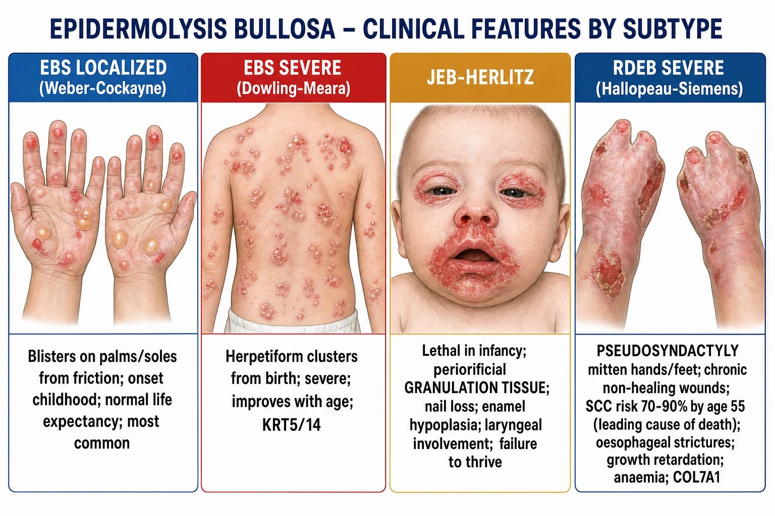
The unifying clinical feature of all EB is mechanical blistering. Blisters and erosions appear after minor trauma, friction, pressure, or heat. The distribution and severity differ by subtype. [1]
EB simplex
- EBS localised (Weber-Cockayne): Blistering on the palms, soles, and sites of friction. Hyperhidrosis and heat can worsen disease. Healing is usually without scarring.
- EBS Dowling-Meara: Neonatal widespread herpetiform grouped blisters on an erythematous base. Severe palmoplantar keratoderma. Oral mucosal involvement. Although neonatal disease can be severe, many patients stabilise and improve with age.
- EBS with PLEC1 mutations: May have associated pyloric atresia, muscular dystrophy, or nail dystrophy. [1]
Junctional EB
- JEB-Herlitz: Widespread blistering and erosions at birth, often with areas of denuded skin. Nails are shed. The hallmark is periorificial granulation tissue around the mouth, nose, ears, eyes, and anus. Hoarseness and stridor reflect laryngeal involvement. Failure to thrive is common, and death usually occurs in the first year of life from sepsis, malnutrition, or airway obstruction.
- JEB-non-Herlitz: Generalised blistering but less severe. Alopecia, enamel hypoplasia, dental caries, and nail dystrophy are common. Patients may survive into adulthood with chronic anaemia, growth retardation, and risk of SCC. [1]
Dystrophic EB
- DDEB: Localised blistering, nail dystrophy, and sometimes albopapuloid lesions. Life expectancy is usually normal.
- RDEB severe (Hallopeau-Siemens): Extensive blistering and erosions from birth. Repeated scarring leads to pseudosyndactyly (fusion of digits into mitten-like hands and feet), contractures, and chronic non-healing wounds. Oesophageal strictures cause dysphagia, and malnutrition is common. SCC is the leading cause of death in young adulthood.
- RDEB inversa: Predominant involvement of the groin, perineum, axillae, and genitourinary tract, with relatively spared skin elsewhere.
- RDEB pruriginosa: Pruritic papules, nodules, and lichenified plaques, often on the shins and forearms. [1]
Extra-cutaneous manifestations
EB is not only a skin disease. Important extra-cutaneous features include: [1]
- Oral and dental: Mucosal blistering, erosions, scarring, ankyloglossia, microstomia, enamel hypoplasia, and dental caries (especially in JEB and DEB).[2]
- Oesophageal: Strictures, dysphagia, gastro-oesophageal reflux, and failure to thrive. Oesophageal strictures are particularly common in RDEB and may require repeated balloon dilatation or gastrostomy feeding.
- Genitourinary: Urethral strictures, urinary retention, and painful genital erosions.
- Ocular: Conjunctival blistering, corneal abrasions, symblepharon, and scarring.
- Laryngeal: Hoarseness and stridor in JEB-Herlitz due to granulation tissue and blistering of the larynx.
- Haematological: Chronic iron-deficiency anaemia from repeated blood loss, often compounded by poor nutrition.
- Skeletal: Osteoporosis and pathological fractures from chronic inflammation, reduced mobility, and vitamin D deficiency.
Differential Diagnosis
A neonate or child with blistering presents a broad differential. The key is to distinguish inherited EB from acquired blistering disorders and infectious mimics. The presence of blistering from birth, a family history, and mechanical induction strongly point to EB. The level of cleavage and the pattern of scarring then help classify the subtype. [1]
EB simplex
- Intraepidermal split
- Positive or variable Nikolsky sign
- No scarring or milia
- KRT5/KRT14 mutations
- Usually autosomal dominant
Junctional EB
- Split at lamina lucida
- Positive Nikolsky sign
- Scarring, nail loss, enamel hypoplasia
- Laminin-332 or COL17A1 mutations
- Autosomal recessive; periorificial granulation in Herlitz
Dystrophic EB
- Sublamina densa split
- Positive Nikolsky sign
- Scarring, milia, pseudosyndactyly
- COL7A1/type VII collagen mutations
- DDEB mild; RDEB severe with SCC risk
Kindler syndrome
- Mixed-plane cleavage
- Photosensitivity and poikiloderma
- Periodontal disease
- FERMT1 mutations
- Autosomal recessive
Acquired blistering diseases
- Bullous pemphigoid — subepidermal blister, but onset is usually in older adults, pruritic urticarial plaques precede blisters, and direct immunofluorescence shows linear IgG and C3 at the basement membrane.
- Pemphigus vulgaris — intraepidermal split, positive Nikolsky sign, mucosal involvement, and autoantibodies against desmoglein 3. It is an acquired autoimmune disease, not inherited.
- Epidermolysis bullosa acquisita (EBA) — an autoimmune subepidermal blistering disease with antibodies against type VII collagen. It mimics DEB clinically but has an adult onset and positive direct immunofluorescence for IgG at the BMZ. Inherited DEB is present from birth and has no circulating anti-type VII collagen antibodies.
- Linear IgA disease — subepidermal blisters with a "string-of-beads" IgA deposition on direct immunofluorescence.
- Pemphigoid gestationis — autoimmune blistering disease of pregnancy; direct immunofluorescence shows C3 linear deposition.
- Bullous impetigo — superficial flaccid bullae caused by Staphylococcus aureus exfoliative toxins. The Nikolsky sign is positive, but the split is superficial and the disease is acute and infectious, not lifelong.
- Staphylococcal scalded skin syndrome (SSSS) — toxin-mediated superficial epidermal split, often with widespread erythema and peeling. Mucous membranes are spared. It is acute and occurs in young children, not present from birth.
- Severe bullous eczema or contact dermatitis — history of atopy or exposure, pruritic eczematous plaques, and not purely mechanical blistering. [1]
Key bedside discriminator
A neonate with widespread blistering from birth, positive Nikolsky sign, mucosal involvement, and periorificial granulation tissue should be presumed to have JEB-Herlitz until proven otherwise. In contrast, a neonate with SSSS has an acute toxic appearance, spared mucosa, and positive cultures for Staphylococcus aureus. [1]
Clinical and Bedside Assessment
Bedside examination in a neonate with suspected EB should be gentle, because every manoeuvre can create new blisters. [1]
Examination sequence
- General inspection — note the extent of blistering, erosions, and denuded skin. Look for signs of sepsis, dehydration, or temperature instability.
- Skin — assess distribution, presence of grouped blisters (Dowling-Meara), scarring, milia, and contractures. Look for non-healing wounds.
- Mucosa — inspect the oral cavity for erosions, ankyloglossia, and microstomia.
- Nails — nail dystrophy or nail loss is common in JEB and DEB.
- Eyes — check for conjunctival blistering, corneal erosions, or symblepharon.
- Airway — listen for hoarseness or stridor, particularly in JEB-Herlitz.
- Hands and feet — assess for pseudosyndactyly, mitten deformities, and contractures in older children and adults with RDEB. [1]
Nikolsky sign
The Nikolsky sign is elicited by applying gentle lateral pressure to the skin with a finger, causing the epidermis to separate and slide. In EBS, the sign is positive because the split is within the epidermis. In JEB and DEB, the sign is also positive because the epidermis separates easily from the dermis. In contrast, bullous pemphigoid is Nikolsky-negative because the epidermis remains intact. The sign helps confirm a mechanobullous disorder but does not distinguish EB subtypes. [1]
Mechanical fragility test
A more formal assessment is the finger-prick or rubbing test, in which gentle mechanical irritation produces a blister or erosion. This is rarely needed in clinical practice because the history is usually sufficient, but it can help confirm a mechanobullous disorder in research or atypical cases. [1]
Assessing pseudosyndactyly
In RDEB, repeated cycles of blistering, scarring, and fusion of adjacent digits produce pseudosyndactyly or "mitten" hands and feet. Assess the degree of fusion, contractures, and functional impairment. Early surgical release, splinting, and occupational therapy are required to preserve hand function. [1]
Investigations
Diagnosis of EB requires confirmation of the level of skin cleavage and, ideally, the underlying genetic defect. Two investigations are central: immunofluorescence mapping (IFM) of a skin biopsy and genetic testing. [1]
Immunofluorescence mapping (IFM)
A skin biopsy is taken from the edge of a fresh blister or induced blister. IFM uses monoclonal antibodies against BMZ proteins to determine the level of cleavage and to identify the missing or reduced protein. Key antibodies include: [1]
- Keratin 5/14 — for EBS.
- Laminin-332 (GB3 antibody) — for JEB.
- Collagen XVII / BP180 — for JEB.
- Type VII collagen (LH7:2 antibody) — for DEB.
- Kindlin-1 — for Kindler syndrome. [1]
IFM is often considered the diagnostic gold standard for initial classification, especially when genetic testing is not immediately available. It can be performed on formalin-fixed tissue and is widely available in specialist dermatopathology laboratories. [1]
Transmission electron microscopy (TEM)
TEM provides ultrastructural detail of the BMZ and can confirm the precise level of cleavage. It is particularly useful when IFM is inconclusive or when research-level phenotyping is required. [1]
Genetic testing
Molecular confirmation by next-generation sequencing or Sanger sequencing of the relevant genes is increasingly the definitive diagnostic test. It identifies the specific mutation, allows prenatal diagnosis in at-risk families, and guides genetic counselling. Genetic testing can sometimes be performed on a saliva or blood sample, avoiding a skin biopsy. [1]
Prenatal diagnosis
For families with a known EB mutation, prenatal diagnosis is available by: [1]
- Chorionic villus sampling at around 11 weeks of gestation.
- Amniocentesis at around 15 weeks of gestation.
- Preimplantation genetic diagnosis for selected mutations, allowing selection of unaffected embryos. [1]
Surveillance for squamous cell carcinoma in RDEB
Because of the very high risk of cSCC, patients with RDEB should undergo full skin examination every 3 to 6 months from around age 10 years (or earlier if clinically indicated).[4] Any new nodule, plaque, persistent ulcer, or change in a chronic wound should be biopsied urgently. Imaging and specialist oncology input are required if SCC is confirmed.
Baseline blood tests in severe EB
In severe EB, baseline and monitoring blood tests should include: [1]
- Full blood count — for anaemia (often iron-deficiency) and infection.
- Ferritin and iron studies — to guide iron replacement.
- 25-hydroxyvitamin D, calcium, phosphate, parathyroid hormone — for bone health.
- Zinc, albumin, pre-albumin — for nutritional status.
- Renal and liver function — as a baseline for drug therapies and for complications such as amyloidosis.
- Thyroid function — if clinically indicated. [1]
Swabs and cultures
Wounds in EB are frequently colonised with bacteria. Surface swabs should be taken when there is clinical evidence of infection, such as increased pain, erythema, warmth, purulent exudate, malodour, or systemic signs. The distinction between colonisation and infection is important: systemic antibiotics are reserved for clinical infection, not for positive swabs alone. [1]
Management — Resuscitation and Acute Emergencies
Neonates with severe EB, particularly JEB-Herlitz and EBS Dowling-Meara, can present with extensive skin loss and are at risk of sepsis, dehydration, electrolyte imbalance, and hypothermia. These patients should be managed in a neonatal intensive care unit with urgent dermatology liaison. [1]
Airway emergency in JEB-Herlitz
Laryngeal involvement with blistering and granulation tissue can cause hoarseness and stridor. Intubation is hazardous because it can precipitate further mucosal blistering. Anaesthetic and ear, nose, and throat expertise should be sought early, and emergency airway equipment should be available. In some cases, a tracheostomy is required, although it also carries significant risks in EB skin. [1]
Septic neonate with severe EB
Management follows the same principles as for any neonate with suspected sepsis and extensive skin loss: [1]
- Airway, breathing, circulation — resuscitate as needed.
- Broad-spectrum intravenous antibiotics — cover Staphylococcus aureus and Gram-negative organisms, guided by local protocols and cultures.
- Fluid management — calculate insensible losses from denuded skin; use burn-regimen fluid resuscitation principles adapted for EB.
- Temperature control — use incubators and minimise heat loss.
- Pain control — adequate analgesia is essential; use intravenous opioids judiciously.
- Wound care — gentle cleansing and non-adherent dressings, avoiding adhesive tapes. [1]
Suspicious wound in RDEB
Any chronic non-healing wound in RDEB should be considered cSCC until proven otherwise. Urgent punch or incisional biopsy of the most representative area is required, followed by urgent dermatology/oncology referral. Treatment often requires wide local excision, but options may be limited by poor wound healing and the need for specialised EB surgery. [1]
Management — Definitive and Stepwise Care
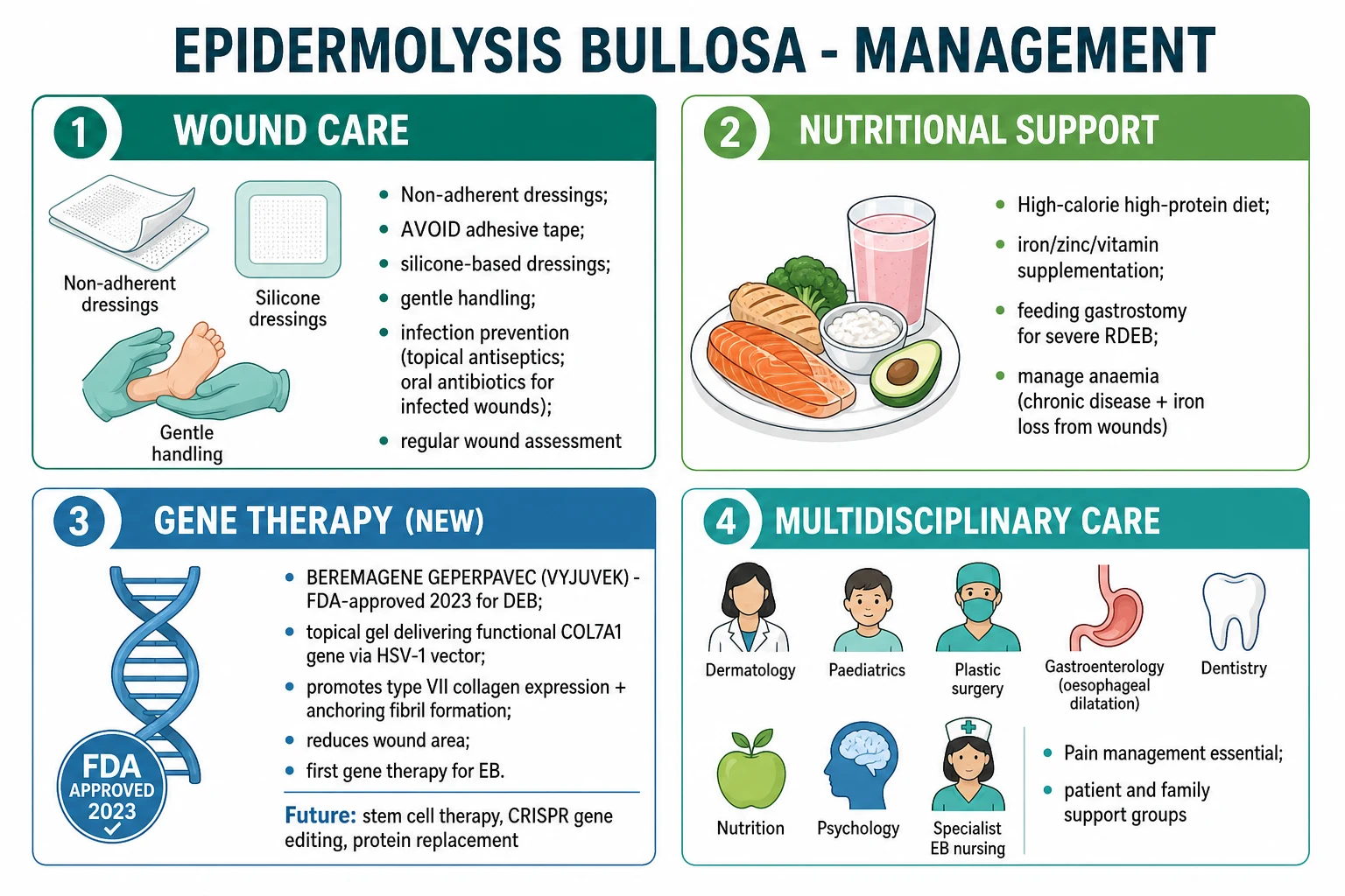
There is no cure for most forms of EB. Management is therefore lifelong, multidisciplinary, and supportive, with the goals of reducing trauma, preventing infection, promoting healing, controlling pain, maintaining nutrition, and detecting complications early. [1]
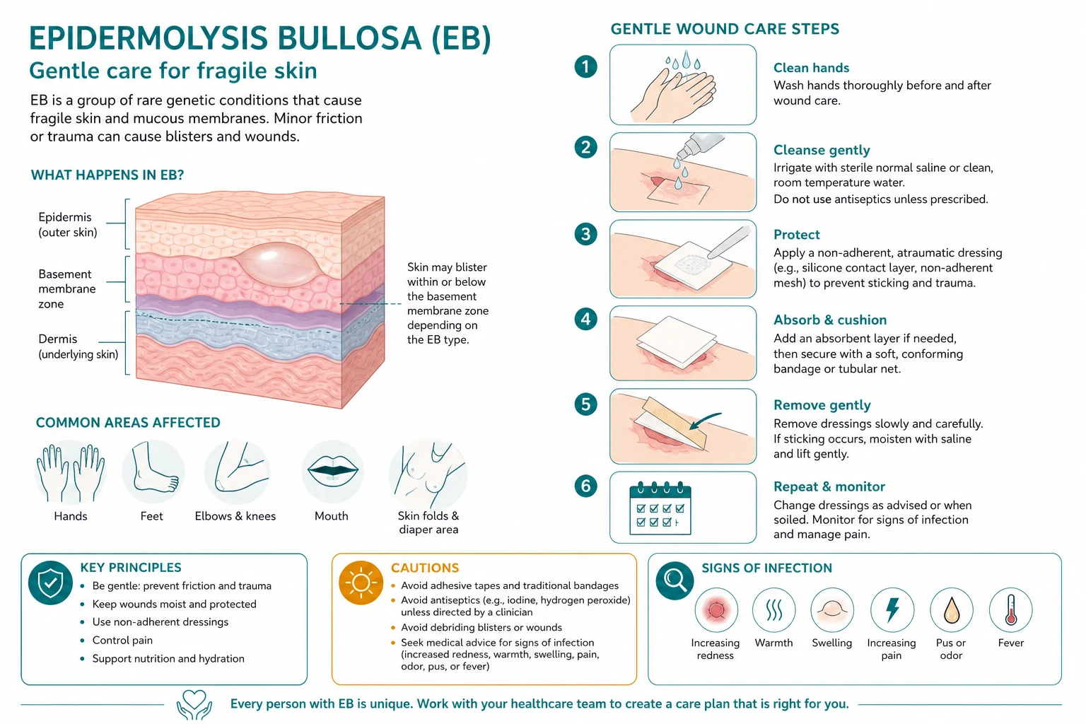
The four pillars of wound care
Effective EB wound care can be summarised as cleanse, de-roof, dress, and retain.[1]
- Cleanse — bathe or shower in lukewarm water; use mild, non-perfumed cleansers. Chlorhexidine 0.05% soaks can reduce bacterial load in infected wounds.
- De-roof — large blisters should be drained gently with a sterile needle, and the blister roof should be removed if it is likely to tear further. Leaving fluid under tension can cause extension of the blister.
- Dress — apply a primary non-adherent contact layer (such as silicone mesh or soft silicone dressing) and a secondary absorbent layer. Avoid adhesive dressings and tapes directly on the skin.
- Retain — use tubular bandages, stockinettes, or loosely wrapped gauze to hold dressings in place. Avoid any adhesive or sticky backing. [1]
Commonly used dressings include Mepitel, Mepilex, Mepilex Border, Urgotul, and lipido-colloid dressings. These minimise trauma on removal and maintain a moist wound environment. [1]
Analgesia
Chronic EB pain is a major burden. The WHO analgesic ladder should be applied, with EB-specific considerations: [1]
- Step 1: Paracetamol and non-steroidal anti-inflammatory drugs, used cautiously because of bleeding risk and renal toxicity.
- Step 2: Gabapentin or pregabalin for neuropathic pain.
- Step 3: Opioids for severe acute or chronic pain, but with close monitoring for constipation, sedation, and dependence.
- Adjuncts: Amitriptyline, lidocaine preparations, and topical anaesthetics for painful wound dressings. [1]
Topical antibiotics
Topical antibiotics are used for clinically infected wounds. Options include fusidic acid 2% three times daily, mupirocin 2% three times daily, and silver sulfadiazine for broader Gram-negative coverage. Prolonged, indiscriminate use should be avoided because it promotes bacterial resistance. Systemic antibiotics are reserved for spreading infection, cellulitis, or systemic signs of sepsis. [1]
Nutritional support
Severe EB patients have high nutritional requirements because of chronic wound exudate, infection, and reduced oral intake from painful mucosal disease. Principles include: [1]
- High-protein, high-calorie diet — often 150% of basal requirements in children.
- Iron, zinc, vitamin D, vitamin C, and multivitamin supplementation — guided by blood levels.
- Gastrostomy feeding — indicated when oral intake is inadequate because of dysphagia, oesophageal stricture, or severe oral disease.
- Regular monitoring of weight, height, BMI, and micronutrient status. [1]
Oesophageal stricture and dysphagia
Oesophageal strictures in RDEB are treated with balloon dilatation under endoscopic guidance. Repeated dilatations are often needed. A gastrostomy may be placed to maintain nutrition, bypassing strictures and reducing the risk of aspiration. [1]
Pseudosyndactyly and hand surgery
Surgical release of fused digits can restore hand function and independence. Surgery must be performed by an experienced EB surgical team, with careful postoperative splinting and non-adhesive dressings to prevent recurrence. [1]
Gene therapy — beremagene geperpavec (Vyjuvek)
Beremagene geperpavec, also known as B-VEC or Vyjuvek, is a topical, redosable gene therapy approved by the United States Food and Drug Administration in 2023 for the treatment of wounds in patients with dystrophic EB. It uses a modified, replication-defective herpes simplex virus type 1 vector to deliver a functional COL7A1 gene directly to wounds.[3] The GEM-3 phase 3 trial showed that B-VEC wounds achieved complete closure in 67% of treated wounds at 6 months, compared with 22% of placebo wounds.[5] It is the first gene therapy approved for EB and represents a major advance, although access remains limited in many regions.
Anti-SCC chemoprevention
High-dose isotretinoin has been used off-label as chemoprevention against cSCC in severe RDEB, particularly in patients with multiple previous SCCs. Evidence is limited to case series and expert opinion, and the drug's side effects — including mucosal dryness, hepatotoxicity, and teratogenicity — require careful monitoring. [1]
Bone protection
Osteoporosis and pathological fractures are common in severe EB. Management includes calcium and vitamin D supplementation, weight-bearing exercise as tolerated, bisphosphonates, and denosumab in selected cases. Bone mineral density should be monitored. [1]
Haematopoietic stem cell transplantation
Allogeneic haematopoietic stem cell transplantation (AHSCT) has been explored as a potential curative therapy for severe RDEB and JEB. The rationale is that donor-derived cells can produce the missing protein in the skin. Results have been mixed, with some patients showing significant benefit but significant procedural risks, including graft-versus-host disease and treatment-related mortality. It remains experimental and is offered only in specialised centres. [1]
Multidisciplinary team (MDT)
Optimal EB care requires a coordinated team including dermatology, paediatrics, plastic surgery, gastroenterology, dentistry, dietetics, ophthalmology, ENT, pain services, psychology, nursing, and palliative care. The EB nurse specialist is often the central coordinator. Families also need psychosocial support, respite care, and education about daily wound care. [1]
Management pillars — SOFT-CARE
SOFT-CARE
Mepitel, Mepilex, Urgotul — non-adherent and trauma-sparing
Maintain barrier function and reduce friction
High-protein, high-calorie diet; supplements and gastrostomy when needed
For infected wounds, not colonisation
Gentle bathing and removal of blister roofs to prevent extension
WHO ladder, including neuropathic agents and opioids when needed
Hand surgery, oesophageal dilatation, physiotherapy, occupational therapy
Family training, EB nurse specialist, psychology, respite
Specific Subtypes and Scenarios
Fellowship and postgraduate exams frequently test subtype recognition. Below is a rapid summary of the most commonly examined variants. [1]
EBS localised (Weber-Cockayne)
Palmoplantar blistering, often worse with heat, friction, and hyperhidrosis. Blistering may decrease in adolescence. Life expectancy is normal. Management focuses on footwear, moisture control, and non-adherent dressings. [1]
EBS generalised severe (Dowling-Meara)
Herpetiform grouped blisters from birth, severe palmoplantar keratoderma, and mucosal involvement. Neonatal risk is high due to sepsis and fluid loss, but many patients improve with age. The prognosis is better than JEB-Herlitz but still guarded. [1]
EBS with pyloric atresia
Caused by mutations in PLEC1. Neonatal blistering is accompanied by congenital pyloric atresia. This combination is often lethal without early surgery and intensive care. [1]
JEB-Herlitz
The classic lethal neonatal form. Widespread blistering, nail loss, periorificial granulation tissue, and hoarseness. Sepsis, failure to thrive, and airway obstruction are the usual causes of death. Intensive multidisciplinary care can prolong survival, but the prognosis remains poor. [1]
JEB intermediate (non-Herlitz)
Survivable but severe. Patients have generalised blistering, enamel hypoplasia, alopecia, and nail dystrophy. They require long-term nutritional support, dental care, and SCC surveillance into adulthood. [1]
DDEB
Mild dominant disease. Localised blistering, nail dystrophy, and sometimes albopapuloid lesions. Life expectancy is usually normal. Genetic counselling is important because of the 50% transmission risk. [1]
RDEB generalised severe (Hallopeau-Siemens)
Extensive blistering from birth, scarring, pseudosyndactyly, contractures, oesophageal strictures, growth retardation, and iron-deficiency anaemia. The cumulative risk of cSCC reaches around 90% by age 55. This is the most morbid DEB subtype and the one with the highest mortality in young adults. [1]
RDEB generalised intermediate
A milder recessive form. Blistering and scarring are present but progression is slower and SCC risk, while elevated, is lower than in Hallopeau-Siemens. [1]
RDEB inversa
Predominant involvement of intertriginous areas, axillae, groin, and genitourinary tract. The skin elsewhere may be relatively spared, but mucosal disease can still be severe. [1]
RDEB pruriginosa
Characterised by intensely pruritic papules and nodules, often on the limbs. It can be mistaken for prurigo nodularis or lichen simplex chronicus until the underlying EB history is recognised. [1]
Kindler syndrome
Photosensitivity, poikiloderma, skin fragility, and periodontal disease. Blisters can occur at multiple levels. Patients are also at increased risk of cSCC, particularly on sun-exposed sites, and require photoprotection and surveillance. [1]
Epidermolysis bullosa acquisita (EBA)
An acquired autoimmune disease, not an inherited EB. Autoantibodies target type VII collagen, producing subepidermal blisters that can be trauma-induced and scarring. It may resemble DEB clinically, but the onset is in adulthood and direct immunofluorescence shows linear IgG at the BMZ. It is treated with immunosuppressants, not EB supportive care. [1]
Complications and Pitfalls
EB complications are numerous and often multisystem. A structured approach reduces the risk of missed diagnoses. [1]
Squamous cell carcinoma
The leading cause of death in young adults with RDEB. It develops in chronic wounds and scarred skin, often in the second or third decade. It is frequently aggressive, multifocal, and prone to metastasis. Biopsy any new nodule or non-healing wound urgently. Surveillance should begin by age 10 years with full skin examination every 3 to 6 months.[4]
Oesophageal strictures
Dysphagia in DEB signals oesophageal stricture. Untreated, it leads to malnutrition, aspiration, and failure to thrive. Balloon dilatation and gastrostomy feeding are mainstays. Oesophageal SCC has also been reported. [1]
Pseudosyndactyly and contractures
Repeated scarring in RDEB fuses digits and limits joint movement. Early surgical release, splinting, and occupational therapy preserve function. Recurrence is common. [1]
Iron-deficiency anaemia
Chronic blood loss from erosions and poor iron absorption produce anaemia. Oral iron may be poorly tolerated; intravenous iron is often needed. Severe cases require transfusion. [1]
Osteoporosis and fractures
Chronic inflammation, vitamin D deficiency, reduced mobility, and long-term steroid use contribute to low bone mineral density. Bisphosphonates, calcium, vitamin D, and physiotherapy are used. [1]
Renal disease
Renal amyloidosis (AA type) can develop from chronic inflammation. Proteinuria and renal impairment should prompt nephrology review. Calcification of the renal pelvis and ureters has also been described in RDEB. [1]
Cardiomyopathy
Cardiomyopathy in EB can result from selenium deficiency, iron overload from repeated transfusions, or chronic anaemia. Cardiac monitoring is part of long-term care. [1]
Common pitfalls
- Missing periorificial granulation tissue — this is the hallmark of JEB-Herlitz and should trigger urgent airway assessment.
- Dismissing a chronic non-healing wound in RDEB as benign — assume SCC until biopsy proven otherwise.
- Using adhesive tape or dressings — these cause new blisters and should be avoided entirely.
- Treating colonisation with systemic antibiotics — reserve systemic therapy for clinical infection.
- Forgetting psychosocial support — EB is a devastating disease for patients and families; mental health support is essential. [1]
Prognosis and Disposition
Prognosis is highly dependent on EB subtype and on the degree of residual protein function. [1]
| Subtype | Prognosis | Disposition |
|---|---|---|
| EBS localised | Excellent; normal life expectancy | Outpatient dermatology; supportive care |
| EBS Dowling-Meara | Severe neonatal course; improves with age | NICU if neonatal; long-term dermatology |
| JEB-Herlitz | Lethal in infancy without intensive care | NICU, EB specialist centre, palliative input |
| JEB intermediate | Survivable with significant morbidity | Long-term MDT; surveillance for SCC |
| DDEB | Generally normal life span | Outpatient; genetic counselling |
| RDEB severe | Median survival often in the 30s; SCC is leading cause of death | Intensive MDT; 3-6 monthly skin surveillance |
| RDEB intermediate | Milder; survival into adulthood likely | Long-term MDT; surveillance for SCC |
| Kindler syndrome | Variable; SCC and photosensitivity risks | Photoprotection and surveillance |
Newborn with blistering
A newborn with widespread blistering should be admitted to the neonatal intensive care unit and reviewed urgently by dermatology. The priorities are sepsis prevention, fluid management, pain control, and early diagnostic biopsy. The family needs urgent genetic counselling and psychological support. [1]
RDEB patient with dysphagia
Dysphagia suggests oesophageal stricture. Urgent gastroenterology review with endoscopy and balloon dilatation should be arranged. If oral intake is inadequate, a gastrostomy should be considered. Barium swallow may be used to delineate strictures, but care must be taken to avoid aspiration. [1]
Suspected SCC lesion
Any suspicious lesion in RDEB should be referred urgently under the local 2-week-wait cancer pathway. An incisional or punch biopsy should be performed promptly. If SCC is confirmed, multidisciplinary management involving dermatology, plastic surgery, oncology, and palliative care is required. Reconstruction is challenging because of poor wound healing and limited donor sites. [1]
Special Populations
Neonates
Neonatal EB is an emergency if widespread. The diagnosis may not be immediately obvious, and the infant must be protected from further trauma. Suctioning, adhesives, blood pressure cuffs, and monitoring leads can all blister the skin. Gentle handling, non-adhesive dressings, and thermoregulation are essential. Genetic diagnosis may be required for counselling. [1]
Children and adolescents
Chronic wound care, pain, school attendance, and social integration are major challenges. Growth and puberty must be monitored closely. Dental disease is common and requires preventive programmes. Transition from paediatric to adult EB services should be planned. [1]
Pregnancy
Pregnancy in EB is rare and high-risk. Caesarean section may be preferred to avoid vaginal delivery trauma, but skin fragility complicates surgical wound healing. Anaesthetic considerations include potential airway fragility, difficult intubation, and limited intravenous access. A multidisciplinary obstetric, anaesthetic, and dermatology team is required. [1]
Bone marrow transplantation
AHSCT is an experimental curative option for severe RDEB and JEB. It is performed in highly specialised centres, often as part of clinical trials. Families must be counselled about risks, benefits, and the uncertain long-term durability of protein restoration. [1]
Immunocompromised EB
Patients on immunosuppressants, with malnutrition, or with long-term wounds are at higher risk of unusual or severe infections. In such patients, the differential for wound deterioration includes bacterial, fungal, and viral infections, as well as SCC. [1]
Evidence, Guidelines, and Regional Considerations
The management of EB is guided by a combination of international consensus, registry data, and emerging trial evidence. [1]
- DEBRA International 2017 wound-care consensus provides the most widely used framework for skin and wound care in EB. It emphasises non-adherent dressings, avoidance of adhesives, gentle handling, and family education.[1]
- Bardhan et al. 2020 (Nature Reviews Disease Primers) provides a comprehensive, modern overview of EB classification, pathophysiology, and management.[1]
- Fine et al. 2009 established the National EB Registry data on SCC cumulative risk, which remains the benchmark for surveillance programmes.[4]
- The GEM-3 trial demonstrated the efficacy of topical beremagene geperpavec (B-VEC / Vyjuvek) for dystrophic EB wounds, leading to FDA approval in 2023.[5]
- Regional access to advanced therapies varies. Vyjuvek is licensed in the United States and has been approved in some other jurisdictions, but access in many low- and middle-income countries, including India, remains limited. Most EB care globally remains supportive, delivered by specialised centres and DEBRA-affiliated patient organisations.
Exam Pearls and High-Yield Points
Split-level mnemonic
S-S-J-D-K
EB simplex (KRT5/KRT14)
Junctional EB (laminin-332/COL17A1)
Dystrophic EB (COL7A1/type VII collagen)
FERMT1/kindlin-1 with photosensitivity
Self-test: a neonate has widespread blistering, nail loss, and granulation tissue around the mouth and nose. What is the most likely diagnosis and immediate concern?
The presentation is classic for JEB-Herlitz. The immediate concern is airway compromise from laryngeal involvement, which can present as hoarseness or stridor. Management includes urgent NICU care, avoidance of further skin trauma, gentle wound care, and early specialist assessment. A skin biopsy for immunofluorescence mapping and genetic testing should be arranged urgently.
Self-test: a 22-year-old with RDEB develops a new nodule in a chronic wound on the forearm. What is your next step?
Urgent biopsy of the lesion to exclude cutaneous squamous cell carcinoma. In RDEB, the cumulative SCC risk reaches around 90% by age 55, and any new nodule or non-healing wound should be considered malignant until proven otherwise. Refer urgently to dermatology/oncology and plan for surgical management with multidisciplinary input.
Red Flags
Exam application bank (NEET-PG / INICET)
One-line answer
Epidermolysis bullosa (EB) is a group of inherited mechanobullous disorders caused by mutations in structural proteins of the skin and mucosa. Blistering follows minor mechanical trauma. Classification is by the level of cleavage: EB simplex (intraepidermal; KRT5/14), junctional EB (lamina lucida; laminin-332/COL17A1), dystrophic EB (sublamina densa; COL7A1/type VII collagen), and Kindler syndrome (mixed plane; FERMT1). Severe recessive dystrophic EB carries a cumulative cutaneous squamous cell carcinoma risk of around 90% by age 55 and remains the leading cause of early death. Diagnosis rests on immunofluorescence mapping and genetic testing; management is multidisciplinary supportive care, with topical gene therapy (beremagene geperpavec / Vyjuvek) now FDA-approved for dystrophic EB.
Worked stems (answer without another resource)
Stem 1 — Classic presentation. Map symptoms to mechanism; name the first investigation and first treatment step with dose/route if drug therapy is standard. [1]
Stem 2 — Unstable / complicated. List red flags that force immediate resuscitation, theatre, ICU, antidote, or reperfusion — and what you do in the first 15 minutes. [1]
Stem 3 — Atypical group. Elderly, pregnancy, child, or immunocompromised: how presentation and thresholds change. [1]
Stem 4 — Differential trap. Name the three closest mimics and one discriminator for each. [1]
Stem 5 — Disposition. Who goes home with safety-netting, who is admitted, who needs HDU/ICU/theatre, and what follow-up is mandatory. [1]
Rapid viva checklist
- Definition + classification
- Pathophysiology chain
- Bedside signs / criteria
- Score with exact components (if any)
- Emergency bundle
- Definitive therapy with doses
- Complications of disease and of treatment
- Special populations
- Guideline/trial name if classic
- Three exam traps
Coverage self-check
If you cannot answer any stem above from this page alone, re-read the matching section — the page is intended to be self-sufficient for final-prof and NEET-PG/INICET questions on Epidermolysis Bullosa.
[1]References
- [1]Bardhan A, Bruckner-Tuderman L, Chapple ILC, et al. Epidermolysis bullosa Nat Rev Dis Primers, 2020.PMID 32973163
- [2]Hon KL, Chu S, Leung AKC. Epidermolysis Bullosa: Pediatric Perspectives Curr Pediatr Rev, 2022.PMID 34036913
- [3]Danescu S, Negrutiu M, Has C. Treatment of Epidermolysis Bullosa and Future Directions: A Review Dermatol Ther (Heidelb), 2024.PMID 39090514
- [4]Fine JD, Johnson LB, Weiner M, et al. The structure-bioavailability approach in antifungal agents Eur J Med Chem, 2009.PMID 19062137
- [5]Gorell ES, Teng JM, Riddle CC, et al. Trial of Beremagene Geperpavec (B-VEC) for Dystrophic Epidermolysis Bullosa N Engl J Med, 2022.PMID 36516090