Dermatology · Medicine
Erythema annulare centrifugum (EAC)
Also known as Erythema annulare centrifugum (EAC) · Erythema gyratum perstans · Deep gyrate erythema · Darier's erythema annulare centrifugum
Erythema annulare centrifugum (EAC) is a figurate (annular or polycyclic) erythema characterised by slowly expanding, erythematous rings with a pathognomonic TRAILING SCALE — a fine scale located just INSIDE the advancing inner edge. It is a reactive type IV hypersensitivity reaction to a distant antigen, most often a dermatophyte (tinea) producing an id reaction, but also drugs, infections, foods, and rarely underlying malignancy (paraneoplastic 'PEACE'). Histology shows the classic 'coat-sleeve' tight perivascular lymphocytic infiltrate. Management is to identify and treat the underlying trigger; EAC is self-limiting but runs a chronic, relapsing course over weeks to months.
On this page & tools
Your progress
Saved locally on this device.
Exam tags
Red flags
Overview & Definition
Erythema annulare centrifugum (EAC) is the prototype and most common of the figurate (annular or gyrate) erythemas — a family of reactive cutaneous reaction patterns in which erythema expands outward in ring, arc, or polycyclic configurations. First described by Darier in 1916, EAC is characterised by slowly expanding, annular or polycyclic, erythematous plaques whose single most distinguishing feature is the trailing scale: a fine, white, subtle scale that lies just inside (trailing behind) the advancing inner edge of the ring. This scale location is the opposite of tinea corporis, where scale leads on the outer advancing border, and it is the feature examiners reward most heavily.[3][1]
EAC is not a disease in itself but a cutaneous reaction pattern — a cutaneous echo of a distant immunological event. The eruption is a type IV (delayed) hypersensitivity response to a remote antigen, most commonly a dermatophyte antigen released from a distant tinea infection (the so-called id reaction). In roughly half of all cases no trigger is identified (idiopathic); the remainder are linked to other infections (bacterial, viral, candidal), drugs, foods, autoimmune disease (notably thyroid), pregnancy, and rarely underlying malignancy. The histological hallmark is the coat-sleeve (tight perivascular, 'cuffing') lymphocytic infiltrate of the superficial and mid-dermis.[1]
EAC matters clinically for three reasons. First, it is often mistaken for tinea corporis and mistreated with topical antifungals; recognising the trailing inner scale and a negative KOH prevents this error. Second, it is a sentinel clue: in a minority of cases it is paraneoplastic (the PEACE syndrome — paraneoplastic erythema annulare centrifugum eruption), and persistent or atypical EAC demands a focused search for occult malignancy, particularly lymphoma. Third, although individual lesions resolve, the course is chronic and relapsing, recurring whenever the trigger recurs, so the long-term solution is trigger eradication rather than suppression of the eruption.[2][4]
Classification
EAC is best understood by situating it within the figurate erythema family and by classifying the eruption itself by its trigger. Both axes are examinable.[3]
The figurate erythema family
The figurate erythemas are a group of reactive erythemas united by their expanding ring or arc morphology. They are distinguished by their scale, distribution, tempo, and clinical associations.[3]
The crucial fellowship-level point is the scale location: EAC alone has the trailing inner scale; the other figurate erythemas either have no scale (erythema marginatum, erythema migrans), concentric wood-grain leading scale (erythema gyratum repens), or target morphology (erythema multiforme).[3]
Classification by trigger
Because EAC is a reactive pattern, the most clinically useful classification is by aetiology:[1]
- Idiopathic — no trigger identified in up to 50 per cent of cases.
- Infective — dermatophyte (tinea) id reaction (the single most common identifiable cause); also Candida, bacterial (streptococcal), viral (EBV, hepatitis, HIV), and in the tropics parasitic (Ascaris, Entamoeba, filaria).
- Drug-induced — penicillins, NSAIDs, allopurinol, hydroxychloroquine, finasteride, oestrogens, among many others.
- Food and food additives — blue cheese (Aspergillus mould), tomatoes, food preservatives and dyes.
- Autoimmune — autoimmune thyroid disease (Hashimoto's, Graves').
- Paraneoplastic (PEACE) — Hodgkin and non-Hodgkin lymphoma, mycosis fungoides / Sézary syndrome, leukaemia, and solid tumours.
- Physiological — pregnancy (benign gestational variant).
- Seasonal — annually recurring EAC, a benign relapsing variant.[1][2][4]
Epidemiology & Risk Factors
Precise incidence figures are not available — EAC is uncommon but not rare, and most epidemiological data come from case series and reviews rather than population studies.[3][5]
Age and sex
EAC can occur at any age, from infancy to old age, but most cases present in adults between the third and sixth decades. There is no sex predominance — men and women are affected equally. A seasonal pattern is described in a minority, with peaks in spring and autumn, and an annually recurring variant recurs at the same time each year in affected individuals.[4]
Risk factors and trigger profile
Because EAC is reactive, "risk factor" is essentially "trigger". The trigger profile shifts with geography and population:[1]
- Dermatophyte infection (tinea) is the most common identifiable trigger worldwide and the dominant driver in regions where dermatophytosis is endemic. EAC appears as a distal id reaction to tinea pedis, tinea cruris, tinea corporis, or tinea unguium — the eruption is immunologically mediated, so it is KOH-negative at the annular border even though the distant tinea is KOH-positive.[1]
- Drug exposure — a careful drug history (including recently started or recently changed medications) is essential. Culprit classes include beta-lactam antibiotics, NSAIDs, allopurinol, antimalarials (hydroxychloroquine), finasteride, and hormonal agents.[5]
- Underlying malignancy — the paraneoplastic association (PEACE) is rare but important. Lymphoma (Hodgkin and non-Hodgkin), mycosis fungoides / Sézary syndrome, leukaemia, and solid tumours (lung, breast, prostate, gastrointestinal) have all been reported. The EAC may precede the diagnosis of malignancy, making it a genuine cutaneous sentinel.[2]
- Autoimmune thyroid disease — an under-recognised association; check thyroid status in idiopathic EAC.[1]
- Pregnancy — a benign gestational form is described and typically resolves postpartum.[5]
- Food additives — blue cheese (containing Aspergillus mould antigens), tomatoes, and preservatives/dyes are reported triggers, particularly in recurrent disease.[1]
Pathophysiology
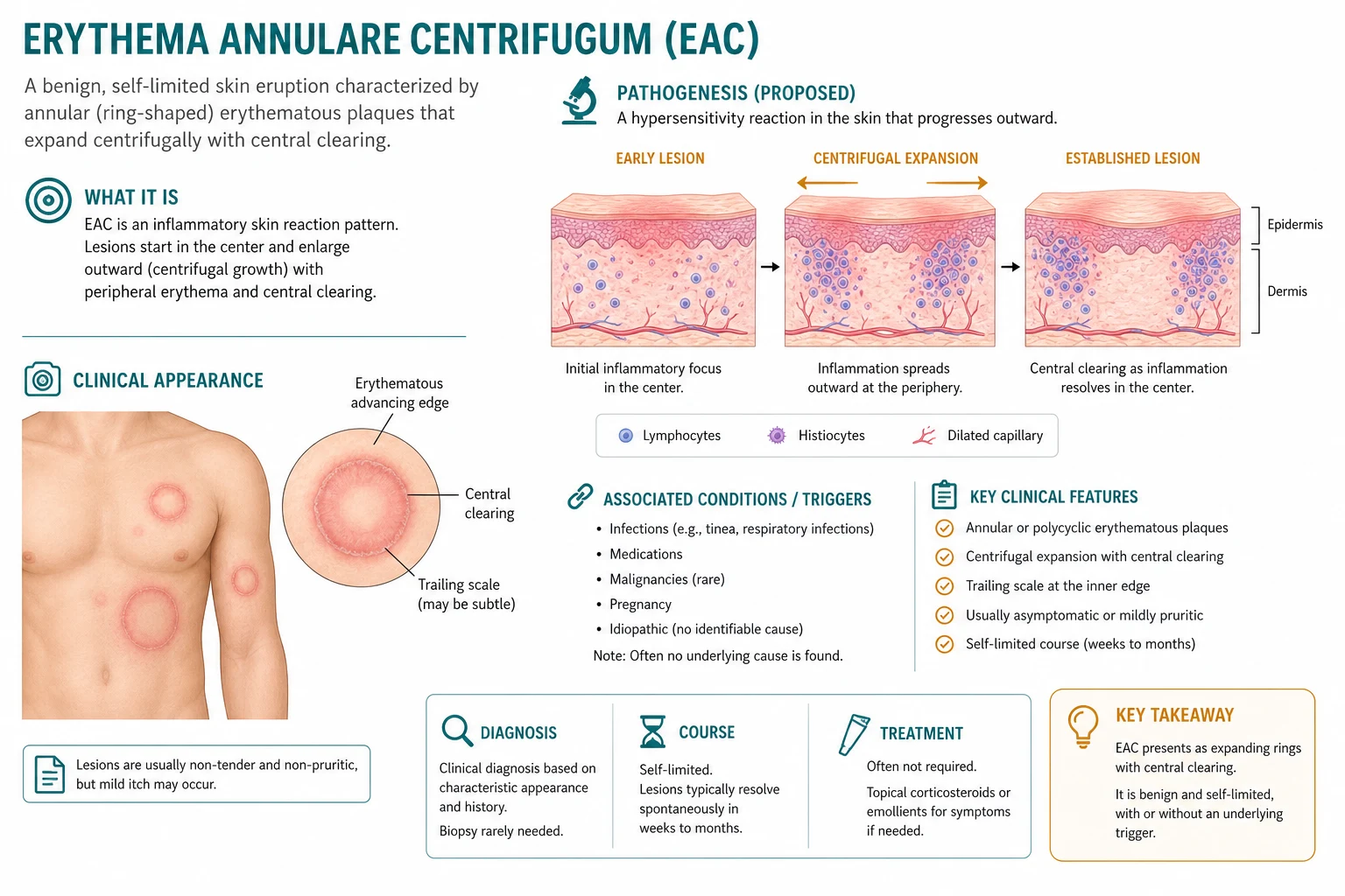
EAC is a type IV (delayed, cell-mediated) hypersensitivity reaction to a distant antigen. The expanding annular morphology is the visible signature of activated T-lymphocytes migrating centrifugally through the skin, releasing cytokines (interferon-gamma, interleukin-2, tumour necrosis factor-alpha) that recruit additional inflammatory cells and produce the advancing erythematous edge. The trailing scale is the desquamating epidermis left behind as the inflammatory front moves outward.[1][3]
Step 1: Antigen exposure and sensitisation
A distant antigen — a dermatophyte antigen released from a focus of tinea, a drug hapten, a food/mould antigen, or a tumour-associated antigen — is processed by antigen-presenting cells (Langerhans cells and dermal dendrocytes) and presented to naive T-lymphocytes in the draining lymph node. This sensitisation step establishes a population of antigen-specific memory T-cells.[1]
Step 2: Cutaneous homing and the id reaction
When the same (or cross-reactive) antigen re-enters the circulation or reaches the skin, sensitised T-cells home to the skin via cutaneous lymphocyte-associated antigen (CLA) binding to E-selectin on dermal endothelium. The critical concept is that of the id reaction: the eruption occurs at a site distant from the antigen source. A patient with tinea pedis may develop EAC on the trunk — the trunk skin is reacting to fungal antigens that have reached it haematogenously, even though the trunk itself is not infected. This is why KOH of the EAC lesion is negative even though the distant tinea is positive.[1]
Step 3: The expanding annular edge
Antigen-specific T-cells cluster around the superficial dermal venules, releasing cytokines that produce the perivascular lymphocytic infiltrate seen on histology — the coat-sleeve or 'cuffing' pattern. This tightly packed perivascular infiltrate, sometimes with eosinophils (particularly in drug-induced or parasite-triggered disease), is the histological hallmark. The inflammatory front then migrates outward through the skin at the characteristic rate of 2 to 5 mm per day, producing the centrifugally expanding ring.[3]
Step 4: The trailing scale
As the advancing edge moves outward, the epidermis it leaves behind desquamates, producing the fine, white, trailing inner scale that is the clinical signature of EAC. The scale trails behind the advancing edge because it is the residue of the inflammatory front that has already passed. This is the inverse of tinea corporis, where the actively growing fungus produces scale on the leading (outer) edge. The trailing scale is sometimes so subtle that it is missed unless the lesion is stretched or viewed in tangential light.[1]
Why lesions resolve centrally
Behind the advancing edge, the inflammatory response wanes as the antigen-specific T-cell clone exhausts or the local antigen is cleared. The central skin returns towards normal, often leaving transient post-inflammatory hyperpigmentation that fades over weeks. New rings appear at the periphery as fresh T-cell clones activate against residual or recurring antigen — producing the migratory, polycyclic pattern that evolves over weeks.[3]
Clinical Presentation
The clinical presentation of EAC is so characteristic that the diagnosis can usually be made at the bedside from the morphology, the scale location, and the distribution — provided the clinician actively looks for the trailing scale and confirms it is not tinea.[1][3]
Morphology
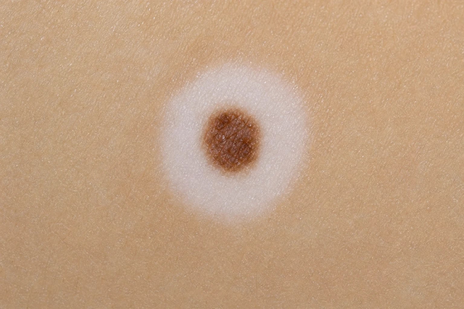
The morphology of EAC is distinctive and centres on the annular configuration and the trailing scale.[1]
- Shape: annular (ring-like) or polycyclic (interlocking arcs formed by coalescing rings). Each lesion starts as a small erythematous macule or papule that expands centrifugally (outward from the centre).[1]
- Border: well-demarcated, raised, erythematous advancing edge with the characteristic fine trailing scale just inside (behind) the edge. The inner edge of the ring is the trailing scale; the outer edge is the actively advancing erythema.[3]
- Rate of expansion: characteristically slow, 2 to 5 mm per day, much slower than urticaria but faster than granuloma annulare.[3]
- Central clearing: as the ring expands, the centre fades — often leaving post-inflammatory hyperpigmentation (brownish discoloration that fades over weeks to months). This produces the classic "ring" or "target-without-a-centre" appearance.[1]
- Number: multiple, usually dozens; new rings appear as old ones expand and resolve, giving a polycyclic, migratory pattern.[3]
Distribution
The distribution is one of the most useful diagnostic clues, with characteristic involved and spared sites:[3]
Symptoms
EAC is frequently asymptomatic. When symptomatic, the dominant symptom is mild pruritus, which is variable and may be absent entirely. Significant pruritus raises the possibility of an alternative diagnosis (tinea, eczema, urticaria) or a coexisting dermatosis. Pain and systemic symptoms are not features of uncomplicated EAC — their presence should prompt investigation for underlying malignancy or an alternative diagnosis.[3]
Timeline and evolution
The timeline of EAC is chronic and migratory, distinct from the acute tempo of urticaria or erythema multiforme:[4]
- Onset: insidious over days to weeks; the patient often cannot date the first lesion precisely.[1]
- Evolution of an individual lesion: each ring expands over days to weeks, reaches a maximum diameter of several centimetres, then resolves centrally over further weeks, leaving hyperpigmentation that fades.[3]
- Evolution of the eruption: chronic and relapsing — new rings continue to appear as old ones resolve, so the eruption migrates across the skin. An untreated episode may last weeks to months, occasionally years.[4]
- Post-inflammatory hyperpigmentation: common but temporary; fades without scarring.[1]
Atypical presentations
Several atypical presentations of EAC are examinable and carry specific management implications:[2]
Differential Diagnosis
The differential diagnosis of an expanding annular erythematous lesion is broad, but the location of the scale, the KOH result, the tempo, and the distribution distinguish the majority of mimics at the bedside. The single most important — and most frequently examined — distinction is EAC versus tinea corporis.[3]
Tinea corporis (the key mimic)
Tinea corporis is the single most important differential and the one examiners test most heavily. Both produce annular, erythematous, expanding lesions. The decisive differences:[1]
- Scale location: tinea has LEADING scale — an active, scaly, raised border on the outer advancing edge, with central clearing. EAC has TRAILING inner scale — a fine scale just inside the advancing edge.[1]
- KOH microscopy: tinea is KOH-POSITIVE (branching septate hyphae); EAC is KOH-NEGATIVE at the active border. This is the single most useful confirmatory test.[1]
- Number and distribution: tinea is usually one or a few lesions, often with a clear source (pet, contact sport, tinea pedis coexisting); EAC is typically multiple and widespread on the trunk and proximal limbs.[3]
- Pruritus: tinea is usually pruritic; EAC is often asymptomatic or only mildly itchy.[1]
Granuloma annulare
Granuloma annulare produces annular plaques composed of small, firm, skin-coloured or erythematous dermal papules with a non-scaly, palpable border. There is no surface scale at all. Lesions are typically on the dorsal hands and feet, fingers, and extensor extremities. The absence of scale, the dermal (rather than epidermal) quality of the border, and the acral distribution distinguish it from EAC. Histology shows palisading granulomatous inflammation and mucin (rather than perivascular lymphocytes).[3]
Urticaria
Urticaria produces transient wheals that appear and resolve within less than 24 hours at any given site. There is no scale, and individual lesions migrate rapidly. EAC lesions are fixed for days to weeks, expand slowly, and carry a trailing scale. The tempo (under 24 hours vs days) is the key discriminator.[3]
Psoriasis
Psoriasis produces well-demarcated, thick, erythematous plaques with a thick, silvery, adherent scale on extensor surfaces (elbows, knees), scalp, and the sacrum, often with nail changes (pitting, onycholysis). The scale is thick and leading, not fine and trailing, and the distribution (extensors) differs from EAC (trunk, proximal limbs).[3]
Erythema migrans (Lyme disease)
Erythema migrans is the expanding annular lesion of early localised Lyme disease (Borrelia burgdorferi). It is usually a single lesion at the site of a tick bite, expands rapidly over days, has a uniform red border without scale, and is accompanied by a history of tick exposure (endemic area, outdoor activity). EAC is multiple, has trailing scale, and has no tick association. Centres for Disease Control / Infectious Diseases Society of America guidance recognises erythema migrans as a clinical diagnosis.[3]
Erythema multiforme
Erythema multiforme produces typical target lesions — three concentric zones: a dusky or bullous centre, a pale oedematous middle ring, and an erythematous outer ring. Lesions favour the extensor extremities, palms, soles, and mucous membranes, and onset is acute (24 to 48 hours), often after herpes simplex or mycoplasma infection or drug exposure. The target morphology and acral/mucosal distribution distinguish it from EAC.[3]
Subacute cutaneous lupus erythematosus (SCLE)
SCLE produces annular or polycyclic erythematous lesions on photosensitive (sun-exposed) skin — the upper back, shoulders, chest, and arms. Lesions may resemble EAC, but SCLE is photosensitive, often has a peripheral scale, and is anti-Ro/SSA antibody positive (in approximately 70 per cent). A photosensitive distribution and a positive antinuclear / anti-Ro antibody distinguish SCLE.[3]
Pityriasis rosea
Pityriasis rosea produces oval, salmon-pink patches with a characteristic collarette (peripheral) scale following Langer's lines in a 'Christmas tree' distribution on the trunk, typically preceded by a herald patch. The collarette scale (peripheral, attached at one edge) and the oval shape along skin lines distinguish it from the annular, trailing-scale rings of EAC.[3]
Mycosis fungoides (cutaneous T-cell lymphoma)
Early mycosis fungoides can present as persistent annular or figurate erythematous patches that mimic EAC and are refractory to topical therapy. The key distinguishing features are persistence beyond months, atypical morphology, and biopsy evidence of atypical lymphocytes with epidermotropism. Any 'EAC' that does not resolve or atypically progresses must be biopsied to exclude mycosis fungoides — a classic fellowship pitfall.[2]
DDx of annular erythema — the SCALPELS mnemonic
Clinical & Bedside Assessment
EAC is fundamentally a clinical diagnosis supported by KOH microscopy. The bedside assessment has two goals: (1) confirm the diagnosis by establishing the trailing-scale morphology and a negative KOH, and (2) identify the trigger, which is the key to definitive management.[1]
History
A structured history should actively probe for the recognised triggers:[1]
- Onset and tempo: when did the eruption start? How fast do individual rings expand? (EAC: days to weeks; urticaria: hours.)[1]
- Symptoms: is the eruption itchy? (EAC: variable, often mild.) Any fever, weight loss, night sweats, lymphadenopathy? (Red flags for lymphoma.)[2]
- Infection history: any tinea pedis, tinea cruris, tinea unguium, tinea corporis, or tinea capitis? Any recent sore throat (streptococcal), viral illness (EBV, hepatitis), or candidal infection?[1]
- Drug history: list all medications started or changed in the preceding weeks — antibiotics (penicillins), NSAIDs, allopurinol, hydroxychloroquine, finasteride, hormones.[5]
- Dietary history: frequent consumption of blue cheese, tomatoes, processed foods with additives/preservatives/dyes.[1]
- Thyroid and autoimmune history: known autoimmune thyroid disease, other autoimmune conditions.[1]
- Pregnancy: in women of reproductive age, consider gestational EAC.[5]
- Malignancy history and red flags: any known malignancy, or B-symptoms (fever, drenching night sweats, weight loss).[2]
Examination
A systematic examination combines lesion assessment with a deliberate search for the trigger and for malignancy red flags:[1]
The KOH test — the decisive bedside manoeuvre
KOH microscopy of the active annular border is the single most useful confirmatory test. Scrape the scale at the advancing edge onto a slide, apply 10 to 20 per cent potassium hydroxide, gently heat, and examine under the microscope.[1]
- Tinea corporis: branching septate hyphae are visible — the test is positive.[1]
- EAC: no hyphae — the test is negative (the eruption is immunological, not infective).[1]
A negative KOH, combined with the trailing-scale morphology, confirms EAC clinically. Always also KOH the distant tinea sites (feet, groin, nails) — these are often positive and identify the antigen source driving the id reaction.[1]
When to suspect underlying malignancy (PEACE)
The following features should raise suspicion of paraneoplastic EAC and trigger a malignancy workup:[2]
- EAC refractory to treatment of an identified trigger (e.g. tinea cleared, eruption persists).[2]
- EAC with systemic symptoms (fever, weight loss, night sweats — B symptoms).[2]
- Lymphadenopathy or hepatosplenomegaly on examination.[2]
- New EAC in an older patient with no obvious trigger.[2]
- EAC with an atypical or rapidly progressive morphology.[2]
Investigations
EAC is a clinical diagnosis. Investigations are directed at (a) excluding tinea (KOH), (b) confirming the diagnosis in atypical cases (biopsy), and (c) identifying the underlying trigger. Routine blood tests are otherwise unremarkable in uncomplicated EAC.[3]
Skin biopsy
Biopsy is not required in the typical case but is indicated when the diagnosis is uncertain, the eruption is atypical or refractory, or mycosis fungoides must be excluded. The characteristic histological finding is:[3]
The 'coat-sleeve' (or 'cuffing') pattern — lymphocytes tightly clustered around the superficial and mid-dermal venules, resembling a coat sleeve around an arm — is the histological hallmark of EAC. It is not entirely specific (a similar pattern can be seen in other inflammatory dermatoses), but in the right clinical context it is strongly supportive. Eosinophils within the infiltrate suggest a drug or parasite trigger. Special stains (PAS or GMS) should be requested to exclude occult dermatophyte in the biopsied skin.[3]
Fungal studies
Fungal studies both exclude tinea at the eruption site and identify the distant focus driving the id reaction:[1]
- KOH microscopy of the EAC border: negative (confirms EAC, excludes tinea at the eruption site).[1]
- KOH / fungal culture of distant sites: examine the feet, groin, nails, and scalp for tinea. A positive distant site identifies the antigen source driving the id reaction and directs definitive treatment.[1]
- Fungal culture of distant tinea sites identifies the organism (Trichophyton rubrum, T. mentagrophytes, etc.) and confirms the trigger.[1]
Trigger workup
When no obvious trigger is found on history and examination, a focused laboratory workup is warranted:[2]
- Full blood count (FBC/CBC) with differential — to screen for leukaemia, lymphoma (abnormal lymphocytes), and eosinophilia (parasite, drug reaction).[2]
- Erythrocyte sedimentation rate (ESR) / C-reactive protein — non-specific, but elevation supports an inflammatory or neoplastic process.[2]
- Thyroid function tests (TSH, free T4) and thyroid antibodies — to detect autoimmune thyroid disease.[1]
- Liver function tests and lactate dehydrogenase (LDH) — LDH elevation raises concern for lymphoma.[2]
- Streptococcal serology (ASO/anti-DNase B) — if post-streptococcal.[1]
- Viral serology (EBV, hepatitis B and C, HIV) — if viral trigger suspected.[1]
- Chest X-ray — baseline screen for occult malignancy, lymphadenopathy, or tuberculosis.[2]
When is malignancy screening indicated?
A full age-appropriate malignancy screen (imaging — CT chest/abdomen/pelvis or PET-CT; lymph node biopsy if lymphadenopathy) is reserved for refractory, atypical, or systemically symptomatic EAC, or where the FBC/LDH/clinical examination raise concern. It is not warranted in every case of idiopathic EAC — the paraneoplastic association, while important, is uncommon.[2]
Management — Resuscitation
EAC is not a dermatological emergency, and there is no time-critical resuscitation. The immediate priority is to identify the trigger and to exclude the dangerous mimics and associations (mycosis fungoides, paraneoplastic EAC with occult lymphoma).[3]
Immediate priorities
The immediate priorities are confirmation of the diagnosis, trigger identification, and exclusion of malignancy:[1]
- Confirm the diagnosis clinically — trailing inner scale + negative KOH. If uncertain, biopsy.[1]
- Search for the trigger — focused history and examination for tinea, drugs, foods, thyroid disease, pregnancy, and malignancy red flags.[1]
- Provide symptomatic relief — topical corticosteroid for pruritus or inflammation; emollient for the trailing scale.[5]
- Reassure the patient — EAC is benign, self-limiting, and non-scarring.[5]
When urgent referral is warranted
Urgent referral is reserved for the red-flag scenarios suggesting malignancy or an atypical mimic:[2]
- Systemic symptoms (B-symptoms) or lymphadenopathy with EAC — urgent haematology/oncology referral for lymphoma workup.[2]
- Refractory or atypical figurate eruption — dermatology referral and biopsy to exclude mycosis fungoides.[2]
- Rapidly progressive erythema gyratum-repens-like eruption with wood-grain scaling — strongly paraneoplastic; urgent malignancy search.[3]
Management — Definitive & Stepwise
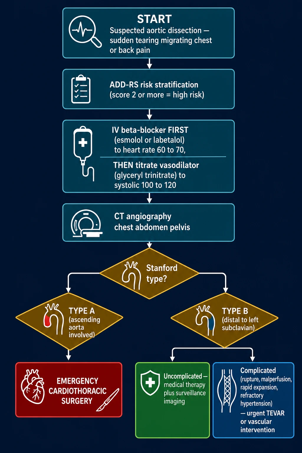
The management of EAC rests on a single principle: the eruption is a reaction; treat the cause, not the skin. Symptomatic treatment of the eruption itself is secondary and often unnecessary, because uncomplicated EAC is self-limiting.[1][5]
Step 1: Treat the underlying trigger (primary, definitive)
Trigger eradication is the only definitive treatment. Without it, the eruption recurs.[1]
| Trigger | Specific treatment |
|---|---|
| Tinea (id reaction) | Oral antifungal — terbinafine 250 mg once daily for 2 to 6 weeks, or itraconazole 200 mg once daily for 1 to 4 weeks. Treat the distant tinea focus (e.g. tinea pedis, onychomycosis). EAC often resolves as the fungal load falls.[1][5] |
| Drug | Withdraw the culprit medication and substitute an alternative if needed. EAC typically resolves over weeks after drug cessation.[5] |
| Bacterial infection (e.g. streptococcal) | Treat the infection with an appropriate antibiotic (e.g. phenoxymethylpenicillin for streptococcal pharyngitis).[1] |
| Candida | Treat candidiasis (topical or oral antifungal as appropriate).[1] |
| Viral (EBV, hepatitis, HIV) | Specific antiviral therapy where indicated; supportive care.[1] |
| Thyroid disease | Restore euthyroidism (levothyroxine for hypothyroidism, antithyroid drugs or radioiodine for hyperthyroidism).[1] |
| Food / food additive | Elimination diet — avoid blue cheese, tomatoes, or the identified additive; challenge to confirm.[1] |
| Malignancy (PEACE) | Treat the underlying neoplasm (chemotherapy, radiotherapy, surgery as appropriate). EAC may resolve with successful cancer treatment.[2] |
Step 2: Symptomatic topical therapy
If the eruption is pruritic or cosmetically distressing, topical therapy can provide symptomatic relief. It does not prevent new lesions and does not alter the underlying course.[5]
- Topical corticosteroids — betamethasone valerate 0.1 per cent or mometasone furoate 0.1 per cent cream/ointment, applied once or twice daily to active lesions for 1 to 2 weeks. Reduces inflammation and pruritus. Avoid prolonged use on the face and intertriginous areas.[5]
- Topical calcineurin inhibitors — tacrolimus 0.1 per cent ointment (or pimecrolimus 1 per cent cream) twice daily. A steroid-sparing alternative, particularly useful on the face, flexures, and for longer courses.[5]
- Emollients — reduce the prominence of the trailing scale and soothe mild irritation.[5]
Step 3: Oral antihistamines
For pruritus that is disturbing sleep or activities, a sedating oral antihistamine at night (e.g. chlorphenamine 4 mg at night, or hydroxyzine 25 mg at night) may help. Non-sedating antihistamines (cetirizine 10 mg daily, loratadine 10 mg daily) are useful for daytime pruritus. EAC is often minimally pruritic, so this is frequently unnecessary.[5]
Step 4: Systemic therapy for refractory disease
A minority of cases are chronic, extensive, or refractory to trigger treatment and topical therapy. The evidence base for systemic therapy is limited (case reports and small case series — no randomised trials).[5]
- Short-course oral corticosteroid — prednisolone 0.5 to 1 mg per kg per day for 1 to 2 weeks, then taper. Effective short-term, but recurrence is common on tapering; reserved for severe, extensive, or disabling disease.[5]
- Hydroxychloroquine 200 mg twice daily — anecdotal benefit in chronic EAC; monitor for retinopathy per local ophthalmology guidance.[5]
- Metronidazole — reported in case reports; mechanism unclear.[5]
- Narrowband UVB phototherapy — useful for extensive disease; courses of 2 to 3 treatments per week for several weeks.[5]
- Ustekinumab (anti-IL-12/23 monoclonal antibody) — reported to resolve refractory EAC in case reports, targeting the T-cell cytokine axis (IL-12 / IL-23) that drives the type IV reaction. Reserved for refractory cases under specialist supervision.[5]
Is specific treatment usually necessary?
No. The single most important principle is that uncomplicated EAC is self-limiting, and in the majority of cases no specific treatment is required beyond trigger identification and eradication. Symptomatic topical therapy is added only if the eruption is pruritic or cosmetically distressing. Systemic therapy is reserved for the rare refractory case. Over-treatment is a pitfall — prolonged topical corticosteroids, in particular, do not alter the course and risk skin atrophy.[5]
Specific Subtypes & Scenarios
Paraneoplastic EAC (PEACE)
Paraneoplastic erythema annulare centrifugum eruption (PEACE) is the rare association of EAC with underlying malignancy. Chodkiewicz and Cohen (2012) consolidated the literature. The malignancies most commonly reported are lymphoma (Hodgkin and non-Hodgkin), leukaemia, mycosis fungoides / Sézary syndrome, and solid tumours (lung, breast, prostate, gastrointestinal, ovarian). EAC may precede the diagnosis of malignancy by months, making it a genuine cutaneous sentinel. The eruption is often extensive, atypical, and refractory to conventional therapy, and it typically resolves with successful treatment of the underlying tumour and recurs with tumour relapse. Any refractory or atypical EAC warrants a malignancy workup (FBC, LDH, lymph node exam, imaging).[2]
Drug-induced EAC
Drug-induced EAC resolves with withdrawal of the culprit drug. The most commonly implicated drugs include penicillins and other beta-lactams, NSAIDs, allopurinol, hydroxychloroquine, finasteride, oestrogens, and thiazides. The mechanism is a drug-induced type IV hypersensitivity reaction. A careful drug history — including over-the-counter and herbal preparations — is essential. Drug withdrawal, with symptomatic topical therapy while the eruption resolves, is usually sufficient.[5]
Tinea-triggered EAC (the id reaction)
Tinea-triggered EAC is the single most common identifiable cause and the prototypical id reaction. The patient has a focus of dermatophyte infection (tinea pedis, tinea cruris, onychomycosis, or tinea corporis) that releases fungal antigens; these reach the skin haematogenously and trigger a distant, KOH-negative, type IV reaction on the trunk. The distant tinea is KOH-positive, but the EAC lesions are KOH-negative. Treatment is oral antifungal therapy directed at the distant focus (terbinafine 250 mg daily for 2 to 6 weeks, or itraconazole 200 mg daily for 1 to 4 weeks); the EAC typically resolves as the fungal load falls over weeks.[1]
Food-additive and pregnancy-associated EAC
- Food-additive EAC — triggers include blue cheese (containing Aspergillus mould antigens), tomatoes, and various food preservatives and dyes. An elimination diet with rechallenge can identify the culprit. This variant is often recurrent and is worth exploring in idiopathic disease.[1]
- Pregnancy-associated EAC — a benign gestational variant described in the second and third trimester. It must be distinguished from the specific dermatoses of pregnancy (polymorphic eruption of pregnancy / PUPPP, pemphigoid gestationis, intrahepatic cholestasis of pregnancy) by morphology and bile acid testing. Gestational EAC typically resolves spontaneously postpartum. Topical corticosteroids (moderate potency) are the symptomatic treatment of choice in pregnancy; systemic therapy is avoided.[5]
Annually recurring EAC
A distinctive seasonal variant recurs each year at the same time (classically spring or autumn), as described by Maurelli and colleagues. It is benign, runs a self-limiting course in each episode, and requires no additional workup once the trigger pattern is recognised. Reassurance is the mainstay.[4]
Complications & Pitfalls
Complications
The complications of EAC are functional and cosmetic rather than destructive, with no scarring:[3]
- Chronic, relapsing course — the main morbidity of EAC is its persistence and recurrence. Untreated episodes may last weeks to months, occasionally years, with new rings appearing as old ones resolve.[4]
- Post-inflammatory hyperpigmentation — lesions resolve leaving brownish discoloration that fades over weeks to months. There is no scarring.[1]
- Psychological distress — extensive or chronic eruptions, particularly on visible sites, may cause anxiety and cosmetic concern.[1]
- Malignancy (PEACE) — the most serious association; EAC may be the first sign of occult lymphoma.[2]
Pitfalls
The classic pitfalls in EAC centre on misreading the scale, missing the trigger, and overlooking malignancy:[2][3]
Pitfalls in EAC — the RINGS mnemonic
- Misdiagnosing mycosis fungoides as EAC — any persistent, atypical, or progressive figurate eruption must be biopsied. Cutaneous T-cell lymphoma can mimic EAC for years.[2]
- Over-treating with prolonged topical corticosteroids — these do not alter the course and risk skin atrophy, telangiectasia, and striae.[5]
- Missing a drug cause — failing to take a thorough drug history, including recently started medications.[5]
- Ignoring thyroid disease — autoimmune thyroid disease is an under-recognised trigger; check TFTs in idiopathic EAC.[1]
Prognosis & Disposition
Natural history of an EAC episode
Prognosis
The prognosis of EAC is benign — self-limiting and non-scarring, though chronic and prone to recurrence:[3]
- Self-limiting — EAC eventually resolves spontaneously, but the course is chronic (weeks to months, occasionally years).[4]
- Recurrence — common, particularly when the trigger persists or recurs (e.g. recurrent tinea pedis, re-exposure to a culprit drug or food, inadequately treated malignancy). Recurrence does not imply treatment failure of the original episode if the trigger was genuinely eradicated.[1]
- No scarring — lesions resolve with post-inflammatory hyperpigmentation that fades; the skin returns to normal.[1]
- Paraneoplastic EAC (PEACE) — the prognosis is that of the underlying malignancy. The EAC itself may resolve with successful cancer treatment and recur with tumour relapse, serving as a tumour marker.[2]
Predictors of a complicated course
Several factors predict a longer or more refractory course and should be addressed proactively:[2]
- Persistence or recurrence of the trigger — the dominant determinant of relapse.[1]
- Failure to identify a trigger (idiopathic EAC) — longer duration; higher chance of chronicity.[4]
- Underlying malignancy (PEACE) — refractory course that mirrors tumour activity.[2]
- Immunocompromise — more extensive, more refractory disease.[2]
Disposition
The disposition depends on the trigger and the presence of red flags:[3]
- Primary care / general practice — most straightforward cases (tinea-triggered, drug-induced) are managed here with trigger identification and symptomatic topical therapy.[1]
- Dermatology referral — for refractory, atypical, or extensive disease; when biopsy is needed to exclude mycosis fungoides; when systemic therapy is contemplated; and for phototherapy or biologic considerations.[2]
- Haematology / oncology referral — when PEACE is suspected (B-symptoms, lymphadenopathy, abnormal FBC/LDH).[2]
Special Populations
The special-population table above summarises the key adjustments, which are all underpinned by the same principle: identify and treat the trigger, and lower the threshold for malignancy workup in the immunocompromised and the elderly.[1][2]
Evidence, Guidelines & Regional Differences
Guidelines
There is no single international guideline for EAC management. Practice is based on narrative reviews, the 2024 systematic review of treatment outcomes, and the principles of figurate erythema diagnosis. Key consensus points:[3][5]
- British Association of Dermatologists / NICE (UK) — no specific guidance; clinical diagnosis with KOH microscopy to exclude tinea; treat the underlying trigger; topical corticosteroids for symptomatic relief; biopsy atypical or refractory lesions.[3]
- American Academy of Dermatology — clinical diagnosis; search for and treat the trigger (especially tinea); consider paraneoplastic workup in refractory disease.[2]
Evidence base
The evidence base is limited and largely observational — case reports, case series, narrative reviews, and one systematic review of treatment outcomes. There are no randomised controlled trials.[5]
- Geng et al. (2024) — the only systematic review of treatment outcomes in EAC. Synthesised the available case-level data; concluded that trigger eradication is the cornerstone, topical corticosteroids provide symptomatic relief, and systemic agents (corticosteroids, hydroxychloroquine, metronidazole, phototherapy, ustekinumab) are reserved for refractory disease with limited high-quality evidence.[5]
- Boehner et al. (2021) — an update and diagnostic approach to the figurate erythemas; reaffirmed the trailing-inner-scale clinical signature of EAC and the coat-sleeve histology, and provided a structured diagnostic algorithm for the figurate erythema family.[3]
- Chodkiewicz and Cohen (2012) — consolidated the paraneoplastic EAC (PEACE) literature; established EAC as a recognised, if rare, cutaneous marker of malignancy, chiefly lymphoma.[2]
- Ilkit et al. (2012) — the definitive review of cutaneous id reactions; established dermatophyte-triggered EAC as the prototypical id reaction and detailed the mechanism and management.[1]
- Maurelli et al. (2021) — a new case series of annually recurring EAC, characterising this benign seasonal variant.[4]
Regional differences
The trigger spectrum and the threshold for malignancy workup vary substantially by region:[1]
Controversies
Several areas of EAC management remain genuinely contested:[5]
- Role of systemic therapy — evidence for hydroxychloroquine, metronidazole, and ustekinumab is limited to case reports; none is established as standard.[5]
- Malignancy workup threshold — there is no consensus on how aggressively to investigate idiopathic EAC for occult malignancy; most authorities reserve imaging for refractory, atypical, or systemically symptomatic disease.[2]
- Whether 'idiopathic' EAC is truly idiopathic — many cases likely have an unrecognised trigger (occult tinea, food additive, thyroid disease); a thorough recurrent search often uncovers a cause.[1]
Exam Pearls
Red flags
Exam application bank (NEET-PG / INICET)
One-line answer
Erythema annulare centrifugum (EAC) is a figurate (annular or polycyclic) erythema characterised by slowly expanding, erythematous rings with a pathognomonic TRAILING SCALE — a fine scale located just INSIDE the advancing inner edge. It is a reactive type IV hypersensitivity reaction to a distant antigen, most often a dermatophyte (tinea) producing an id reaction, but also drugs, infections, foods, and rarely underlying malignancy (paraneoplastic 'PEACE'). Histology shows the classic 'coat-sleeve' tight perivascular lymphocytic infiltrate. Management is to identify and treat the underlying trigger; EAC is self-limiting but runs a chronic, relapsing course over weeks to months.
Worked stems (answer without another resource)
Stem 1 — Classic presentation. Map symptoms to mechanism; name the first investigation and first treatment step with dose/route if drug therapy is standard. [1]
Stem 2 — Unstable / complicated. List red flags that force immediate resuscitation, theatre, ICU, antidote, or reperfusion — and what you do in the first 15 minutes. [1]
Stem 3 — Atypical group. Elderly, pregnancy, child, or immunocompromised: how presentation and thresholds change. [1]
Stem 4 — Differential trap. Name the three closest mimics and one discriminator for each. [1]
Stem 5 — Disposition. Who goes home with safety-netting, who is admitted, who needs HDU/ICU/theatre, and what follow-up is mandatory. [1]
Rapid viva checklist
- Definition + classification
- Pathophysiology chain
- Bedside signs / criteria
- Score with exact components (if any)
- Emergency bundle
- Definitive therapy with doses
- Complications of disease and of treatment
- Special populations
- Guideline/trial name if classic
- Three exam traps
Coverage self-check
If you cannot answer any stem above from this page alone, re-read the matching section — the page is intended to be self-sufficient for final-prof and NEET-PG/INICET questions on Erythema annulare centrifugum (EAC).
References
- [1]Ilkit M, Durdu M, Karakas M, et al. Cutaneous id reactions: a comprehensive review of clinical manifestations, epidemiology, etiology, and management Crit Rev Microbiol, 2012.PMID 22300403
- [2]Chodkiewicz HM, Cohen PR, et al. Paraneoplastic erythema annulare centrifugum eruption: PEACE Am J Clin Dermatol, 2012.PMID 22320680
- [3]Boehner A, Neuhauser R, Zink A, et al. Figurate erythemas - update and diagnostic approach J Dtsch Dermatol Ges, 2021.PMID 34046996
- [4]Maurelli M, Gisondi P, Colato C, et al. Annually Recurring Erythema Annulare Centrifugum: A New Case Series with Review of the Literature Case Rep Dermatol, 2021.PMID 34248533
- [5]Geng RSQ, Sood S, Lee A, et al. Treatment Outcomes in Erythema Annulare Centrifugum: A Systematic Review J Cutan Med Surg, 2024.PMID 39323045