Dermatology · Medicine
Erythema multiforme
Also known as Erythema multiforme · EM · Erythema multiforme minor · Erythema multiforme major · MIRM
Erythema multiforme (EM) is an acute, immune-mediated, usually self-limiting mucocutaneous eruption characterised by typical raised target lesions with three concentric colour zones. It is distinct from Stevens-Johnson syndrome / toxic epidermal necrolysis (SJS/TEN). Herpes simplex virus (HSV) is the most common trigger for EM minor; Mycoplasma pneumoniae drives Mycoplasma-induced rash and mucositis (MIRM), an EM-spectrum disorder in children. Management focuses on trigger identification and treatment, supportive care, and recurrent HSV suppression.
On this page & tools
Your progress
Saved locally on this device.
Exam tags
Red flags
Erythema multiforme is one of the most recognisable dermatological emergencies on exam papers because of its target lesions, but it is also one of the most misdiagnosed conditions in real practice. The name itself contains the key morphological clue: multiforme describes the varied appearances (macules, papules, vesicles, bullae), while erythema reminds the candidate that the lesions are red. The core exam task is to separate EM from SJS/TEN, identify the trigger, and choose the correct management — especially suppressive antiviral therapy for recurrent HSV-driven disease.[2][7]
Overview and Definition
Erythema multiforme is an acute, usually self-limited, mucocutaneous hypersensitivity reaction that presents with characteristic target lesions. It is now accepted as a distinct clinicopathological entity from SJS/TEN, rather than the mild end of the same spectrum.[2][7] The lesions are classically palpable (raised), symmetrical, and favour acral sites — the palms, soles, dorsal hands and feet, extensor forearms, elbows, knees and face. Mucosal involvement may occur but is usually milder and more limited than in SJS/TEN.
The disorder is immune-mediated rather than infectious. It represents a host response to an antigen — most commonly HSV DNA deposited in the skin after a herpetic outbreak — and resolves once the offending antigen is cleared. Prompt recognition prevents unnecessary treatment, hospitalisation and patient anxiety. The typical episode lasts 1–4 weeks, and most patients recover without scarring or long-term sequelae. Mortality is negligible in uncomplicated EM, which is a critical distinguishing feature from SJS/TEN.[1][7]
The condition was first described in the early nineteenth century and has been reclassified repeatedly. The crucial modern development was the recognition that the clinical morphology and prognosis of EM are so different from SJS/TEN that they should be separated conceptually. This distinction has important therapeutic implications: EM does not require the aggressive supportive measures, burn-unit care and intensive monitoring that SJS/TEN often does, and the prognosis is dramatically better. [1]
[1]Classification
Modern terminology divides EM into two main clinical forms. The historical classification of Bastuji-Garin and Roujeau (1993) separated EM minor, EM major, SJS and TEN using the proportion of body surface area with epidermal detachment and the morphology of the lesions. Current consensus keeps EM major as a distinct entity from SJS/TEN: EM major has mucosal involvement but still shows typical raised three-zone target lesions, little or no epidermal detachment, and an infection-related trigger. SJS/TEN, by contrast, is drug-related, shows atypical flat purpuric macules, and has high mortality.[2][7]
EM minor
EM major
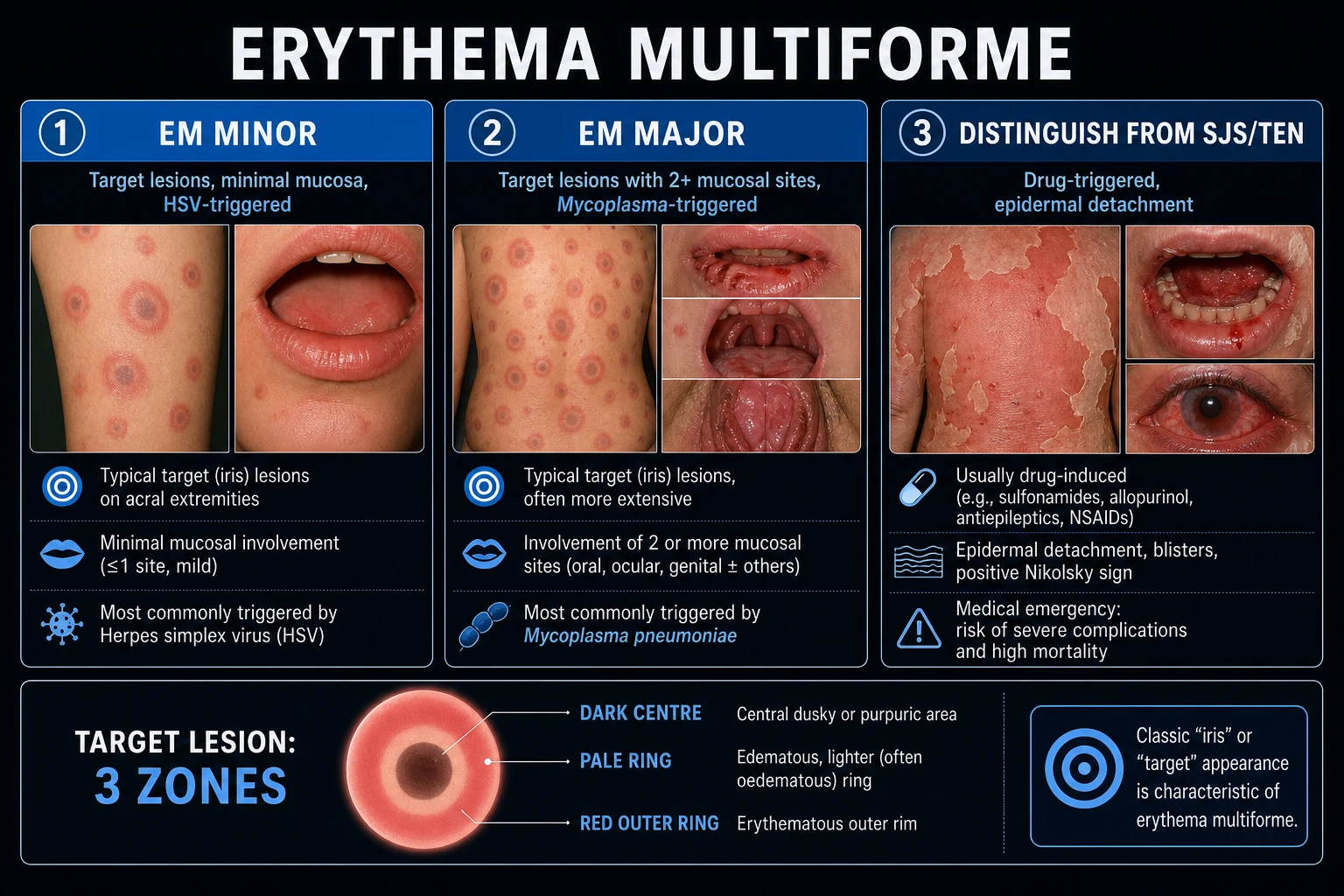
EM minor is the classic teaching form. It affects the skin only, or a single mucosal site so mildly that it is barely noticeable. EM major shares the same skin morphology but adds mucosal disease at one or more sites. The presence of mucosal involvement does not shift the disease into the SJS/TEN category. SJS/TEN is defined by its own morphology, drug-related trigger, epidermal detachment and high mortality. This conceptual firewall is important because it prevents the unnecessary hospitalisation and anxiety that comes from mislabelling a self-limited infection-triggered eruption as a life-threatening drug reaction. [1]
Epidemiology and Risk Factors
EM occurs in all age groups, with a slight male predominance. It is rare: the exact incidence is unknown, but EM is far less common than other mucocutaneous eruptions such as urticaria or simple drug exanthems. The condition is recurrent in a substantial minority of patients, and when it recurs the trigger is almost always HSV reactivation.[7]
The most important risk factor is recent infection with HSV-1 or HSV-2. HSV triggers roughly 70% of all cases of EM and up to 90% of recurrent cases. The skin eruption typically appears 1–2 weeks after the herpetic outbreak, sometimes after a subclinical recurrence. Other triggers include Mycoplasma pneumoniae (especially in children and young adults), other infections (streptococcal pharyngitis, adenovirus, EBV, CMV, parvovirus B19, hepatitis viruses, fungi, tuberculosis), and a growing list of drugs — although true drug-induced EM is rare and many drug-triggered cases are reclassified as SJS/TEN on closer inspection.[1][2][4]
Drugs reported with EM-like eruptions include NSAIDs, anticonvulsants (carbamazepine, phenytoin), sulfonamides, penicillins, allopurinol, and barbiturates. However, drug-induced cases often have atypical flat lesions and mucosal involvement that overlap with SJS/TEN, so the diagnosis must be made with caution. Mycoplasma-associated disease is the dominant trigger in children and may be severe enough to mimic SJS/TEN. Recurrent episodes tend to occur in the same individual, suggesting a genetic predisposition to HSV-directed immune responses, though no single HLA allele has been consistently identified across all populations. [1]
Pathophysiology
EM is a type IV (cell-mediated) hypersensitivity reaction. The central event is the presentation of an antigen — usually HSV DNA or a drug hapten — to T lymphocytes in the skin, followed by a cytotoxic immune response that damages individual keratinocytes and produces the characteristic epidermal changes and target morphology.[2][7]
In HSV-associated EM, the pathogenetic sequence is well characterised. HSV replicates at a mucocutaneous site such as the lip or genitalia. Viral particles and DNA then travel to the dorsal root ganglia, where the virus establishes latency. Reactivation leads to local replication and, in susceptible individuals, deposition of HSV DNA, antigens or immune complexes in the skin. Keratinocytes process these antigens and present them to HSV-specific CD4+ T cells. Cytotoxic CD8+ T cells then kill infected keratinocytes, producing individual necrotic keratinocytes and a lymphocytic infiltrate. The three-zone target lesion is thought to reflect concentric rings of immune response intensity: central epidermal necrosis, surrounding oedema from cytokine-mediated vascular leakage, and an outer rim of erythema from perivascular inflammation.[2][7]
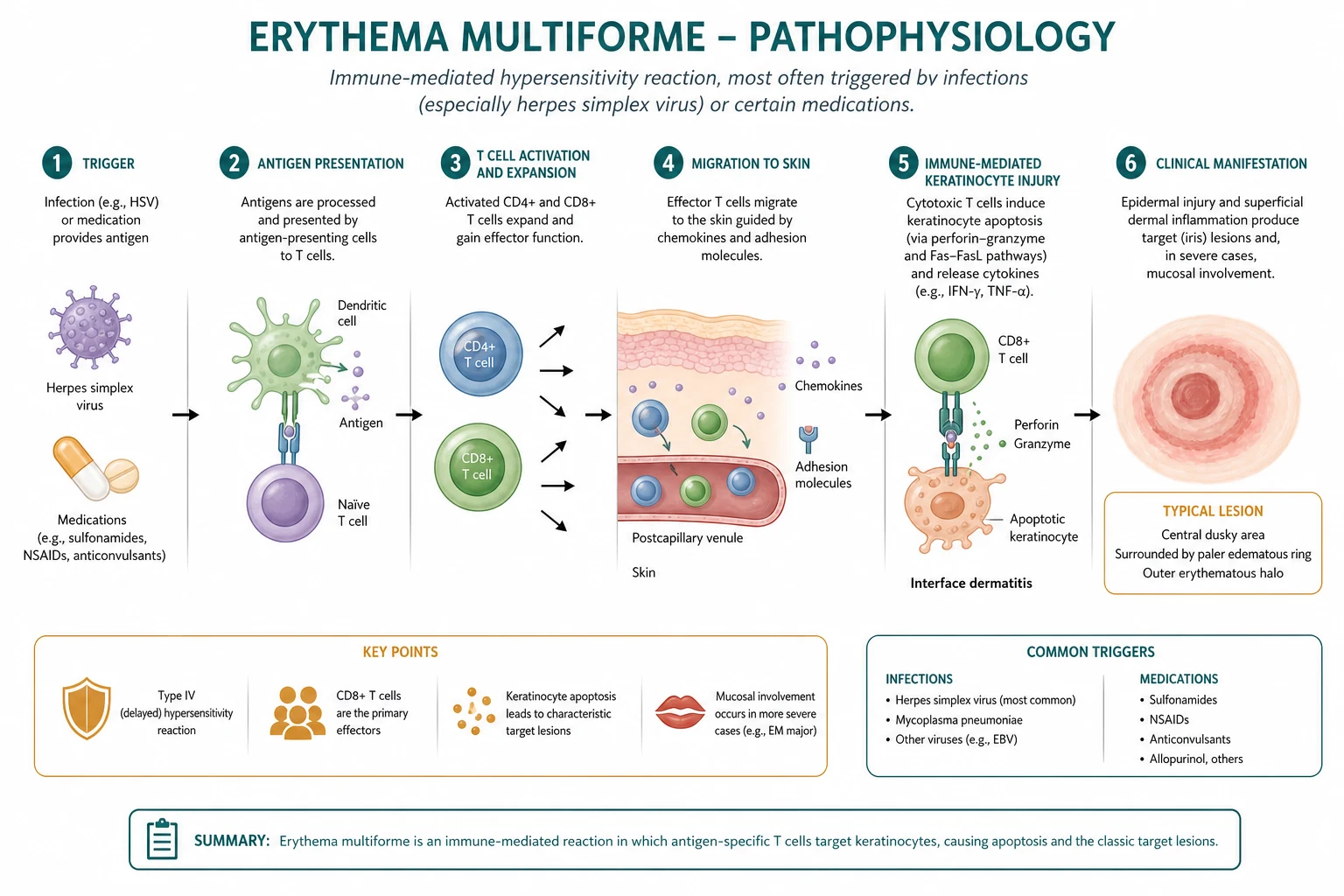
The molecular cascade involves several effector mechanisms. HSV DNA fragments persist in the epidermis for days after the active herpetic lesion has healed. CD4+ T cells recognise HSV peptides presented by HLA class II on local antigen-presenting cells, producing interferon-gamma and tumour necrosis factor-alpha that activate cytotoxic CD8+ T cells. These CD8+ T cells kill keratinocytes that display HSV antigens on HLA class I, through perforin/granzyme release and Fas-Fas ligand interactions. The resulting keratinocyte apoptosis is scattered rather than confluent, which is why EM shows individual necrotic keratinocytes rather than the full-thickness epidermal necrosis seen in SJS/TEN. Adhesion molecules such as ICAM-1 and E-selectin are upregulated on dermal endothelial cells, allowing T cells to migrate into the epidermis and producing the perivascular and intraepidermal infiltrate that defines the histology.[2]
Mycoplasma pneumoniae is thought to trigger disease through a different mechanism. The organism produces a superantigen-like or cross-reactive immune response that causes reactive infectious mucocutaneous eruption (RIME), of which MIRM is the classic example. The mucositis is typically more prominent than the skin rash, and the skin lesions may be targetoid, vesicular, bullous or polymorphic. The prominent mucosal involvement reflects the affinity of Mycoplasma antigens or cross-reactive antibodies for mucosal epithelium, especially conjunctival, oral and urogenital surfaces.[3][8][9]
Drug-induced EM is less clearly defined. In the classic type IV hypersensitivity model, a drug or its metabolite binds to a self-peptide, forming a hapten that activates cytotoxic T cells. Some drug reactions also involve the p-i (pharmacological interaction with immune receptors) concept, where the drug binds directly to the T-cell receptor or HLA molecule without antigen processing. However, many cases previously labelled "drug-induced EM" are now considered SJS/TEN with flat atypical targets and epidermal necrosis. The key distinction is that true EM almost always has typical raised targets, whereas drug-induced severe mucocutaneous reactions have flat purpuric macules and epidermal detachment.[2][4]
Clinical Presentation
Skin lesions
The hallmark of EM is the target lesion. A typical target lesion has three zones: [1]
- Central zone: dusky, violaceous, purpuric or bullous; may be crusted or eroded. This represents the focus of epidermal injury.
- Middle zone: a pale, oedematous, raised ring. This corresponds to dermal oedema and is the reason the lesion is palpable.
- Outer zone: an erythematous halo. This is the peripheral inflammatory flare. [1]
The lesions are usually symmetrical, begin as erythematous macules and evolve into papules and target lesions over 24–72 hours. They favour the extensor surfaces of the forearms and legs, the dorsal hands and feet, the palms and soles, the elbows and knees, and the face. The trunk is relatively spared in classic EM, which is another important distinction from SJS/TEN.[1][7]
Atypical EM may present with targetoid lesions lacking the full three-zone architecture, vesicular or bullous lesions, or lesions that are widespread and involve the trunk. These atypical cases are more difficult to distinguish from SJS/TEN and often require a biopsy. Dermoscopy can sometimes help: EM targets show a central violaceous or brownish area, a whitish ring and a peripheral erythematous rim, while SJS/TEN lesions show diffuse purpuric dots and an absence of the structured target pattern. [1]
EM has been described in association with radiotherapy, malignancy (especially as a paraneoplastic phenomenon), autoimmune disease and inflammatory bowel disease. There are also rare reports of EM-like eruptions triggered by contact allergens, insect bites and certain foods. In immunocompromised hosts, the lesions may be more widespread, less targetoid and slower to resolve. [1]
Mucosal involvement
EM minor has no mucosal involvement, or only a single mild site. EM major involves one or more mucosal surfaces, most commonly the oral mucosa (lips, buccal mucosa, tongue, palate). The erosions are usually painful but are shallower and less extensive than in SJS/TEN. Other mucosal sites include the conjunctivae, genitalia and, rarely, the anorectal mucosa. Ocular involvement is a red flag because it can lead to scarring, symblepharon and vision loss; early ophthalmology review is essential.[5][7]
Prodromal and systemic symptoms
Many patients with EM have a prodromal phase of low-grade fever, malaise, arthralgia and, in HSV-related cases, a recent herpetic lesion. The rash appears 1–3 weeks after HSV infection and 1–2 weeks after Mycoplasma respiratory infection. The lesions may evolve in crops, with new lesions appearing over several days. Pruritus is variable; some patients report burning or tenderness rather than itch. Systemic symptoms are usually mild in EM and disproportionate systemic toxicity should raise suspicion for SJS/TEN or severe MIRM.[2][7]
Differential Diagnosis
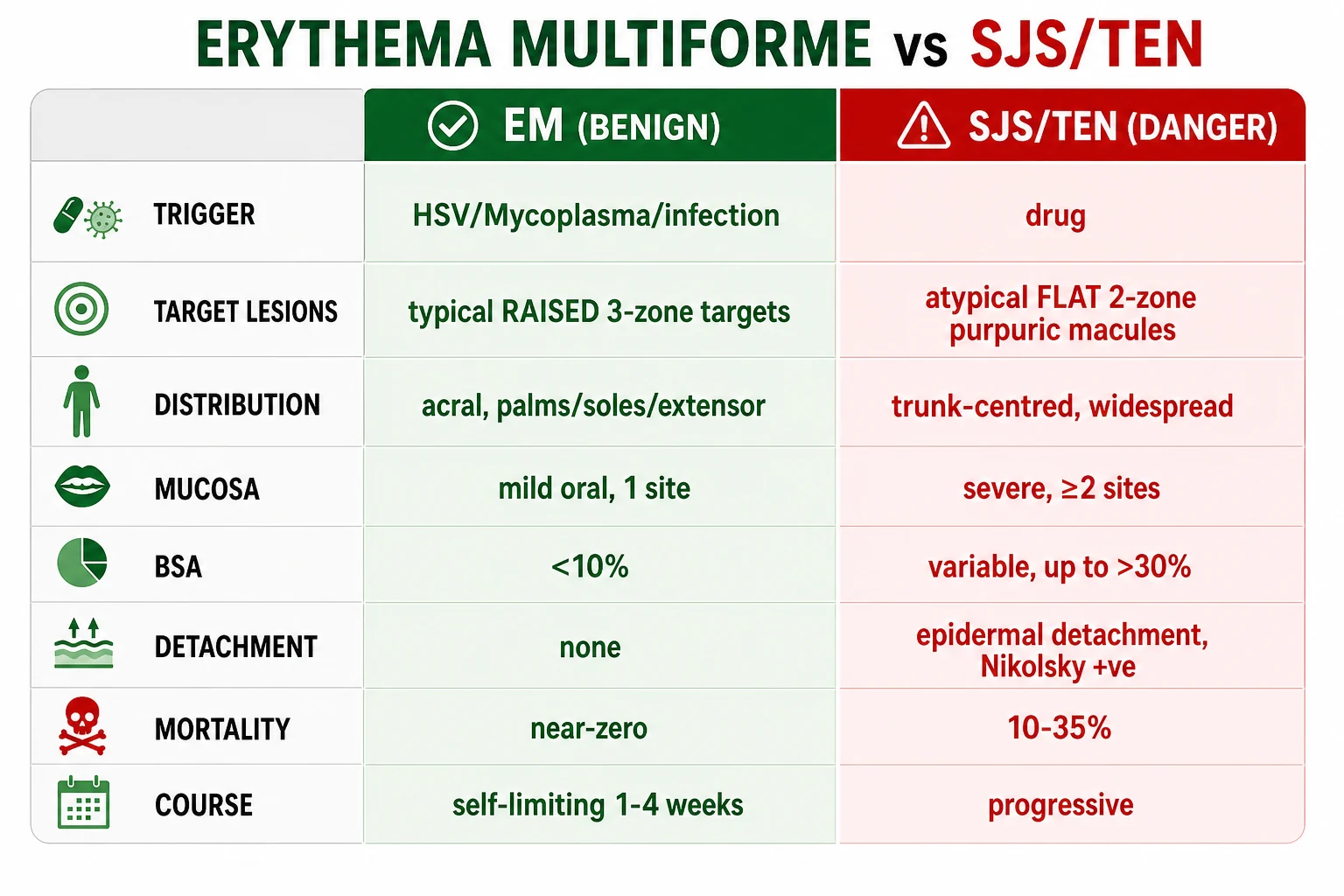
The differential diagnosis of EM is broad and includes every targetoid or annular eruption. The most important distinction is from SJS/TEN, because misclassification can lead to unnecessary aggressive therapy or, conversely, undertreatment of a life-threatening condition. The key discriminators are the trigger, morphology, distribution and prognosis.[2][7]
Urticaria multiforme
Fixed drug eruption
Annular psoriasis
- Stevens-Johnson syndrome / TEN: flat, purpuric, atypical two-zone macules; trunk-centred; drug trigger; epidermal detachment; positive Nikolsky sign; high mortality. Mucosal involvement is usually severe and multi-site, and the patient looks toxic.
- Urticaria multiforme: transient migratory wheals that disappear within 24 hours, often with central dusky clearing but no true blistering; lesions move from site to site; responds to antihistamines and is not associated with mucosal scarring.
- Fixed drug eruption: round, dusky, well-demarcated plaques that recur at exactly the same site with each exposure to the offending drug; lesions lack the three-zone architecture and acral distribution; post-inflammatory hyperpigmentation is common.
- Lyme disease erythema migrans: expanding annular erythema with central clearing, often after a tick bite; typically a single or few lesions rather than multiple acral targets; associated with fever, arthralgia, lymphadenopathy and a history of tick exposure; serology confirms the diagnosis.
- Annular psoriasis: chronic, scaly, well-demarcated plaques; no true target morphology; Auspitz sign and Koebner phenomenon are present; biopsy shows regular acanthosis, neutrophils and diminished granular layer.
- Serum sickness-like reaction: fever, arthralgia, urticarial rash and lymphadenopathy occurring 1–3 weeks after drug exposure; lacks true target lesions and has a different systemic pattern.
- Sweet syndrome: tender erythematous plaques or nodules, fever, neutrophilia; histology shows dense neutrophilic infiltrate; no target lesions.
- Kawasaki disease: fever, mucositis, rash, conjunctivitis, lymphadenopathy and extremity changes in children; coronary artery complications are the major concern; rash is polymorphous rather than targetoid.
- Acute generalised exanthematous pustulosis (AGEP): drug-induced, sterile pustules on an erythematous base, fever and neutrophilia; pustular morphology, not target lesions. [1]
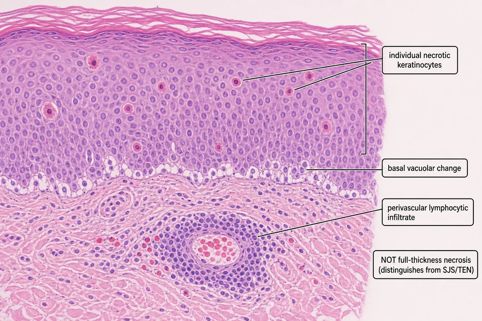
When the diagnosis is uncertain, a punch biopsy is the definitive discriminator. EM shows individual necrotic keratinocytes, basal vacuolar change and a perivascular lymphocytic infiltrate; SJS/TEN shows full-thickness epidermal necrosis, sparse infiltrate and extensive keratinocyte apoptosis. The absence of full-thickness necrosis on biopsy is therefore reassuring, but the biopsy must be taken from an early lesion because established lesions may show only non-specific changes. [1]
Another important mimic is DRESS (drug reaction with eosinophilia and systemic symptoms), which presents with fever, facial oedema, lymphadenopathy, eosinophilia and a morbilliform rash that may become purpuric. Unlike EM, DRESS has systemic organ involvement and a long latency after drug exposure. Pityriasis rosea can produce oval plaques with a collarette of scale but lacks the three-zone target morphology and mucosal involvement. [1]
On direct immunofluorescence, EM usually shows no immune complex deposition, or only scattered IgM and C3 around necrotic keratinocytes. This helps to distinguish EM from autoimmune blistering diseases such as bullous pemphigoid and pemphigus vulgaris, which show linear IgG and C3 at the basement membrane or intercellular IgG respectively. In paraneoplastic pemphigus, direct immunofluorescence shows intercellular and basement membrane staining and the clinical picture is more severe and refractory.[2][5]
Clinical and Bedside Assessment
The bedside assessment in suspected EM is designed to answer three questions: Is this EM?, Is it severe enough to need hospitalisation?, and What is the trigger?. [1]
Start with the focused skin examination. Look for target lesions, document their distribution, and test whether they are raised and palpable. Count the number of mucosal sites involved. Assess for epidermal detachment by gentle lateral pressure — Nikolsky sign is negative in EM because the epidermis is intact. In SJS/TEN, Nikolsky sign is positive and the epidermis slips off with lateral pressure. The presence of a positive Nikolsky sign should shift the working diagnosis strongly away from EM.[7]
Examine the mouth, eyes and genitalia in every patient. Oral erosions are common in EM major; check the lips, buccal mucosa, tongue and palate. Ocular involvement is an emergency: look for conjunctival injection, discharge, pseudomembrane, or corneal ulceration. Genital erosions can cause urinary retention and are easily missed unless the patient is specifically asked. [1]
Vital signs and systemic assessment are important because MIRM in children can be accompanied by pneumonia and respiratory distress. Listen for cough, wheeze or crepitations, and look for signs of dehydration if oral intake is poor. Check for lymphadenopathy, fever and arthralgia. A full drug history is mandatory, including the exact dates of initiation and cessation of all medications, over-the-counter drugs and herbal remedies in the preceding 2–8 weeks. Ask about a history of cold sores, genital herpes, recent respiratory illness or sore throat, and any previous similar episodes. Dermoscopy is not required but can help in atypical cases: EM targets show a central brown or violaceous disc, a whitish oedematous ring and a peripheral erythematous rim, whereas SJS/TEN shows diffuse purpuric dots without the concentric target structure. [1]
Investigations
The diagnosis of EM is clinical in most cases. A patient with typical raised three-zone target lesions on acral sites, a compatible trigger and mild mucosal disease does not need a biopsy. Investigations are used to confirm the trigger, exclude dangerous mimics, and guide management.[1][7]
Bedside and laboratory tests
- Skin biopsy (punch): reserved for atypical or severe cases where the distinction from SJS/TEN is uncertain. Histology shows individual necrotic keratinocytes, basal vacuolar change, lymphocytic exocytosis, subepidermal blistering in bullous cases, and a superficial perivascular lymphocytic infiltrate. Direct immunofluorescence is usually negative or non-specific. Full-thickness epidermal necrosis, sparse infiltrate and extensive detachment would point to SJS/TEN.[5][7]
- HSV PCR or serology: if HSV is suspected, especially in recurrent EM. PCR from a fresh herpetic lesion is most sensitive. Serology is less useful because many adults are HSV-seropositive; paired acute and convalescent titres are rarely needed for management.
- Mycoplasma pneumoniae PCR or serology: in children and young adults with respiratory symptoms and mucositis. PCR from a respiratory sample is preferred in the acute phase; serology can show rising titres on paired samples. Cold agglutinins may be positive but are non-specific.
- Targeted infectious work-up: throat swab for streptococcus, EBV/CMV serology, hepatitis serology, HIV test if risk factors, and chest X-ray if respiratory symptoms.
- Drug review: establish a clear timeline of drug exposure relative to rash onset. A drug started within the last 1–3 weeks with flat purpuric lesions should raise suspicion for SJS/TEN rather than EM.
Management — Resuscitation
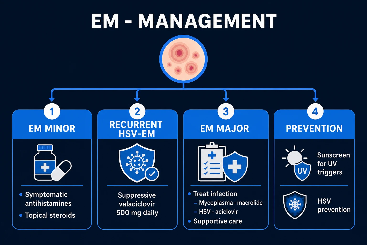
Most EM is not a resuscitation emergency. However, EM major with extensive mucosal involvement, MIRM with respiratory compromise, or any patient with dehydration, haemodynamic instability, or ocular involvement requires urgent hospital assessment and sometimes admission.[1][6]
The immediate priorities are: [1]
- Airway and breathing: assess for respiratory distress, especially in Mycoplasma-associated disease; provide oxygen if needed; obtain chest imaging if pneumonia is suspected.
- Fluid resuscitation: patients with painful oral erosions may be unable to drink. Start intravenous crystalloid if there is dehydration, tachycardia or reduced urine output.
- Nutrition: maintain oral intake where possible; if not possible, use nasogastric feeding or parenteral nutrition.
- Pain control: oral analgesics are often inadequate for severe mucosal erosions; use stepwise analgesia including opioids if necessary.
- Eye protection: any ocular involvement warrants urgent ophthalmology review to prevent corneal ulceration and scarring.
- Infection prevention: severe mucosal erosions can become secondarily infected; institute aseptic skin care and treat infection early. [1]
Hospitalisation is indicated for EM major with significant mucosal involvement, inability to maintain oral hydration, eye involvement, extensive skin disease, or diagnostic uncertainty with concern for SJS/TEN.[6]
Management — Definitive and Stepwise
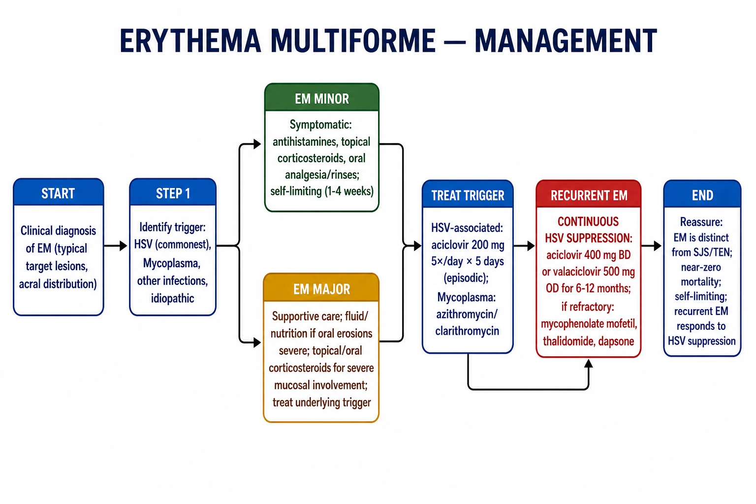
Management of EM has two pillars: treat the trigger and provide supportive care. The specific trigger determines the definitive therapy, while the severity of mucosal disease determines the level of supportive care needed.[1][6][7]
Step 1: Identify and remove the trigger
- HSV-associated EM: treat active herpes with antiviral therapy. For recurrent EM, the key is long-term suppressive antiviral therapy rather than episodic treatment.
- Mycoplasma-associated EM / MIRM: treat the infection with a macrolide antibiotic such as azithromycin or clarithromycin. Azithromycin is commonly given as 500 mg on day 1 followed by 250 mg daily for 4 days, or as 500 mg daily for 3 days; clarithromycin is an alternative where azithromycin is unavailable or contraindicated.
- Drug-associated EM: stop the suspected drug immediately. If the morphology is atypical or severe, manage as SJS/TEN until proven otherwise.
- Idiopathic EM: supportive care only. [1]
Step 2: Symptomatic care for EM minor
EM minor is managed as an outpatient. The mainstays are: [1]
- Topical corticosteroids: a moderate-potency steroid cream or ointment (for example, betamethasone 0.1% or mometasone furoate 0.1%) applied thinly to the skin lesions twice daily for 1–2 weeks reduces inflammation and pruritus. Superpotent steroids are rarely needed and should be avoided on the face and flexures.
- Antihistamines: a non-sedating antihistamine such as cetirizine 10 mg once daily or loratadine 10 mg once daily can help with pruritus and discomfort. A sedating antihistamine at night may be useful if sleep is disturbed.
- Emollients: frequent application of bland emollients to protect the skin barrier and reduce dryness.
- Analgesia: paracetamol 1 g four times daily, or NSAIDs if not contraindicated, for arthralgia and skin tenderness. [1]
Step 3: Supportive care for EM major
EM major with mucosal involvement requires more active management: [1]
- Topical corticosteroids for skin lesions as above.
- Mucosal care: topical anaesthetic mouthwashes (for example, benzocaine or lidocaine) before meals, saline or chlorhexidine mouth rinses, and topical corticosteroid dental paste for oral erosions. Sucralfate suspension can be used as a protective coating. Maintain oral hygiene with a soft toothbrush and avoid spicy, acidic, rough or salty foods.[5]
- Eye care: lubricating eye drops, topical corticosteroids or antibiotic drops under ophthalmology guidance, and tarsorrhaphy or amniotic membrane in severe cases. Fluorescein staining is used to detect corneal epithelial defects.
- Systemic corticosteroids: the evidence is mixed and controversial. Some clinicians use a short course of oral prednisolone 0.5–1 mg/kg/day for severe EM major with painful mucosal disease, but this is not supported by high-quality evidence and may be harmful if the true diagnosis is SJS/TEN. Steroids are generally reserved for severe, refractory cases and should be tapered rapidly.[6]
- Other systemic agents: dapsone, azathioprine, mycophenolate mofetil, thalidomide, ciclosporin or cyclophosphamide have been used for refractory or recurrent EM that does not respond to antiviral suppression. These are specialist-led treatments and require monitoring for adverse effects. Intravenous immunoglobulin and plasmapheresis have been reported in very severe, refractory cases but are not standard of care.[6]
Supportive nursing and nutritional care
Good supportive care shortens the symptomatic phase and prevents complications. The skin should be cleansed with lukewarm water and a mild soap-free cleanser; crusted erosions can be soaked with saline compresses. A bland, fragrance-free emollient should be applied several times daily. Mucosal care is labour-intensive: saline mouth rinses after meals, topical anaesthetic before eating, and a soft diet can prevent weight loss. Patients with eye involvement may need hourly lubricating drops during the day and a paraffin-based ointment at night. Genital erosions should be managed with sitz baths, barrier creams and topical anaesthetic. A multidisciplinary team including dermatology, ophthalmology, dietetics and nursing is valuable for severe EM major or MIRM. [1]
Step 4: Recurrent EM — antiviral prophylaxis
Recurrent EM, defined as more than six episodes per year, is almost always HSV-driven. The cornerstone of management is continuous oral antiviral suppression: [1]
- Aciclovir 400 mg twice daily for 6–12 months; or
- Valaciclovir 500 mg once daily for 6–12 months. [1]
This suppressive regimen reduces HSV reactivation and dramatically decreases the frequency and severity of EM recurrences. It is often continued for 6–12 months and then tapered; if relapse occurs, it can be restarted. For patients who continue to have breakthrough episodes, dose escalation or the addition of an immunomodulatory agent may be considered.[1][7]
Specific Subtypes and Scenarios
EM minor
EM minor is the classic form. It presents with typical target lesions on acral sites and no or only one mild mucosal site. The trigger is usually HSV. It is self-limited and managed with supportive care alone. Patients can be reassured that the prognosis is excellent and mortality is negligible. The main challenge is to distinguish it from urticaria multiforme, fixed drug eruption and early SJS/TEN if the lesions are atypical. [1]
EM major
EM major includes one or more mucosal sites but retains the characteristic typical raised target lesions. The mucosal disease can be painful and may require hospital admission for fluid and nutrition support. It is still distinct from SJS/TEN because the lesions are raised targets, the trigger is usually infection, and there is little or no epidermal detachment. Systemic corticosteroids may be used for severe mucosal disease but are controversial. Ophthalmology review is essential if the eyes are involved.[2][7]
Mycoplasma-induced rash and mucositis (MIRM)
MIRM is an EM-spectrum condition triggered by Mycoplasma pneumoniae. It predominantly affects children and young adults, often after a prodromal respiratory illness of cough, fever and malaise. The disease is characterised by prominent mucositis (oral, ocular, genital, urethral or anal) with relatively sparse skin lesions. The skin lesions may be targetoid, vesicular, bullous, papular or polymorphic. MIRM is generally milder than SJS/TEN and has a lower mortality, but it can be severe enough to require hospitalisation and ophthalmology input. Management includes macrolide therapy for the underlying infection, supportive care, and careful ophthalmological assessment. In some centres, MIRM is grouped under the broader term reactive infectious mucocutaneous eruption (RIME), which includes mucocutaneous eruptions triggered by Mycoplasma and other infections such as Chlamydia pneumoniae, group A streptococcus and adenovirus.[3][8][9]
Recurrent HSV-associated EM
Recurrent EM is usually HSV-driven. The eruption follows a herpetic outbreak by 1–2 weeks and may be more severe than the initial episode. Some patients have a clear history of cold sores, while others have only subclinical HSV reactivation. Continuous suppressive antiviral therapy is the mainstay of prevention. For patients with breakthrough recurrences despite antivirals, mycophenolate mofetil, thalidomide, dapsone, azathioprine or ciclosporin may be used under specialist supervision. The patient should be taught to recognise early herpetic lesions and to seek treatment promptly, although suppressive therapy usually prevents the need for episodic antivirals.[1][6]
Drug-induced EM-like eruption
Drug-induced cases should be viewed with caution. A drug started 1–3 weeks before the rash, especially with flat atypical targets, trunk predominance and mucosal involvement, is more likely to be SJS/TEN than true EM. The safest approach is to stop the drug, admit the patient if severe, and biopsy if the diagnosis is uncertain. True drug-induced EM is rare and usually mild. The offending drug must be permanently avoided and the patient should be warned about cross-reactivity with structurally related agents where relevant.[4]
Idiopathic EM
Up to half of EM cases have no identifiable trigger. These are managed supportively. Recurrent idiopathic EM that does not respond to antivirals is uncommon and may warrant specialist review for alternative diagnoses such as paraneoplastic pemphigus, lupus erythematosus, Behçet disease or MAGIC syndrome (mouth and genital ulcers with inflamed cartilage). A thorough biopsy, immunofluorescence studies and autoimmune work-up may be needed in these refractory cases.[2]
Complications and Pitfalls
The most important complication is misdiagnosis as SJS/TEN. This can lead to unnecessary aggressive treatment, prolonged hospitalisation and unnecessary anxiety. Conversely, failing to recognise SJS/TEN because the lesions are targetoid can lead to fatal undertreatment. The key safeguards are to look for typical raised three-zone targets, acral distribution, infection trigger and absence of epidermal detachment.[2][7]
Other complications include: [1]
- Ocular sequelae: conjunctival scarring, symblepharon, corneal ulceration and vision loss in EM major with ocular involvement. Early ophthalmology review is essential.
- Dehydration and malnutrition: from painful oral erosions, especially in children and the elderly.
- Secondary bacterial infection: of skin erosions or mucosal ulcers, particularly Staphylococcus aureus and Streptococcus pyogenes.
- Urinary retention: from genital erosions, especially in MIRM.
- Respiratory compromise: in MIRM associated with Mycoplasma pneumoniae pneumonia.
- Psychological distress: recurrent EM, especially when visible on the face or hands, can significantly affect quality of life and schooling or employment.
- Post-inflammatory pigmentary change: although scarring is uncommon, darker skin types may develop transient hyperpigmentation or hypopigmentation after the acute lesions resolve. Sun protection during recovery helps to minimise dyspigmentation. [1]
Common pitfalls include: [1]
- Prescribing systemic corticosteroids routinely for all EM. Evidence is limited, and steroids may be harmful if the diagnosis is SJS/TEN.
- Using only topical aciclovir cream for recurrent HSV-associated EM. This is insufficient; suppressive oral therapy is needed.
- Forgetting to examine all mucosal sites and to involve ophthalmology early.
- Missing the Mycoplasma trigger in children and therefore missing the opportunity to treat with a macrolide.
- Failing to counsel patients about permanent avoidance of the offending drug when a true drug-induced severe eruption is diagnosed. [1]
Prognosis and Disposition
The prognosis of EM is excellent. EM minor and uncomplicated EM major are self-limited, resolving within 1–4 weeks without scarring. Mortality is near zero in typical EM. Recurrent HSV-associated EM can be effectively prevented with continuous antiviral suppression. MIRM usually has a good prognosis, although severe cases can require hospitalisation and may have mucosal sequelae, particularly ocular scarring.[1][7][8]
Disposition depends on severity. EM minor can be managed in the community with a clear safety-net. EM major with mucosal involvement or systemic symptoms may need short-term hospital admission for supportive care. Patients with ocular involvement should be referred to ophthalmology urgently. All patients with recurrent EM should be followed up to confirm HSV association and adjust suppressive therapy. Recurrence after stopping suppressive antivirals is common and should prompt restarting therapy or adding an immunomodulator. [1]
Long-term follow-up
Patients with recurrent EM benefit from a structured follow-up plan. Document the frequency and severity of episodes, photograph the lesions, and keep a diary of triggers including herpetic outbreaks, respiratory infections, new medications and stress. Periodically review the need for suppressive antivirals and consider stepping down after 6–12 months if the disease is quiescent. If recurrences are debilitating, patch testing and specialist review may identify rare drug or contact triggers. Psychological support can be helpful for patients whose lesions are visible and recurrent. [1]
Special Populations
Children
MIRM is the most important paediatric consideration. Children present with respiratory symptoms followed by mucositis and a sparse rash. Always consider Mycoplasma pneumoniae and treat with a macrolide. Children are also more prone to dehydration because of painful oral erosions, so admission for fluid support is often appropriate. Dosing of medications must be weight-based: for example, azithromycin is typically given as 10 mg/kg on day 1 (maximum 500 mg) followed by 5 mg/kg daily for 4 days (maximum 250 mg). Topical steroid potency should be adjusted for body surface area and the thinner paediatric skin. [1]
Pregnancy
EM is uncommon in pregnancy. If HSV is the trigger, aciclovir and valaciclovir are considered safe in pregnancy and can be used for treatment or suppression if clinically indicated. Systemic corticosteroids should be avoided unless absolutely necessary. Any drug suspected of triggering the rash should be stopped. Pregnant patients with severe mucosal disease should be managed jointly with obstetrics. [1]
Immunocompromised patients
Immunocompromised patients may have atypical or more severe disease, and a wider range of triggers including CMV, EBV and other opportunistic infections. HSV reactivation is common and may be subclinical. Antiviral therapy is important, but prophylactic doses may need adjustment. Biopsy is more often required because the differential diagnosis is broader and includes graft-versus-host disease, paraneoplastic pemphigus and lupus. Drug-induced severe reactions are also more common in this group, so SJS/TEN must be carefully excluded. [1]
Elderly patients
The elderly are more likely to be on multiple medications, so drug-induced severe mucocutaneous reactions must be carefully excluded. They are also more vulnerable to dehydration, malnutrition and secondary infection. Management should be cautious, with early hospitalisation if oral intake is poor. Renal function should be considered when dosing antivirals and other agents. [1]
HIV and other immunosuppressive states
Patients with HIV may have more frequent and severe HSV reactivation, leading to recurrent or persistent EM. They may also develop EM-like eruptions from opportunistic infections, including CMV and EBV, and from antiretroviral therapy. In these patients, biopsy and extensive infectious work-up are often required. Optimising antiretroviral therapy and providing suppressive antivirals are key management strategies. Solid-organ transplant recipients, patients on biologics and those with haematological malignancy may have atypical presentations, slower resolution and a higher risk of drug-induced severe reactions; multidisciplinary care is essential. [1]
Evidence, Guidelines and Regional Differences
The modern classification of EM rests on the Bastuji-Garin and Roujeau system proposed in 1993, which separated EM into minor and major and distinguished it from SJS/TEN. Subsequent international consensus has reinforced this distinction based on trigger, morphology, distribution and prognosis.[2][7]
Regional differences include: [1]
- Antiviral prophylaxis: aciclovir 400 mg twice daily and valaciclovir 500 mg once daily are widely used in Europe, North America, Australia and South Asia for recurrent HSV-associated EM. In resource-limited settings, aciclovir is often preferred because of lower cost and wider availability. Famciclovir is an alternative in some regions but is less commonly used.
- Mycoplasma treatment: azithromycin is the macrolide of choice in most regions, including India, the UK, the US and Australia, because of its once-daily dosing and favourable safety profile. Clarithromycin is an acceptable alternative where azithromycin is unavailable or contraindicated. Doxycycline is generally avoided in children under 8 years and in pregnancy.
- Systemic corticosteroids: there is no universal guideline. Some British and European centres avoid steroids for EM, while some North American dermatologists use short courses for severe mucosal disease. The evidence base remains low quality and expert opinion varies. The lack of a proven benefit, combined with the risk of harm if the true diagnosis is SJS/TEN, makes routine steroid use unwise. [1]
The key controversy is whether drug-induced EM truly exists as a distinct entity separate from SJS/TEN. The current view is that most severe drug-triggered mucocutaneous eruptions with atypical flat targets are better classified as SJS/TEN, and true drug-induced EM is rare and mild. This controversy matters for prognosis, drug counselling and medicolegal documentation. Another area of debate is the role of systemic corticosteroids: although widely used for severe mucosal disease in some countries, randomised trial evidence is lacking, and steroids do not alter the course of uncomplicated EM. Some experts advocate early, short courses only when the diagnosis is secure and mucosal disease is severe. The use of tumour necrosis factor inhibitors and other biologics for refractory EM remains experimental and is reserved for specialist centres.[4]
Regional guideline notes:
- United Kingdom: EM is managed supportively in primary care unless mucosal involvement is severe; NICE guidance focuses on excluding SJS/TEN and identifying triggers. Aciclovir and valaciclovir are used for HSV suppression under specialist advice.
- United States: American Academy of Dermatology educational resources support the EM–SJS/TEN distinction and antiviral prophylaxis for recurrent HSV-associated EM. Corticosteroids are not recommended routinely.
- Australia and New Zealand: The Australasian guidelines for severe cutaneous adverse reactions advise early specialist referral when SJS/TEN is suspected; EM is generally treated in the community or dermatology day-case setting.
- India and South Asia: Infectious triggers are emphasised; macrolides are first-line for Mycoplasma. Aciclovir is preferred for suppression because of cost and availability. [1]
Exam Pearls and High-Yield Minutiae
TARGET
Click for the three most examiner-loved scenarios
- A young adult with cold sores then target lesions on palms and soles — EM minor, HSV-triggered. Treat supportively; if recurrent, start aciclovir 400 mg twice daily.
- A child with cough, fever and oral erosions plus sparse target lesions — MIRM / Mycoplasma pneumoniae-associated EM. Start azithromycin and admit for supportive care if dehydrated.
- A patient on a new drug with flat purpuric trunk lesions, mucosal erosions and positive Nikolsky sign — this is SJS/TEN, not EM. Stop the drug and manage as an emergency.
- Typical target lesions are raised and have three zones; atypical SJS/TEN targets are flat and have two zones.
- HSV is the commonest trigger (around 70% of cases, up to 90% of recurrent cases).
- Mycoplasma pneumoniae is the key paediatric trigger; think of MIRM when respiratory symptoms precede mucositis.
- EM major is not SJS/TEN: EM major has typical targets, minimal detachment, infection trigger and near-zero mortality.
- Recurrent EM needs suppressive antivirals: aciclovir 400 mg twice daily or valaciclovir 500 mg once daily for 6–12 months.
- Systemic corticosteroids are controversial and should not be used automatically.
- Nikolsky sign is negative in EM; positive Nikolsky points to SJS/TEN or other blistering disorders.
- Histology: individual necrotic keratinocytes with a lymphocytic infiltrate; full-thickness necrosis means SJS/TEN.
- Dapsone, azathioprine, mycophenolate mofetil, thalidomide and ciclosporin are options for refractory or recurrent EM under specialist supervision.
- Ocular involvement is an emergency requiring urgent ophthalmology review and fluorescein examination. [1]
Exam application bank (NEET-PG / INICET)
One-line answer
Erythema multiforme (EM) is an acute, immune-mediated, usually self-limiting mucocutaneous eruption characterised by typical raised target lesions with three concentric colour zones. It is distinct from Stevens-Johnson syndrome / toxic epidermal necrolysis (SJS/TEN). Herpes simplex virus (HSV) is the most common trigger for EM minor; Mycoplasma pneumoniae drives Mycoplasma-induced rash and mucositis (MIRM), an EM-spectrum disorder in children. Management focuses on trigger identification and treatment, supportive care, and recurrent HSV suppression.
Worked stems (answer without another resource)
Stem 1 — Classic presentation. Map symptoms to mechanism; name the first investigation and first treatment step with dose/route if drug therapy is standard. [1]
Stem 2 — Unstable / complicated. List red flags that force immediate resuscitation, theatre, ICU, antidote, or reperfusion — and what you do in the first 15 minutes. [1]
Stem 3 — Atypical group. Elderly, pregnancy, child, or immunocompromised: how presentation and thresholds change. [1]
Stem 4 — Differential trap. Name the three closest mimics and one discriminator for each. [1]
Stem 5 — Disposition. Who goes home with safety-netting, who is admitted, who needs HDU/ICU/theatre, and what follow-up is mandatory. [1]
Rapid viva checklist
- Definition + classification
- Pathophysiology chain
- Bedside signs / criteria
- Score with exact components (if any)
- Emergency bundle
- Definitive therapy with doses
- Complications of disease and of treatment
- Special populations
- Guideline/trial name if classic
- Three exam traps
Coverage self-check
If you cannot answer any stem above from this page alone, re-read the matching section — the page is intended to be self-sufficient for final-prof and NEET-PG/INICET questions on Erythema multiforme.
[1]References
- [1]Trayes KP, Love G, Studdiford JS. Erythema Multiforme: Recognition and Management Am Fam Physician, 2019.PMID 31305041
- [2]Kechichian E, Dupin N, Wetter DA, et al. Erythema multiforme EClinicalMedicine, 2024.PMID 39583748
- [3]Canavan TN, Mathes EF, Frieden I, et al. Mycoplasma pneumoniae-induced rash and mucositis as a syndrome distinct from Stevens-Johnson syndrome and erythema multiforme: a systematic review J Am Acad Dermatol, 2015.PMID 25592340
- [4]Del Pozzo-Magaña BR, Liy-Wong C. Drugs and the skin: A concise review of cutaneous adverse drug reactions Br J Clin Pharmacol, 2024.PMID 35974692
- [5]Fitzpatrick SG, Cohen DM, Clark AN. Ulcerated Lesions of the Oral Mucosa: Clinical and Histologic Review Head Neck Pathol, 2019.PMID 30701449
- [6]Soares A, Sokumbi O. Recent Updates in the Treatment of Erythema Multiforme Medicina (Kaunas), 2021.PMID 34577844
- [7]Sokumbi O, Wetter DA. Clinical features, diagnosis, and treatment of erythema multiforme: a review for the practicing dermatologist Int J Dermatol, 2012.PMID 22788803
- [8]Lofgren D, Salgado A, Wetter DA. Mycoplasma Pneumoniae-Induced Rash and Mucositis: A Systematic Review of the Literature Spartan Med Res J, 2021.PMID 34532621
- [9]Frantz GF, Janniger CK, Micali G, et al. Mycoplasma pneumoniae–Induced Rash and Mucositis (MIRM) 2026.PMID 30247835