Dermatology · Medicine
Erythrasma
Also known as Erythrasma · Corynebacterium minutissimum infection · Coral-red fluorescence dermatosis · Kocuria infection of skin folds
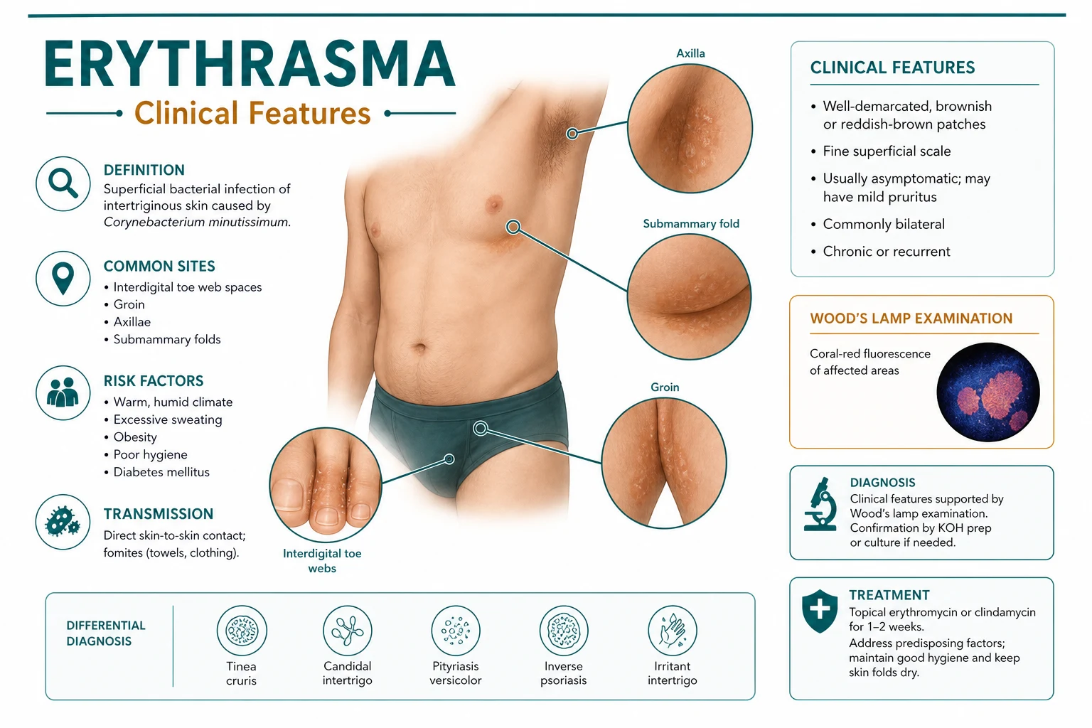
Erythrasma is a chronic, superficial bacterial infection of intertriginous skin caused by Corynebacterium minutissimum (reclassified by some taxonomic schemes as Kocuria or retained within Corynebacterium), presenting as well-demarcated, reddish-brown, finely scaly macules and patches in the groin, axillae, toe webs, submammary folds and gluteal cleft. The pathognomonic diagnostic sign is coral-red fluorescence under Wood's lamp (UV-A 365 nm) produced by coproporphyrin III. NEET-PG/INICET high-yield topics include differentiation from tinea cruris, candidal intertrigo and inverse psoriasis; the fact that imidazole antifungals are effective against this bacterial infection; and oral erythromycin or clarithromycin for extensive disease.
On this page & tools
Your progress
Saved locally on this device.
Practise this topic
Exam tags
Red flags
Overview & Definition
Erythrasma is a chronic, superficial bacterial infection of the intertriginous skin — the warm, moist, occluded skin folds of the groin, axillae, toe webs, submammary folds and gluteal cleft. The causative organism is Corynebacterium minutissimum, a Gram-positive, aerobic, pleomorphic diphtheroid that is a normal commensal of human skin flora but becomes pathogenic under favourable local conditions. In recent taxonomic revisions the organism has been reassigned by some authors to the genus Kocuria, while many dermatology and microbiology texts continue to refer to it as Corynebacterium minutissimum. For examination purposes either name may be accepted, and the clinical syndrome is unchanged. The organism is sometimes called a "diphtheroid" because of its resemblance to the Corynebacterium diphtheriae group on Gram stain, but it does not produce diphtheria toxin and is non-toxigenic. [1]
The disease is one of the most common superficial bacterial infections of the skin, particularly in tropical and humid climates. It is frequently asymptomatic or mildly symptomatic, and patients often present because of the cosmetic appearance or mild pruritus rather than significant discomfort. Despite its benign nature, erythrasma is an important diagnostic entity because it is easily mistaken for tinea cruris, candidal intertrigo or inverse psoriasis, and the pathognomonic coral-red fluorescence under Wood's lamp allows instant bedside diagnosis when the clinical picture is unclear.[1][6]
The condition was first clearly described in the late nineteenth century, and the bacterial cause was established in the early twentieth century. The organism was long known as Corynebacterium minutissimum because of its minute size and diphtheroid morphology. Modern molecular taxonomy has questioned this placement, and some authorities have reclassified the agent as a species of Kocuria, a genus of Gram-positive cocci within the family Micrococcaceae. However, because the clinical literature, board examinations and routine microbiology reports overwhelmingly continue to use the name Corynebacterium minutissimum, a candidate should be prepared to recognise both names and should not be thrown by either term. The key examination fact is that the organism is a Gram-positive diphtheroid that produces coproporphyrin III. [1]
[1]Classification
Erythrasma is classified clinically by anatomical distribution and extent of disease. Understanding the categories matters because the approach to treatment and the differential diagnosis shift with the site. [1]
1. Localised erythrasma A single small patch in one intertriginous site, most commonly the groin or a toe web. These lesions respond reliably to topical therapy over 2–4 weeks. Localised disease is the form most often seen in primary care and outpatient dermatology clinics. [1]
2. Extensive erythrasma Multiple contiguous or bilateral intertriginous sites are involved, such as both groins, the axillae and the submammary folds. Extensive disease often signals an underlying predisposition — most importantly diabetes mellitus, obesity or hyperhidrosis — and usually requires systemic therapy in addition to topical measures. [1]
3. Disseminated erythrasma Widespread patches extend beyond the classic skin folds onto the trunk and proximal limbs. This uncommon pattern is strongly associated with diabetes, immunosuppression and residence in warm humid climates. Systemic macrolide therapy is usually necessary. [1]
4. Toe-web (interdigital) erythrasma The most common anatomical site overall. The lateral 4th interdigital space is classically affected. Toe-web disease often coexists with dermatophyte infection (tinea pedis) and candidal intertrigo, producing maceration, fissuring and malodour. [1]
5. Intertriginous erythrasma A broader category that includes groin, axillae, submammary folds, gluteal cleft and umbilicus. All are characterised by the same warm, moist, occlusive microenvironment that allows C. minutissimum to flourish. [1]
Epidemiology & Risk Factors
Erythrasma is distributed worldwide, but its prevalence rises sharply in hot, humid climates where occlusion and sweating are constant. Up to one in five people may carry or clinically express the infection in tropical regions, and all age groups can be affected, although adults are more commonly symptomatic than children. The higher adult prevalence reflects apocrine sweat activity, greater body mass, occupational exposure to occlusive footwear and the increased prevalence of diabetes and obesity with age.[1]
The major risk factors are best understood as local microenvironmental factors plus systemic predisposition. Warmth, moisture and occlusion raise skin surface pH, encourage maceration of the stratum corneum, and allow C. minutissimum to proliferate. Obesity creates deeper, more occlusive folds; hyperhidrosis supplies continuous moisture; and occlusive clothing traps heat and sweat. Diabetes mellitus predisposes through immunological dysfunction, impaired neutrophil activity and the glucose-rich cutaneous environment that favours bacterial overgrowth. Immunosuppression from HIV, corticosteroids, chemotherapy or biologic agents can produce more extensive or refractory disease. Poor hygiene, tight synthetic clothing and communal bathing are additional environmental contributors. Toe-web erythrasma is particularly common in swimmers, athletes and military personnel who wear occlusive boots or footwear for long periods. [1]
The seasonal pattern is also notable: erythrasma is more common in summer and in tropical monsoon climates, and often improves during cooler, drier months. Occupations that involve prolonged standing, heavy sweating or occlusive uniforms increase risk. A family history is not usually relevant, although shared living conditions and footwear can lead to clustering of toe-web disease in households. The condition is not regarded as highly contagious in the traditional sense, because C. minutissimum is a commensal organism that requires the right local environment to become pathogenic; nevertheless, close contact and shared occlusive environments can facilitate transmission of the organism between individuals who share the same predisposing factors. [1]
Pathophysiology
Corynebacterium minutissimum is a Gram-positive, aerobic, pleomorphic rod (diphtheroid) that is part of the normal cutaneous flora. It is a non-spore-forming, non-motile organism that thrives in the lipid-rich environment of the stratum corneum. Under favourable conditions — warmth, moisture, occlusion, friction and an alkaline pH — it multiplies within the superficial stratum corneum without invading living tissue. This superficial confinement explains why erythrasma is rarely complicated by deeper infection in immunocompetent hosts, and why it leaves no scarring after treatment.[1][9]
The organism produces two clinically important classes of substances: [1]
-
Coproporphyrin III, a water-soluble porphyrin. This compound accumulates in the stratum corneum and emits the pathognomonic coral-red (salmon-pink) fluorescence when illuminated by Wood's lamp (UV-A, 365 nm). The fluorescence is not present in every skin disease that affects the folds, so it is the single most useful bedside diagnostic clue. The colour arises because coproporphyrin III has a characteristic absorption and emission spectrum in the red-orange range under long-wave ultraviolet light. Even after treatment, fluorescence may persist for several days as the porphyrin is cleared from the stratum corneum. The ability to produce porphyrin is not universal among cutaneous corynebacteria, which explains why C. minutissimum causes this distinctive sign and other common diphtheroids do not. [1]
-
Proteolytic and keratolytic enzymes that digest the cornified layer. These enzymes produce the fine, powdery scale and mild maceration seen clinically. Unlike dermatophytes, which invade keratin with hyphae, C. minutissimum remains confined to the surface and produces a more subtle, less inflammatory scale. The organism does not produce true hyphae or pseudohyphae, so potassium hydroxide microscopy is negative in pure erythrasma. [1]
The host inflammatory response is remarkably mild. Invasion of living tissue is not part of the usual disease process, so neutrophilic infiltrates, pustules and ulceration are absent unless a secondary infection supervenes. The superficial confinement and minimal inflammation explain why erythrasma is usually asymptomatic and why it heals without scarring. [1]
The combination of bacterial overgrowth and porphyrin production gives the lesions their characteristic colour and fluorescence. Invasion below the stratum corneum is extremely rare; when it occurs, it is usually in profoundly immunocompromised hosts and can present with bacteraemia, osteomyelitis or visceral infection. Such systemic disease is not "erythrasma" in the usual sense but rather invasive corynebacterial infection caused by the same organism.[5]
Clinical Presentation
Erythrasma is usually a slowly progressive, chronic eruption that patients may have ignored for weeks or months. The lesions are typically asymptomatic or mildly pruritic, and the lack of significant itch is an important clue distinguishing erythrasma from tinea cruris or candidal intertrigo, both of which are usually more symptomatic. In some patients the first clue is a faint brownish discolouration noticed incidentally, while in others mild itching, burning or a feeling of moisture in the fold brings them to medical attention. The lesions do not blister, weep or crust unless there is secondary infection or coexisting dermatitis. [1]
Distribution
The classic sites are all intertriginous: [1]
- Toe webs — the most common site overall, classically the 4th interdigital space. Patients notice maceration, scaling, fissuring and sometimes malodour. The toe web may be the only site of disease, or it may coexist with tinea pedis or candidal intertrigo.
- Groin and crural folds — well-demarcated patches extending from the inguinal crease onto the inner thighs. The border is typically sharp and geometric. Lesions may be bilateral or asymmetrical and can extend onto the scrotum or labia, although mucosal surfaces are spared.
- Axillae — less common than the groin but clinically similar; may be bilateral and may coexist with trichomycosis axillaris, another corynebacterial infection.
- Submammary folds — more common in women with obesity, producing erythematous-brown patches beneath the breasts.
- Gluteal cleft and umbilicus — less common but well described, especially in obese or diabetic individuals. [1]
Disseminated erythrasma produces more widespread patches beyond the classic folds onto the trunk and proximal limbs and is strongly associated with diabetes, immunosuppression and tropical residence. In this rare pattern the lesions retain the same reddish-brown colour and fine scale but are more numerous and less obviously confined to folds. [1]
Morphology
The lesions are macules and patches with the following features: [1]
- Well-demarcated, sharp borders — a hallmark that contrasts with the more diffuse, less geometric edges of intertrigo or eczematous dermatitis.
- Reddish-brown to coppery colour — the brownish hue is characteristic and distinguishes erythrasma from the bright red of candidal intertrigo or the salmon-pink of inverse psoriasis.
- Fine, powdery scale — usually subtle; much less prominent than the active, advancing border of tinea cruris.
- Mild pruritus or no symptoms — often discovered incidentally.
- Long-standing, slowly progressive lesions when untreated. [1]
Differential Diagnosis
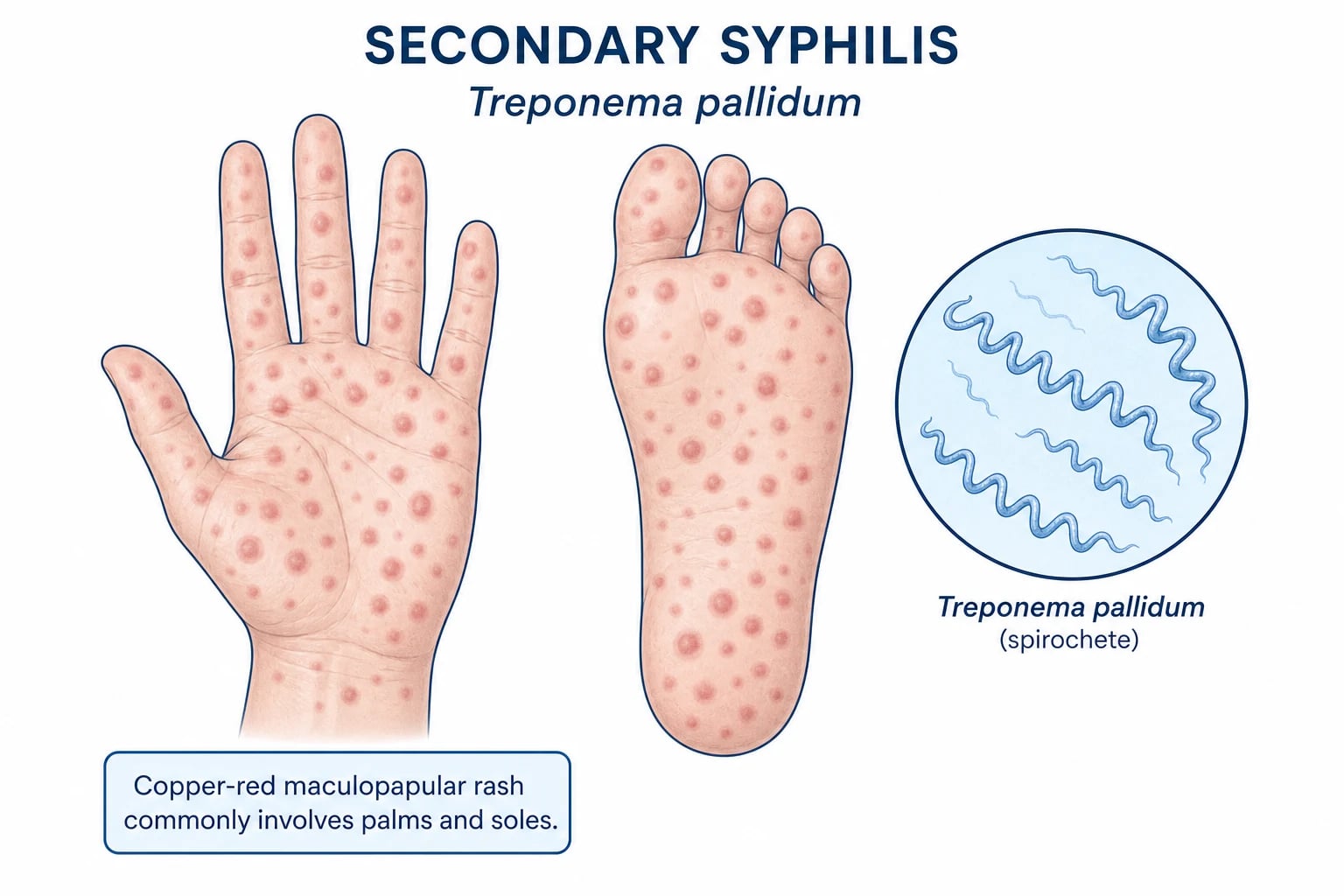
The main diagnostic challenge is distinguishing erythrasma from the other common causes of intertriginous erythema and scale. The Wood's lamp is the single most useful discriminator because the coral-red fluorescence is pathognomonic. A careful history of the onset, duration, associated symptoms and response to prior treatments often points to the correct diagnosis before any investigation is performed. When doubt remains, a skin scraping for potassium hydroxide microscopy is the next step. [1]
Erythrasma
Tinea cruris
Candidal intertrigo
Inverse psoriasis
Irritant intertrigo
Contact dermatitis
Erythrasma-like dermatitis is a term sometimes used for clinically similar eruptions that lack the characteristic fluorescence. These cases may represent an eczematous process, irritant intertrigo or early erythrasma before porphyrin production becomes detectable. If Wood's lamp is negative, the diagnosis should be reconsidered and KOH microscopy performed to exclude dermatophyte or candidal infection. Seborrhoeic dermatitis and acanthosis nigricans can also affect folds but are usually distinguishable by their distribution, scale quality and associated findings. [1]
Clinical & Bedside Assessment
The focused examination in suspected erythrasma should be performed in a warm, darkened room to allow accurate Wood's lamp assessment. The clinician should inspect all major intertriginous sites because disease is often multifocal. [1]
Focused physical examination
- Inspect the toe webs, groin, axillae, submammary folds, gluteal cleft and umbilicus for well-demarcated reddish-brown patches with fine scale.
- Palpate gently to confirm superficial scale and exclude tenderness or deeper induration (which would suggest secondary infection).
- Examine the surrounding skin for clues to alternative diagnoses: satellite pustules (candidiasis), active scaly border (tinea cruris), smooth salmon-pink plaques elsewhere (psoriasis).
- Perform Wood's lamp examination in a darkened room after allowing the patient to adapt to the dark for a minute or two. The lamp should be held close to the skin and the lesions inspected for coral-red fluorescence. [1]
The Wood's lamp sign
The coral-red (salmon-pink) fluorescence of erythrasma is pathognomonic. The fluorescence may be visible as a uniform glow across the patch or as a more intense rim. If the diagnosis is uncertain, KOH microscopy of skin scrapings is performed to exclude fungal infection. In resource-limited settings, Wood's lamp examination alone is usually sufficient to start treatment. [1]
Documentation and patient explanation
When documenting the examination, note the anatomical sites involved, the presence of well-demarcated reddish-brown patches with fine scale, and the Wood's lamp findings. A brief diagram in the notes can be helpful for follow-up. Patients should be told that erythrasma is a superficial bacterial infection, not a fungal infection, and that it is treatable but may recur if the skin folds remain moist. Explaining the Wood's lamp fluorescence as a harmless pigment produced by the bacteria often reassures patients and helps them understand why treatment needs to continue even after the rash looks better. [1]
Investigations
Wood's lamp examination
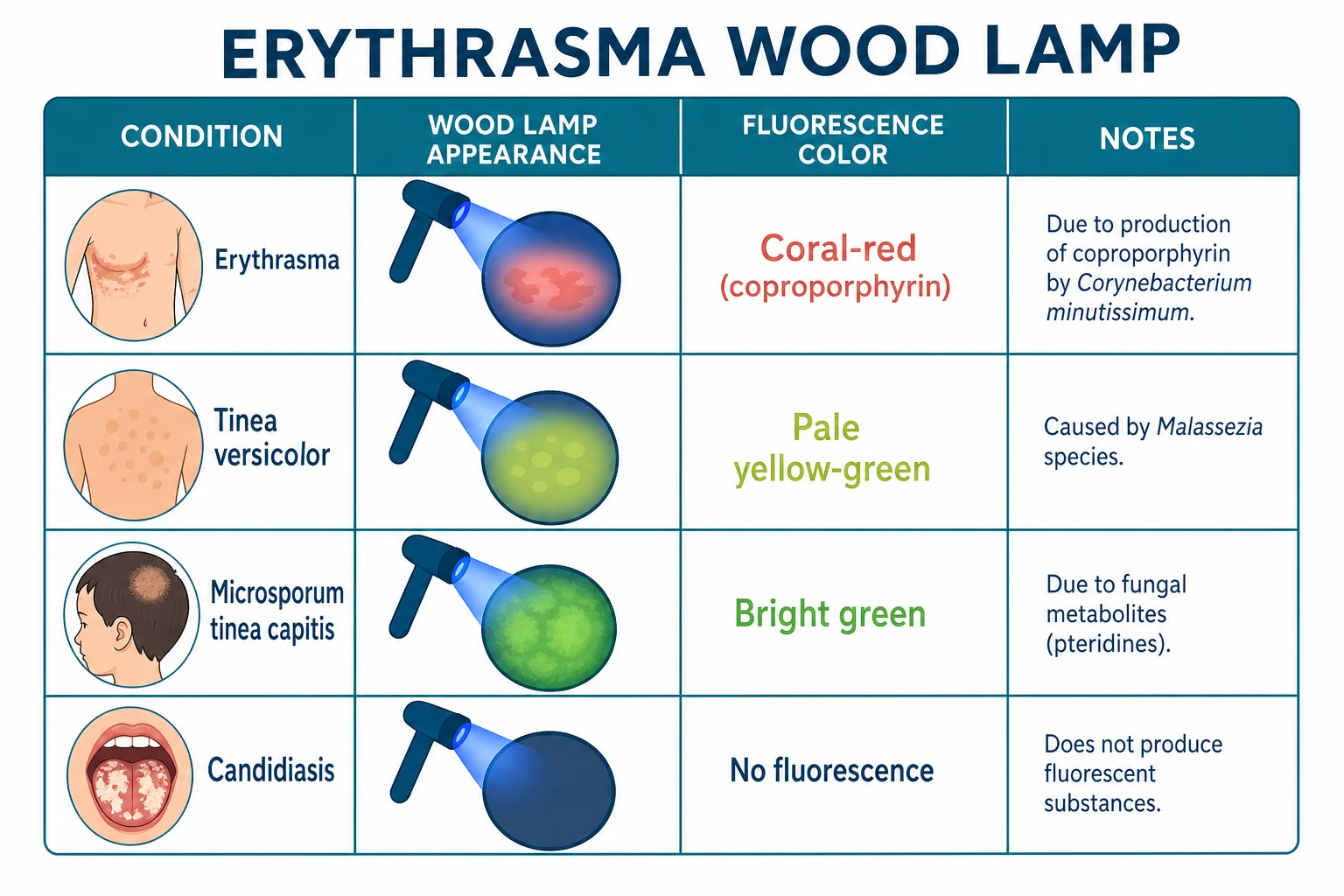
Wood's lamp examination (long-wave ultraviolet-A, 365 nm) is the diagnostic test of choice. The coral-red fluorescence is caused by coproporphyrin III produced by C. minutissimum. The test is rapid, inexpensive, non-invasive and highly specific. False negatives can occur if the room is not sufficiently dark, if the lamp is the wrong wavelength, if the skin is wet, or if the patient has recently washed the area (coproporphyrin is water-soluble).[4][6][7]
The key Wood's lamp differential is worth memorising: [1]
| Condition | Wood's lamp fluorescence | Source of fluorescence |
|---|---|---|
| Erythrasma | Coral-red (salmon-pink) | Coproporphyrin III from C. minutissimum |
| Tinea versicolor | Pale yellow-green / golden | Pteridine from Malassezia species |
| Tinea capitis (Microsporum) | Bright green | Fungal metabolites in infected hairs |
| Tinea capitis (Trichophyton) | No fluorescence | — |
| Pseudomonas infection | Yellow-green | Pyocyanin and pyoverdin |
| Candidal intertrigo | No fluorescence | — |
| Porphyria cutanea tarda | Coral-red urine and blister fluid | Uroporphyrin and coproporphyrin |
Wood's lamp fluorescence colours
COFFEE-GREEN
Erythrasma (coproporphyrin III)
Porphyria cutanea tarda (urine/blister fluid)
Tinea versicolor (pteridine)
Microsporum tinea capitis
Pseudomonas pyocyanin
No green in erythrasma or candidiasis
Remember coral-red = erythrasma
Coral-red = the key exam pearl
False negatives if not dark enough
False-negative Wood's lamp examinations can occur if the room is not sufficiently dark, if the lamp is the wrong wavelength, if the skin is wet or recently washed, or if the patient has applied a topical preparation that obscures fluorescence. The examiner should allow the eyes to adapt to darkness for at least a minute, hold the lamp close to the skin, and inspect the lesions carefully. Some examiners clean the area gently with alcohol before testing to remove surface contaminants and moisture, though this is not always necessary. The fluorescence is best appreciated against a dark background; even faint coral-pink hue is clinically significant. [1]
Microscopy and culture
- Skin scraping + KOH preparation — performed to exclude dermatophyte infection or candidiasis when Wood's lamp findings are equivocal. Erythrasma itself is KOH negative.
- Gram stain — may reveal Gram-positive pleomorphic diphtheroid rods in the stratum corneum. This is rarely needed in routine practice because Wood's lamp is so characteristic.
- Culture — C. minutissimum can be cultured on nutrient agar under aerobic conditions, but it is slow and clinically unnecessary in most cases. Culture is reserved for atypical presentations, treatment failure, or suspected systemic infection.
- Skin biopsy — not routine. Histology shows a mild superficial inflammatory infiltrate with organisms in the stratum corneum. [1]
Screening for underlying disease
In extensive, recurrent, disseminated or treatment-resistant erythrasma, screen for: [1]
- Diabetes mellitus — fasting plasma glucose and HbA1c.
- Obesity — body mass index and waist circumference.
- Immunosuppression — consider HIV testing, medication review, corticosteroid use.
- Hyperhidrosis — clinical history and examination. [1]
These investigations are not required for an isolated toe-web patch in an otherwise healthy adult, but they are essential when the disease is widespread or keeps returning despite adequate therapy. [1]
Management — Resuscitation
Erythrasma is not a medical emergency in its usual superficial form. There is no immediate resuscitation required, and the disease is managed in the outpatient setting. However, the clinician must be alert to two important situations that do require urgent action: [1]
-
Secondary bacterial cellulitis or erysipelas — if the intertriginous area becomes acutely tender, warm, swollen, with spreading erythema, fever or systemic symptoms, this suggests deeper bacterial infection (usually Streptococcus pyogenes or Staphylococcus aureus) rather than uncomplicated erythrasma. Management then becomes urgent: blood cultures, intravenous antibiotics (e.g., flucloxacillin, cephalexin or clindamycin according to local guidelines), and assessment for sepsis. [1]
-
Systemic infection with C. minutissimum — extremely rare, reported in immunocompromised hosts. Presentation may include fever, bacteraemia, osteomyelitis, endocarditis or visceral abscess. Blood cultures, imaging and intravenous antibiotics guided by susceptibility are required, with infectious-disease input. [1]
For the vast majority of patients, the management ladder is elective and outpatient. [1]
Management — Definitive & Stepwise
Treatment is dictated by the extent of disease, the presence of co-infection, and the underlying predisposing conditions. [1]
Step 1: Topical therapy for localised disease
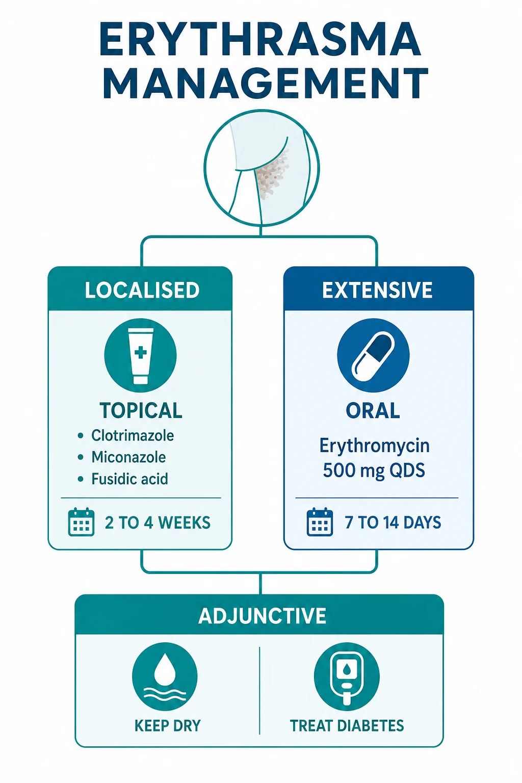
For localised or limited erythrasma, topical therapy is first-line. Treatment should be continued for 2–4 weeks, even if clinical improvement is seen earlier, to reduce recurrence. The patient should be warned that Wood's lamp fluorescence may linger for a few days after the rash has clinically cleared, because coproporphyrin III remains within the shedding stratum corneum. [1]
- Clotrimazole 1% cream applied twice daily for 2–4 weeks. Imidazole antifungals are effective against C. minutissimum despite it being a bacterium, because they interfere with the fungal-type cytoplasmic membrane synthesis pathways that are also present in this organism. This is a favourite examination point. Clotrimazole is inexpensive, widely available, and generally well tolerated. Mild local irritation is uncommon.
- Miconazole 2% cream applied twice daily for 2–4 weeks — a similar imidazole alternative with comparable efficacy and tolerability.
- Fusidic acid 2% cream applied twice daily for 2 weeks — a topical antibacterial with reliable activity against Gram-positive organisms. It is widely used in the United Kingdom and many Commonwealth countries. A small randomised trial in general practice found fusidic acid cream comparable to systemic erythromycin for localised erythrasma, supporting topical-first management.
- Erythromycin 2% topical solution or gel applied twice daily for 2 weeks — directly targets C. minutissimum and is a logical topical choice when available.
- Clindamycin 1% topical solution or gel applied twice daily for 2 weeks — an alternative topical antibiotic, especially useful in regions where macrolide resistance is a concern or where clindamycin is preferred.
- Mupirocin 2% ointment applied twice daily for 1–2 weeks — effective against Gram-positive cocci and coryneforms; useful in toe-web disease or when mixed bacterial infection is suspected. [1]
The choice among these agents should be guided by local availability, cost, patient tolerance and co-infection. If the patient has coexisting tinea pedis or tinea cruris, an imidazole or allylamine antifungal may treat both the fungal infection and the erythrasma, simplifying therapy. [1]
Step 2: Oral antibiotics for extensive, recurrent or disseminated disease
When disease is widespread, multifocal, recurrent or unresponsive to topical agents, systemic therapy is indicated. The macrolides are the traditional agents of choice. Macrolides inhibit bacterial protein synthesis by binding to the 50S ribosomal subunit and are highly active against Gram-positive diphtheroids. [1]
- Erythromycin 500 mg orally four times daily for 7–14 days — the most commonly cited systemic regimen in older and current literature. Erythromycin directly inhibits C. minutissimum protein synthesis and produces rapid clinical clearance. Gastrointestinal side effects are common, including nausea, abdominal cramping and diarrhoea; taking the dose with food can help. Erythromycin is also associated with drug interactions through CYP3A4 inhibition.
- Clarithromycin 500 mg orally twice daily for 7 days — an alternative macrolide with better gastrointestinal tolerability and a simpler twice-daily schedule. Some clinicians prefer clarithromycin over erythromycin for this reason, and it is a reasonable first-line systemic choice where available.
- Tetracycline 500 mg orally four times daily for 14 days — a historical alternative, generally considered less effective than macrolides and less commonly used today because of photosensitivity, gastrointestinal upset and contraindications in pregnancy and children. Doxycycline 100 mg orally twice daily may be used as a tetracycline substitute in adults, but evidence is limited.
- Azithromycin has also been used in some reports, though it is less well established than erythromycin or clarithromycin for erythrasma. A possible regimen is 500 mg on day 1 followed by 250 mg daily for 4 days, but this is not supported by high-quality trial data. [1]
Systemic therapy should be accompanied by the same topical and preventive measures. The combination of oral macrolide plus topical imidazole or antibacterial is reasonable for extensive disease with mixed infection or when rapid clearance is desired. [1]
Step 3: Adjunctive and preventive measures
Relapse is common if the local environment is not corrected. Every patient should receive advice on: [1]
- Keep folds dry — use absorbent cotton clothing, change sweaty clothes promptly, and dry skin folds thoroughly after bathing.
- Drying powders — zinc oxide, talc or antifungal-drying powders reduce moisture and friction in toe webs and groin folds.
- Weight reduction — reduces depth and occlusion of skin folds.
- Diabetes control — optimising glycaemic control reduces recurrence risk and allows faster clearance.
- Treat hyperhidrosis — aluminium chloride 20% antiperspirant, iontophoresis, or botulinum toxin for severe cases.
- Treat co-infections — check for and treat concomitant tinea pedis, tinea cruris or candidal intertrigo, which often coexist and can mimic or perpetuate erythrasma.
- Antibacterial washes — chlorhexidine 4% or benzoyl peroxide washes may reduce cutaneous Corynebacterium colonisation and help prevent relapse.
- Avoid occlusive clothing and footwear — breathable cotton underwear and shoes that allow ventilation reduce the moist microenvironment. [1]
Escalation and follow-up
- Escalate to oral therapy if lesions are extensive, multifocal, recurrent, or not improved after 4 weeks of appropriate topical therapy.
- Reassess the diagnosis if Wood's lamp was not performed or if there is no response to two adequate courses; repeat KOH microscopy and consider biopsy.
- Screen for diabetes and obesity in recurrent or extensive cases.
- Refer to dermatology if the diagnosis is uncertain, if there is treatment failure, or if there is coexisting extensive dermatophyte infection requiring oral antifungal therapy. [1]
Specific Subtypes & Scenarios
Toe-web erythrasma
This is the most common clinical presentation. The lateral 4th interdigital space is the classic site, but any toe web can be involved. The web space shows white maceration, fissuring and fine scale. Patients may complain of malodour, itching or burning, especially after exercise or wearing occlusive footwear. Toe-web erythrasma commonly coexists with tinea pedis and candidal intertrigo; management should therefore include treatment of all identified co-infections. Keeping the toe webs dry is essential: advise patients to dry thoroughly between the toes, use cotton or wool socks, rotate shoes to allow drying, and consider antifungal or drying powders. [1]
Erythrasma in diabetes mellitus
Diabetic patients are prone to more extensive, recurrent and treatment-resistant erythrasma. Hyperglycaemia, immune dysfunction and obesity create a permissive environment. In a diabetic patient with extensive erythrasma, the approach should include oral macrolide therapy, rigorous glycaemic control, weight management and screening for fungal co-infection. Recurrence is the rule unless the diabetes is well controlled. [1]
Erythrasma in pregnancy
Pregnancy increases skin fold moisture and occlusion, and the physiological changes can predispose to erythrasma. Topical agents are preferred because systemic absorption is low. Clotrimazole, miconazole, fusidic acid and erythromycin topical are generally considered safe in pregnancy. Oral macrolides such as erythromycin or clarithromycin are generally considered safe in pregnancy if needed, but topical therapy should be exhausted first. Tetracycline is contraindicated in pregnancy and breastfeeding because of effects on fetal bone and teeth. As always, check local formulary guidance. [1]
Erythrasma in immunocompromised hosts
HIV infection, immunosuppressive therapy, corticosteroids and chemotherapy can all predispose to more extensive, atypical or persistent disease. In these patients, be alert to systemic infection (rare) and to coexisting opportunistic infections such as candidiasis, dermatophytosis or bacterial superinfection. Culture may be more useful in this population. [1]
Disseminated erythrasma
Rarely, patients develop widespread patches beyond the classic folds. This pattern is strongly associated with diabetes, immunosuppression and tropical residence. Oral macrolides are required, and the underlying predisposition must be addressed aggressively. [1]
Complications & Pitfalls
The main complications of erythrasma are diagnostic and recurrent rather than immediately dangerous. Because the lesions are asymptomatic or mildly symptomatic, patients may not seek treatment, allowing the infection to persist for months. Chronic colonisation can spread to multiple intertriginous sites, and the altered stratum corneum can become a portal for other organisms. The psychological impact of a chronic, discoloured rash in the groin or axillae should not be underestimated; many patients experience embarrassment, social avoidance and reduced quality of life. [1]
Diagnostic pitfalls
- Misdiagnosis as tinea cruris — leading to unnecessary oral antifungal therapy (e.g., terbinafine) while the bacterial infection persists. The key is to perform Wood's lamp examination before starting empiric antifungal therapy in any intertriginous rash.
- Misdiagnosis as candidal intertrigo — leading to prolonged azole therapy without addressing the bacterial component. Candidiasis has satellite pustules and bright-red moist erythema; erythrasma has fine scale and coral-red fluorescence.
- Missing extensive or disseminated disease — if only the presenting patch is examined, other intertriginous sites may be missed. Always examine the toe webs, groin, axillae and submammary folds.
- Attributing all disease to erythrasma when co-infection exists — toe-web disease often has concurrent dermatophyte or candidal infection. If erythrasma improves but maceration persists, reconsider co-infection. [1]
Complications of untreated or recurrent disease
- Secondary bacterial infection — macerated skin folds can become superinfected with Staphylococcus aureus or Streptococcus pyogenes, producing cellulitis, erysipelas or lymphangitis. This requires systemic antibacterial therapy.
- Chronicity and cosmetic concern — lesions may persist for months or years, causing embarrassment and affecting quality of life.
- Fissuring and discomfort — especially in toe webs, where fissures can be painful and predispose to bacterial entry.
- Rare systemic infection — bacteraemia, endocarditis, osteomyelitis or visceral infection in severely immunocompromised hosts. Blood cultures and imaging should be obtained if systemic infection is suspected.[5]
Prognosis & Disposition
The prognosis of uncomplicated erythrasma is excellent. With appropriate topical or systemic therapy, clinical improvement is usually evident within 1–2 weeks, and clearance is often achieved by 2–4 weeks. No scarring occurs because the infection is confined to the superficial stratum corneum. Wood's lamp fluorescence may persist for a short period after clinical clearance because residual coproporphyrin takes time to shed; this does not indicate treatment failure. [1]
The major determinant of recurrence is the local environment. Patients who continue to experience warmth, moisture and occlusion in skin folds will often relapse. Addressing obesity, hyperhidrosis, occlusive clothing and diabetes is therefore as important as prescribing antimicrobial therapy. Even with optimal treatment, recurrence is common when these factors are not modified, and patients should be counselled about this at the outset. Long-term preventive strategies, including regular drying, breathable clothing and weight management, are often more important than the initial antibiotic choice. Patient education should be documented at each visit. [1]
Disposition is almost always outpatient. Patients can be managed in primary care or dermatology outpatient clinics. Referral to dermatology is appropriate when the diagnosis is uncertain, the disease is extensive or recurrent, there is coexisting fungal infection requiring systemic therapy, or the patient fails two adequate treatment courses. Infectious-disease referral is reserved for the rare systemic infection or for immunocompromised patients with atypical features. [1]
Special Populations
Children
Erythrasma can occur in children, particularly in the groin and toe webs, but it is less common than in adults. Children may also have atopic dermatitis, irritant napkin dermatitis or candidal napkin dermatitis that mimics erythrasma. Wood's lamp examination is safe and helpful, and children often find the glowing light interesting rather than frightening. Topical therapy is preferred; oral macrolides are reserved for extensive disease. Weight-based dosing is required if oral macrolides are used: erythromycin 30–50 mg/kg/day divided into 2–4 doses, or clarithromycin 15 mg/kg/day divided twice daily, for 7–14 days. Tetracycline is contraindicated in children under 8 years of age because of permanent dental staining and effects on bone growth. Parents should be counselled about hygiene, drying, and appropriate footwear, especially for children who swim or play sports. [1]
Pregnancy and breastfeeding
Topical therapy with imidazoles or topical antibacterials is first-line in pregnancy and breastfeeding because systemic absorption is minimal. Clotrimazole, miconazole, fusidic acid and topical erythromycin are generally considered safe in pregnancy. Systemic erythromycin and clarithromycin are generally considered acceptable in pregnancy if topical therapy fails, but topical therapy should be exhausted first. Tetracycline is contraindicated in pregnancy and breastfeeding because of effects on fetal bone and teeth. When breastfeeding, topical agents are preferred because of minimal systemic absorption; if oral antibiotics are necessary, erythromycin is generally compatible with breastfeeding, but observe the infant for gastrointestinal upset or rash. Lactating women with submammary erythrasma should keep the fold dry and use breast pads that wick moisture away from the skin. [1]
Elderly
Older adults have more skin folds, reduced mobility, higher rates of diabetes and obesity, and may have difficulty drying skin folds. They are therefore more prone to extensive and recurrent erythrasma. Incontinence, limited mobility and institutional living can all contribute. Management should be practical: ensure patients can reach and dry affected areas, review footwear and incontinence, and screen for diabetes. Carers should be taught to dry skin folds during personal care. Topical therapy is usually adequate; oral macrolides are used for extensive disease with attention to drug interactions and cardiac risks. [1]
Immunocompromised patients
As discussed, immunocompromised patients may have more extensive, persistent or atypical disease. HIV infection, immunosuppressive therapy, corticosteroids and chemotherapy can all predispose to erythrasma and can alter the clinical appearance. Culture is more useful in this population, and the threshold for systemic therapy is lower. Always consider co-infection and the rare possibility of systemic C. minutissimum infection. In transplant recipients, drug interactions between macrolides and calcineurin inhibitors or mammalian target of rapamycin inhibitors must be considered; in such cases, topical therapy or a non-macrolide alternative may be preferred with specialist input. [1]
Evidence, Guidelines & Regional Differences
A 2025 systematic review of interventions for erythrasma concluded that the evidence base is limited but supports the use of topical imidazoles, topical antibacterials, and oral macrolides. No single regimen was clearly superior for all patients, and treatment choice should be guided by extent, site, patient preference and local availability. The review emphasised that the diagnosis is usually made clinically with Wood's lamp, and that many cases can be managed without microscopy or culture. The authors also noted that the quality of randomised controlled trial evidence is low, with many studies being small, old and methodologically heterogeneous, so current recommendations rely heavily on clinical tradition and the pharmacological spectrum of the agents.[2]
Older reviews (Holdiness 2002) established erythromycin — both topical and oral — as the historical gold standard, and noted that imidazole antifungals were also effective because of their activity against this diphtheroid organism. The Scandinavian primary-care trial by Hamann and Thorn (1991) found that topical fusidic acid cream was comparable to systemic erythromycin for localised disease, supporting topical-first management when feasible. A more recent narrative review (Forouzan and Cohen 2020) summarised the differential diagnosis and management comprehensively for clinicians, while case reports and clinical images repeatedly underscore the diagnostic value of the coral-red Wood's lamp fluorescence.[1][3][4][8]
Regional treatment preferences
- India and LMIC settings — topical clotrimazole and miconazole are inexpensive, widely available and effective; oral erythromycin is commonly used when topical therapy fails or disease is extensive.
- United Kingdom — the British National Formulary lists topical fusidic acid, mupirocin and topical erythromycin as options; oral erythromycin or clarithromycin are reserved for widespread disease.
- United States — topical clindamycin solution and erythromycin are available; oral clarithromycin is often preferred over erythromycin for better tolerability.
- Australia — topical agents are first-line; oral macrolides for extensive disease. Drying powders and hygiene advice are emphasised in the hot, humid climate. [1]
Controversies
- The exact taxonomic classification of the organism remains debated; some authorities classify it as Kocuria, while clinical texts continue to use Corynebacterium. Examiners usually accept either name.
- Optimal duration of therapy is not well defined by randomised trials; 2–4 weeks of topical therapy and 7–14 days of oral macrolides are widely used.
- Whether to screen all patients for diabetes is debated; most authorities recommend screening only in extensive, recurrent or treatment-resistant cases, not for an isolated toe-web patch in a healthy young adult. [1]
Exam Pearls
The following points represent the high-yield knowledge that examiners consistently test on erythrasma. They should be memorised and practised in viva and MCQ formats. [1]
- The causative organism is Corynebacterium minutissimum, a Gram-positive diphtheroid; some taxonomic schemes place it in the genus Kocuria.
- The pathognomonic bedside sign is coral-red (salmon-pink) fluorescence under Wood's lamp (365 nm), caused by coproporphyrin III.
- Clinical lesions are well-demarcated, reddish-brown, finely scaling macules and patches in intertriginous sites.
- The 4th interdigital toe web is the most common single site.
- Imidazole antifungals (clotrimazole, miconazole) are effective against erythrasma despite it being a bacterial infection — a classic exam trick.
- First-line topical therapy includes clotrimazole, miconazole, fusidic acid, erythromycin, clindamycin and mupirocin, used for 2–4 weeks.
- Oral therapy for extensive disease is a macrolide: erythromycin 500 mg four times daily for 7–14 days, or clarithromycin 500 mg twice daily for 7 days.
- Tetracycline is contraindicated in pregnancy and children under 8 years.
- Recurrence is common unless predisposing factors are addressed; screen for diabetes in extensive, recurrent or resistant cases.
- The key differentials are tinea cruris (Wood's lamp negative, KOH positive), candidal intertrigo (satellite pustules, no fluorescence, KOH positive) and inverse psoriasis (salmon-pink smooth plaques, no fluorescence). [1]
Exam application bank (NEET-PG / INICET)
One-line answer
Erythrasma is a chronic, superficial bacterial infection of intertriginous skin caused by Corynebacterium minutissimum (reclassified by some taxonomic schemes as Kocuria or retained within Corynebacterium), presenting as well-demarcated, reddish-brown, finely scaly macules and patches in the groin, axillae, toe webs, submammary folds and gluteal cleft. The pathognomonic diagnostic sign is coral-red fluorescence under Wood's lamp (UV-A 365 nm) produced by coproporphyrin III. NEET-PG/INICET high-yield topics include differentiation from tinea cruris, candidal intertrigo and inverse psoriasis; the fact that imidazole antifungals are effective against this bacterial infection; and oral erythromycin or clarithromycin for extensive disease.
Worked stems (answer without another resource)
Stem 1 — Classic presentation. Map symptoms to mechanism; name the first investigation and first treatment step with dose/route if drug therapy is standard. [1]
Stem 2 — Unstable / complicated. List red flags that force immediate resuscitation, theatre, ICU, antidote, or reperfusion — and what you do in the first 15 minutes. [1]
Stem 3 — Atypical group. Elderly, pregnancy, child, or immunocompromised: how presentation and thresholds change. [1]
Stem 4 — Differential trap. Name the three closest mimics and one discriminator for each. [1]
Stem 5 — Disposition. Who goes home with safety-netting, who is admitted, who needs HDU/ICU/theatre, and what follow-up is mandatory. [1]
Rapid viva checklist
- Definition + classification
- Pathophysiology chain
- Bedside signs / criteria
- Score with exact components (if any)
- Emergency bundle
- Definitive therapy with doses
- Complications of disease and of treatment
- Special populations
- Guideline/trial name if classic
- Three exam traps
Coverage self-check
If you cannot answer any stem above from this page alone, re-read the matching section — the page is intended to be self-sufficient for final-prof and NEET-PG/INICET questions on Erythrasma.
[1]References
- [1]Forouzan P, Cohen PR. Erythrasma Revisited: Diagnosis, Differential Diagnoses, and Comprehensive Review of Treatment Cureus, 2020.PMID 33145138
- [2]Radhakrishnan S, Logamoorthy R, Karthikeyan K, et al. Erythrasma: a systematic review of interventions Clin Exp Dermatol, 2025.PMID 40635638
- [3]Holdiness MR. Management of cutaneous erythrasma Drugs, 2002.PMID 12010076
- [4]Blasco-Morente G, Arias-Santiago S, Pérez-López I, et al. Coral-Red Fluorescence of Erythrasma Plaque Sultan Qaboos Univ Med J, 2016.PMID 27606125
- [5]Sebaratnam DF, Lee S. <em>Corynebacterium minutissimum</em> infection: erythrasma Med J Aust, 2017.PMID 28987127
- [6]Robles-Silva C, Hidalgo L, Vera-Kellet C, et al. Erythrasma and the role of Wood's light: A simple diagnosis pearl Med Clin (Barc), 2023.PMID 37517932
- [7]Garcia-Souto F, et al. Visual Dermatology: Erythrasma Fluorescence Under Wood's Lamp J Cutan Med Surg, 2020.PMID 31994922
- [8]Hamann K, Thorn P. Systemic or local treatment of erythrasma? A comparison between erythromycin tablets and Fucidin cream in general practice Scand J Prim Health Care, 1991.PMID 2041927
- [9]Sarkany I, Taplin D, Blank H. The etiology and treatment of erythrasma J Invest Dermatol, 1961.PMID 14497222