Dermatology · Medicine
Excision margins and elliptical excision
Also known as Elliptical excision · Fusiform excision · Surgical margins · Wide local excision · WLE
Elliptical (fusiform) excision removes a lesion with a planned clinical margin and closes primarily along relaxed skin tension lines, classically with an approximate 3:1 length-to-width ratio and layered closure. Margin width is disease-specific: melanoma follows Breslow thickness; low-risk BCC is often taught at about 4 mm; SCC uses risk-stratified 4–6 mm or wider/Mohs pathways. Clinical margin is not identical to histologic clearance under bread-loaf sectioning.
On this page & tools
Your progress
Saved locally on this device.
Exam tags
Red flags
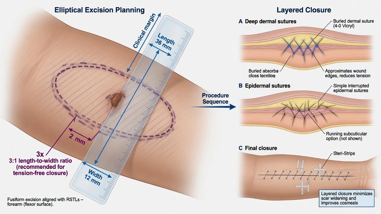
Overview and Definition
Elliptical (fusiform) excision is the workhorse of office dermatologic surgery: the lesion is removed as a full-thickness skin specimen with a planned clinical margin, and the wound is closed primarily as a linear scar. The design aims to remove disease completely while distributing tension so the scar lies in or parallel to relaxed skin tension lines (RSTLs) and dog-ear (standing cone) deformity is minimised.[8]
Two different “margins” must never be confused:
- Clinical surgical margin — the measured distance on living skin from the visible (or dermoscopically mapped) edge to the incision.
- Histologic margin — whether tumour reaches the inked peripheral or deep edge on pathology sections.[7]
Standard vertical “bread-loaf” sectioning examines representative slices, not 100% of the margin — unlike Mohs micrographic surgery, which examines the entire peripheral and deep margin in staged horizontal sections. [1]
Classification of Excisions
| Pattern | Intent | Typical use |
|---|---|---|
| Diagnostic ellipse / narrow excision | Full-thickness histology | Suspected melanoma, uncertain tumours |
| Therapeutic ellipse with defined clinical margin | Cure + reconstruction in one stage | Low-risk NMSC, many trunk/extremity tumours |
| Wide local excision (WLE) | Melanoma clearance by Breslow-based width | After diagnostic biopsy of melanoma |
| Mohs / margin-controlled surgery | 100% margin examination, tissue sparing | High-risk facial NMSC, selected recurrent tumours |
Geometry and Pathophysiology of Closure
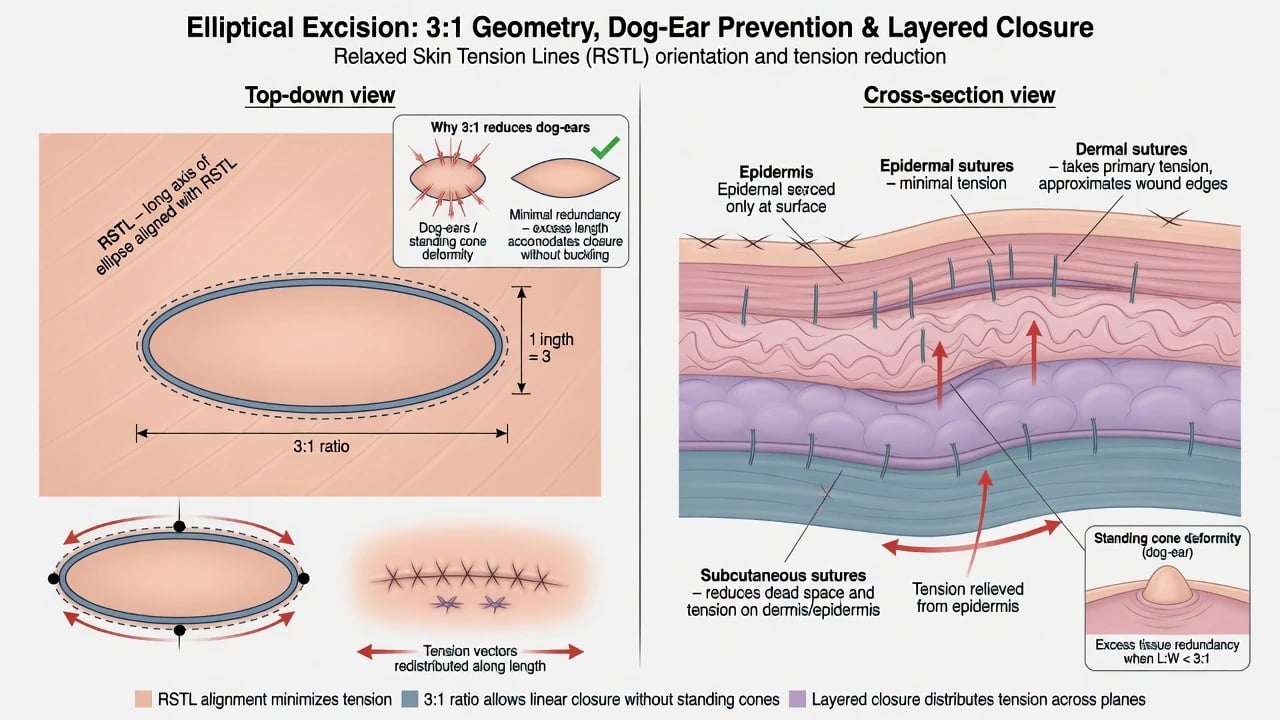
A short, wide defect forces excess tissue into conical standing cones at the apices. Lengthening the ellipse to roughly three times the width redistributes that excess so apices close flat — the classic 3:1 teaching ratio (site and elasticity modify the ideal).[8] Small residual dog-ears may regress with time depending on location.[9]
Layered closure places absorbable sutures in dermis/subcutis to approximate the deep dead space and offload the epidermis; fine epidermal sutures or subcuticular techniques finish alignment. Undermining is used judiciously to mobilise edges without devascularising tips. [1]
Epidemiology and Clinical Context
Most elective cutaneous ellipses are performed for keratinocyte cancers, atypical pigmented lesions, and melanoma WLE after biopsy. Facial free margins (eyelid, lip, alar rim, helical rim) convert a “simple ellipse” into a functional reconstructive problem — examiners expect early recognition and referral rather than heroic linear closure under tension. [1]
Preoperative Assessment
- Diagnosis and thickness/stage data — especially Breslow for melanoma planning.[1][10]
- Site, phototype, keloid history, smoking, diabetes, anticoagulation.
- Mark the lesion, planned clinical margin, and RSTL-aligned ellipse with the patient upright when gravity matters (face/neck).
- Consent: scar length (often longer than the lesion), numbness, infection, bleeding, recurrence, possible re-excision, and reconstructive alternatives.
Recommended Clinical Margins
Melanoma (thickness-based clinical margins)
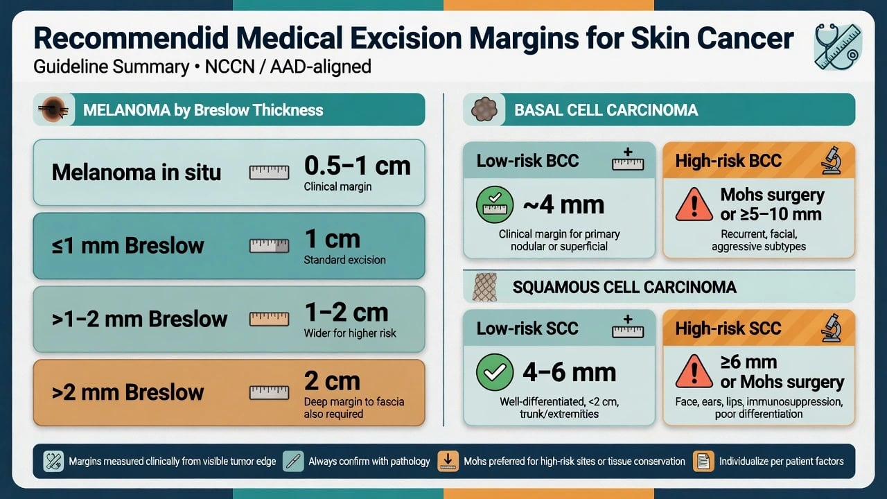
AAD guidelines of care and the trial lineage that informed modern NCCN-style tables teach width by Breslow thickness after adequate microstaging:[1][2][10]
| Breslow thickness | Typical clinical margin teaching |
|---|---|
| Melanoma in situ | 0.5–1 cm |
| ≤1.0 mm | 1 cm |
| >1.0–2.0 mm | 1–2 cm |
| >2.0 mm | 2 cm |
Depth includes subcutaneous tissue appropriate to site; anatomic constraints (digit, face) may require specialised surgical oncology planning. Meta-analytic work continues to refine how much width is enough, but board answers still start from the thickness ladder.[2]
Basal cell carcinoma
For many well-defined, primary, low-risk BCCs, a clinical margin in the order of 4 mm is the classic teaching figure associated with high rates of histologic clearance in standard excision series.[5][3] High-risk features (H-zone face, recurrent disease, poorly defined borders, aggressive histology such as infiltrative/morpheaform, large size, immunosuppression) push the pathway toward Mohs or wider excision and careful reconstruction — not a minimal ellipse alone.[3]
Squamous cell carcinoma
Low-risk invasive SCC is commonly planned with about 4–6 mm clinical margins; high-risk SCC (face/ear/lip, poor differentiation, deep invasion, perineural risk, recurrence, immunosuppression) needs ≥6 mm clinical margins or preferably Mohs/specialist care, with attention that depth matters as much as peripheral width.[4][6]
Differential: Ellipse vs Alternatives
Standard ellipse
- One-stage removal + linear scar
- Best when closure is simple
- Bread-loaf margin sampling
- Ideal trunk/extremity low-risk tumours
Mohs surgery
- 100% margin control
- Tissue sparing on face
- Staged same-day clearance
- Preferred high-risk NMSC
Destructive C&E/cryo
- No orientated margins
- Low-risk selected only
- Faster office destruction
- Never for melanoma
Technique — Stepwise Elliptical Excision
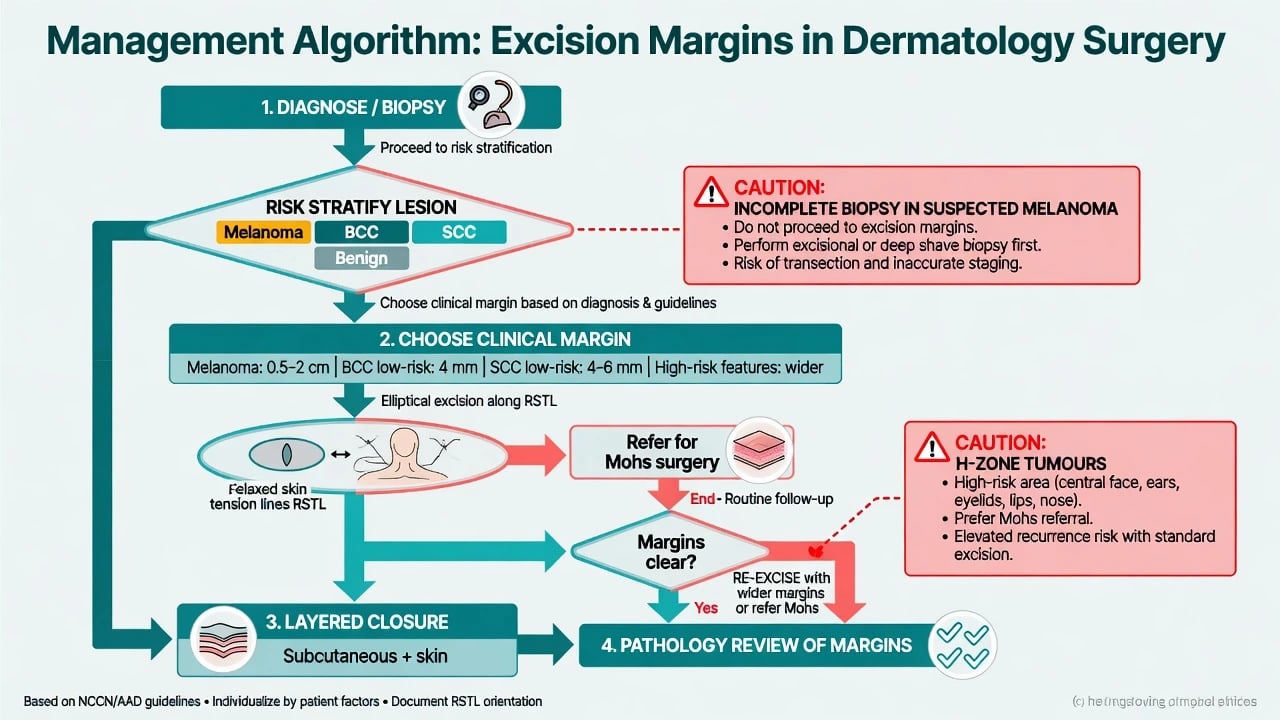
- Mark lesion border, clinical margin, and fusiform tips along RSTLs.
- Anaesthetise (field or nerve blocks as indicated); prepare sterile field.
- Incise vertically to the planned deep plane; avoid beveling that falsely narrows the deep face unless intentionally designing a Mohs bevel.
- Remove specimen; orient with suture/ink for pathology when needed.
- Haemostasis — precise; avoid charred edges.
- Undermine only as needed; deep dermal sutures then epidermal alignment.
- Dress, counsel activity restrictions, and schedule suture removal by site (face often 5–7 days; trunk/extremities longer). [1]
If linear closure would distort a free margin or require extreme tension, redesign with flap, graft, or delayed reconstruction rather than forcing a short ellipse. [1]
Pathology Correlation and Positive Margins
Vertical sectioning can miss focal peripheral positivity between bread-loaf slices.[7] Report reading must specify peripheral vs deep involvement and tumour type. Management of a positive margin is context-dependent: re-excision or Mohs for residual NMSC; multidisciplinary planning for melanoma; observation only in highly selected low-risk scenarios with informed consent — not as a default after incomplete high-risk clearance.
Complications
Special Populations and Sites
- Face: prioritise RSTLs, aesthetic subunits, and Mohs when risk is high.[3]
- Acral/digit: functional margins and referral thresholds.
- Anticoagulated elderly: continue most anticoagulants for low-risk cutaneous surgery after individualised risk discussion; meticulous haemostasis.
- Skin of colour: scar and dyspigmentation counselling; keloid-prone sites need tension-minimising design.
Prognosis and Follow-Up
Complete excision of low-risk NMSC with clear margins carries excellent local control; melanoma follow-up is stage-based. Patients need a written plan for suture care, infection signs, and pathology result review — do not assume “no news is clear margins.” [1]
Evidence, Guidelines, and Regional Practice
AAD melanoma guidelines and the historical trial base underpin Breslow-linked WLE widths.[1][10] Systematic reviews explore margin adequacy and outcomes.[2][5] European interdisciplinary guidelines structure BCC and SCC risk and surgical priority.[3][4] UK SCC excision commentary emphasises adequate depth as well as peripheral width.[6] Classic surgical geometry papers still teach dog-ear prevention and natural regression patterns.[8][9]
Exam Pearls
- 3:1 fusiform design along RSTLs; layered closure.
- Melanoma: MIS 0.5–1 cm; ≤1 mm → 1 cm; 1–2 mm → 1–2 cm; >2 mm → 2 cm.[1]
- Low-risk BCC ~4 mm; high-risk → Mohs/wider.[3][5]
- Low-risk SCC 4–6 mm; high-risk ≥6 mm or Mohs.[4]
- Clinical margin ≠ 100% histologic guarantee with bread-loafing.[7]
Red Flags
Exam application bank (NEET-PG / INICET)
One-line answer
Elliptical (fusiform) excision removes a lesion with a planned clinical margin and closes primarily along relaxed skin tension lines, classically with an approximate 3:1 length-to-width ratio and layered closure. Margin width is disease-specific: melanoma follows Breslow thickness; low-risk BCC is often taught at about 4 mm; SCC uses risk-stratified 4–6 mm or wider/Mohs pathways. Clinical margin is not identical to histologic clearance under bread-loaf sectioning.
Worked stems (answer without another resource)
Stem 1 — Classic presentation. Map symptoms to mechanism; name the first investigation and first treatment step with dose/route if drug therapy is standard. [1]
Stem 2 — Unstable / complicated. List red flags that force immediate resuscitation, theatre, ICU, antidote, or reperfusion — and what you do in the first 15 minutes. [1]
Stem 3 — Atypical group. Elderly, pregnancy, child, or immunocompromised: how presentation and thresholds change. [1]
Stem 4 — Differential trap. Name the three closest mimics and one discriminator for each. [1]
Stem 5 — Disposition. Who goes home with safety-netting, who is admitted, who needs HDU/ICU/theatre, and what follow-up is mandatory. [1]
Rapid viva checklist
- Definition + classification
- Pathophysiology chain
- Bedside signs / criteria
- Score with exact components (if any)
- Emergency bundle
- Definitive therapy with doses
- Complications of disease and of treatment
- Special populations
- Guideline/trial name if classic
- Three exam traps
Coverage self-check
If you cannot answer any stem above from this page alone, re-read the matching section — the page is intended to be self-sufficient for final-prof and NEET-PG/INICET questions on Excision margins and elliptical excision.
Expanded exam teaching (depth pass)
Clinical reasoning
For Excision margins and elliptical excision, examiners test whether you can prioritise life threats, choose the right first test, and give specific therapy (agent, dose, route, timing). Generic phrases without numbers score poorly.
Mechanism → feature map
Build a short chain: cause → pathophysiologic intermediate → clinical feature → complication. Every major symptom in the classic vignette should sit on that chain.
Investigation strategy
- Bedside/first-line tests that change immediate management
- Confirmatory or staging tests
- What a normal result does not exclude
- When not to delay treatment for imaging (unstable patient)
Management ladder
- Resuscitation / ABC / sepsis or haemorrhage bundle as relevant
- Specific antidote / procedure / antimicrobial / reperfusion / surgery
- Supportive care and monitoring targets
- Definitive long-term therapy and secondary prevention
- Disposition and safety-net advice
Special populations
Always prepare one line each for children, pregnancy, elderly, renal/hepatic impairment, and immunocompromised patients when the topic allows.
Pitfalls that fail candidates
- Treating the number not the patient
- Missing pregnancy status when relevant
- Imaging before stabilisation
- Wrong empiric cover or wrong antidote timing
- Incomplete counselling on recurrence, adherence, or red-flag return
Elliptical (fusiform) excision removes a lesion with a planned clinical margin and closes primarily along relaxed skin tension lines, classically with an approximate 3:1 length-to-width ratio and layered closure. Margin width is disease-specific: melanoma follows Breslow thickness; low-risk BCC is often taught at about 4 mm; SCC uses risk-stratified 4–6 mm or wider/Mohs pathways. Clinical margin is not identical to histologic clearance under bread-loaf sectioning. [1]
Structured revision sheet
Must-know numbers and names
List every score, size threshold, dose, and time window from this topic on a blank page from memory, then check against the sections above.
Three classic MCQ angles
- Most likely diagnosis given a vignette
- Next best step in management
- Most appropriate investigation
Three classic SAQ angles
- Pathophysiology in five steps
- Management algorithm with doses
- Complications and prevention
Clinical station flow
Greet → focused history → targeted exam → investigations → explain diagnosis → emergency care → definitive plan → safety-net / follow-up → answer examiner questions on mechanism and pitfalls.
[1]- Minimal ellipse for infiltrative facial BCC.
- Ignoring a positive deep margin report.
- Linear closure that ectropions an eyelid or elevates a lip.
- Destructive treatment substituted for margin-controlled surgery in high-risk disease. [1]
Exam anchors
References
- [1]Swetter SM, Tsao H, Bichakjian CK, et al. Guidelines of care for the management of primary cutaneous melanoma J Am Acad Dermatol, 2019.PMID 30392755
- [2]Hanna S, Lo SN, Saw RP. Surgical excision margins in primary cutaneous melanoma: A systematic review and meta-analysis Eur J Surg Oncol, 2021.PMID 33722422
- [3]Peris K, Fargnoli MC, Garbe C, et al. Diagnosis and treatment of basal cell carcinoma: European consensus-based interdisciplinary guidelines Eur J Cancer, 2019.PMID 31288208
- [4]Stratigos A, Garbe C, Lebbe C, et al. Diagnosis and treatment of invasive squamous cell carcinoma of the skin: European consensus-based interdisciplinary guideline Eur J Cancer, 2015.PMID 26219687
- [5]Quazi SJ, Aslam N, Saleem H, et al. Surgical Margin of Excision in Basal Cell Carcinoma: A Systematic Review of Literature Cureus, 2020.PMID 32821563
- [6]Khan AA, Potter M, Cubitt JJ, et al. Guidelines for the excision of cutaneous squamous cell cancers in the United Kingdom: the best cut is the deepest J Plast Reconstr Aesthet Surg, 2013.PMID 23352886
- [7]Weinstein MC, Brodell RT, Bordeaux J, et al. The art and science of surgical margins for the dermatopathologist Am J Dermatopathol, 2012.PMID 23000878
- [8]Hudson-Peacock MJ, Lawrence CM. Comparison of wound closure by means of dog ear repair and elliptical excision J Am Acad Dermatol, 1995.PMID 7896954
- [9]Jennings TA, Keane JC, Varma R, et al. Observation of Dog-Ear Regression by Anatomical Location Dermatol Surg, 2017.PMID 28930788
- [10]Sharib J, Slingluff CL Jr, Beasley GM. Melanoma trials that defined surgical management: Overview of trials that established NCCN margin guidelines J Surg Oncol, 2022.PMID 34897715