Dermatology · Medicine
Folliculitis decalvans
Also known as Folliculitis decalvans (FD) · Quinquaud disease · Quinquaud folliculitis decalvans · Tufted folliculitis · Folliculitis decalvans capillitii
Folliculitis decalvans (Quinquaud disease) is a chronic, relapsing, PRIMARY NEUTROPHILIC cicatricial (scarring) alopecia of the scalp, defined by recurrent crops of follicular pustules on the scalp vertex and occiput together with the pathognomonic sign of TUFTED FOLLICULITIS (multiple hair shafts, classically 5-20, emerging from a single dilated follicular opening, the so-called 'doll's hair'). Staphylococcus aureus is cultured from lesional pustules in approximately 70% of cases, but FD is an abnormal, persistent neutrophilic inflammatory response to S. aureus (or its superantigens) and follicular contents rather than simple pyoderma. Untreated, the inflammatory cascade destroys follicular stem cells and replaces them with fibrous tissue, producing PERMANENT scarring alopecia. First-line systemic therapy is the COMBINATION of oral rifampicin 300 mg twice daily PLUS clindamycin 300 mg twice daily for 10-12 weeks, achieving durable remission in roughly 50-70% of patients; alternatives include oral isotretinoin, dapsone, intralesional triamcinolone and Nd:YAG laser hair removal for tufted follicles.
On this page & tools
Your progress
Saved locally on this device.
Exam tags
Red flags
Overview & Definition
Folliculitis decalvans (FD), eponymously Quinquaud disease after Paul Guillaume Charles Quinquaud who described it in 1888, is a chronic, relapsing, primary cicatricial (scarring) alopecia of the scalp characterised by recurrent crops of follicular papules and pustules on the scalp vertex and occiput, accompanied by the hallmark tufted hair folliculitis — the clustering of multiple hair shafts (classically 5 to 20) emerging from a single dilated follicular opening, the so-called doll's hair" sign.[1][3][7]
The defining biological features are threefold. First, FD is a primary neutrophilic cicatricial alopecia — that is, the destructive infiltrate around the follicle is dominated by neutrophils rather than lymphocytes, which places it firmly within the neutrophilic subgroup of the cicatricial alopecias and immediately distinguishes it from the lymphocytic scarring alopecias (lichen planopilaris, frontal fibrosing alopecia, central centrifugal cicatricial alopecia, and discoid lupus).[2][4] Second, Staphylococcus aureus is recovered from lesional pustule cultures in approximately 70% of cases, yet FD is not a simple pyogenic folliculitis: it represents an abnormal, persistent neutrophilic inflammatory response to S. aureus (possibly superantigen-driven) and to retained follicular contents. Third, the inflammatory cascade inexorably destroys follicular stem cells located in the bulge region and replaces them with fibrous tissue, producing permanent, irreversible scarring alopecia in the affected areas.[1][8]
Tufted folliculitis deserves special attention. Historically debated as either a distinct entity, a subtype, or merely a clinical sign, it is now regarded as the pathognomonic morphological expression of folliculitis decalvans, occurring whenever the inflammatory destruction of adjacent follicular walls causes multiple adjacent follicles to fuse into a single dilated infundibulum through which their combined hair shafts then emerge.[3]
[1]Classification
FD is best understood within the working classification of primary cicatricial alopecias, which is divided by the dominant inflammatory cell on histology into three groups. This classification is examinable because it dictates the biopsy interpretation and the differential diagnosis.[2][4]
- Neutrophilic|Neutrophils|Folliculitis decalvans; dissecting cellulitis of the scalp; acne keloidalis nuchae|Pustules, crusting, tufting, S. aureus
- Lymphocytic|Lymphocytes|Lichen planopilaris; frontal fibrosing alopecia; central centrifugal cicatricial alopecia (CCCA); discoid lupus erythematosus; pseudopelade of Brocq|Perifollicular erythema and hyperkeratosis; NO pustules
- Mixed|Neutrophils + lymphocytes + plasma cells|Acne necrotica; erosive pustular dermatosis|Both pustules and scarring in atypical distribution
Within FD itself, three temporal clinical stages are recognised — they describe the same disease at different points in its natural history rather than separate diseases:[1][7]
- Early / papulopustular stage. Discrete follicular papules and yellow pustules on an erythematous base over the scalp vertex and occiput, occurring in crops; early tufting of three to five hairs may already be visible.
- Plaque / active stage. Coalescence of lesions into boggy, crusted, actively pustular plaques with the fully developed tufted folliculitis sign (8 to 15 hairs per dilated follicle); the active advancing edge surrounds a central area of evolving scarring.
- Late / cicatricial (burnt-out) stage. Smooth, shiny, atrophic, hairless scalp that is devoid of follicular markings; a few residual tufts may persist at the periphery where inflammation is still smouldering. The scarring is permanent. [1]
Follicular occlusion spectrum overlap. FD shares pathophysiological kinship with the follicular occlusion spectrum (formerly "tetrad"): hidradenitis suppurativa, dissecting cellulitis of the scalp, acne conglobata, and pilonidal sinus. Acne keloidalis nuchae is increasingly grouped with these as well. A single patient may exhibit two or more of these conditions simultaneously — most commonly FD coexisting with dissecting cellulitis or with AKN — and the overlap is so consistent that some authors argue they are one follicular-destructive process expressed at different anatomical sites.[5]
The cicatricial alopecias — neutrophilic vs lymphocytic
Epidemiology & Risk Factors
FD is uncommon but not rare. Precise incidence is unknown because most data come from specialist hair clinics and case series; it accounts for a substantial minority of primary cicatricial alopecias seen in tertiary referral.[4]
- Age: young and middle-aged adults; typical onset 20-40 years, although paediatric and elderly cases are reported.[10]
- Sex: slight male predominance (approximately 1.5-2:1), although women are increasingly recognised.
- Ethnicity / skin type: over-represented in skin of colour (Fitzpatrick IV-VI), particularly individuals of African, South Asian and Hispanic ancestry; the reason is incompletely understood but relates to hair shaft geometry, follicular density, sebum composition, and increased S. aureus nasal carriage.
- Site of onset: scalp vertex and occiput most commonly; rare extra-scalp involvement of the beard, axillae, pubic area, and gluteal cleft.
Risk factors. [1]
- Staphylococcus aureus nasal and scalp carriage — the single strongest associated factor; carriers have higher relapse rates and benefit from decolonisation strategies.[8]
- Follicular occlusion tendency — coexisting dissecting cellulitis, hidradenitis, AKN or nodulocystic acne implies a shared occlusion-driven pathology.[5]
- Friction and trauma — tight headgear, helmets, vigorous scratching, harsh chemical relaxers, and tight braiding increase follicular damage and S. aureus entry.
- Immunosuppression — HIV, transplant recipients, and patients on biologics may develop refractory or atypical neutrophilic folliculitis that overlaps with FD; an evaluation for immunodeficiency is warranted when FD is severe, paediatric, or multi-site.
- Seborrhoeic / atopic background — increased sebum and an altered scalp microbiome favour S. aureus overgrowth.[8]
- Genetic predisposition — rare familial clusters; severe early-onset disease warrants consideration of STAT3 gain-of-function and Hyper-IgE (Job) syndrome, both of which produce refractory staphylococcal folliculitis with scarring.
In Indian practice, FD commonly presents late with extensive cicatricial alopecia because of delayed referral, widespread use of over-the-counter topical steroids (which mask pustules and favour S. aureus), and prior treatment with keratolytic oils that promote follicular occlusion. Malassezia and dermatophyte co-colonisation are common and warrant antifungal shampoos as an adjunct.
Pathophysiology
FD is a neutrophil-mediated follicular-destructive process. The currently accepted pathogenic cascade has four linked steps.[1][2][8]
Step 1 — Follicular occlusion and S. aureus colonisation. Keratinous material and altered sebum obstruct the follicular infundibulum, creating an anaerobic milieu that favours overgrowth of Staphylococcus aureus. The scalp microbiome of FD patients shows reduced microbial diversity and enrichment of Staphylococcus and Corynebacterium biofilm-forming taxa.[8]
Step 2 — Abnormal neutrophilic inflammatory response. S. aureus and its superantigens (staphylococcal enterotoxins A/B/C and toxic shock syndrome toxin-1) trigger polyclonal T-cell activation and a florid neutrophilic infiltrate that is disproportionate and persistent. The host response — not the organism — is the proximate cause of tissue destruction. This explains why simple antistaphylococcal antibiotics alone often fail: the inflammatory machinery is already self-sustaining.[2][8]
Step 3 — Follicular wall rupture and TUFTING. The intense neutrophilic infiltrate and proteolytic enzymes (elastase, MMPs) destroy the epithelial walls of adjacent follicles, causing them to fuse into a single, dilated, common infundibulum. The surviving hair shafts from each contributing follicle then emerge together through this one enlarged opening, producing the tufted folliculitis sign (5-20 hairs per opening).[3]
Step 4 — Follicular stem-cell destruction and fibrosis. The inflammation targets follicular stem cells in the bulge region (the insertion of the arrector pili muscle). Once these stem cells are destroyed, the follicle cannot regenerate, and the space is replaced by dense fibrous (scar) tissue in which no follicular structures remain. This is why the alopecia is permanent and irreversible: hair does not regrow in established scar, and even successful treatment only halts further loss.[1]
Why tufting is pathognomonic. Tufting requires the simultaneous destruction and fusion of several adjacent follicular walls — a phenomenon unique to a neutrophilic, follicle-rupturing process. Lymphocytic scarring alopecias destroy follicles one at a time and never produce tufting; non-scarring folliculitides do not destroy walls at all. Hence a single tuft at the bedside is diagnostic.[3]
Biofilm and chronicity. S. aureus within FD lesions forms biofilm on the hair shaft and within the dilated infundibulum, shielding bacteria from antibiotics and host immunity. This is the biological basis for the chronicity, the high relapse rate, and the need for combination antibiotic therapy that can penetrate biofilm and prevent resistance.[8]
Overlap with dissecting cellulitis and AKN. Histologically all three show follicular occlusion, neutrophilic rupture and granulomatous fibrosis; the differences are anatomical (FD = scalp vertex pustules and tufts; DCS = boggy nodules and sinus tracts; AKN = nape keloidal papules). Doche and colleagues' retrospective study found AKN and FD coexisted in a substantial proportion of patients, supporting a shared follicular-destructive process.[5]
Clinical Presentation
- Follicular papules and pustules arising in crops on an erythematous base; pustules are small (2-5 mm), yellow, often umbilicated by a central hair.
- Honey-coloured crusting overlying active pustules.
- Tufted folliculitis — clusters of 5 to 20 hair shafts emerging from a single dilated follicular opening, the doll's hair" sign; tufts are surrounded by an erythematous, often boggy, halo.
- Progressive scarring alopecia — smooth, shiny, atrophic, hairless scalp devoid of follicular markings; the scar is pale or hypopigmented centrally with an active, erythematous, pustular advancing edge. [1]
Distribution. Begins on the scalp vertex (most common) and occiput; spreads centrifugally. Extra-scalp involvement (beard, moustache, axillae, pubic area, gluteal cleft) is rare but recognised. [1]
The "burning edge". A cardinal teaching sign: at any given time there is a central zone of burnt-out scar surrounded by a rim of active inflammation (erythema, pustules, crusting, fresh tufts). Treatment is directed at the active edge; once an area has scarred, no intervention will regrow hair there. [1]
Symptoms. [1]
- Pain, burning, and tenderness in active lesions (often disproportionate to the visible findings).
- Pruritus of variable severity.
- Discharge of pus or serosanguineous fluid from pustules and boggy areas.
- Cosmetic and psychological distress — visible hair loss on the scalp vertex produces major quality-of-life impairment; psychiatric comorbidity (depression, anxiety) is significantly higher in scarring than non-scarring alopecia.[9]
Atypical presentations.[10]
- Paediatric FD — rare; presents identically but raises the question of underlying immunodeficiency (Hyper-IgE) when severe or recurrent.
- Immunosuppressed / HIV — more extensive, more refractory, may overlap with eosinophilic folliculitis; biopsy mandatory.
- Keloidal FD (skin of colour) — instead of atrophic scar, the burnt-out phase forms keloidal or hypertrophic plaques, particularly on the nape and occiput, blurring the line with AKN.
- Generalised / multi-site FD — simultaneous scalp, beard and pubic involvement suggests follicular occlusion syndrome and warrants screening for HS, dissecting cellulitis and acne conglobata. [1]
Differential Diagnosis
The differential diagnosis divides into (A) the other neutrophilic cicatricial alopecias, (B) the lymphocytic scarring alopecias, and (C) non-scarring causes that can be confused clinically before scarring develops.[2][4][7]
- Dissecting cellulitis of scalp (DCS)|Boggy nodules, fluctuant abscesses, interconnected SINUS TRACTS discharging pus; no tufting; part of follicular occlusion tetrad; young Black men
- Acne keloidalis nuchae (AKN)|Nape of neck/lower occiput only; firm keloidal papules and plaques; foreign-body granuloma to ingrown hair; never the vertex proper
- Lichen planopilaris (LPP)|Lymphocytic; perifollicular erythema and hyperkeratosis; NO pustules; hair pull positive; painful; classic "footprints in the snow" scarring
- Frontal fibrosing alopecia (FFA)|Lymphocytic; postmenopausal women; progressive FRONTOTEMPORAL hairline recession with loss of eyebrows/body hair; no pustules
- Central centrifugal cicatricial alopecia (CCCA)|Lymphocytic; Black women; CENTRAL crown; slowly expanding; associated with tight hairstyles and hot-comb
- Discoid lupus erythematosus (DLE)|Lymphocytic; well-demarcated erythematous plaques with FOLLICULAR PLUGGING, atrophy, and scarring; face/conchal bowl involvement; positive ANA / DIF (lupus band)
- Pseudopelade of Brocq|End-stage "footprints in the snow" non-inflammatory scarring; no pustules; diagnosis of exclusion
- Tinea capitis / kerion|Non-scarring (usually) but boggy kerion mimics pus; KOH positive; fungal culture positive; lymphadenopathy; children; AVOID steroid before culture
- Alopecia areata|Non-scarring; exclamation-mark hairs; normal scalp with follicular markings preserved; no pustules
- Traction alopecia|Non-scarring early; pattern matches tight hairstyle; no pustules
Three distinguishing features separating FD from dissecting cellulitis of the scalp. [1]
- FD = follicular pustules with tufted hairs in a relatively flat plaque; DCS = boggy, fluctuant nodules and abscesses.
- FD has no sinus tracts; DCS is defined by interconnecting sinus tracts that discharge pus.
- FD is distributed on the vertex; DCS favours vertex AND occiput with larger confluent boggy plaques and is more strongly associated with the full occlusion tetrad.[5]
Three features separating FD from acne keloidalis nuchae. [1]
- FD = scalp vertex; AKN = nape of neck and lower occiput only.
- FD = pustules and doll's-hair tufts; AKN = firm keloidal papules and plaques (foreign-body granuloma to ingrown hair).
- AKN is essentially confined to young men of African descent with tightly curled hair; FD is broader. [1]
Critical miss not to make. Tinea capitis and kerion are non-scarring (or only reversibly scarring) and are made dramatically worse by topical or intralesional corticosteroid. Always send fungal culture and KOH before starting any steroid, particularly in a child with boggy, pustular scalp disease.[7]
Clinical & Bedside Assessment
The pathognomonic bedside sign: tufted folliculitis. Inspection of the scalp vertex for clusters of 5 to 20 hair shafts emerging from a single dilated follicular opening is the single most useful bedside manoeuvre. The surrounding skin is erythematous and may be crusted or pustular. A single unequivocal tuft, in the right clinical context, confirms FD.[3]
Assess activity vs burnt-out scar. Map the scalp into zones: [1]
- Active (burning) edge — erythema, pustules, crusting, fresh tufting, positive hair-pull (anagen hairs come out easily and painlessly). This is the zone that will progress to scar if untreated.
- Burnt-out centre — smooth, shiny, atrophic, hairless, no follicular markings, no erythema, negative hair-pull. This will not recover regardless of treatment. [1]
Trichoscopy (dermoscopy of the scalp).[6][7]
- Tufted hairs — 5 to 20 hair shafts from one follicular opening (diagnostic).
- Perifollicular pustules and yellow-yellow-orange crusts.
- Perifollicular hyperkeratosis ("tubular" scaling encircling emerging hair tufts).
- Empty follicles and absent follicular openings in scarred zones.
- Low-density, randomly distributed vessels in scar; contrast with the peripilar sign of LPP. [1]
Hair pull test. Grasp approximately 50 hairs at the active margin and pull firmly: extraction of more than a few anagen hairs indicates active disease. In burnt-out scar the pull is negative. [1]
Examine the rest of the body. Screen for follicular occlusion spectrum signs: nape keloidal papules (AKN), axillary and groin sinus tracts (HS), nodulocystic face/chest/back acne (acne conglobata), natal cleft sinus (pilonidal). Their presence confirms the spectrum and changes management.[5]
Severity and documentation. Standardised serial photographs (same lighting, same angle, vertex and occiput), measurement of the active edge with a ruler, and assessment of the percentage scalp surface area affected allow objective tracking of response. [1]
Investigations
Diagnosis is primarily clinical — characteristic pustular folliculitis with tufted hairs and scarring alopecia in the right demographic. Investigations serve three purposes: (i) confirm the clinical impression; (ii) exclude mimics (especially fungal infection and lymphocytic scarring alopecias); and (iii) guide antibiotic choice.[1][2]
Bacterial culture and sensitivity. Swab (or, better, aspirate with a fine needle) the contents of a fresh pustule. Staphylococcus aureus is recovered in approximately 70%; always request MRSA screening so that empirical therapy can be adjusted. S. aureus nasal swabs identify carriers who benefit from decolonisation (mupirocin nasal ointment + chlorhexidine body wash) to reduce relapse.[8]
Fungal studies — KOH and fungal culture. Mandatory in any case where tinea or kerion is plausible (children, animal contact, boggy plaque, lymphadenopathy). Brush-scalp culture or pulled-hair culture is more sensitive than surface swab. Send BEFORE applying any topical or intralesional corticosteroid, which can convert a florid kerion into a quiet, scarring, culture-negative mimic. [1]
Trichoscopy. As described above — provides supportive, non-invasive confirmation and distinguishes FD from the lymphocytic scarring alopecias (which show peripilar white/grey halos, perifollicular hyperkeratosis without pustules, and absence of tufting).[6]
Scalp biopsy — when and what. Biopsy two sites: (i) the active advancing edge (a 4-mm punch through a fresh pustule and surrounding erythema) and (ii) the burnt-out centre to document the extent of scarring.[2][4]
- Early/active histopathology: dense perifollicular neutrophilic infiltrate around the infundibulum and isthmus; follicular rupture with extruded keratin and hair shaft fragments; admixed lymphocytes, plasma cells and foreign-body giant cells; perifollicular abscess.
- Late/burnt-out histopathology: loss of sebaceous glands, perifollicular concentric fibrosis ("onion-skin"), replacement of follicular units by dense collagenous tissue, and reduced total follicle count. The interfollicular epidermis is normal or atrophic.
- The neutrophilic infiltrate is the single most important discriminator from lymphocytic scarring alopecias. [1]
Direct immunofluorescence (DIF). Send when DLE is in the differential (well-demarcated scaly plaques, conchal involvement, photosensitivity). DLE shows granular IgG/IgM/C3 deposition along the dermo-epidermal junction ("lupus band"); FD is negative.[7]
Laboratory baseline before systemic therapy. Full blood count, liver function tests, urea and electrolytes before starting rifampicin + clindamycin, isotretinoin or dapsone. G6PD assay is mandatory before dapsone. Pregnancy test (hCG) before isotretinoin or tetracyclines. HIV serology if risk factors or atypical/refractory course. [1]
Management — Resuscitation
FD is a chronic disease, not a medical emergency, but several acute scenarios require prompt action at the first visit. [1]
Acute scenarios warranting urgent action. [1]
- Severe pain or constitutional symptoms with an acutely boggy, fluctuant plaque — aspirate or incise and drain any abscess; send pus for culture.
- Suspected squamous cell carcinoma in a chronic FD scar (non-healing ulcer, proliferative nodule, rapid growth, pain) — urgent 4-mm punch biopsy; do not assume it is a "flare".[1]
- Rapidly progressive scarring with active pustular inflammation despite topical therapy — start the definitive rifampicin + clindamycin combination without further delay; every additional week of active inflammation destroys more follicles permanently.
Empirical therapy at the first visit (while awaiting culture). Swab pustules, screen for MRSA, and start an empirical anti-staphylococcal regimen — oral flucloxacillin 500 mg four times daily (or, in MRSA-positive settings, doxycycline 100 mg BD or clindamycin 300 mg QDS) for one to two weeks as a bridge to definitive combination therapy. [1]
Counselling at diagnosis — non-negotiable.[1]
- The disease is chronic and relapsing; the goal of treatment is to halt further scarring, not to regrow hair in established scar.
- Scarring is permanent; early aggressive treatment prevents further follicular destruction.
- Combination systemic therapy is needed; monotherapy causes resistance and relapse.
- Psychological support — scarring alopecia carries a significantly elevated risk of depression and anxiety; offer counselling, support-group information, and cosmetic camouflage or wig referral.[9]
When to suspect immunodeficiency. Severe, recurrent, multi-site, or paediatric FD warrants screening for Hyper-IgE (Job) syndrome and STAT3 gain-of-function: serum IgE, eosinophil count, and genetic testing if characteristic facies, eczema, pneumonia or skeletal abnormalities coexist. [1]
Management — Definitive & Stepwise
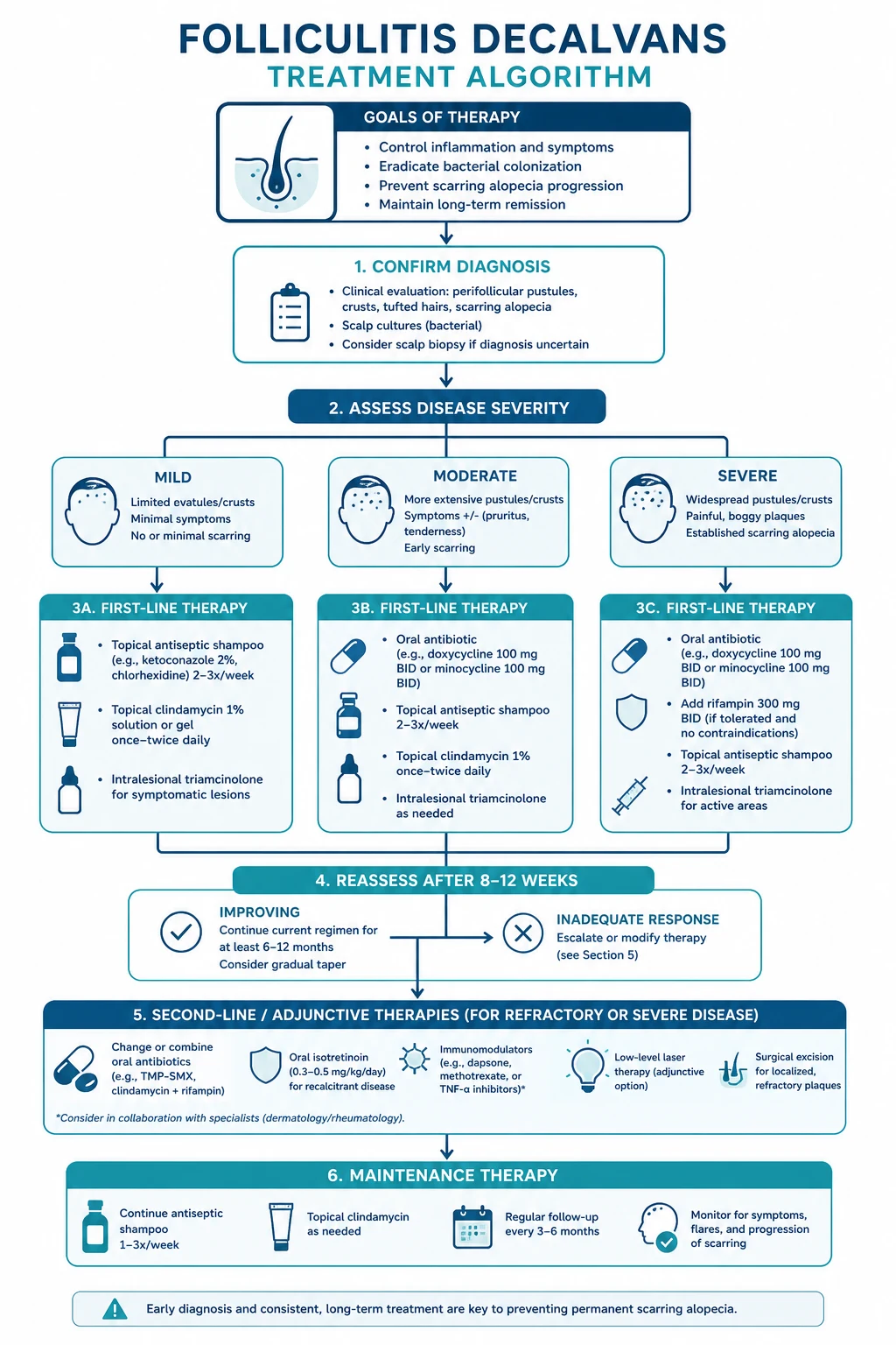
FD management is a stepwise escalation driven by disease activity and response. There is no consensus first-line regimen validated by randomised trials — the evidence base is almost entirely case series and expert opinion — but the rifampicin + clindamycin combination is the most consistently effective regimen reported and is regarded as first-line systemic therapy.[1][2]
Step 1 — Topical therapy (mild / localised disease and adjunct). [1]
- Topical corticosteroids: clobetasol propionate 0.05% lotion or foam OR betamethasone dipropionate 0.05%, applied BD to active edges for up to 4 weeks, then tapered.
- Topical antibiotics: clindamycin 1% lotion OR erythromycin 2% solution, BD.
- Antiseptic / keratolytic shampoos: ketoconazole 2% twice weekly (reduces Malassezia and S. aureus), chlorhexidine 4% wash daily during flares.
- Mupirocin 2% nasal ointment BD for 5 days each month for documented S. aureus nasal carriers, with chlorhexidine body wash — reduces relapse. [1]
Step 2 — First-line systemic therapy: the rifampicin + clindamycin combination.[1]
RifClinda 10-12 — the first-line regimen
Step 3 — Second-line / alternative systemic agents (refractory, relapsing, or contraindicated combination).[1][2]
- Oral isotretinoin 0.5 mg/kg/day for 4-6 months (some authors escalate to 1 mg/kg/day). Reduces sebum production and follicular occlusion; particularly useful when FD overlaps with dissecting cellulitis or acne conglobata. Contraception mandatory (teratogen); monitor lipids and LFTs; avoid concurrent tetracyclines (benign intracranial hypertension).
- Dapsone 50-150 mg daily — anti-neutrophilic via myeloperoxidase inhibition. Check G6PD before starting; monitor haemoglobin and methaemoglobin at 4 weeks and monthly; counsel for haemolysis, especially in patients of Mediterranean, African or South-East Asian ancestry.
- Oral tetracyclines — doxycycline 100 mg BD or lymecycline 408 mg daily; mainly anti-inflammatory (MMP inhibition) and useful for long-term maintenance after rifampicin + clindamycin. Avoid in pregnancy and in children under 8 years (tooth discolouration).
- Oral zinc sulphate 400 mg daily — small series suggest benefit in refractory disease via anti-inflammatory and anti-androgenic effects. [1]
Step 4 — Intralesional therapy. [1]
- Triamcinolone acetonide 5-10 mg/mL (up to 40 mg/mL for keloidal/hypertrophic variants), 0.05-0.1 mL per site into active inflammatory nodules and plaques every 4-6 weeks. Reduces perifollicular inflammation rapidly; useful for symptomatic, painful, or cosmetically distressing lesions. [1]
Step 5 — Physical / procedural therapy. [1]
- Laser hair removal — long-pulsed Nd:YAG 1064 nm is the preferred device in skin of colour (Fitzpatrick IV-VI); diode and alexandrite lasers are alternatives in lighter skin types. A course of 6-10 sessions reduces follicular density, abolishes tufting, and lowers relapse rate by removing the substrate for ongoing inflammation. Nd:YAG is the safest choice in skin of colour because its longer wavelength bypasses epidermal melanin.
- Photodynamic therapy (PDT) with MAL or ALA plus red light — case series show benefit in refractory FD; mechanism is anti-bacterial and immunomodulatory. [1]
Step 6 — Surgery (rare). [1]
- Excision with primary closure or grafting of a small, stable, burnt-out plaque — occasionally used for cosmesis, but wide recurrence is the rule if excised during active disease.
- Scalp reduction — historical, largely abandoned. [1]
Maintenance therapy.[1]
- Long-term topical clindamycin 1% lotion OR erythromycin 2% solution BD to active areas.
- Low-dose oral doxycycline 40 mg daily (sub-antimicrobial dose) for anti-inflammatory maintenance.
- Ketoconazole 2% / chlorhexidine 4% shampoo twice weekly.
- S. aureus decolonisation in carriers (mupirocin nasal + chlorhexidine body wash).
- Serial photography every 3-6 months to detect relapse early. [1]
Specific Subtypes & Scenarios
Tufted folliculitis as a clinicopathological pattern. Saleh and Sathe review tufted folliculitis as a clinical pattern that overlaps almost completely with FD; where the two differ, tufted folliculitis refers to the morphological sign and FD to the disease spectrum. In practice, a patient with tufting has FD until proven otherwise.[3]
Paediatric folliculitis decalvans. Rare; presentation is identical to adult disease. Management differences:[10]
- Weight-based dosing: rifampicin 15-20 mg/kg/day (max 600 mg/day) in two divided doses; clindamycin 8-25 mg/kg/day in three to four divided doses.
- Avoid tetracyclines under 8 years (tooth discolouration) and avoid isotretinoin in young children except under specialist guidance.
- Investigate immunodeficiency (Hyper-IgE/STAT3) when disease is severe, recurrent, or accompanied by eczema, pneumonia or skeletal anomalies. [1]
Follicular occlusion syndrome overlap. When FD coexists with dissecting cellulitis, hidradenitis, AKN or acne conglobata, treatment should address the whole spectrum. Oral isotretinoin is the agent that crosses all members of the spectrum and is often preferred when overlap is present; rifampicin + clindamycin remains effective for the FD component.[5]
Skin of colour — keloidal FD. In Fitzpatrick IV-VI skin, the burnt-out phase may form keloidal or hypertrophic plaques rather than atrophic scar, particularly on the nape and occiput, blurring the boundary with AKN. Treatment adds intralesional triamcinolone 10-40 mg/mL into keloidal plaques and prefers Nd:YAG 1064 nm for laser epilation (safest in dark skin). Post-inflammatory hyperpigmentation is a major cosmetic issue; sun protection and hydroquinone-based camouflage may help.[5]
Immunosuppressed / HIV-associated neutrophilic folliculitis. In HIV or transplant recipients, an atypical papulopustular eruption on the scalp and trunk may mimic or coexist with FD. Biopsy with bacterial, fungal and mycobacterial cultures is mandatory; eosinophilic folliculitis (Ofuji) is an important mimic that requires different therapy (topical corticosteroids, phototherapy, anti-IL-4/13). Antiretroviral optimisation is central. [1]
End-stage / pseudopelade-like FD. A long-standing FD plaque may evolve into a clinically "burnt-out", non-inflammatory, smooth, scarred patch indistinguishable from pseudopelade of Brocq. No active treatment is useful at this stage; management focuses on cosmetic camouflage and surveillance for SCC. [1]
Complications & Pitfalls
Complications. [1]
- Permanent scarring alopecia — the principal complication; once scarred, hair does not regrow.
- Keloidal / hypertrophic scarring in skin of colour.
- Secondary bacterial infection and rarely cellulitis of the scalp.
- Squamous cell carcinoma (Marjolin's ulcer) in chronic cicatricial plaques — rare but reported; any non-healing ulcer, proliferative nodule, or persistent pain in a long-standing scar mandates biopsy.[1]
- Psychological morbidity — depression, anxiety, body-image disturbance, social withdrawal; psychiatric comorbidity is significantly higher in scarring than non-scarring alopecia.[9]
- Drug adverse effects — rifampicin hepatotoxicity and drug interactions; clindamycin C. difficile colitis; isotretinoin teratogenicity, mucocutaneous dryness, lipid abnormalities; dapsone haemolysis and methaemoglobinaemia.
Common pitfalls. [1]
- Treating FD as simple staphylococcal folliculitis with a short course of flucloxacillin — relapse is inevitable because the underlying neutrophilic, biofilm-driven, follicular-destructive process is unaddressed.
- Using monotherapy — single-antibiotic regimens (rifampicin alone OR clindamycin alone) drive rapid resistance and produce near-universal relapse.
- Missing tinea / kerion by starting topical or intralesional corticosteroid before sending fungal culture — converts a treatable infection into a scarring, culture-negative mimic.
- Biopsy of the burnt-out centre only — yields non-specific fibrosis and misses the diagnostic neutrophilic infiltrate; always biopsy the active advancing edge.
- Confusing FD with a lymphocytic scarring alopecia — leads to corticosteroid-based regimens that do not address the neutrophilic driver.
- Forgetting rifampicin's CYP3A4 induction — leads to contraceptive failure, warfarin under-anticoagulation, and calcineurin-inhibitor under-exposure. [1]
Prognosis & Disposition
Natural history. FD is chronic, relapsing, and difficult to cure completely. The disease smoulders over years to decades, with episodes of active pustular inflammation interspersed with quiescent periods; each active episode converts more scalp into permanent scar.[1]
Response to rifampicin + clindamycin. Approximately 50-70% of patients achieve a durable remission after a single 10-12 week course; relapse, when it occurs, usually responds to a repeat course or to escalation to isotretinoin/dapsone.[1]
Cosmetic and quality-of-life burden. Visible scarring alopecia on the scalp vertex produces major cosmetic and psychological burden. A substantial minority require cosmetic camouflage (spray-on hair-fibre concealers, scalp micropigmentation), a wig or hairpiece, or surgical reconstruction (excision of a small stable plaque with primary closure, or follicular unit extraction into burnt-out scar — although transplanted follicles can re-activate disease and the failure rate is significant).[9]
Hair transplantation in FD scar. Controversial; only considered when disease has been quiescent for at least 6-12 months on no active therapy, and even then carries a real risk of koebnerisation / disease reactivation. Most surgeons avoid transplanting into actively inflamed FD. [1]
Follow-up plan. [1]
- Review at 4 weeks of combination therapy (LFTs, adherence, response), at end of therapy (12 weeks), then 3-monthly for the first year and 6-12-monthly lifelong.
- Serial photography at each visit to detect relapse early.
- Maintenance topical therapy for at least 6-12 months after remission.
- Patient education on the relapsing nature, the need for prompt review of any new pustule, and the irreversibility of established scar. [1]
Special Populations
Paediatric.[10]
- Weight-based dosing: rifampicin 15-20 mg/kg/day in two divided doses (max 600 mg/day); clindamycin 8-25 mg/kg/day in three to four divided doses.
- Avoid tetracyclines under 8 years (permanent tooth discolouration) and avoid doxycycline under 12 years.
- Avoid isotretinoin in prepubertal children except under specialist guidance.
- Screen for immunodeficiency (Hyper-IgE, STAT3 gain-of-function) when disease is severe, recurrent, or accompanied by eczema, pneumonia, coarse facies or skeletal anomalies. [1]
Pregnancy and breastfeeding. [1]
- SAFE: topical clindamycin, erythromycin, mupirocin, ketoconazole shampoo, topical corticosteroids (low to mid potency).
- CONTRAINDICATED: isotretinoin (severe teratogen — iPLEDGE-style programme), tetracyclines (doxycycline, lymecycline — tooth and bone effects in the fetus), and avoid rifampicin unless essential (relative caution; rifampicin is used for tuberculosis in pregnancy but the FD indication rarely justifies it).
- Dapsone is generally avoided in the third trimester (theoretical neonatal haemolysis); check G6PD if used.
- Intralesional triamcinolone is acceptable for focal symptomatic lesions. [1]
Elderly / polypharmacy. The principal concern is rifampicin's potent CYP3A4 induction. Major interactions to screen before starting: [1]
- Warfarin and DOACs — rifampicin markedly reduces anticoagulant levels; intensify INR monitoring or switch DOAC.
- Oral contraceptive pill — contraceptive failure; counsel barrier method.
- Statins (especially simvastatin, atorvastatin) — reduced efficacy; lipid check.
- Calcineurin inhibitors (ciclosporin, tacrolimus) — sub-therapeutic levels in transplant recipients; close monitoring.
- Anticonvulsants and sulfonylureas — altered levels.
- Clindamycin adds C. difficile risk in the elderly — counsel regarding diarrhoea. [1]
Skin of colour (Fitzpatrick IV-VI). Keloidal tendency in the burnt-out phase; prominent post-inflammatory hyper- and hypopigmentation. Prefer Nd:YAG 1064 nm over alexandrite or diode lasers (deeper penetration, less epidermal melanin absorption, lower dyspigmentation risk). Test-patch any laser before full treatment.[5]
Immunocompromised / HIV. Lower threshold for biopsy and culture to exclude infective mimics (eosinophilic folliculitis, dermatophytic folliculitis Majocchi, bacterial and mycobacterial folliculitis). Antiretroviral optimisation; consider biopsy-proven FD before committing to prolonged rifampicin + clindamycin. [1]
Evidence, Guidelines & Regional Differences
Evidence base — weak but consistent. There are no randomised controlled trials of FD therapy. The largest evidence synthesis is the systematic review by Dlela Kh and colleagues (JAAD 2019), which pooled case series and case reports and concluded that the rifampicin + clindamycin combination is the most consistently effective regimen, with isotretinoin, dapsone and laser as alternatives for refractory disease. Subsequent narrative reviews (Svara 2025) reach the same conclusion.[1][2]
Svara et al. (2025) narrative review. Synthesises the conventional armamentarium and surveys emerging targets: anti-TNF (adalimumab, infliximab), anti-IL-17 (secukinumab), anti-IL-1 (anakinra), and JAK inhibitors (baricitinib, tofacitinib) — all currently at case-report level only, with no trial-grade evidence.[2]
Zorlu et al. (2025) — 15-year tertiary cohort. Of 109 primary cicatricial alopecia patients, the neutrophilic group (FD and dissecting cellulitis) was the largest, confirming the demographic and clinical pattern and reinforcing FD's place as a major cause of scarring alopecia in specialist practice.[4]
Doche et al. (2019) — AKN and FD overlap. A retrospective series demonstrating that AKN and FD coexisted in a substantial proportion of patients, supporting the view that they are one follicular-destructive process expressed at different anatomical sites.[5]
Scalp-microbiome literature (Xu 2024). Demonstrates reduced diversity and enrichment of biofilm-forming staphylococci and corynebacteria in FD, providing the biological rationale for combination antibiotic therapy and antiseptic shampoos.[8]
Controversy: cause, coloniser, or bystander? Whether S. aureus is the primary pathogen, a secondary coloniser that aggravates an innate-immune defect, or an innocent bystander is unresolved. The clinical implication is that antibiotic therapy alone is insufficient — anti-inflammatory and physical measures (laser) are needed to interrupt the self-sustaining inflammatory loop. [1]
Regional deltas. [1]
NAHRS (North American Hair Research Society) classifies FD as a neutrophilic primary cicatricial alopecia and recommends rifampicin + clindamycin as first-line systemic therapy. Isotretinoin and laser hair removal are accepted alternatives.
BAD favours a stepwise approach beginning with topical and oral antibiotics, reserving combination rifampicin + clindamycin for refractory disease — a more conservative starting point than NAHRS.
European guidance is broadly concordant with NAHRS; Nd:YAG laser and PDT are more widely used in European centres as second-line.
IADVL guidance emphasises the late presentation common in India, the overuse of topical steroids (masking and worsening disease), and the role of antifungal shampoos given frequent Malassezia and dermatophyte co-colonisation. Cost and access to laser and biologics limit their routine use; rifampicin + clindamycin is the practical first-line systemic regimen.
Why the evidence base is weak. FD is rare (no single centre accrues enough patients for RCTs), there is no validated outcome measure, the disease is relapsing-remitting (confounding short-term assessments), and the most effective regimen (rifampicin + clindamycin) cannot easily be blinded. Progress will require multicentre registries. [1]
Exam Pearls
[1]FD in a single mnemonic REACT
Cicatricial alopecia SLIP-N-DAD (lymphocytic vs neutrophilic)
Exam application bank (NEET-PG / INICET)
One-line answer
Folliculitis decalvans (Quinquaud disease) is a chronic, relapsing, PRIMARY NEUTROPHILIC cicatricial (scarring) alopecia of the scalp, defined by recurrent crops of follicular pustules on the scalp vertex and occiput together with the pathognomonic sign of TUFTED FOLLICULITIS (multiple hair shafts, classically 5-20, emerging from a single dilated follicular opening, the so-called 'doll's hair'). Staphylococcus aureus is cultured from lesional pustules in approximately 70% of cases, but FD is an abnormal, persistent neutrophilic inflammatory response to S. aureus (or its superantigens) and follicular contents rather than simple pyoderma. Untreated, the inflammatory cascade destroys follicular stem cells and replaces them with fibrous tissue, producing PERMANENT scarring alopecia. First-line systemic therapy is the COMBINATION of oral rifampicin 300 mg twice daily PLUS clindamycin 300 mg t [1]
Worked stems (answer without another resource)
Stem 1 — Classic presentation. Map symptoms to mechanism; name the first investigation and first treatment step with dose/route if drug therapy is standard. [1]
Stem 2 — Unstable / complicated. List red flags that force immediate resuscitation, theatre, ICU, antidote, or reperfusion — and what you do in the first 15 minutes. [1]
Stem 3 — Atypical group. Elderly, pregnancy, child, or immunocompromised: how presentation and thresholds change. [1]
Stem 4 — Differential trap. Name the three closest mimics and one discriminator for each. [1]
Stem 5 — Disposition. Who goes home with safety-netting, who is admitted, who needs HDU/ICU/theatre, and what follow-up is mandatory. [1]
Rapid viva checklist
- Definition + classification
- Pathophysiology chain
- Bedside signs / criteria
- Score with exact components (if any)
- Emergency bundle
- Definitive therapy with doses
- Complications of disease and of treatment
- Special populations
- Guideline/trial name if classic
- Three exam traps
Coverage self-check
If you cannot answer any stem above from this page alone, re-read the matching section — the page is intended to be self-sufficient for final-prof and NEET-PG/INICET questions on Folliculitis decalvans.
[1] [1]References
- [1]Dlela Kh ZB, Cervantes J, Hoss E, et al. Updates in therapeutics for folliculitis decalvans: A systematic review with evidence-based analysis J Am Acad Dermatol, 2019.PMID 30092322
- [2]Svara F, Bortone G, Ambrosio L, et al. Diagnostic Challenges and Treatment Strategies in Neutrophilic Cicatricial Alopecias: A Narrative Review from Conventional Therapies to New Therapeutic Targets Life (Basel), 2026.PMID 42195390
- [3]Saleh HM, Sathe NC Tufted Hair Folliculitis 2026.PMID 28613486
- [4]Zorlu Ö, Albayrak H, Aytekin S Clinical features of 109 patients with primary cicatricial alopecia: a 15-year retrospective study Acta Dermatovenerol Alp Pannonica Adriat, 2026.PMID 41915583
- [5]Doche I, Coelho EQ, Quaresma MV, et al. Acne keloidalis nuchae and folliculitis decalvans: same process affecting the follicle or coexisting diseases? A retrospective study Int J Dermatol, 2019.PMID 31241169
- [6]Rakowska A, Czuwara J, Zaremba I, et al. The dermatoscope in the hair clinic: Trichoscopy of scarring and nonscarring alopecia J Am Acad Dermatol, 2023.PMID 37591567
- [7]Lepe V, Micalizzi C, Zampino MR Common causes of hair loss - clinical manifestations, trichoscopy and therapy J Eur Acad Dermatol Venereol, 2021.PMID 33290611
- [8]Xu Z, Wang T, Chen H, et al. Scalp microbiome: a guide to better understanding scalp diseases and treatments Arch Dermatol Res, 2024.PMID 39073596
- [9]Baker NJ, Alomary SA, Ogunleye T, et al. Risk of Psychiatric Comorbidities in Non-Scarring Versus Scarring Alopecia: A Retrospective Cohort Study J Cutan Med Surg, 2026.PMID 41925130
- [10]Motsios D, Sotiropoulou K, Karabarba S, et al. Comprehensive Case Report on Folliculitis Decalvans in a Pediatric Patient Acta Dermatovenerol Croat, 2025.PMID 42370657