Dermatology · Medicine
Folliculitis, furuncle and carbuncle
Also known as Folliculitis · Furuncle (boil) · Furunculosis · Carbuncle · Hot tub folliculitis (Pseudomonas) · Eosinophilic folliculitis
Folliculitis, furuncles, and carbuncles are a spectrum of staphylococcal (and occasionally other) infections of the hair follicle, progressing from superficial folliculitis to a deeper, painful nodule (furuncle) and to a coalescent multiloculated abscess draining through several follicles (carbuncle). Fellowship-level assessment demands mastery of the follicle-centred morphology, the common and special pathogens (S. aureus including PVL-producing and MRSA strains, Pseudomonas hot-tub folliculitis, gram-negative and eosinophilic/HIV-associated variants), incision-and-drainage as primary therapy for furuncles/carbuncles, the role and limitations of adjunctive antibiotics, and the prevention of recurrent furunculosis through S. aureus decolonisation.
On this page & tools
Your progress
Saved locally on this device.
Exam tags
Red flags
Overview
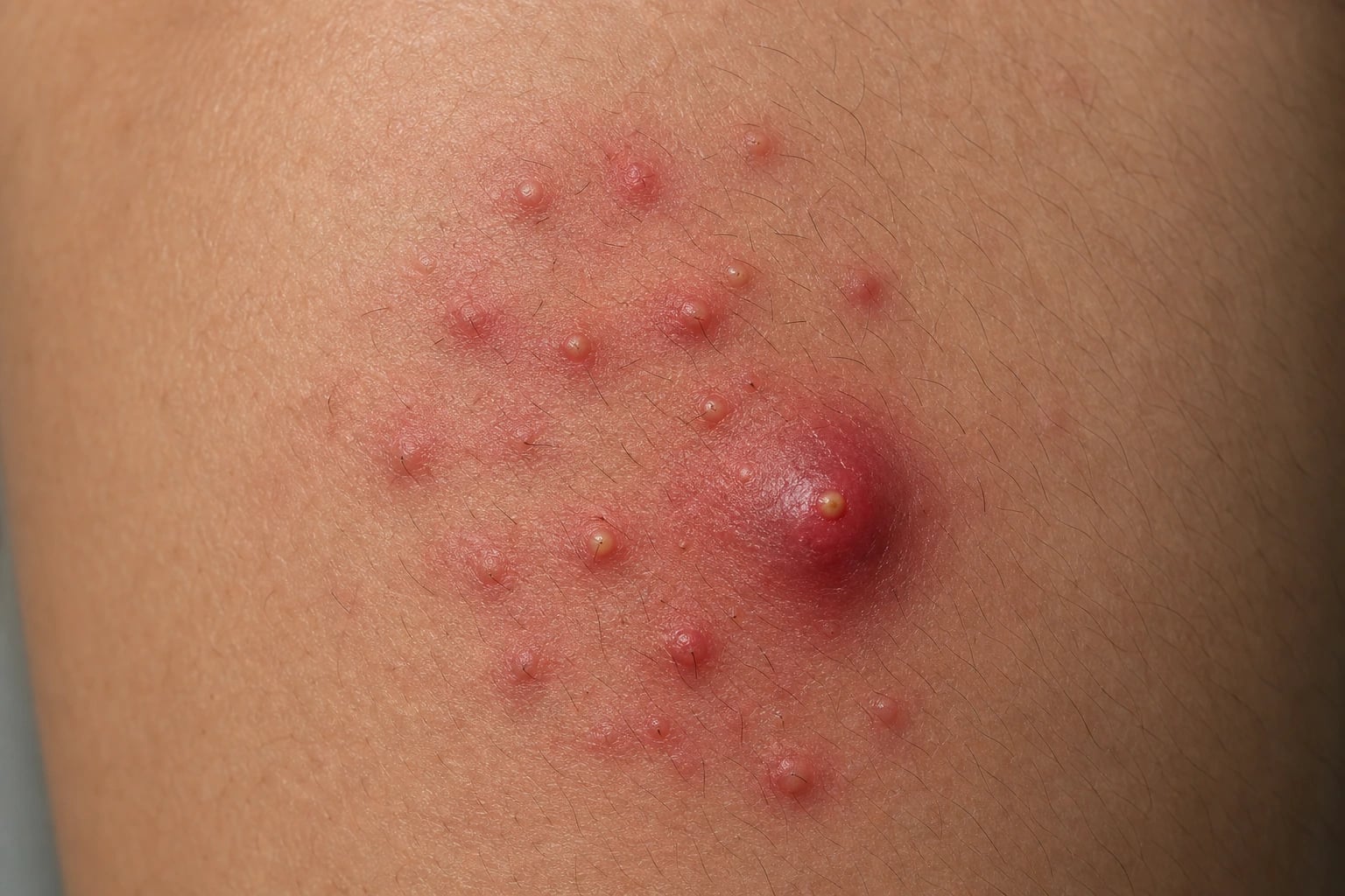
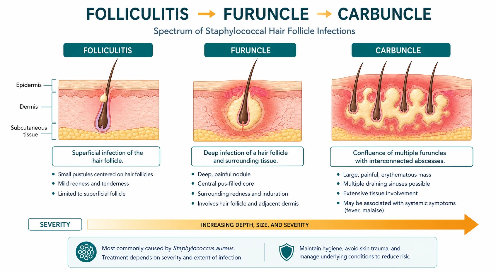
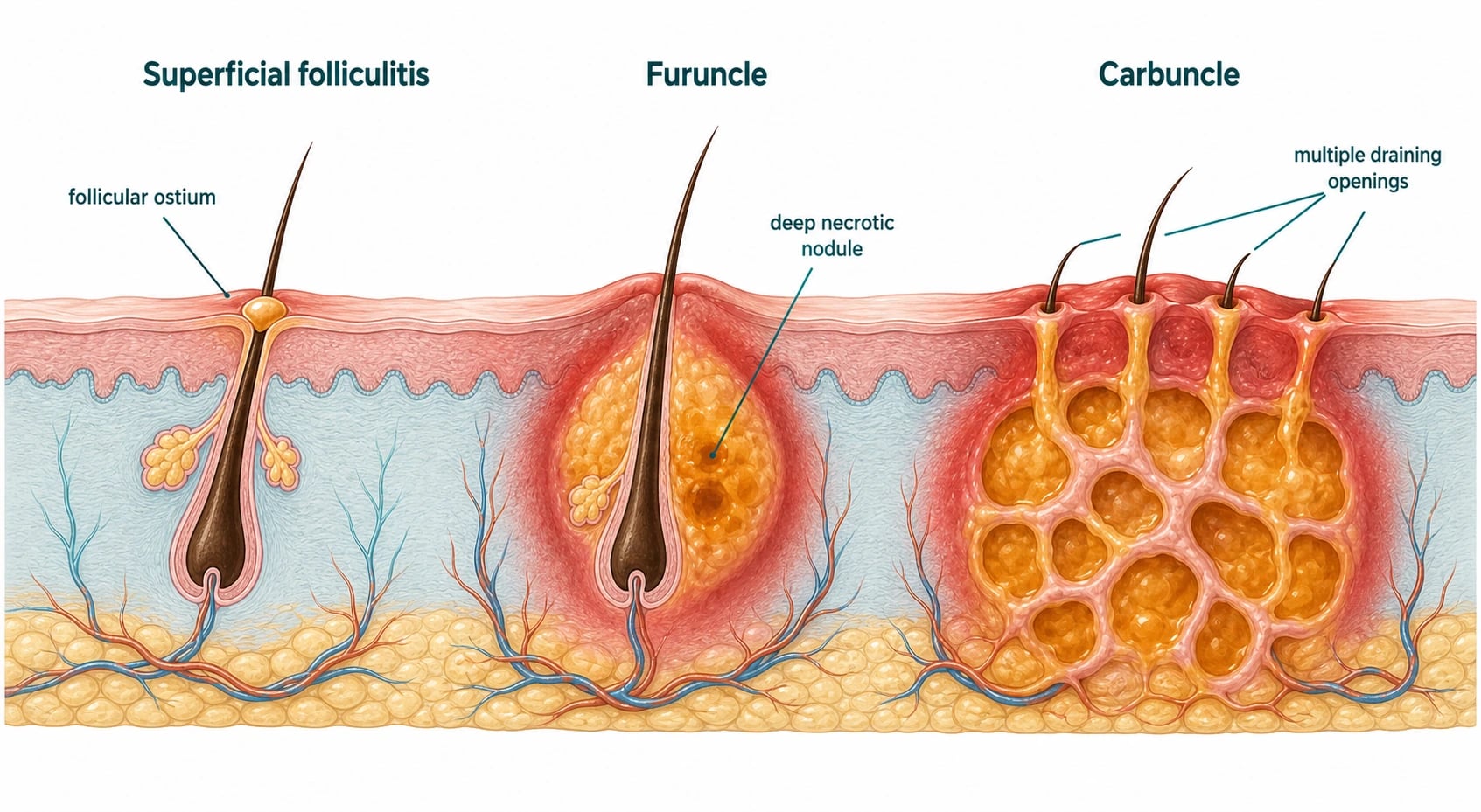
Folliculitis, furuncles, and carbuncles form a spectrum of follicle-centred bacterial infections, most commonly caused by Staphylococcus aureus. Superficial folliculitis is a tiny pustule at the follicular ostium; a furuncle (boil) is an acute, deep, painful, necrotic nodule of a hair follicle and surrounding tissue; and a carbuncle is a coalescence of interconnected furuncles draining through several openings, typically on the nape, back, or thighs, often with systemic upset and scarring. Fellowship-level competence requires confident phenotyping, recognition of special pathogens (PVL-producing S. aureus, MRSA, Pseudomonas hot-tub folliculitis, gram-negative folliculitis of acne patients, and eosinophilic folliculitis of HIV), primary incision and drainage for abscess-forming lesions, the selective and evidence-based use of adjunctive antibiotics, and a structured approach to recurrent furunculosis via S. aureus decolonisation and treatment of predisposing conditions.[1][2][3]
Pathophysiology and microbiology
- Staphylococcus aureus is the cause of the great majority of folliculitis, furuncles, and carbuncles. Infection begins with follicular occlusion and bacterial proliferation, progressing through a neutrophilic folliculitis to a deep dermal/subcutaneous abscess. The cilium-bearing follicle provides a niche that protects bacteria from surface antiseptics; shaving, friction, and occlusion disrupt the follicular barrier and seed the inoculum. Bacterial adhesins (teichoic acid, fibronectin-binding protein A) tether S. aureus to the corneocyte around the follicular ostium, where the neutrophil influx that follows underlies the follicular pustule. Rupture of the follicular epithelium spills keratin, bacteria, and lipoid contents into the dermis, generating the deep inflammatory nodule characteristic of a furuncle; coalescence of multiple such lesions defines a carbuncle.[1][2]
- Panton-Valentine leucocidin (PVL)-producing S. aureus (both MSSA and MRSA strains) is associated with recurrent, multiple, necrotic furunculosis and, more dangerously, with necrotising pneumonia; PVL strains should be suspected in young, otherwise well patients with severe or recurrent furunculosis. PVL is a pore-forming, bi-component leucocidin encoded by the lukS-PV and lukF-PV genes (prophage ΦPVL) that lyses neutrophils and macrophages — both by membrane pore formation and by apoptosis induction — driving the necrotic phenotype. PVL-positive strains are also linked to bone and joint infections in young athletes and to severe community-acquired pneumonia following influenza. [6][7]
- MRSA is an increasingly important cause, particularly in endemic regions, contact sports, IVDU, and institutional settings. CA-MRSA clones (USA300 in North America; ST80 in Europe) frequently carry ΦPVL, while HA-MRSA strains are typically multi-resistant and lack PVL but bring resistance pressure into the community through decolonisation failure.
- Pseudomonas aeruginosa causes hot-tub (spa) folliculitis, appearing 8-48 hours after exposure to contaminated warm water (hot tubs, pools, spas), as itchy, erythematous follicular papules and pustules, often on occluded areas (axillae, groins, under swimwear); it is usually self-limiting but occasionally causes serious infection, particularly in immunocompromised patients, who may develop ecthyma gangrenosum or bacteraemia.[10]
- Gram-negative folliculitis (Escherichia coli, Klebsiella, Proteus, Pseudomonas) complicates long-term acne therapy (especially tetracyclines), presenting as perinasal pustules. Long-term antibiotics suppress the susceptible resident flora, allowing overgrowth of resistant Gram-negative organisms in the peri-nasal and perioral skin. Withdrawing the tetracycline and switching to isotretinoin or an amoxicillin-clavulanate usually controls the breakout.[14][15]
- Eosinophilic pustular folliculitis (Ofuji disease, and the HIV-associated variant) presents with intensely pruritic, recurrent, follicular papules and pustules, often on the face and trunk, with peripheral eosinophilia; it is a marker of advanced HIV immunosuppression (CD4 < 200-300) and of certain haematological disorders. Histology shows eosinophil-rich follicular infiltrates ± flame figures. It is driven by an IL-5/eotaxin axis and improves dramatically when the underlying HIV is treated with antiretroviral therapy.[11][12][13]
- Malassezia folliculitis (Pityrosporum) is a yeast-driven, itchy, acne-like folliculitis on the trunk, treated with antifungals. It is more common in hot, humid climates and is often misdiagnosed as acne; potassium hydroxide microscopy of pustule contents reveals the characteristic 'spaghetti and meatballs' yeast forms. It responds to topical ketoconazole shampoo or a short course of oral itraconazole.
- Drug-induced folliculitis is increasingly recognised — notably an acneiform folliculitis from EGFR inhibitors and BTK inhibitors (ibrutinib and related agents), which is follicle-centred and dose-dependent rather than infectious. EGFRi-induced eruption is more monomorphic in the seborrheic zone; BTK inhibitor lesions can mimic acneiform eruption or suppurative folliculitis. Management centres on topical corticosteroids and oral doxycycline 100 mg BD for anti-inflammatory benefit, not incision and drainage.[16]
Clinical features
- Superficial folliculitis — small, follicle-centred, erythematous papules topped by a tiny pustule, often itchy and occurring in areas of friction, shaving, occlusion (heat, sweat), or after hot-tub exposure. Common on the beard area (sycosis barbae), scalp, axillae, buttocks, and thighs. It is typically self-limiting but recurs in carriers of S. aureus.
- Furuncle (boil) — a tender, firm, red nodule that enlarges and becomes fluctuant, with a central yellow punctum that ruptures to discharge pus and a necrotic core; may heal with scarring. Lesions on the face (danger triangle), especially around the nose and upper lip, carry a risk of cavernous sinus thrombosis via the valveless facial veins draining to the cavernous sinus. Fluctuant lesions should be incised and drained; spontaneous rupture can occur but leaves a residual cavity and an unroofed necrotic roof.
Diagnosis & pathophysiology of the spectrum [1]
The diagnosis is clinical, anchored in recognising the follicle-centred morphology at every stage of the spectrum. Bacterial folliculitis starts as a small superficial follicular pustule; the contents (neutrophils, bacteria, occasional eosinophils) sit below the stratum corneum around the ostium. Furunculosis is the next depth step — spontaneous rupture of the follicular epithelium drains the bacterial cargo into the dermis, producing the deep, well-circumscribed, tender, fluctuant nodule that defines the boil. Carbunculosis is the lateral coalescence of multiple contiguous follicles into a multiloculated, draining, deep abscess, classically on the back, nape or thighs, with systemic features and significant scarring. Recognising the depth and lateral extent of a lesion at the bedside is what dictates therapy: superficial lesions respond to topical therapy, deep fluctuant lesions require I&D, carbuncles also need I&D plus adjunctive antibiotics in most patients. A simple bedside 'diabetic' classification by clicking the lesion with the tip of a cotton bud is unreliable — use the needle-aspiration test (18 G needle on 10 mL syringe) to confirm fluctuance.[1][2][9]
Microbial investigation is routine in selected scenarios only — for isolated superficial disease, MCS is wasteful and changes management little. Swab expressed pus or exudate in Amies transport medium; send for Gram stain, culture, sensitivity. Add PVL PCR in any patient with recurrent or severe necrotic furunculosis, household clusters, athletes, or institutional outbreaks. PVL-positive S. aureus can be a MSSA or MRSA — knowing the PVL status is more important than knowing MSSA vs MRSA for clinical management, since PVL-positive strains of either background require toxin-suppressing antibiotics (clindamycin or linezolid). In recurrent furunculosis, screen for the systemic predispositions: HbA1c/fasting glucose, FBC, serum iron and ferritin, HIV, immunoglobulins ± neutrophil oxidative burst (NBT/DHR if chronic granulomatous disease is suspected), and a nasal/perineal carriage swab. [2][9]
Biopsy with H&E plus PAS, GMS, Gram staining is reserved for atypical or unresponsive lesions, or when the morphology suggests eosinophilic folliculitis, Majocchi granuloma, deep fungal infection, folliculotropic lymphoma, or in immunocompromised hosts. The biopsy is critical in distinguishing infection from non-infectious folliculocentric inflammation (drug-induced EGFRi/BTKi eruption, lupus, dermatosis that mimics infection).[16]
Differential diagnosis
Acne vulgaris, acneiform drug eruptions (corticosteroids, androgens, EGFR/BTK inhibitors), hidradenitis suppurativa, keratosis pilaris, Majocchi granuloma (dermatophyte folliculitis), Malassezia folliculitis, atopic dermatitis, scabies, insect bites, cutaneous anthrax (the historical "malignant pustule"), and inflamed epidermoid/pilar cysts. The follicular, pustular morphology and culture help distinguish bacterial folliculitis. The differential can be stratified by the depth and tempo of the lesion. Superficial folliculitis with monomorphic pustules at the follicular ostium raises the differential of Pseudomonas hot-tub folliculitis, Pityrosporum folliculitis, eosinophilic folliculitis (HIV), subcorneal pustular dermatosis, halogenoderma, and pseudofolliculitis barbae (a foreign-body inflammatory reaction to ingrown hair after close shaving — not infectious). Furuncle must be distinguished from the inflamed epidermoid cyst, which is also fluctuant but centred on a punctum expressing keratinous debris (not pus); from hidradenitis suppurativa, which sits in the apocrine zones (axillae/groin/perianal) and forms sinus tracts over years, not weeks; from cutaneous anthrax, which has a painless black eschar surrounded by oedema; and from deep mycoses (sporotrichoid nodular lesions; immunocompromised host). Carbuncle can mimic an epidermal inclusion cyst abscess, a pilonidal abscess (midline sacral pit rather than nape/back), and a necrotising soft-tissue infection — the last is the critical dont-miss because necrotising fasciitis spreads faster, is exquisitely painful out of proportion, and produces systemic sepsis with crepitus/bullae/anaesthesia.[16]
Management
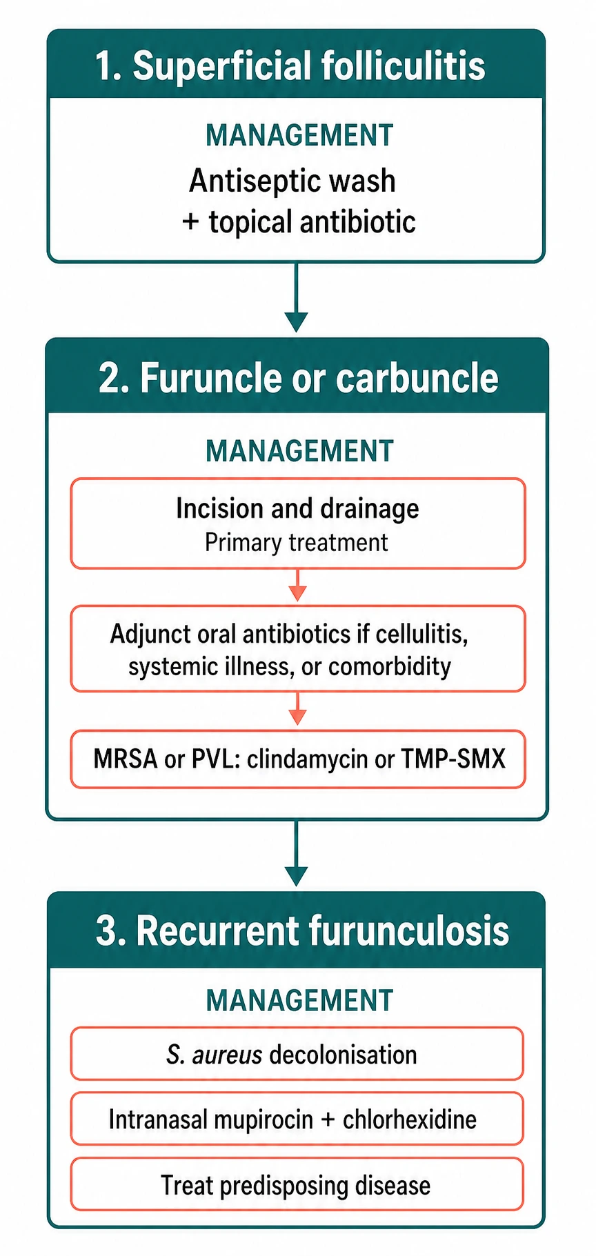
Often self-limiting; warm compresses and antiseptic wash (chlorhexidine 4% once daily for 1-2 weeks). Topical mupirocin 2% ointment applied to individual lesions BD-TDS 7-10 days, or topical clindamycin 1% solution for limited staphylococcal disease with broader coverage of Gram-positive cocci. Address shaving technique and occlusion: leave 1-2 mm of stubble, use a sharp single-edge blade, shave in the direction of hair growth, and avoid hot towels immediately before shaving (which dilates follicles and increases bacterial seeding). Anti-odourant and antiperspirant overuse can worsen axillary disease; pause for 4 weeks.[1]
-
Incision and drainage (I&D) is the primary and most important therapy for a fluctuant furuncle or carbuncle; antibiotics are adjunctive. Cochrane evidence supports I&D as the cornerstone, and shows that the benefit of adjunctive antibiotics over placebo is modest for uncomplicated lesions, with antibiotic choice guided by severity, immune status, and local resistance. The technique involves:
- Pre-procedure: topical or local anaesthetic (e.g., 1% lidocaine without adrenaline); chlorhexidine antisepsis; assessment of carbuncle lateral extent with the 'pinch test' to map multiloculated cavities.
- Incision: a No. 11 blade makes a linear stab wound across the area of maximum fluctuance; for a carbuncle, a cruciate (cross) incision allows drainage of multiple connected loculi.
- Evacuation: express pus with gloved pressure; a 14-16 Fr Yates drain or iodoform wick prevents premature closure of the cavity.
- Sample: send the central core to microbiology.
- Post-procedure: irrigation of the cavity with normal saline; close follow-up at 48-72 hours; replace drain in 24-48 hours; cavity heals by secondary intention.[1]
-
Adjunctive antibiotics are indicated for: surrounding cellulitis, systemic symptoms (fever), immunocompromise, diabetes, large lesions, facial/danger-triangle lesions, failure of I&D alone, and recurrent disease — an oral anti-staphylococcal agent (flucloxacillin/cephalexin; clindamycin, TMP-SMX, or doxycycline for suspected MRSA or PVL strains).[5][3]
-
Severe disease or immunocompromise may require IV antibiotics and surgical assessment.[8]
Specific and special-pathogen therapy
- PVL-producing S. aureus — combine drainage with an antibiotic that suppresses toxin production (clindamycin or linezolid) plus a rifampicin-based regimen, and treat carriage, often with specialist input; PVL strains warrant eradication because of recurrence and the risk of necrotising pneumonia.[6][7]
- Hot-tub folliculitis (Pseudomonas) — usually self-limiting; a fluoroquinolone (ciprofloxacin) for severe, persistent, or immunocompromised cases.[10]
- Gram-negative folliculitis — oral isotretinoin (or amoxicillin-clavulanate/a fluoroquinolone); discontinue the implicated long-term tetracycline where possible.[14][15]
- Eosinophilic folliculitis — treat the underlying cause (highly active antiretroviral therapy for HIV); topical corticosteroids, topical permethrin, UVB phototherapy, and oral antihistamines provide symptomatic relief; indomethacin or itraconazole in selected cases.[11][12]
- Malassezia folliculitis — topical or oral antifungal (ketoconazole, fluconazole, itraconazole).
Prevention of recurrent furunculosis
A structured S. aureus decolonisation strategy underpins prevention: intranasal mupirocin (twice daily for 5 days), chlorhexidine or dilute bleach body wash, treatment of household contacts (who may be reservoirs), and attention to hygiene, fomite/linen washing, and avoidance of sharing personal items.[3][4] Treat predisposing disease (diabetes, HIV, iron deficiency). For proven PVL disease, eradication regimens combine systemic antibiotics with topical decolonisation. Cochrane and recent reviews support decolonisation for reducing recurrence, though reinfection from carriers and the environment remains a challenge.[1][4]
Complications
Cellulitis, abscess, bacteraemia and S. aureus bacteraemia with metastatic infection (endocarditis, osteomyelitis, septic pulmonary emboli — particularly with PVL strains), cavernous sinus thrombosis from facial lesions, scarring, and the vicious cycle of recurrent disease with ongoing carriage.[8][9]
- Cavernous sinus thrombosis (CST): facial furuncle in the danger triangle (corners of mouth to bridge of nose) drains via the superior ophthalmic and inferior ophthalmic veins (valveless) into the cavernous sinus. Onset 24-72 h after the lesion, with progressive periorbital swelling, proptosis, ophthalmoplegia, papilloedema, headache, fever, sepsis, and possibly seizures or focal neurology. Confirm with MRI/MRV — look for filling defects in the cavernous sinus, distention, loss of flow voids. CSF may show neutrophilic/mononuclear pleocytosis; do not delay treatment for diagnostic confirmation. Treat with IV vancomycin/teicoplanin + linezolid (or clindamycin) for toxin suppression, ophthalmology and ENT input, anticoagulation per neurology (controversial but supported by case series). Mortality remains 20-30% even with treatment.[8]
- S. aureus bacteraemia with metastatic infection: take two peripheral blood culture sets in any patient with fever or systemic features. Especially aggressive in PVL-positive infections, which have an alarming tendency to produce endocarditis, vertebral osteomyelitis, septic pulmonary emboli, and septic arthritis. Echocardiography is indicated for persistent bacteraemia or new murmur; MRI of the spine for back pain; PET-CT to find occult foci when fever persists >7 days.[9]
- Carbuncle-related sepsis: particularly in diabetes or other immunocompromise; mortality rises sharply when systemic sepsis accompanies a delayed presentation. Admit for IV antibiotics and surgical assessment.
- Scarring: particularly on the scalp (folliculitis decalvans), beard (sycosis barbae), upper trunk, and back. Cosmetic scarring from carbuncles can require secondary excision once infection has resolved.
- Recurrence without decolonisation: ~50% within 12 months. Combined decolonisation + household contact treatment reduces this to ~10-20% per Ibler and Kromann (2014).
- Quality of life impact is comparable to mild psoriasis or eczema; chronic recurrent disease warrants dermatology referral.
- Misdiagnosis: incision of herpetic whitlow, inflamed epidermoid cyst, hidradenitis suppurativa, or deep mycosis can complicate disease. Always review the morphology before an incision.
Clinical pearl
[1]Red flags
Exam application bank (NEET-PG / INICET)
One-line answer
Folliculitis, furuncles, and carbuncles are a spectrum of staphylococcal (and occasionally other) infections of the hair follicle, progressing from superficial folliculitis to a deeper, painful nodule (furuncle) and to a coalescent multiloculated abscess draining through several follicles (carbuncle). Fellowship-level assessment demands mastery of the follicle-centred morphology, the common and special pathogens (S. aureus including PVL-producing and MRSA strains, Pseudomonas hot-tub folliculitis, gram-negative and eosinophilic/HIV-associated variants), incision-and-drainage as primary therapy for furuncles/carbuncles, the role and limitations of adjunctive antibiotics, and the prevention of recurrent furunculosis through S. aureus decolonisation.
Worked stems (answer without another resource)
Stem 1 — Classic presentation. Map symptoms to mechanism; name the first investigation and first treatment step with dose/route if drug therapy is standard. [1]
Stem 2 — Unstable / complicated. List red flags that force immediate resuscitation, theatre, ICU, antidote, or reperfusion — and what you do in the first 15 minutes. [1]
Stem 3 — Atypical group. Elderly, pregnancy, child, or immunocompromised: how presentation and thresholds change. [1]
Stem 4 — Differential trap. Name the three closest mimics and one discriminator for each. [1]
Stem 5 — Disposition. Who goes home with safety-netting, who is admitted, who needs HDU/ICU/theatre, and what follow-up is mandatory. [1]
Rapid viva checklist
- Definition + classification
- Pathophysiology chain
- Bedside signs / criteria
- Score with exact components (if any)
- Emergency bundle
- Definitive therapy with doses
- Complications of disease and of treatment
- Special populations
- Guideline/trial name if classic
- Three exam traps
Coverage self-check
If you cannot answer any stem above from this page alone, re-read the matching section — the page is intended to be self-sufficient for final-prof and NEET-PG/INICET questions on Folliculitis, furuncle and carbuncle.
Expanded exam teaching (depth pass)
Clinical reasoning
For Folliculitis, furuncle and carbuncle, examiners test whether you can prioritise life threats, choose the right first test, and give specific therapy (agent, dose, route, timing). Generic phrases without numbers score poorly.
Mechanism → feature map
Build a short chain: cause → pathophysiologic intermediate → clinical feature → complication. Every major symptom in the classic vignette should sit on that chain.
Investigation strategy
- Bedside/first-line tests that change immediate management
- Confirmatory or staging tests
- What a normal result does not exclude
- When not to delay treatment for imaging (unstable patient)
Management ladder
- Resuscitation / ABC / sepsis or haemorrhage bundle as relevant
- Specific antidote / procedure / antimicrobial / reperfusion / surgery
- Supportive care and monitoring targets
- Definitive long-term therapy and secondary prevention
- Disposition and safety-net advice
Special populations
Always prepare one line each for children, pregnancy, elderly, renal/hepatic impairment, and immunocompromised patients when the topic allows.
Pitfalls that fail candidates
- Treating the number not the patient
- Missing pregnancy status when relevant
- Imaging before stabilisation
- Wrong empiric cover or wrong antidote timing
- Incomplete counselling on recurrence, adherence, or red-flag return
Folliculitis, furuncles, and carbuncles are a spectrum of staphylococcal (and occasionally other) infections of the hair follicle, progressing from superficial folliculitis to a deeper, painful nodule (furuncle) and to a coalescent multiloculated abscess draining through several follicles (carbuncle). Fellowship-level assessment demands mastery of the follicle-centred morphology, the common and special pathogens (S. aureus including PVL-producing and MRSA strains, Pseudomonas hot-tub folliculiti [1]
[1]Clinical & bedside assessment
The diagnosis is usually clinical. Focus on the follicle-centred morphology, the distribution (beard, scalp, axillae, buttocks, thighs, hot-tub occluded areas), and the presence of fluctuance. Always seek:
- Portal of entry: shaving or waxing in the last 2 weeks, occlusive clothing, recent hot tub/spa exposure, contact sports, abrasion or insect bite, IVDU sites, surgical wound, chronic eczema.
- Carrier status: examine the anterior nares with a gloved finger or sterile swab if the patient reports frequent touching of nose before rubbing a lesion; swab the perineum and axillae when there is household spread.
- Predisposing disease: ask about diabetes, HIV, immunosuppression, eczema/atopic dermatitis, eczema herpeticum history, malignancy, IVDU, iron deficiency (heavy menstrual bleeding, chronic GI blood loss), and recent antibiotics (for Gram-negative folliculitis).
- PVL risk: young patient (under ~40), recurrent necrotic furuncles, household spread, severe solitary lesion, athlete (wrestling, rugby), close contact with a confirmed PVL case.
- Severity screen: fluctuant abscess, surrounding cellulitis >5 cm, systemic fever, lymphangitis, immunocompromise, facial lesion in the danger triangle, large carbuncle, recurrent or rapidly progressive disease.
- Hot-tub exposure history with timing: 8-48 hours after contact strongly suggests Pseudomonas folliculitis. [1]
Examine the oropharynx, axillae, umbilicus, groin, and perineum — these are reservoirs for S. aureus and benefit from decolonisation. [1]
Investigations
Pathology and bedside
- Pus swab culture and sensitivity (MCS) is indicated for recurrent disease, severe or extensive infection, treatment failure, suspected MRSA or PVL, immunocompromise, outbreaks, and any lesion of the danger triangle. Use a sterile cotton/rayon swab to sample expressed pus or exudate; transport in Amies medium to microbiology.
- PVL PCR (reference lab; not all sites stock it) is warranted when suspicion is high: recurrent necrotic furuncles, household spread, severe solitary lesion, atypical pneumonia in a contact, or community MRSA-cluster scenario.
- HIV serology in any patient under ~50 with recurrent furunculosis; do not wait for severe disease.
- HbA1c or fasting glucose, FBC, ferritin/Iron studies, immunoglobulins for recurrent disease to look for the predisposing conditions named above.
- Skin biopsy with H&E/special stains (PAS, GMS, Gram) when the morphology suggests eosinophilic folliculitis, Majocchi granuloma, deep fungal infection, or atypical immunosuppression.
- Blood cultures when systemic sepsis, facial furuncle with systemic features, immunocompromised host, or prosthetic material in situ.
- Nasal/perineal swab for S. aureus carriage with culture ± spa-typing or PCR at decolonisation evaluation.
Recognition of the spectrum in clinic
The single most useful bedside distinction is fluctuance: a fluctuant lesion of any size needs drainage, not antibiotics alone. Use the needle-aspiration test: an 18 G needle on a 10 mL syringe enters the cavity and returns pus under pressure; aspirate ≥1 mL of pus to confirm abscess. Aspirated material goes to MCS. [1]
Specific scenarios
Recurrent furunculosis — the half-life of disease
Recurrent furunculosis is defined as ≥3 episodes in 12 months or persistent disease across body sites. The cornerstone of prevention is a structured S. aureus decolonisation strategy:
- Intranasal mupirocin 2% ointment to both anterior nares twice daily for 5 days (kills carriage).
- Chlorhexidine 4% body wash or dilute bleach baths (1/4 cup household bleach per full bath) — daily for 5-7 days, then weekly maintenance.
- Wash all towels, sheets, and flannels at 60 °C daily during the regimen.
- Treat household contacts if nose swabs return positive.
- Optimise predisposing factors: glycaemic control, ART adherence, iron replacement, weight, hygiene at work, avoid sharing razors.
- Failure (recurrence within 3-6 months): repeat the regimen; consider topical mupirocin bd for the rest of the month; consider low-dose oral clindamycin 150 mg nocte for 3 months or TMP-SMX 480 mg BD 7 days repeated monthly per Cochrane. [1]
PVL-positive S. aureus — a special pathogen
PVL-positive strains are common in community-onset recurrent furunculosis, in household clusters, and in athletes in close-contact sports. The lesions are usually small, tender, and have a central necrotic centre ("bull's eye" appearance). Therapy must combine effective drainage with an antibiotic that suppresses toxin production:
- Clindamycin 300 mg QDS orally for 7-10 days (suppresses toxin synthesis and is highly penetrant).
- Add rifampicin 300 mg BD in confirmed PVL with multiple lesions.
- Hospitalise for IV therapy in cellulitis/systemic sepsis: linezolid 600 mg BD IV has very high anti-toxin activity.
- Eradicate carriage aggressively (see above) once lesions have healed; consider neomycin/chlorhexidine nasal cream if mupirocin resistant. [1]
Hot-tub (Pseudomonas) folliculitis
Onset 8-48 hours after exposure; 1-4 mm erythematous follicular papules and pustules on covered sites under swimwear or in occluded axillae/groins. May have low-grade fever. Often self-resolves in 7-10 days without specific therapy. For severe/immunocompromised cases: ciprofloxacin 500 mg BD orally for 7-10 days. Public-health reporting is advised when commercial pool/spa exposure is implicated. [1]
Eosinophilic folliculitis (Ofuji / HIV-associated)
Intensely pruritic, urticarial, follicular papules on face/trunk with peripheral eosinophilia. Histology shows eosinophil-rich follicular infiltrates ± flame figures. Treat the underlying cause: ART for HIV (often clears within weeks). Topical corticosteroids (mometasone), topical permethrin, and oral antihistamines for symptomatic relief; refractory disease may require UVB phototherapy, indomethacin, or itraconazole. Avoid incision.
Gram-negative folliculitis
Long-term tetracyclines (used for acne) suppress susceptible resident flora. Patients on minocycline/doxycycline for >6 months who develop perinasal/perioral pustules should switch therapy. Isotretinoin 0.5 mg/kg for 4-6 months is the most reliable therapy; alternatives include amoxicillin-clavulanate 875 mg BD 14 days or ciprofloxacin. [1]
Drug-induced acneiform folliculitis
EGFR inhibitors (cetuximab, erlotinib, gefitinib, panitumumab) and BTK inhibitors (ibrutinib, acalabrutinib) cause a non-infectious, follicle-centred acneiform eruption in 60-90% of patients, usually in the face, scalp, and upper chest. Management is with topical corticosteroids, doxycycline 100 mg BD (anti-inflammatory dose), and, where severe, oral isotretinoin. Do not incise — these are sterile. EGFR skin toxicity predicts treatment response in colorectal/lung cancer, so dose adjustments are usually preferred over discontinuation. [1]
Complications & prognosis
- Cavernous sinus thrombosis — danger-triangle lesion with progressive periorbital swelling, headache, ophthalmoplegia, sepsis. MRI/CTV confirms; treatment with IV vancomycin + linezolid, anticoagulation per neurology.
- S. aureus bacteraemia with metastatic infection — endocarditis, osteomyelitis, septic pulmonary emboli, especially with PVL strains.
- Carbuncle-related sepsis in diabetes or immunocompromised.
- Scarring — particularly on the scalp, beard, and upper trunk.
- Recurrence rate ~50% within 12 months in untreated carriers; decolonisation + household contact treatment reduces this to ~10-20% per the Ibler and Kromann review.
- Quality of life impact is comparable to mild psoriasis or eczema; chronic recurrent disease warrants dermatology referral. [1]
Special populations
- Children — usually limited disease. Avoid fluoroquinolones; avoid tetracyclines under ~8 years; topical antiseptics and judicious I&D suffice for most paediatric carbuncles. S. aureus carriage is common in the household; family decolonisation usually breaks the cycle.
- Pregnancy — flucloxacillin, cephalexin, and erythromycin are safe; avoid tetracyclines and fluoroquinolones; I&D remains primary.
- Diabetes — tighter glycaemic control reduces recurrence. Pre-treatment HbA1c trajectory is a useful talking point in the clinic; tighten monitoring during systemic antibiotic courses to avoid interactions (e.g., rifampicin reduces sulphonylurea effect).
- Athletes — rugby/wrestling teams see cluster outbreaks of PVL furunculosis. Exclude from contact sport until lesions are healed, scabbed over, and culture-negative; wash kit at 60 °C; reinforce hand hygiene; chlorhexidine body wash weekly.
- Homeless/MRSA colonisation — in resource-limited settings, oral clindamycin 300 mg QDS or TMP-SMX 960 mg BD are reasonable first-line empirical choices when decolonisation facilities are inaccessible.
- IVDU — always consider MRSA, bacteraemia, and endocarditis when there is systemic illness. Two sets of blood cultures and an echocardiogram if fever persists after 72 hours. [1]
Regional deltas
[1]High-yield management cues for a furuncle
High-yield microcard
Quick numbers for the examiner
Exam pearls
References
- [1]Lin HS, Lin PT, Tsai YS, et al. Interventions for bacterial folliculitis and boils (furuncles and carbuncles) Cochrane Database Syst Rev, 2021.PMID 33634465
- [2]Ibler KS, Kromann CB. Recurrent furunculosis - challenges and management: a review Clin Cosmet Investig Dermatol, 2014.PMID 24591845
- [3]Sharara SL, Maragakis LL, Cosgrove SE. Decolonization of Staphylococcus aureus Infect Dis Clin North Am, 2021.PMID 33303331
- [4]Piewngam P, Otto M. Staphylococcus aureus colonisation and strategies for decolonisation Lancet Microbe, 2024.PMID 38518792
- [5]David MZ, Daum RS. Treatment of Staphylococcus aureus Infections Curr Top Microbiol Immunol, 2017.PMID 28900682
- [6]Touaitia R, Mairi A, Ibrahim NA, et al. Staphylococcus aureus: A Review of the Pathogenesis and Virulence Mechanisms Antibiotics (Basel), 2025.PMID 40426537
- [7]Jiang J-H, Cameron DR, Nethercott C, et al. Virulence attributes of successful methicillin-resistant Staphylococcus aureus lineages Clin Microbiol Rev, 2023.PMID 37982596
- [8]Tong SYC, Fowler VG Jr, Skalla L, et al. Management of Staphylococcus aureus Bacteremia: A Review JAMA, 2025.PMID 40193249
- [9]Toschi A, Giannella M, Viale P. Recurrence of skin and soft tissue infections: identifying risk factors and treatment strategies Curr Opin Infect Dis, 2025.PMID 39882704
- [10]Spernovasilis N, Psichogiou M, Poulakou G. Skin manifestations of Pseudomonas aeruginosa infections Curr Opin Infect Dis, 2021.PMID 33492004
- [11]Parker SR, Parker DC, McCall CO. Eosinophilic folliculitis in HIV-infected women: case series and review Am J Clin Dermatol, 2006.PMID 16734507
- [12]Bobotsis R, Brathwaite S, Eshtiaghi P, et al. HIV: Inflammatory dermatoses Clin Dermatol, 2024.PMID 38142786
- [13]Long H, Zhang G, Wang L, et al. Eosinophilic Skin Diseases: A Comprehensive Review Clin Rev Allergy Immunol, 2016.PMID 25876839
- [14]Fulton JE Jr, McGinley K, Leyden J, et al. Gram-negative folliculitis in acne vulgaris Arch Dermatol, 1968.PMID 4234087
- [15]Plewig G, Jansen T. Acneiform dermatoses Dermatology, 1998.PMID 9557242
- [16]Sibaud V, Beylot-Barry M, Protin C, et al. Dermatological Toxicities of Bruton's Tyrosine Kinase Inhibitors Am J Clin Dermatol, 2020.PMID 32613545
- [17]Zha M, Usatine R. Common Skin Conditions in Children and Adolescents: Bacterial Infections FP Essent, 2024.PMID 38896826
- [18]Clebak KT, Malone MA. Skin Infections Prim Care, 2018.PMID 30115333