Dermatology · Medicine
Fox-Fordyce disease (apocrine miliaria)
Also known as Fox-Fordyce disease · Apocrine miliaria · Apocrine retention miliaria · Fox-Fordyce syndrome
Fox-Fordyce disease (apocrine miliaria) is a chronic, intensely pruritic papular dermatosis of apocrine gland-bearing skin — the axillae, anogenital area, areolae, periumbilical and sternal regions — produced by keratinous obstruction of the apocrine sweat duct at its entry into the hair follicle, retention of secretion, ductal dilatation and rupture, and a perifollicular inflammatory reaction. It predominantly affects women aged 15 to 35 years (post-pubertal, reproductive age), is rare in men, children and post-menopausal women, and frequently worsens premenstrually. The hallmark is intensely pruritic, dome-shaped, skin-coloured, 1 to 3 mm papules with anhidrosis and overlying hair loss in the affected follicles. Diagnosis is clinical, supported by histology (apocrine duct spongiosis, parakeratotic plug, retention cyst with foam cells). First-line treatment is topical clindamycin 1 percent lotion BD plus a topical retinoid (tretinoin 0.025 to 0.1 percent nocte); combined oral contraceptive pills with an anti-androgenic progestin are the hormonal mainstay; photodynamic therapy, electrocautery, CO2 laser ablation and botulinum toxin are options for resistant disease.
On this page & tools
Your progress
Saved locally on this device.
Exam tags
Red flags
Overview & Definition
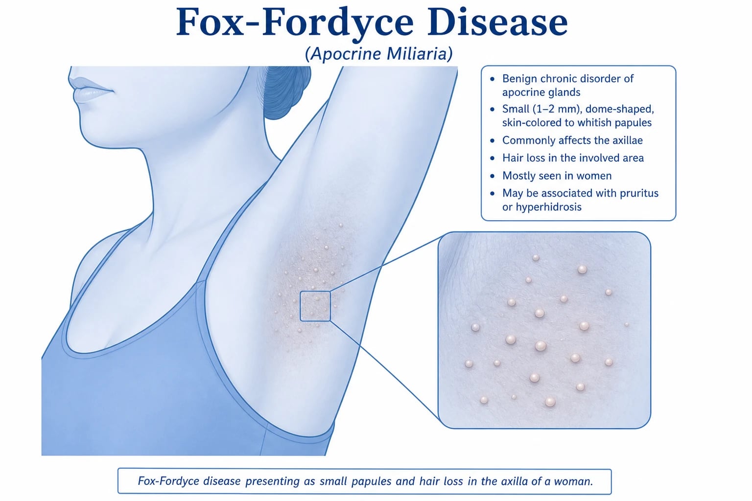
Fox-Fordyce disease — synonymously apocrine miliaria or apocrine retention miliaria — is a chronic, intensely pruritic, papular dermatosis confined to apocrine gland-bearing skin. It is produced by keratinous obstruction of the apocrine sweat duct at its entry into the hair follicle, with subsequent retention of apocrine secretion, ductal dilatation and rupture, and a perifollicular lymphohistiocytic inflammatory reaction that generates the characteristic pruritic papule. The condition was first described by the American dermatologists George Henry Fox and John Addison Fordyce in 1902 in two young women with intensely itchy axillary papules; the eponym is attached to both names.[1][5]
The condition is best understood as the apocrine counterpart of eccrine miliaria (prickly heat). Where eccrine miliaria results from obstruction of the eccrine sweat duct that opens directly onto the skin surface, Fox-Fordyce results from obstruction of the apocrine sweat duct which — uniquely — empties into the hair follicle rather than directly onto the epidermis. This single anatomical fact (apocrine duct enters the follicle) explains every clinical feature of the disease: the follicular distribution, the associated hair loss (the duct and the hair shaft share the follicle), the anhidrosis (the obstructed gland cannot deliver its secretion), and the predilection for the axillae, anogenital region, areolae and periumbilical skin where apocrine glands are concentrated.[1][3]
Why the name "apocrine miliaria"
The term apocrine miliaria emphasises the mechanistic parallel with eccrine miliaria (the obstructive sweat retention disorder of eccrine glands). Both disorders share the same pathogenic triad — duct obstruction → sweat retention → intra-dermal leakage → inflammation — but differ in the gland affected and therefore in distribution, symptom, and demographic.[3]
| Feature | Eccrine miliaria (prickly heat) | Apocrine miliaria (Fox-Fordyce) |
|---|---|---|
| Gland obstructed | Eccrine | Apocrine |
| Duct opens onto | Skin surface directly | Hair follicle |
| Distribution | Widespread — trunk, neck, flexures, occluded skin | Restricted — axillae, anogenital, areolae, periumbilical |
| Demographic | Any age; infants, hot-climate adults | Women 15 to 35 years |
| Trigger | Heat, humidity, occlusion, fever | Hormonal (puberty, menstrual cycle); heat, emotional stress |
| Symptom | Prickling, stinging | Intense pruritus, often premenstrual |
| Hair involvement | Absent | Hair loss in affected follicles |
Classification
Fox-Fordyce disease is not formally subtyped in any widely used classification scheme, but the disease is usefully classified along three axes: [1]
-
By glandular pathology — it belongs to the apocrine gland disorders, alongside hidradenitis suppurativa (obstruction and rupture of the folliculo-apocrine unit with sinus tract formation), bromhidrosis (malodorous apocrine secretion), chromhidrosis (coloured apocrine secretion) and apocrine chromhidrosis. Fox-Fordyce is the purest retention disorder of the apocrine unit — the gland is structurally intact but its duct is obstructed. [1]
-
By clinical course — chronic persistent (the common form, with exacerbations and remissions over years) versus chronic relapsing with prolonged remissions (the form that resolves with pregnancy or menopause). Acute Fox-Fordyce is essentially not described. [1]
-
By site predominance — axillary (over ninety percent of cases, often the only site), anogenital, areolar, periumbilical, and the rare sternal / inframammary / circummary forms. Generalised disease affecting all apocrine areas simultaneously is uncommon; most patients have one or two dominant sites. [1]
A fourth, rarer variant reported in case literature is linear / unilateral Fox-Fordyce disease, in which papules follow a Blaschkoid distribution — probably a mosaic manifestation rather than a true subtype.[2]
Position within the follicular occlusion spectrum
Fox-Fordyce sits at the least destructive end of the follicular occlusion spectrum — a group of conditions driven by occlusion and rupture of the pilosebaceous–folliculo-apocrine unit: [1]
| Disorder | Unit obstructed | Hallmark | Severity |
|---|---|---|---|
| Fox-Fordyce disease | Apocrine duct → follicle | Pruritic papules, anhidrosis, hair loss | Mild–moderate, non-scarring |
| Acne vulgaris | Pilosebaceous follicle | Comedones, inflammatory papules/pustules | Variable |
| Hidradenitis suppurativa | Folliculo-apocrine unit | Painful nodules, abscesses, sinus tracts, scarring | Severe, scarring |
| Dissecting cellulitis of scalp | Scalp follicle | Boggy nodules, sinus tracts, alopecia | Severe, scarring |
| Pilonidal sinus | Sacral follicle | Sacral sinus, abscess | Moderate, recurrent |
The shared pathway is occlusion → rupture → inflammation, but Fox-Fordyce uniquely produces pruritus rather than pain and no sinus tract formation or scarring — features that anchor it at the benign end of the spectrum.[1]
Epidemiology & Risk Factors
Fox-Fordyce disease is uncommon. Exact incidence and prevalence are not established because the condition is under-reported (many patients tolerate the pruritus or are misdiagnosed), but dermatology outpatient series suggest it accounts for a small fraction of pruritic axillary presentations.[1]
Why women 15 to 35
The tight demographic restriction to post-pubertal women of reproductive age is the single most important epidemiological clue, and reflects the androgen-dependent maturation of the apocrine gland: [1]
- Apocrine glands are quiescent until puberty. They enlarge and begin active secretion only under androgenic stimulation at puberty (testosterone and dihydrotestosterone). In prepubertal children the glands are inactive and the duct is not yet a secretory conduit — hence the disease does not occur before puberty.
- Women are over-represented despite both sexes having apocrine glands, suggesting that the oestrogen–androgen balance modulates ductal keratinisation and gland secretion in a way that favours obstruction in women. Premenstrual and pregnancy-related fluctuations in disease activity support a hormonal driver.
- Post-menopausal involution. Apocrine glands involute after the menopause as sex-hormone levels fall — explaining the rarity of new-onset Fox-Fordyce in post-menopausal women and the tendency of established disease to remit after the menopause. [1]
Risk factors and triggers
| Factor | Mechanism |
|---|---|
| Female sex, reproductive age | Hormonal drive to apocrine secretion |
| Heat, humidity, occlusive clothing | Increased apocrine secretion against an obstructed duct |
| Emotional stress | Adrenergic and cholinergic stimulation of apocrine secretion |
| Exercise | Increased sweat output |
| Premenstrual phase | Hormonal modulation of apocrine gland activity — classic premenstrual exacerbation |
| Shaving / depilation / antiperspirants | Mechanical and chemical irritation of the follicular infundibulum |
| Co-existing apocrine / follicular disorders | Hyperhidrosis, follicular occlusion tetrad — overlap reported |
A positive family history is occasionally reported but no Mendelian pattern is established; the disease is not heritable in the usual sense.[2][3]
Pathophysiology
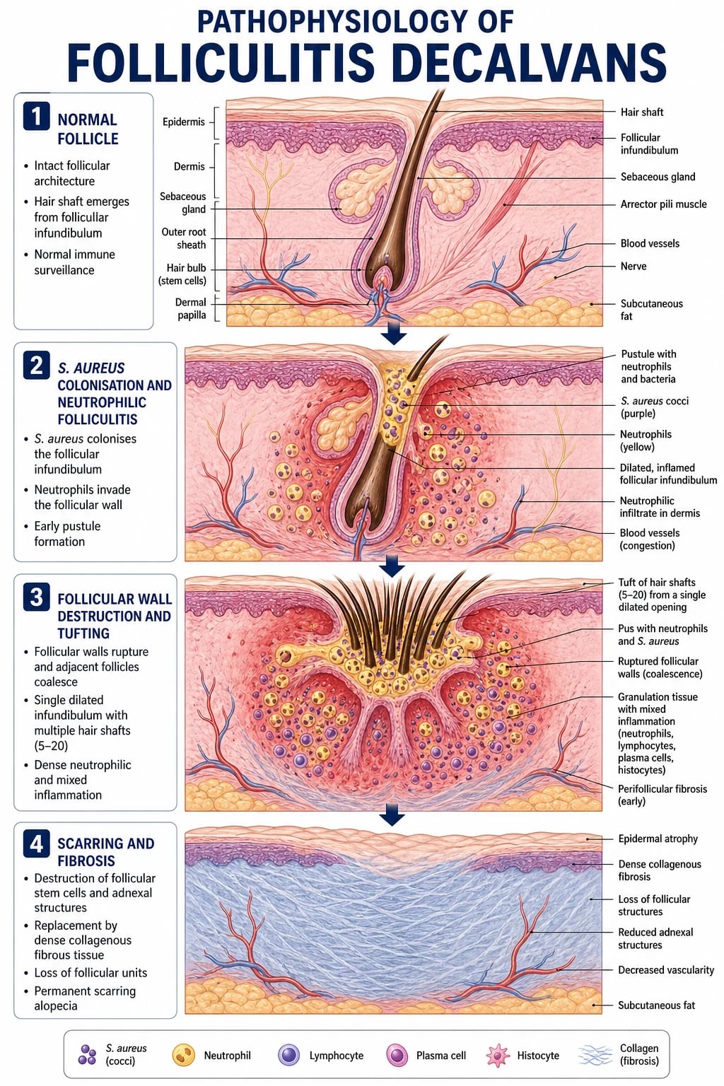
Fox-Fordyce disease is a mechanical retention disorder of the apocrine sweat gland, analogous to miliaria of the eccrine gland. The entire clinical picture flows from a single event — keratinous obstruction of the apocrine sweat duct at its entry into the hair follicle. [1]
Step 1 — Apocrine duct obstruction
Apocrine glands are large, deep-dermal coiled tubular glands that empty their secretion through a duct that opens into the hair follicle (above the entry of the sebaceous duct), in contrast to eccrine glands, whose ducts open directly onto the skin surface. A keratinous plug forms at the intra-follicular portion of the apocrine duct (the infundibular entry point), most plausibly driven by: [1]
- Androgen-mediated ductal keratinisation (the same hormonal milieu that activates the gland at puberty);
- Follicular infundibular hyperkeratosis (mechanically aggravated by shaving, depilation, antiperspirants);
- Possibly an idiopathic local keratinisation defect confined to the apocrine duct epithelium. [1]
Step 2 — Retention of secretion
With the duct obstructed but the gland still actively secreting (under hormonal drive), apocrine secretion accumulates behind the plug, producing progressive ductal dilatation and an intra-dermal retention vesicle / cyst. The retained secretion is rich in protein, lipid and pheromone precursors, and is mildly irritating to surrounding tissue. [1]
Step 3 — Ductal rupture and inflammation
Pressure within the obstructed duct eventually forces focal rupture of the duct wall, with extravasation of apocrine secretion into the surrounding dermis. This elicits a perifollicular lymphohistiocytic inflammatory infiltrate with characteristic foam cells (lipid-laden macrophages that have phagocytosed apocrine lipid). The inflammation is sterile — it is not driven by infection but by the chemical irritancy of the extravasated secretion. The release of inflammatory mediators (prostaglandins, leukotrienes, neuropeptides) is the proximate cause of the intense pruritus.[3]
Step 4 — Clinical lesion formation
The combination of (a) a dilated obstructed duct, (b) a perifollicular inflammatory infiltrate, and (c) overlying infundibular hyperkeratosis produces the firm, dome-shaped, skin-coloured papule characteristic of the disease. Because the obstructed duct shares the follicle with the hair shaft, hair growth is disrupted and the overlying hair is shed — producing the characteristic localised alopecia over each papule. And because the obstructed gland cannot deliver its secretion to the surface, the affected skin is anhidrotic / hypohidrotic.[1]
The hormonal link
The tight restriction of Fox-Fordyce to post-pubertal reproductive-age women, the premenstrual exacerbation, the improvement during pregnancy, and the remission after menopause all point to a hormonal modulation of apocrine gland activity: [1]
- Androgens (testosterone, DHT) drive apocrine gland enlargement and secretion at puberty — necessary but not sufficient for disease.
- Oestrogen–progestogen cyclical changes appear to modulate ductal keratinisation and gland secretion, with many women reporting a premenstrual flare.
- Pregnancy (high oestrogen and progesterone) and menopause (low sex hormones) both tend to suppress apocrine secretion, producing improvement or remission.
- The therapeutic efficacy of combined oral contraceptive pills (OCPs) with anti-androgenic progestins (cyproterone acetate, drospirenone) and of oral isotretinoin (which shrinks sebaceous and apocrine glands) further supports the hormonal aetiology.[1][2]
Clinical Presentation
The clinical picture is stereotyped: a young woman with intensely itchy papules in one or both axillae, often with associated decreased sweating and loss of axillary hair over the affected skin. [1]
Morphology of the lesion
- Size: 1 to 3 mm in diameter.
- Shape: dome-shaped, smooth, firm, rounded papules.
- Colour: skin-coloured or slightly hyperpigmented / yellowish; never brightly erythematous unless secondarily inflamed or infected.
- Surface: smooth; a central keratotic plug or hairless orifice may be visible with a hand lens.
- Arrangement: grouped or scattered clusters, often follicular in distribution; lesions do not coalesce into plaques, do not form sinus tracts, and do not ulcerate.
- Consistency: firm, due to the underlying retention cyst and inflammatory infiltrate.
- Number: typically dozens to over a hundred in a single axilla.[1][4]
Distribution — apocrine gland-bearing skin only
[1]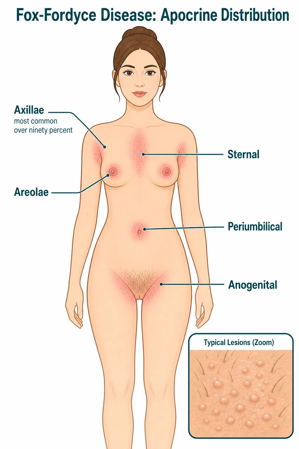
Symptoms
- Intense pruritus is the hallmark and the presenting complaint in the majority. Itching is often severe enough to disturb sleep and significantly impair quality of life.
- Premenstrual exacerbation — classically the itch and the papule count worsen in the days before menstruation, reflecting hormonal modulation of apocrine secretion.
- Heat-, emotion- and exercise-induced worsening — any stimulus that increases apocrine secretion (sympathetic / cholinergic drive, raised body temperature) aggravates symptoms, because more secretion is being driven into an obstructed duct.
- Decreased sweating (anhidrosis / hypohidrosis) in the affected areas — the patient may volunteer that the axilla no longer sweats normally.
- Cosmetic concern — the visible papules and the patchy hair loss cause significant distress. [1]
Hair changes
Because the apocrine duct empties into the hair follicle, obstruction of the duct also disrupts the hair shaft. The affected follicles show thin, brittle hair or complete hair loss, producing a patchy, follicular alopecia within the affected axillary, pubic or periareolar skin. Restoration of hair growth may follow successful treatment. [1]
Natural history and course
- Onset is gradual, usually in the late teens or early twenties.
- Course is chronic, with exacerbations and remissions over months to years.
- Spontaneous remission may occur, especially with pregnancy and menopause (hormonal suppression of apocrine glands).
- No systemic features, no scarring, no sinus tract formation, no malignant transformation — the disease is confined to the skin and is benign. [1]
Atypical presentations
- In men — rare; usually axillary and perineal disease in obese, hirsute young men; the premenstrual / hormonal features are absent and treatment response is poorer.[2]
- Perianal or periumbilical predominance — easily misdiagnosed as folliculitis or contact dermatitis; biopsy may be required.
- Linear / unilateral (Blaschkoid) Fox-Fordyce — rare mosaic form.
- Periareolar / circummary presentation — confused with eczema or Paget disease; the symmetric, papular, follicular morphology and the intense pruritus point to Fox-Fordyce.
- In post-menopausal women — diagnosis should be questioned; consider alternative diagnoses (Darier disease, contact dermatitis, scabies) and biopsy.[3]
Differential Diagnosis
Fox-Fordyce disease must be distinguished from other papular or inflammatory disorders of the axilla, anogenital region and flexures. The single best discriminator is the symptom: Fox-Fordyce itches; hidradenitis suppurativa hurts. [1]
Other differentials
- Folliculitis — pustules (not papules) centred on hair follicles, often with a positive bacterial culture (Staphylococcus aureus); painful rather than intensely pruritic; responds to antibacterial therapy.
- Erythrasma — well-demarcated red-brown, finely scaling patches in the axilla and groin; coral-red fluorescence under Wood's lamp; caused by Corynebacterium minutissimum; not pruritic.
- Darier disease (keratosis follicularis) — greasy, crusted, yellow-brown papules in a seborrhoeic distribution (chest, back, scalp, face, flexures); nail changes (V-shaped notching, longitudinal ridging); palmar pits; autosomal dominant inheritance (ATP2A2 mutation). Differentiated by family history, nail signs and histology (suprabasal acantholysis with dyskeratosis).
- Hailey-Hailey disease (benign chronic pemphigus) — relapsing, moist, crusted plaques in intertriginous areas (neck, axillae, groin); erosions and fissures; autosomal dominant (ATP2C1); painful rather than pruritic; histology shows full-thickness acantholysis ("dilapidated brick wall").
- Granulosis rubra nasi — rare erythema and papules on the nose in children with hyperhidrosis; an eccrine gland disorder; different demographic and site.
- Scabies — intense generalized pruritus, worse at night, with burrows in finger webs, wrists, waistline, genitals; household contacts affected; not confined to apocrine areas.
- Lichen simplex chronicus — lichenified plaque(s) from chronic rubbing; not follicular; not associated with anhidrosis.
- Apocrine carcinoma (very rare) — a rapidly growing, firm, dermal or subcutaneous nodule in an apocrine area; ruled out by biopsy when morphology or behaviour is atypical.[1][3]
The bedside discriminator is the triad of intense pruritus + dome-shaped skin-coloured follicular papules + anhidrosis with hair loss in apocrine areas of a reproductive-age woman — when all four are present the diagnosis is secure. [1]
Clinical & Bedside Assessment
Fox-Fordyce disease is a clinical diagnosis. A focused history and examination are usually sufficient; biopsy is reserved for atypical or refractory cases. [1]
Focused history
- Symptom character — onset, intensity, diurnal pattern (often worse at night), relationship to menstrual cycle (premenstrual exacerbation), heat, emotional stress, exercise.
- Site of onset — usually axillary; ask about other apocrine areas.
- Sweating pattern — has the patient noticed reduced sweating in the affected area?
- Hair pattern — has the patient noticed hair loss over the papules?
- Menstrual and gynaecological history — cycle regularity, premenstrual symptoms, oral contraceptive use (and response, including OCP-triggered or OCP-improved disease), pregnancies, menopausal status.
- Aggravating exposures — antiperspirants, deodorants, shaving, depilation, occlusive clothing.
- Quality-of-life impact — sleep disturbance, work / school impairment, psychological distress. [1]
Examination — a systematic approach
- Inspect both axillae in good light. Look for the typical dome-shaped, skin-coloured, follicular papules; assess distribution, count, and the overlying hair.
- Inspect the anogenital region, areolae, periumbilical and sternal skin. Even if the patient reports only axillary symptoms, examine all apocrine areas — silent involvement is common.
- Assess sweating. The affected axilla feels drier than the contralateral side. An optional starch-iodine test (paint iodine, dust starch — sweat turns the starch blue-black) demonstrates the anhidrotic patch.
- Assess hair. Look for patchy follicular alopecia over the papules.
- Wood's lamp examination of the axilla — to exclude erythrasma (coral-red fluorescence).
- General skin examination — to identify coexisting apocrine or follicular disorders (hidradenitis suppurativa, acne, hyperhidrosis, follicular occlusion tetrad).
- Look for scabies, contact dermatitis, Darier or Hailey-Hailey signs that would redirect the diagnosis. [1]
Named clinical signs and observations
- Fox-Fordyce papule — the firm, dome-shaped, skin-coloured, follicular papule (1 to 3 mm) with central keratotic plug and overlying hair loss; the unit lesion.
- Axillary anhidrosis — the affected axilla is drier than the contralateral side on inspection and on starch-iodine testing.
- Follicular alopecia within the papule — the obstructed apocrine duct disrupts the shared hair follicle, producing a hairless papule.
- Premenstrual exacerbation — a temporal pattern of increased pruritus and papule count in the luteal phase.[4]
Investigations
Diagnosis is clinical in the great majority of cases. Investigations are reserved for confirmation in atypical cases, exclusion of mimics, assessment of secondary infection, and hormonal workup of refractory disease. [1]
Skin biopsy (the definitive test)
A punch biopsy of a characteristic papule shows the diagnostic triad of Fox-Fordyce disease:[3]
- Apocrine duct spongiosis — intercellular oedema (spongiosis) of the intra-epidermal portion of the apocrine duct epithelium at its entry into the follicular infundibulum.
- Parakeratotic plug — a keratinous plug with parakeratosis obstructing the apocrine duct orifice within the follicular infundibulum.
- Retention cyst / dilated apocrine duct filled with retained secretion, with foam cells (lipid-laden macrophages) in the duct lumen and surrounding dermis. [1]
Supporting features include a perifollicular lymphohistiocytic inflammatory infiltrate and follicular hyperkeratosis. The biopsy is diagnostic when it captures an affected apocrine duct, but because the duct is small and sampling is patchy, a single non-diagnostic biopsy does not exclude Fox-Fordyce — serial sectioning or re-biopsy of a fresh papule may be needed. [1]
Dermoscopy
Dermoscopy, as characterised by Srivastava et al. (2020), shows:[4]
- Perifollicular papules with central keratotic plugging.
- Sparse or absent hair within each papule.
- Pale yellowish background corresponding to the retention cyst.
- Absence of the vascular patterns seen in basal cell carcinoma or melanocytic lesions. [1]
Dermoscopy supports a clinical diagnosis non-invasively and helps distinguish Fox-Fordyce from folliculitis, Darier disease and contact dermatitis. [1]
Microbiology
- Bacterial swab of any crusted or pustular lesion — to identify S. aureus secondary infection (which alters management: add a topical or oral antibiotic).
- Wood's lamp examination — to exclude erythrasma (coral-red fluorescence).
- Fungal scraping (KOH) — if tinea corporis / cruris is in the differential. [1]
Hormonal workup (selected cases)
In refractory disease or when hyperandrogenism is suspected (hirsutism, acne, irregular menses), check: [1]
- LH, FSH, total and free testosterone, DHEA-S, prolactin, 17-hydroxyprogesterone, sex-hormone-binding globulin — to identify polycystic ovary syndrome, congenital adrenal hyperplasia, or androgen-secreting tumour as a driver of apocrine hyperactivity. [1]
Patch testing
If contact dermatitis is a serious consideration, patch testing to standard series (and to the patient's own deodorant / antiperspirant) is appropriate before committing to hormonal therapy. [1]
What is NOT needed routinely
- No blood tests are required in a typical case.
- No imaging is required.
- No allergy testing is required. [1]
Management — Resuscitation
Fox-Fordyce disease is not a dermatological emergency. There is no time-critical bundle. "Resuscitation" in this context means acute symptom control and patient counselling at the first visit. [1]
Acute pruritus control
- Topical corticosteroid — betamethasone valerate 0.1 percent cream or clobetasol propionate 0.05 percent cream applied twice daily for two to four weeks to the affected axilla to settle the inflammatory reaction and itch. Use a moderate or potent steroid for short courses only, because the axilla is an intertriginous (thin-skinned) area prone to atrophy and striae with prolonged potent steroid use.
- Oral antihistamine — cetirizine 10 mg nocte or chlorphenamine 4 mg at night for itch-related sleep disturbance; sedating antihistamines are preferred for nocturnal itch.
- Avoidance of triggers — heat, occlusive clothing, sweating, harsh deodorants / antiperspirants, and aggressive shaving until the acute flare settles. [1]
Patient counselling at the first visit
- Diagnosis and natural history — explain the chronic relapsing nature, the hormonal influence, and the tendency to improve with pregnancy and menopause.
- Reassurance — Fox-Fordyce is benign, non-contagious, non-scarring and carries no malignancy risk.
- Quality of life — acknowledge the impact of severe pruritus on sleep and mood; offer ongoing support.
- Treatment ladder — set realistic expectations: topical therapy controls but rarely cures; recurrence is common after stopping therapy. [1]
When to suspect and treat secondary infection
- Yellow crust, surrounding erythema, tenderness, pustule formation, regional lymphadenopathy — suggests Staphylococcus aureus secondary infection from scratching.
- Swab the lesion.
- Topical antibiotic — mupirocin 2 percent ointment or fusidic acid 2 percent cream TDS for 7 to 10 days for localised infection.
- Oral antibiotic — flucloxacillin 500 mg QDS (or cefalexin 500 mg QDS in penicillin-allergic patients without anaphylaxis) for 7 days for cellulitis or extensive infection. [1]
When to refer urgently
- Suspicion of an alternative diagnosis — painful abscesses / sinus tracts (hidradenitis suppurativa), rapidly growing nodule (apocrine carcinoma), atypical morphology (Darier, Hailey-Hailey) — refer to dermatology for biopsy.
- Severe or refractory disease unresponsive to topical therapy — refer for systemic or procedural options.
- Psychological distress — refer for counselling or psychological support if pruritus and cosmetic concern are causing depression or anxiety. [1]
Management — Definitive & Stepwise
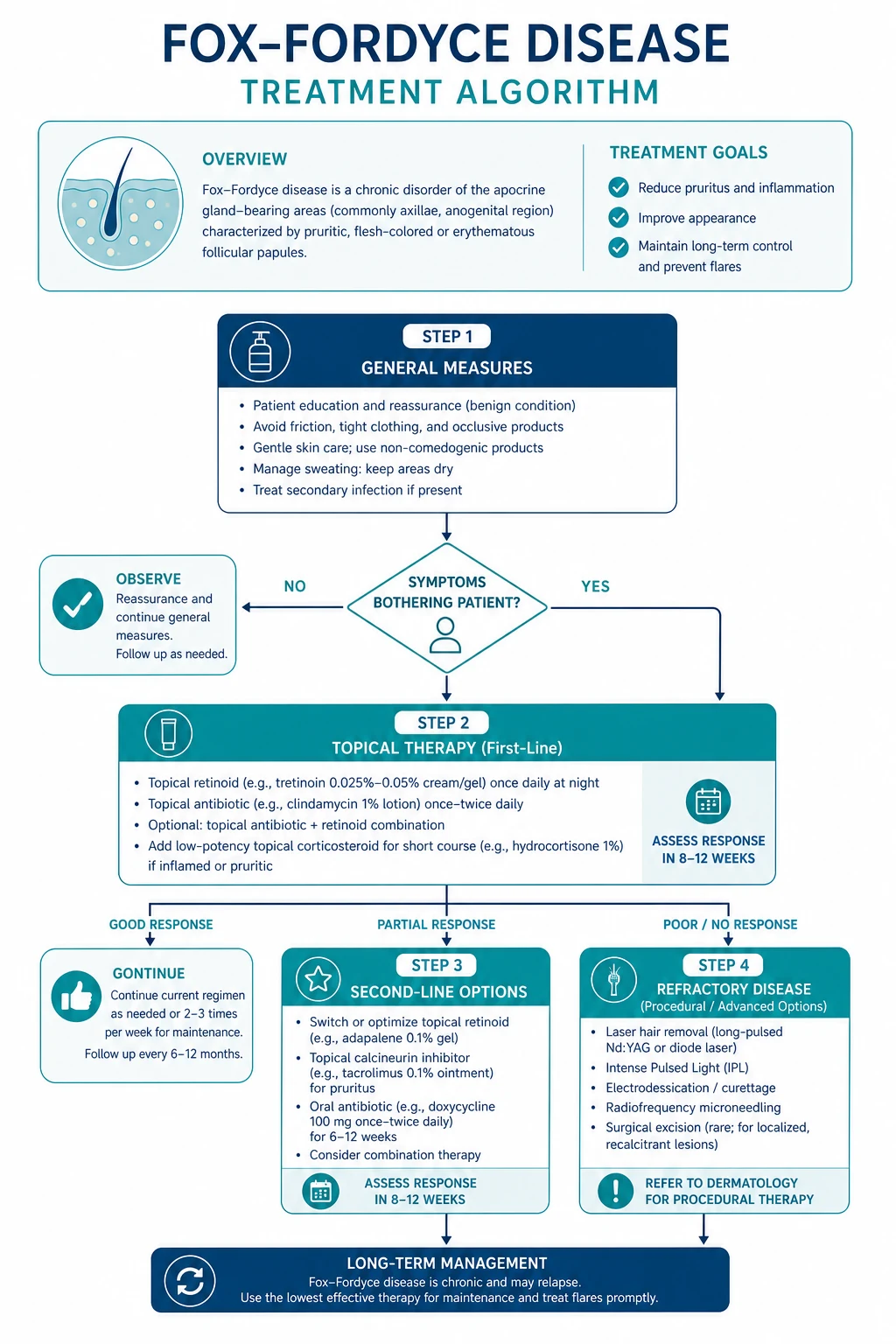
Treatment is stepwise, escalating from topical to hormonal to systemic to procedural therapies. No treatment reliably cures the disease; the goal is control of pruritus and reduction of papule count with the least invasive effective regimen.[1]
First-line therapy
- Topical clindamycin 1 percent lotion BD — the anti-inflammatory and antibacterial action reduces the perifollicular inflammatory infiltrate and any secondary staphylococcal colonisation. Clindamycin 1 percent lotion BD is widely recommended as first-line therapy in the European / UK and Indian literature.[1]
- Topical retinoid — tretinoin 0.025 to 0.1 percent cream applied nocte, or adapalene 0.1 percent gel nocte. Retinoids normalise follicular keratinisation, reduce the keratinous plug that obstructs the apocrine duct, and have additional anti-inflammatory effects. Begin at the lower strength to limit irritation; combine with a steroid-sparing emollient. Avoid in pregnancy (topical retinoids are contraindicated in pregnancy — switch to clindamycin alone).
- Topical corticosteroid — betamethasone valerate 0.1 percent or clobetasol propionate 0.05 percent cream twice daily for two to four weeks to settle acute pruritus and inflammation. Use short courses only to avoid axillary atrophy and striae; taper to a mild steroid (hydrocortisone 1 percent) or to calcineurin inhibitor (tacrolimus 0.1 percent ointment) for maintenance.
- Intralesional triamcinolone acetonide — 5 to 10 mg/mL injected into a few stubborn individual papules that resist topical therapy.[1]
Second-line therapy (refractory or relapsing disease)
-
Combined oral contraceptive pill (COCP) with an anti-androgenic progestin — the hormonal mainstay. Examples:
- Ethinylestradiol 35 micrograms + cyproterone acetate 2 mg (co-cyprindiol, "Diane-35") — once daily for 21 days of a 28-day cycle.
- Ethinylestradiol 30 micrograms + drospirenone 3 mg — once daily for 21 of 28 days.
- Mechanism: suppresses pituitary LH / FSH → reduces ovarian androgen production → reduces androgen-driven apocrine gland secretion and ductal keratinisation.
- Indication: women with premenstrual exacerbation, hyperandrogenic features, or disease resistant to topical therapy. Note: OCPs may improve Fox-Fordyce in most women but have paradoxically triggered the disease in case reports — review the response at three months.
- Cautions: standard COCP contraindications (smoker over 35, migraine with aura, prior VTE, breast cancer, uncontrolled hypertension). Avoid in pregnancy.[1][2]
-
Topical calcineurin inhibitor — tacrolimus 0.1 percent ointment or pimecrolimus 1 percent cream BD as a steroid-sparing anti-inflammatory for long-term use in intertriginous skin, where they avoid the atrophy of long-term topical corticosteroid. [1]
Third-line / refractory therapy
-
Oral isotretinoin — 0.5 to 1 mg/kg/day for 4 to 6 months. Isotretinoin shrinks sebaceous and apocrine glands and normalises follicular keratinisation; it has been reported effective in refractory Fox-Fordyce. However:
- Recurrence is common after discontinuation.
- Teratogenicity (Category X) — the population most affected (women of reproductive age) is precisely the population in whom isotretinoin is most problematic; rigorous contraception (two methods) and pregnancy testing before, during and after therapy are mandatory.
- Monitoring — lipids (fasting triglycerides, cholesterol) and LFTs at baseline, 1 month, and 3-monthly; assess for depression and suicidal ideation; expect mucocutaneous dryness (lip balm, moisturiser, artificial tears).
- Paradoxical initial flare is recognised — may require a short course of oral prednisolone cover.[2]
-
Oral prednisolone — short course (e.g. 0.5 mg/kg/day for 1 to 2 weeks, tapering) for acute severe flares that are unresponsive to topical therapy and that disrupt sleep or work. [1]
Physical / procedural treatments (resistant localised disease)
- Electrocautery / electrodessication of individual papules — destroys the obstructed duct; useful for a few refractory papules; may leave small scars.
- CO2 laser ablation — vaporises the papules and the obstructed duct; effective in resistant disease; risk of scarring and post-inflammatory pigment change, particularly in darker skin types.
- Photodynamic therapy (PDT) with methyl aminolevulinate (MAL) or 5-aminolaevulinic acid (5-ALA) — case series report good efficacy in refractory Fox-Fordyce; mechanism is selective destruction of the inflamed, photosensitised apocrine unit. Limited availability in resource-poor settings.[1]
- Botulinum toxin A injection — intradermal injection (e.g. 50 units per axilla distributed across 10 to 20 sites) blocks the cholinergic stimulation of apocrine secretion; reduces pruritus and papule count for 3 to 6 months. Off-label; main use is in patients who cannot take systemic therapy.[1]
- Surgical excision — radical excision of severely affected axillary skin; rarely required and reserved for the most refractory localised disease; risks scarring and contracture.
Stepwise escalation ladder
The Fox-Fordyce treatment ladder — 'CRISP-H'
Specific Subtypes & Scenarios
Premenstrual-exacerbating Fox-Fordyce
The most common pattern. History of cyclical flare in the luteal phase; the COCP with an anti-androgenic progestin (cyproterone acetate or drospirenone) is the specific therapy — it abolishes the cyclical hormonal surge and reduces apocrine secretion. Topical therapy continues in parallel.[1]
Fox-Fordyce in men
Rare. Usually axillary and perineal disease in young, often obese or hirsute men. The premenstrual / hormonal features are absent, the disease may be more refractory, and OCPs are obviously not an option. Treatment relies on topical clindamycin + retinoid, intralesional steroid, oral isotretinoin, and procedural options. Identify and treat any underlying hyperandrogenism if present.[2]
Fox-Fordyce and the follicular occlusion tetrad
Fox-Fordyce may co-exist with hidradenitis suppurativa, dissecting cellulitis of the scalp, pilonidal sinus and acne conglobata — the follicular occlusion tetrad / triad. When the two diseases co-exist, the clinical picture is mixed (pruritic papules plus painful sinus tracts) and management must address both. Recognising the overlap prevents mislabelling the pruritic papules as "early HS" and withholding effective topical therapy. [1]
Resistant / refractory Fox-Fordyce
Disease unresponsive to 3 to 6 months of optimal topical + hormonal therapy should be referred to a dermatologist for consideration of oral isotretinoin, photodynamic therapy, CO2 laser ablation or botulinum toxin. Re-verify the diagnosis with biopsy before committing to systemic or procedural therapy — a treatment-resistant "Fox-Fordyce" may in fact be Darier disease, contact dermatitis or folliculitis.[3]
Paediatric / peri-pubertal Fox-Fordyce
Rare but reported in early puberty. Avoid oral isotretinoin in younger adolescents unless under specialist supervision. Treat with topical clindamycin + a mild topical corticosteroid; reserve topical retinoid for older adolescents; consider COCP (with gynaecology input) in post-menarche girls with premenstrual flares.[3]
Complications & Pitfalls
Complications
- Severe pruritus — sleep disturbance, irritability, impaired work or school performance, anxiety and depression. Quality-of-life impact is the principal morbidity.
- Secondary bacterial infection — Staphylococcus aureus infection of excoriated papules, producing impetiginisation, crusting, regional lymphadenopathy, occasionally cellulitis.
- Lichenification — chronic rubbing produces thickened, leathery, hyperpigmented skin in the affected axilla.
- Post-inflammatory hyperpigmentation — especially in darker skin types, after excoriation or procedural treatment.
- Scarring — generally not a feature of Fox-Fordyce itself, but may follow secondary infection or aggressive procedural treatment (electrocautery, CO2 laser).
- Steroid atrophy and striae — from prolonged potent topical corticosteroid use in the axilla.
- Cosmetic and psychological distress from the visible papules and patchy axillary alopecia. [1]
Pitfalls
Pitfalls in Fox-Fordyce disease — 'DIAGNOSE'
The most common diagnostic pitfall is mislabelling hidradenitis suppurativa as Fox-Fordyce (or vice versa). The cardinal discriminator — pruritus (Fox-Fordyce) versus pain (HS) — resolves the dilemma at the bedside; sinus tracts, abscesses and scarring are absent in Fox-Fordyce and present in HS.[1]
Prognosis & Disposition
Prognosis
- Chronic, relapsing course with exacerbations and remissions over years.
- Spontaneous remission may occur, particularly with pregnancy and menopause (hormonal suppression of apocrine glands).
- No cure — treatment controls symptoms and papule count but recurrence is common after therapy is stopped, especially after topical or systemic retinoids.
- No systemic involvement, no scarring (in untreated disease), no malignant transformation — the disease is confined to the skin and is benign throughout its course.
- Quality of life is the principal determinant of severity; intractable pruritus may cause significant morbidity.[1]
Predictors of improvement
| Factor | Effect |
|---|---|
| Pregnancy | Usually improves (high oestrogen / progesterone suppresses apocrine secretion) |
| Menopause | Often remits (involution of apocrine glands) |
| Effective hormonal therapy (OCP) | Improves most women; may trigger paradoxically in a minority |
| Sustained topical therapy | Controls but rarely cures |
| Co-existing hyperhidrosis | May worsen disease activity |
Disposition
- Primary care / general practice — initiate first-line topical therapy and patient counselling.
- Dermatology referral — for refractory disease (failure of 3 to 6 months of topical therapy), diagnostic uncertainty (atypical morphology requiring biopsy), procedural options (PDT, CO2 laser, botulinum toxin, electrocautery), and consideration of oral isotretinoin.
- Gynaecology / reproductive medicine — for OCP selection in women with contraindications or those planning pregnancy.
- Psychology / counselling — for patients with significant sleep, mood or quality-of-life impact. [1]
Special Populations
Pregnancy
Fox-Fordyce disease often improves during pregnancy (the high oestrogen and progesterone environment suppresses apocrine secretion). Management in pregnancy is conservative: [1]
- First line: topical clindamycin 1 percent lotion BD (safe in pregnancy).
- Avoid topical and oral retinoids (tretinoin, adapalene, isotretinoin — Category X, absolutely contraindicated).
- Avoid oral corticosteroid in the first trimester (oral cleft risk); short courses after the first trimester are acceptable if essential.
- Topical corticosteroid — use low-potency (hydrocortisone 1 percent) or moderate-potency short courses; avoid potent and very potent steroids, especially in the third trimester (risk of low birth weight with high cumulative dose).
- OCPs are contraindicated in pregnancy — discontinue on conception.
- Reassure: the disease is benign, does not affect the fetus, and is expected to improve.[1]
Women planning pregnancy
- Discontinue oral isotretinoin at least one month (ideally three months) before conception — teratogenicity (craniofacial, cardiac, thymic, CNS defects).
- Switch to topical clindamycin ± a low-potency topical corticosteroid.
- Consider post-partum return to retinoid or OCP therapy if breast-feeding is completed. [1]
Breast-feeding
- Topical clindamycin — safe.
- Topical corticosteroid — low-potency preferred; moderate potency short courses acceptable.
- Avoid topical / oral retinoids.
- Avoid COCP in the first 6 weeks post-partum (VTE risk); progestogen-only pill is acceptable after 6 weeks. [1]
Men
Rare disease; treat as for refractory non-hormonal Fox-Fordyce — topical clindamycin + retinoid ± intralesional steroid ± oral isotretinoin ± procedural options. Identify and treat any underlying hyperandrogenism. OCPs are not applicable.[2]
Adolescents / peri-pubertal
- Initiate with topical clindamycin + mild-to-moderate topical corticosteroid.
- Add a topical retinoid (start at the lowest strength) for older adolescents.
- Consider COCP (with paediatric / gynaecology input) for post-menarche girls with premenstrual flares.
- Reserve oral isotretinoin for severe refractory disease under specialist supervision; ensure robust contraception in sexually active adolescents. [1]
Post-menopausal women
Fox-Fordyce is rare in this group — the diagnosis should be questioned. Consider and exclude contact dermatitis, scabies, Darier disease, lichen simplex chronicus and (for atypical nodular lesions) apocrine carcinoma. Biopsy if there is any diagnostic doubt. [1]
Immunocompromised patients
Approach is unchanged, but:
- Secondary staphylococcal infection is more common and more aggressive — have a low threshold for swab and antibiotic therapy.
- Topical therapy is preferred to minimize systemic immunosuppression.
- OCPs, isotretinoin and procedural options are used with the standard cautions.[1]
Evidence, Guidelines & Regional Differences
Strength of evidence
The evidence base for Fox-Fordyce disease is limited — the disease is uncommon, no randomised controlled trials exist, and most recommendations rest on case series, retrospective reviews and expert opinion.[1]
[1]Key evidence sources
- Borman et al. (StatPearls, 2026, PMID 31424791) — comprehensive, regularly updated review that lays out the topical-clindamycin-first, retinoid-second, OCP-third, isotretinoin-and-procedural-fourth ladder; the de facto reference standard.[1]
- Tetzlaff et al. (Dermatology Online Journal, 2016, PMID 26909204) — emphasises the histopathological triad (apocrine duct spongiosis, parakeratotic plug, retention cyst with foam cells) and reviews the differential diagnosis from hidradenitis suppurativa and Darier disease.[3]
- Srivastava et al. (Indian Dermatology Online Journal, 2020, PMID 32903893) — characterised the dermoscopic features (perifollicular papules with central keratotic plugging; sparse or absent hair within each papule; pale yellowish background) and added a non-invasive diagnostic tool.[4]
- Sahin et al. (2018, PMID 29641729) — reviewed the role of oral isotretinoin in refractory disease and noted the high recurrence rate after discontinuation, the teratogenicity constraint, and the need for combined topical + hormonal therapy.[2]
- Fox and Fordyce (1902 / 1936, PMID 19990737) — the original description of the disease in two young women with intensely pruritic axillary papules.[5]
Controversies
- Clindamycin resistance — long-term topical clindamycin selects for resistant staphylococci; rotating with benzoyl peroxide or a topical retinoid mitigates resistance.
- Role of isotretinoin — effective but teratogenic in the population most affected; some authors reserve it strictly for refractory disease, others use it earlier in non-reproductive-age or reliably-contracepting patients.
- Botulinum toxin — limited case-series evidence; cost and repeat-injection requirement restrict its use.
- OCP as trigger versus therapy — OCPs usually improve Fox-Fordyce but have paradoxically triggered the disease in case reports; the individual response should be reviewed at three months and the OCP switched or stopped if worsening occurs. [1]
Exam Pearls
[1]Red Flags
Exam application bank (NEET-PG / INICET)
One-line answer
Fox-Fordyce disease (apocrine miliaria) is a chronic, intensely pruritic papular dermatosis of apocrine gland-bearing skin — the axillae, anogenital area, areolae, periumbilical and sternal regions — produced by keratinous obstruction of the apocrine sweat duct at its entry into the hair follicle, retention of secretion, ductal dilatation and rupture, and a perifollicular inflammatory reaction. It predominantly affects women aged 15 to 35 years (post-pubertal, reproductive age), is rare in men, children and post-menopausal women, and frequently worsens premenstrually. The hallmark is intensely pruritic, dome-shaped, skin-coloured, 1 to 3 mm papules with anhidrosis and overlying hair loss in the affected follicles. Diagnosis is clinical, supported by histology (apocrine duct spongiosis, parakeratotic plug, retention cyst with foam cells). First-line treatment is topical clindamycin 1 perc
Worked stems (answer without another resource)
Stem 1 — Classic presentation. Map symptoms to mechanism; name the first investigation and first treatment step with dose/route if drug therapy is standard. [1]
Stem 2 — Unstable / complicated. List red flags that force immediate resuscitation, theatre, ICU, antidote, or reperfusion — and what you do in the first 15 minutes. [1]
Stem 3 — Atypical group. Elderly, pregnancy, child, or immunocompromised: how presentation and thresholds change. [1]
Stem 4 — Differential trap. Name the three closest mimics and one discriminator for each. [1]
Stem 5 — Disposition. Who goes home with safety-netting, who is admitted, who needs HDU/ICU/theatre, and what follow-up is mandatory. [1]
Rapid viva checklist
- Definition + classification
- Pathophysiology chain
- Bedside signs / criteria
- Score with exact components (if any)
- Emergency bundle
- Definitive therapy with doses
- Complications of disease and of treatment
- Special populations
- Guideline/trial name if classic
- Three exam traps
Coverage self-check
If you cannot answer any stem above from this page alone, re-read the matching section — the page is intended to be self-sufficient for final-prof and NEET-PG/INICET questions on Fox-Fordyce disease (apocrine miliaria).
[1]References
- [1]Borman K, et al. Fox-Fordyce Disease (Apocrine Miliaria) 2026.PMID 31424791
- [2]Sahin MO, et al. Fox-Fordyce disease An Bras Dermatol, 2018.PMID 29641729
- [3]Tetzlaff MT, et al. Fox-Fordyce Disease Sultan Qaboos Univ Med J, 2016.PMID 26909204
- [4]Srivastava N, et al. Fox-Fordyce Disease: Dermoscopic Perspective Skin Appendage Disord, 2020.PMID 32903893
- [5]Fox GH, Fordyce JA. Fox-Fordyce Disease Proc R Soc Med, 1936.PMID 19990737