Dermatology · Medicine
Frontal fibrosing alopecia (FFA)
Also known as Frontal fibrosing alopecia (FFA) · Kossard's alopecia
Frontal fibrosing alopecia (FFA) is a primary lymphocytic scarring (cicatricial) alopecia characterised by progressive, symmetric recession of the frontal and temporal hairline, predominantly in post-menopausal Caucasian women. First described by Kossard in 1994, it is considered a clinical variant of lichen planopilaris (LPP). Clinical hallmarks include perifollicular erythema and hyperkeratosis at the active hairline margin, loss of eyebrows (approximately 50 to 80 percent), and lonely hairs (isolated terminal hairs within the receded zone). Histology reveals a perifollicular lymphocytic (lichenoid) infiltrate around the upper follicle with basaloid degeneration, loss of sebaceous glands, and destruction of bulge-region stem cells, resulting in irreversible scarring. Treatment with topical and intralesional corticosteroids, 5-alpha-reductase inhibitors (finasteride or dutasteride), and hydroxychloroquine aims to halt progression, as existing hair loss is permanent.
On this page & tools
Your progress
Saved locally on this device.
Exam tags
Red flags
Overview & Definition
Frontal fibrosing alopecia (FFA), eponymously Kossard's alopecia after Steven Kossard who first described the condition in 1994 in a series of 16 post-menopausal women from Sydney, is a primary lymphocytic scarring (cicatricial) alopecia characterised by progressive, symmetric recession of the frontal and temporal hairline.[5] It is now the most commonly encountered primary cicatricial alopecia in specialist hair clinics worldwide, and its incidence has risen dramatically over the past two decades for reasons that are incompletely understood.[1]
FFA is classified within the lymphocytic subgroup of primary cicatricial alopecias and is widely regarded as a clinical variant of lichen planopilaris (LPP), sharing the same histopathological signature of a perifollicular lymphocytic (lichenoid) interface dermatitis. What distinguishes FFA from classic LPP is its anatomical distribution: FFA produces a band-like recession of the frontotemporal hairline (and frequently the eyebrows), whereas classic LPP affects the scalp in a patchy, multifocal pattern with no predilection for the frontal margin.[3]
The defining biological principle is that FFA is a scarring (cicatricial) alopecia. Once a follicle has been destroyed and replaced by fibrous tissue, it cannot regenerate — the hair loss is permanent and irreversible. This is because the inflammatory attack targets follicular stem cells in the bulge region (located at the insertion of the arrector pili muscle), which are the regenerative niche responsible for cyclic hair renewal. Destruction of these stem cells abolishes the follicle's capacity to produce a new hair shaft. The primary goal of treatment is therefore to halt further recession, not to regrow lost hair.[1][2]
Classification
FFA is best understood within the working classification of primary cicatricial alopecias, which is divided by the dominant inflammatory cell on histology into three groups. This classification is examinable because it dictates biopsy interpretation, differential diagnosis, and treatment approach.[3][5]
- Lymphocytic|Lymphocytes (CD8+ T-cells)|Lichen planopilaris; frontal fibrosing alopecia; central centrifugal cicatricial alopecia (CCCA); discoid lupus erythematosus; pseudopelade of Brocq|Perifollicular erythema and hyperkeratosis; NO pustules
- Neutrophilic|Neutrophils|Folliculitis decalvans; dissecting cellulitis of the scalp; acne keloidalis nuchae|Pustules, crusting, tufting, S. aureus
- Mixed|Neutrophils + lymphocytes + plasma cells|Acne necrotica; erosive pustular dermatosis|Both pustules and scarring in atypical distribution
FFA sits squarely in the lymphocytic group. The practical consequence is that the clinical morphology shows perifollicular erythema and hyperkeratosis at the active margin (not pustules), and the treatment strategy targets the lymphocytic inflammatory cascade with corticosteroids, antimalarials, and 5-alpha-reductase inhibitors — not antibiotics or anti-neutrophilic agents.[3]
Within the lymphocytic group, FFA is distinguished from its siblings by its band-like frontotemporal distribution and its demographic predilection for post-menopausal women. Classic LPP produces patchy, multifocal scarring anywhere on the scalp; CCCA produces centrifugal scarring from the crown in women of African descent; DLE produces well-demarcated inflammatory plaques with follicular plugging; and pseudopelade of Brocq produces non-inflammatory "footprints in the snow" scarring.[5]
[1]Epidemiology & Risk Factors
FFA has undergone a remarkable epidemiological shift since its original description. Kossard's 1994 series described a rare condition; today, FFA is the most frequently diagnosed primary cicatricial alopecia in many specialist hair clinics, and its incidence continues to rise globally.[1][4]
- Age: almost exclusively post-menopausal women, with a mean age at onset of 56 to 70 years. Rare cases in pre-menopausal women and men are reported but are the exception.
- Sex: strong female predominance, with a female-to-male ratio of approximately 6 to 1. Male FFA tends to present with a different pattern (more temple involvement, less eyebrow loss) and is easily missed.
- Ethnicity: predominantly Caucasian women of European descent, particularly those with Fitzpatrick skin types I to III. However, FFA is increasingly reported in all ethnicities, including South Asian, East Asian, African, and Hispanic populations, paralleling the global rise.
- Geography: the highest reported incidences are from Europe (particularly the United Kingdom, Spain, France, and Germany), Australia (reflecting Kossard's original Sydney cohort), and North America. [1]
The rising incidence. The dramatic increase in FFA diagnoses over the past 20 years is one of the most discussed phenomena in trichology. Several factors contribute:[4]
- Increased awareness and recognition among dermatologists and trichologists — what was previously misdiagnosed as traction alopecia or androgenetic alopecia is now correctly identified as FFA.
- An apparent true increase in incidence, beyond what recognition alone explains. Large cohort studies from Spain, the United Kingdom, and Singapore show a sustained year-on-year rise in new diagnoses.
- A putative environmental trigger — the leading hypothesis implicates leave-on facial products and sunscreens applied to the forehead and frontal hairline. The temporal correlation between the widespread adoption of daily sunscreen use (particularly among Caucasian women from the 1990s onward) and the rise of FFA is striking, but a definitive causal link has not been proven.[4]
Risk factors. [1]
- Post-menopausal status — the single strongest demographic association; oestrogen deficiency may increase follicular vulnerability to autoimmune attack.[1]
- Use of leave-on facial products, particularly sunscreens — applied daily to the forehead and frontal hairline; several case-control studies have found a statistically significant association. The specific causative ingredient has not been identified (candidates include titanium dioxide, zinc oxide, and various preservatives).[4]
- Coexisting autoimmune disease — a personal or family history of autoimmune thyroid disease, vitiligo, or alopecia areata is reported in a subset of patients, suggesting an autoimmune diathesis.
- Family history — rare familial clusters of FFA have been reported; HLA associations are under investigation but no consistent HLA allele has been confirmed.[1]
In Indian and South Asian practice, FFA is increasingly recognised but often presents late with extensive hairline recession (3 to 5 cm) because of delayed referral and the common misattribution of frontal recession to traction from traditional hairstyles (oiling, tight braiding, judas). The rising use of facial cosmetics and sunscreens in urban Indian women is paralleling the Western epidemiological trend. Pigmentary change (post-inflammatory hyperpigmentation in the receded zone) is more pronounced in skin of colour and is a common presenting complaint.
Pathophysiology
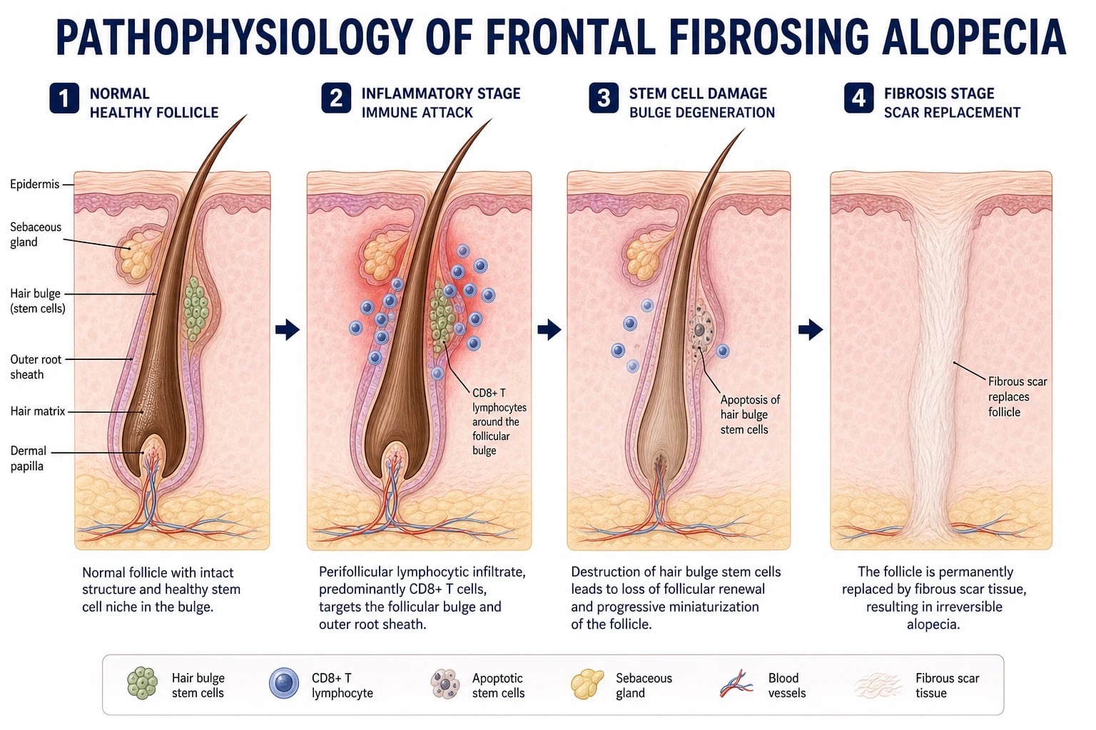
FFA is a lymphocytic scarring alopecia mediated by a perifollicular lichenoid interface dermatitis. The currently accepted pathogenic cascade has four linked steps, although the precise initiating antigen remains unknown.[4]
Step 1 — Antigenic trigger at the upper follicle. An unknown antigen initiates a lichenoid inflammatory response directed against the upper portion of the hair follicle (the infundibulum and isthmus). The leading hypothesis is that a component of leave-on facial products or sunscreens (applied to the forehead and frontal hairline, where the disease begins) acts as the trigger, either directly or via a hapten mechanism. The striking anatomical correlation — disease begins precisely where facial cosmetics are applied — supports this. An alternative (or complementary) hypothesis invokes hormonal factors: the post-menopausal state (oestrogen deficiency) may render the upper follicle more vulnerable to immune attack.[4]
Step 2 — Lymphocytic (lichenoid) infiltrate around the upper follicle. A dense perifollicular infiltrate of T-lymphocytes — predominantly CD8+ cytotoxic T-cells — accumulates in a band-like pattern around the upper follicle, producing a lichenoid interface dermatitis. The lymphocytes attack the basal layer of the outer root sheath, causing vacuolar (basaloid) degeneration of the follicular epithelium and apoptosis of follicular keratinocytes. This is the same histopathological process seen in classic lichen planopilaris and cutaneous lichen planus — the reason FFA is classified as an LPP variant.[3][4]
Step 3 — Destruction of bulge-region stem cells and sebaceous glands. The inflammatory attack extends to the hair follicle bulge — the region at the insertion of the arrector pili muscle that houses the follicular stem cell niche. These stem cells are essential for cyclic hair regeneration (each hair cycle requires repopulation of the follicular matrix from the bulge). Their destruction means the follicle cannot initiate a new anagen phase. Concurrently, the sebaceous glands are destroyed, contributing to the clinical appearance of dry, shiny, scarred skin.[1]
Step 4 — Fibrosis and permanent scarring. Once the stem cells and follicular epithelium are destroyed, the space is replaced by dense fibrous (collagen) tissue. No follicular structures remain — no hair shafts, no follicular ostia, no sebaceous glands. This is why the alopecia is permanent and irreversible: there is no regenerative capacity left. Treatment at this stage can only halt further recession at the active margin; the scarred zone cannot recover.[1][2]
The role of androgens and 5-alpha-reductase. The observation that 5-alpha-reductase inhibitors (finasteride, dutasteride) are among the most effective treatments for FFA — despite the disease predominantly affecting post-menopausal women with low androgen levels — points to a role for the androgen pathway in pathogenesis. The hair follicle expresses 5-alpha-reductase, and local dihydrotestosterone (DHT) may sustain the inflammatory or degenerative process. Blocking DHT production appears to modulate the perifollicular inflammatory milieu, even in the setting of systemic oestrogen deficiency. This is an area of active investigation.[1]
The pathophysiology of FFA in four steps
Clinical Presentation
The clinical presentation of FFA is highly characteristic and, in a classic case, the diagnosis can be made clinically at the bedside. The tempo is slowly progressive, typically unfolding over 3 to 6 years before the patient presents.[5]
Hallmark: progressive, symmetric frontotemporal hairline recession. The defining feature is a slowly advancing, symmetric band of recession affecting the frontal and temporal hairline. The hairline moves posteriorly, creating a broad, high forehead. The band of recession is typically 0.5 to 3 cm wide at presentation, though severe cases may show more than 5 cm of recession. The recession is symmetric in most cases and follows the natural contour of the original hairline, preserving the temporal peaks in early disease and obliterating them as it advances. A key feature is that hair density behind the frontal band is preserved — the disease affects the leading edge, not the entire scalp.[1]
Perifollicular erythema and hyperkeratosis at the active margin. At the leading edge of the receding hairline (the active margin), individual hair follicles show perifollicular erythema (pink-red halos around emerging hair shafts) and perifollicular hyperkeratosis (white scale or keratotic plugs around the hair shaft). These are the trichoscopic and clinical correlates of active inflammation — they indicate disease activity and guide where intralesional corticosteroid injections should be targeted.[5]
Lonely hairs. A characteristic and diagnostically helpful sign is the presence of isolated terminal hairs standing alone within the receded zone — the so-called lonely hair sign. These are remnants of the original hairline that have survived the inflammatory destruction, standing as lone sentinels in an otherwise smooth, hairless band. Their presence strongly supports FFA over traction alopecia (where lonely hairs are absent).[1]
Eyebrow loss. Partial or complete loss of the eyebrows occurs in approximately 50 to 80 percent of patients and is one of the most useful distinguishing features. Eyebrow loss typically begins laterally and progresses medially. Crucially, eyebrow loss may precede the scalp hair loss by months or years, and patients may attribute it to ageing. The combination of eyebrow loss plus frontal hairline recession in a post-menopausal woman is virtually pathognomonic for FFA.[1][5]
Body hair loss. Loss of body hair — particularly the axillary, pubic, and limb hair — is reported in a subset of patients and reflects the systemic nature of the follicular autoimmune process. This is easily overlooked unless specifically asked about.[1]
Facial papules. Small, flesh-coloured to erythematous perifollicular papules on the temples, cheeks, and forehead are seen in some patients and represent follicular involvement of facial vellus hairs. This FFA-plus phenotype is associated with more aggressive disease.[1]
Pigmentary change. The receded zone may show hypopigmentation (pale, shiny skin suggesting scar) or hyperpigmentation (post-inflammatory pigmentation, particularly in skin of colour).[5]
Symptoms. FFA is frequently asymptomatic and the patient presents for cosmetic reasons. However, pruritus (itching) and trichodynia (hair pain or tenderness) are reported by up to 50 percent of patients at the active margin and correlate with disease activity.[5]
Scalp skin in the receded zone. The skin within the receded band appears smooth, shiny, and devoid of follicular markings — the clinical hallmark of scarring. There are no follicular ostia visible, confirming that the follicles have been replaced by fibrous tissue. This contrasts with the non-scarring alopecias (alopecia areata, telogen effluvium), where follicular openings remain visible.[5]
Differential Diagnosis
The differential diagnosis of progressive frontotemporal hairline recession is broad, but the combination of demographic (post-menopausal woman), morphological (perifollicular erythema, lonely hairs, eyebrow loss), and trichoscopic findings usually narrows it quickly.[3][5]
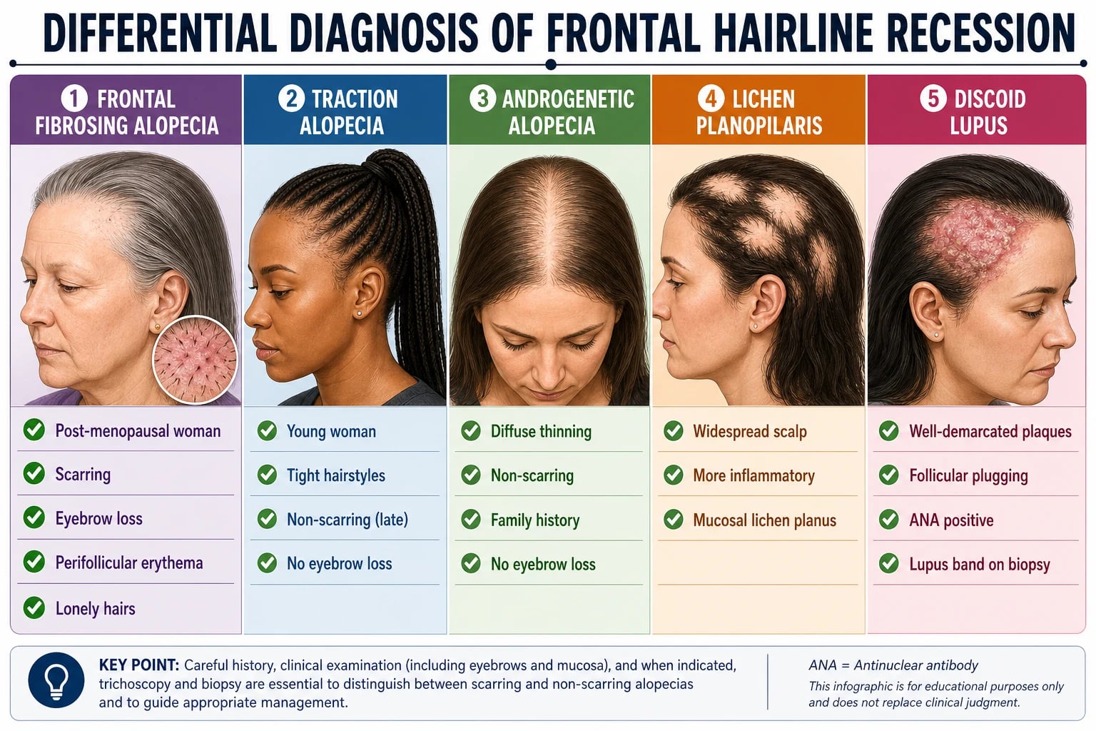
- Frontal fibrosing alopecia|Post-menopausal woman (55 to 70 yr)|Perifollicular erythema, lonely hairs, eyebrow loss (50 to 80%), body hair loss|Yes (cicatricial)
- Traction alopecia|Young woman with tight hairstyles|History of braids, weaves, extensions; no eyebrow loss; no perifollicular erythema in late phase|Late stage: yes; early: no
- Androgenetic alopecia|Men and women of any age post-puberty|Diffuse thinning at crown and vertex; family history; non-scarring; no eyebrow loss|No
- Alopecia areata (ophiasis)|Any age, autoimmune background|Non-scarring; exclamation-mark hairs; sudden onset; no perifollicular hyperkeratosis|No
- Classic lichen planopilaris|Middle-aged women (40 to 60 yr)|Patchy multifocal scarring anywhere on scalp; more inflammatory; oral/nail lichen planus|Yes (cicatricial)
- Discoid lupus erythematosus|Women (20 to 50 yr), photodistributed|Well-demarcated plaques with scale and follicular plugging; ANA positive; lupus band on DIF|Yes (cicatricial)
The three most tested bedside discriminating features separating FFA from traction alopecia are:[3]
- Eyebrow loss — present in FFA (50 to 80%), absent in traction alopecia.
- Perifollicular erythema and hyperkeratosis — present at the active margin in FFA, absent in late traction alopecia (though present in early traction).
- Demographic — FFA affects post-menopausal women; traction alopecia typically affects younger women with a clear hairstyle history. [1]
Atypical presentations that warrant biopsy. When the clinical picture does not fit neatly — for example, a pre-menopausal woman, a man, rapidly progressive recession, or atypical morphology (plaques with follicular plugging suggesting DLE) — a scalp biopsy from the active margin is indicated to confirm the diagnosis and exclude mimics.[3]
Clinical & Bedside Assessment
The clinical assessment of suspected FFA combines a focused history, a systematic scalp and hairline examination, and trichoscopy (dermoscopy).[5]
History. Ask about:
- Tempo of hairline recession — when did the patient first notice the hairline moving back? How fast? Measure recession from a fixed landmark (the glabella or the original hairline position marked in old photographs).
- Eyebrow and body hair loss — is this present? When did it start relative to scalp loss?
- Symptoms — itching, burning, tenderness at the hairline (indicates active inflammation).
- Hair care and cosmetic practices — use of leave-on facial products, sunscreens, facial moisturisers applied to the forehead and hairline; tight hairstyles, hair extensions, chemical treatments.
- Menopausal status and hormonal history.
- Autoimmune history — thyroid disease, vitiligo, alopecia areata, lichen planus.
- Family history of hair loss or autoimmune disease. [1]
Scalp examination. Inspect the frontal and temporal hairline in good lighting. Assess:
- The width of the recession band — measure from the glabella to the current hairline and compare with old photographs.
- The active margin — look for perifollicular erythema (pink halos around follicles) and perifollicular hyperkeratosis (white scale around hair shafts).
- Lonely hairs — isolated terminal hairs in the receded zone.
- The receded zone — smooth, shiny skin devoid of follicular markings (confirming scarring).
- Pigmentary change — hypo- or hyperpigmentation.
- Facial papules — small perifollicular papules on temples and cheeks. [1]
Eyebrow, body hair, nail, and mucosal examination. Examine the eyebrows (lateral loss first), axillary and limb hair, and the nails (looking for lichen planus changes — pterygium, longitudinal ridging, onycholysis). Inspect the oral mucosa for Wickham striae (suggesting systemic lichen planus and classic LPP overlap).[3]
Trichoscopy (dermoscopy). Trichoscopy is the single most useful bedside investigation. Use a dermoscope (polarised, 10x to 20x) on the active hairline margin.[5]
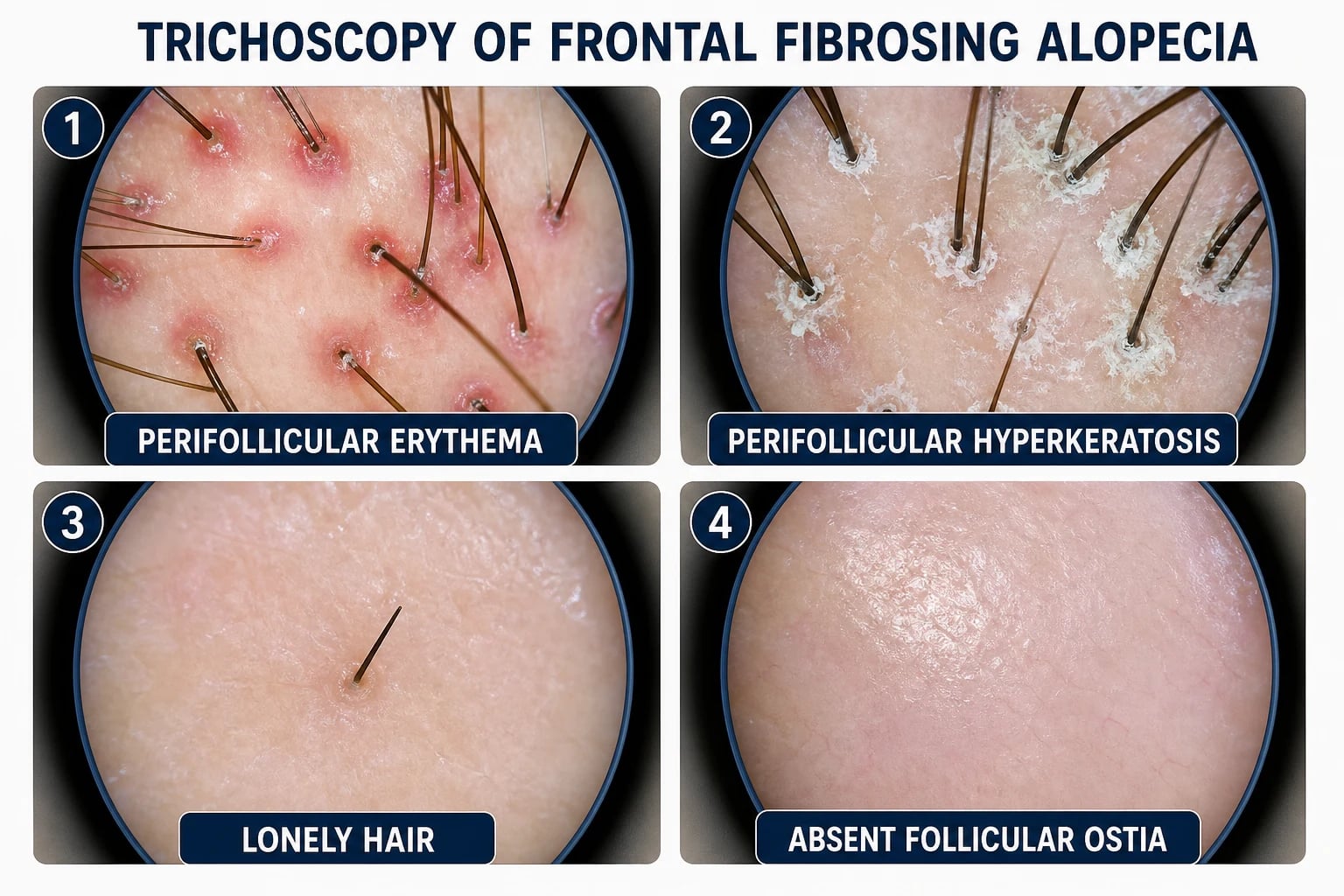
The four key trichoscopy findings of FFA are:[5]
- Perifollicular erythema — pink-red halos around individual hair follicles at the active margin. This is the trichoscopic correlate of active inflammation and is the most reliable sign of disease activity.
- Perifollicular hyperkeratosis — white or yellow-white scale and keratotic plugs surrounding the hair shaft at the follicular ostium. Together with erythema, this reflects the lichenoid inflammatory process.
- Lonely hair — a single isolated terminal hair shaft in an otherwise smooth, hairless area of scarred scalp.
- Absent follicular ostia in the scarred zone — smooth skin with no visible follicular openings, plus scattered white dots representing fibrotic follicular tracts. This confirms the scarring (cicatricial) nature of the alopecia. [1]
Assessing disease activity. The presence of perifollicular erythema and hyperkeratosis at the leading edge indicates active disease; their absence (with only smooth, shiny scar) indicates burnt-out disease. This distinction guides treatment: active disease warrants escalation of anti-inflammatory therapy; burnt-out disease needs no further treatment (scarred follicles cannot recover).[1]
The FFA Severity Index (FFASI). A validated severity score exists (FFASI), grading recession width, eyebrow loss, body hair loss, perifollicular erythema/hyperkeratosis, and symptoms. It is used mainly in research and specialist practice to document progression and treatment response.[1]
Photographic documentation. Standardised frontal, temporal, and vertex photographs at each visit are essential to document progression objectively, as patients' subjective reports of change are unreliable. [1]
Investigations
FFA is primarily a clinical diagnosis, but trichoscopy and, in selected cases, scalp biopsy confirm the diagnosis and exclude mimics.[3][5]
When is biopsy indicated? Biopsy is not necessary in a classic presentation (post-menopausal woman with typical clinical and trichoscopic features). It is indicated when:[3]
- The presentation is atypical (pre-menopausal woman, man, rapidly progressive, or morphology atypical).
- There are features suggesting an alternative diagnosis — erythematous plaques with follicular plugging (suggesting DLE), pustules (suggesting folliculitis decalvans), or exclamation-mark hairs (suggesting alopecia areata).
- The patient is being considered for systemic therapy and histological confirmation is desired before committing to long-term treatment. [1]
Skin biopsy technique. Take a 4 mm punch biopsy from the active margin (the leading edge of the receding hairline, where perifollicular erythema is visible on trichoscopy). Biopsying the burnt-out centre yields only fibrosis and is non-diagnostic. Send for routine histopathology (H&E). Direct immunofluorescence (DIF) is indicated if DLE is in the differential.[3]
Histopathology of FFA. The characteristic findings are:[3][4]
- Perifollicular lymphocytic infiltrate — a band-like (lichenoid) accumulation of lymphocytes (predominantly CD8+ T-cells) around the upper follicle (infundibulum and isthmus), sparing the lower follicle and dermal papilla in early disease.
- Basaloid (vacuolar) degeneration — vacuolar changes and apoptosis in the basal layer of the outer root sheath of the follicular epithelium (the lichenoid interface dermatitis pattern, identical to classic LPP).
- Loss of sebaceous glands — sebaceous glands adjacent to affected follicles are destroyed; their absence is a helpful diagnostic clue.
- Reduced follicle count — fewer terminal follicles per unit area compared with normal scalp.
- Perifollicular fibrosis — concentric lamellar fibrosis around the upper follicle, progressing to complete replacement of the follicle by dense collagen (scar) in late-stage lesions.
- Absence of neutrophils, plasma cells, and organisms — this distinguishes FFA from the neutrophilic cicatricial alopecias (folliculitis decalvans) and infectious causes. [1]
Differentiating FFA from classic LPP on histology. The histology of FFA and classic LPP is identical — both show the perifollicular lymphocytic lichenoid infiltrate. The distinction is clinical (anatomical distribution: frontal band versus patchy multifocal), not histological. This is why FFA is considered a variant of LPP rather than a separate disease.[3]
Direct immunofluorescence (DIF). In FFA, DIF is typically negative or shows only non-specific deposition (occasional Civatte bodies). This distinguishes FFA from discoid lupus erythematosus, where DIF shows a positive lupus band (granular deposition of IgG, IgM, and C3 along the dermo-epidermal junction). DIF is indicated whenever DLE is in the differential.[3]
Blood tests. No blood tests are routinely needed for FFA — it is not associated with systemic autoimmune disease (unlike DLE). However, a screening thyroid function test (TSH) and ANA are reasonable if there is clinical suspicion of coexisting autoimmune disease or if the presentation is atypical. FFA does not cause systemic symptoms.[5]
Management — Resuscitation and Counselling
FFA is a chronic condition, not a medical emergency, but the counselling at diagnosis is critical and is often mishandled. The following must be addressed at the first visit:[2]
Set realistic expectations. The single most important message is that existing hair loss is permanent and irreversible (scarring). Treatment aims to halt further recession, not to regrow lost hair. Patients who are promised regrowth will be disappointed and lose trust. Frame the goal as "protecting the hair you still have."[2]
Escalation triggers for urgent action. [1]
- Rapidly progressive recession (more than 1 cm in 6 months) — escalate to systemic therapy immediately; delay leads to further irreversible scarring.
- Extensive eyebrow loss — functionally and cosmetically significant; warrants early systemic treatment to preserve remaining eyebrows.
- Active perifollicular erythema and hyperkeratosis across a wide margin — indicates smouldering, widespread inflammation that will produce further recession if not treated. [1]
Discontinue potential environmental triggers. Advise the patient to stop applying leave-on facial products and sunscreens directly to the forehead and frontal hairline. This is a low-risk, potentially high-yield intervention. Sun protection should continue using physical barriers (hats, caps, clothing) rather than chemical sunscreens on the skin near the hairline. While the causal link is unproven, the risk of discontinuation is negligible and the potential benefit is real.[4]
Address the psychological impact. Hair loss, particularly of the frontal hairline and eyebrows, has a significant impact on identity, self-esteem, and quality of life. Acknowledge this, offer camouflage options (hairpieces, fringes, hair styling to cover the receded hairline, eyebrow tattooing or microblading for eyebrow loss), and refer for psychological support if distress is significant.[1]
Management — Definitive and Stepwise
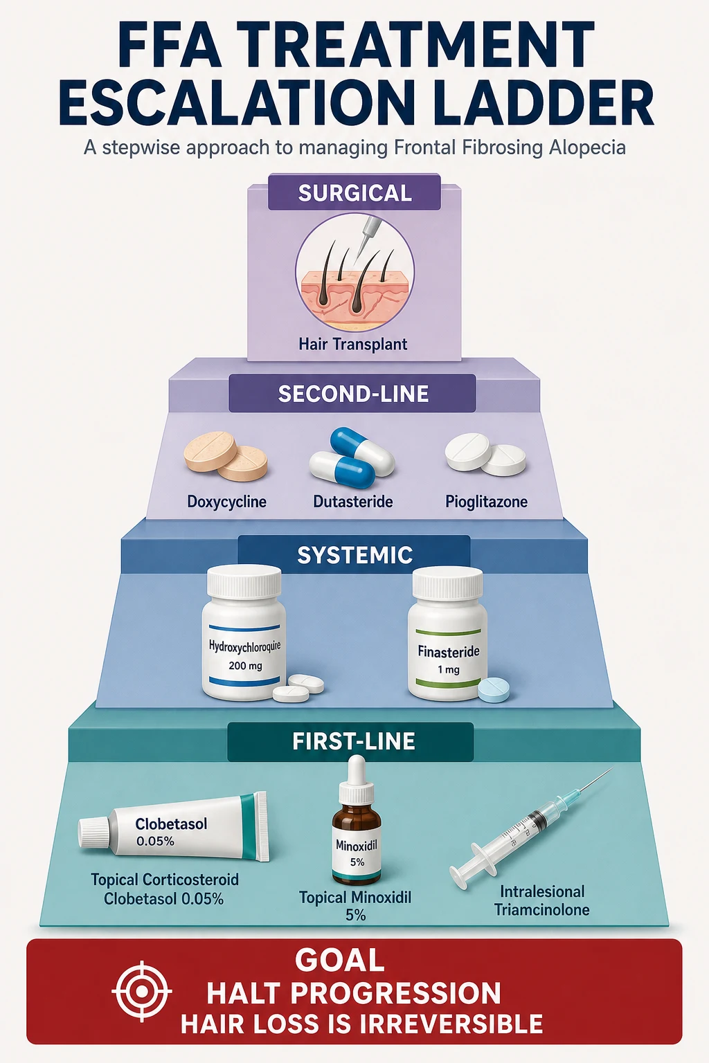
Treatment of FFA is guided by disease activity (the presence of perifollicular erythema and hyperkeratosis at the active margin) and the rate of progression. There are no randomised controlled trials to guide therapy; recommendations are based on expert consensus, retrospective case series, and the shared pathophysiology with lichen planopilaris.[1][2]
First-line: topical and intralesional therapy
Topical corticosteroids. Clobetasol propionate 0.05% lotion or foam applied to the active hairline margin once daily for the first 4 to 6 weeks, then tapering to alternate days. This reduces the perifollicular lymphocytic inflammation at the active edge. Monitor for skin atrophy with prolonged use; limit continuous daily use to 4 to 6 weeks, then switch to weekend pulsing or a less potent steroid.[5]
Intralesional corticosteroids. Triamcinolone acetonide 10 mg/mL injected into the active leading edge of the hairline using a 27-gauge needle, 0.05 to 0.1 mL per injection site at 1 cm intervals, repeated every 4 to 6 weeks for 3 to 6 sessions. This delivers a concentrated anti-inflammatory dose directly to the site of active disease. Warn patients about transient skin atrophy and hypopigmentation at injection sites.[1]
Topical calcineurin inhibitors. Tacrolimus 0.1% ointment applied to the active margin twice daily is a steroid-sparing alternative, particularly useful for long-term maintenance. Unlike corticosteroids, it does not cause skin atrophy.[5]
Topical minoxidil. Minoxidil 5% lotion or foam applied twice daily to the frontal scalp. Minoxidil does not halt the scarring process but may stimulate remaining viable follicles to produce thicker shafts, improving cosmetic density in non-scarred areas. Low-dose oral minoxidil (0.25 to 1.25 mg daily) is increasingly used as an alternative to topical application.[1]
First-line systemic therapy (for progressive disease)
Hydroxychloroquine. Hydroxychloroquine 200 to 400 mg daily (typically 200 mg twice daily) is the traditional first-line systemic agent for FFA, reflecting its role as first-line therapy for lichen planopilaris. It is an antimalarial with immunomodulatory (anti-lymphocytic) properties. Monitor for retinal toxicity: perform a baseline ophthalmological assessment (including colour vision and visual fields) at initiation, and annual screening thereafter (more frequent in patients over 60 or on treatment for more than 5 years). Therapeutic effect is typically delayed by 8 to 12 weeks.[1][3]
5-alpha-reductase inhibitors. Finasteride 1 to 5 mg daily or dutasteride 0.5 mg daily (or weekly) are now considered by many specialists to be the most consistently effective systemic agents for FFA. They block the conversion of testosterone to dihydrotestosterone (DHT), modulating the androgen-driven component of the follicular inflammatory process. Despite the disease predominantly affecting post-menopausal women with low androgen levels, the response rate is high (approximately 50 to 70 percent of patients show disease stabilisation). The mechanism is thought to involve local DHT reduction in the follicle rather than systemic androgen effects. Side effects are uncommon in post-menopausal women but include decreased libido and mood changes; liver function should be monitored.[1][2]
Second-line systemic therapy
Doxycycline. Doxycycline 100 mg twice daily — used for its anti-inflammatory (matrix metalloproteinase-inhibiting) rather than its antibiotic effect. Particularly useful if there is coexisting perifollicular inflammation. Monitor for photosensitivity (advise sun protection) and gastrointestinal upset.[1]
Pioglitazone. Pioglitazone 15 to 30 mg daily — a PPAR-gamma agonist (thiazolidinedione) with anti-inflammatory properties. Emerging evidence from small case series suggests benefit in FFA. Monitor for fluid retention, weight gain, and (rarely) bladder cancer risk with long-term use.[1]
Refractory disease
Mycophenolate mofetil. 500 mg to 1 g twice daily — an immunosuppressant that inhibits lymphocyte proliferation. Reserved for disease progressive despite first- and second-line agents. Monitor full blood count and liver function monthly for the first 3 months, then 3-monthly.[1]
Methotrexate. 10 to 25 mg once weekly (oral or subcutaneous) with folic acid supplementation — an antimetabolite immunosuppressant. Monitor full blood count, liver function, and renal function. Effective in a subset of patients with progressive LPP/FFA.[1]
Surgical management
Hair transplantation. Follicular unit extraction (FUE) or follicular unit transplantation (FUT) can be attempted to reconstruct the frontal hairline in patients with burnt-out, inactive FFA. Critical caveats:[2]
- The disease must be clinically and trichoscopically inactive for at least 2 years before surgery. Transplanting into active disease risks Koebnerisation — the surgical trauma can reactivate the inflammatory process, destroying the transplanted follicles.
- Graft survival is often disappointing (lower than in androgenetic alopecia) because the scarred recipient bed has reduced vascularity and altered tissue mechanics.
- The patient must understand that transplantation is cosmetic reconstruction of the hairline, not treatment of the underlying disease — the disease can reactivate at any time and threaten the transplanted follicles. [1]
Finasteride
Dose
1 to 5 mg orally once daily
Adjunctive measures
- Discontinue leave-on facial products and sunscreens applied to the forehead and hairline.[4]
- Physical sun protection (hats, caps) rather than chemical sunscreens on the skin near the hairline.
- Scalp camouflage — hairpieces, fringes, hair styling, and eyebrow microblading or tattooing for eyebrow loss.
- Psychological support for patients with significant distress.
Specific Subtypes and Scenarios
FFA in men. Male FFA is rare (approximately 15 percent of cases in large series) and presents differently: more temple involvement, less eyebrow loss, and frequently misdiagnosed as androgenetic alopecia (male pattern baldness). The key to recognition is the band-like frontotemporal recession (rather than the bitemporal recession of androgenetic alopecia) and the presence of perifollicular erythema at the active margin. 5-alpha-reductase inhibitors are effective in men (as they are for androgenetic alopecia), though sexual side effects may limit adherence.[1]
FFA in pre-menopausal women. Rare and frequently misdiagnosed as traction alopecia. The key discriminators are the same: perifollicular erythema, lonely hairs, and eyebrow loss. Finasteride and dutasteride are strictly teratogenic and must not be prescribed to women who may become pregnant without robust contraception. Hydroxychloroquine and topical therapies are the preferred first-line agents in this group.[1]
FFA with facial papules (FFA-plus phenotype). A subset of patients develop small perifollicular papules on the temples, cheeks, and forehead, representing involvement of facial vellus hair follicles. This phenotype is associated with more aggressive disease and a higher likelihood of body hair loss. Treatment escalation should be earlier.[1]
FFA overlapping with classic lichen planopilaris. Some patients exhibit features of both FFA (frontal band recession) and classic LPP (patchy scarring elsewhere on the scalp, oral mucosal Wickham striae, nail lichen planus). This overlap supports the view that FFA is a variant of LPP rather than a separate entity. Management is the same — a combination of corticosteroids, antimalarials, and 5-alpha-reductase inhibitors.[3]
FFA in skin of colour. In Fitzpatrick skin types IV to VI, post-inflammatory hyperpigmentation in the receded zone is more pronounced and may be the presenting complaint. The scarring may also show a keloidal or hypertrophic tendency. Tailor topical steroid potency (avoid very potent steroids on facial skin of colour due to risk of hypopigmentation) and address pigmentary change with sun protection and, if needed, topical depigmenting agents once the inflammatory process is controlled.[1]
Complications and Pitfalls
Complications of the disease.[1][5]
- Permanent scarring alopecia — the defining complication. Once the follicle is destroyed and replaced by fibrous tissue, it cannot regenerate. This is the primary reason for early diagnosis and treatment.
- Eyebrow loss — cosmetically significant and usually permanent. Eyebrow microblading or tattooing is often the only satisfactory solution.
- Body hair loss — axillary, pubic, and limb hair may be permanently lost.
- Psychological distress — the cosmetic impact of frontal hairline recession and eyebrow loss on identity and quality of life is substantial. Anxiety, depression, and social withdrawal are common. Psychological support should be offered proactively.
- Disease progression despite treatment — a subset of patients (approximately 20 to 30 percent) continue to progress despite optimal therapy. This is not necessarily a treatment failure but reflects the aggressive biology of the disease in some individuals. [1]
Classic pitfalls. [1]
Exam application bank (NEET-PG / INICET)
One-line answer
Frontal fibrosing alopecia (FFA) is a primary lymphocytic scarring (cicatricial) alopecia characterised by progressive, symmetric recession of the frontal and temporal hairline, predominantly in post-menopausal Caucasian women. First described by Kossard in 1994, it is considered a clinical variant of lichen planopilaris (LPP). Clinical hallmarks include perifollicular erythema and hyperkeratosis at the active hairline margin, loss of eyebrows (approximately 50 to 80 percent), and lonely hairs (isolated terminal hairs within the receded zone). Histology reveals a perifollicular lymphocytic (lichenoid) infiltrate around the upper follicle with basaloid degeneration, loss of sebaceous glands, and destruction of bulge-region stem cells, resulting in irreversible scarring. Treatment with topical and intralesional corticosteroids, 5-alpha-reductase inhibitors (finasteride or dutasteride), and
Worked stems (answer without another resource)
Stem 1 — Classic presentation. Map symptoms to mechanism; name the first investigation and first treatment step with dose/route if drug therapy is standard. [1]
Stem 2 — Unstable / complicated. List red flags that force immediate resuscitation, theatre, ICU, antidote, or reperfusion — and what you do in the first 15 minutes. [1]
Stem 3 — Atypical group. Elderly, pregnancy, child, or immunocompromised: how presentation and thresholds change. [1]
Stem 4 — Differential trap. Name the three closest mimics and one discriminator for each. [1]
Stem 5 — Disposition. Who goes home with safety-netting, who is admitted, who needs HDU/ICU/theatre, and what follow-up is mandatory. [1]
Rapid viva checklist
- Definition + classification
- Pathophysiology chain
- Bedside signs / criteria
- Score with exact components (if any)
- Emergency bundle
- Definitive therapy with doses
- Complications of disease and of treatment
- Special populations
- Guideline/trial name if classic
- Three exam traps
Coverage self-check
If you cannot answer any stem above from this page alone, re-read the matching section — the page is intended to be self-sufficient for final-prof and NEET-PG/INICET questions on Frontal fibrosing alopecia (FFA).
[1]Prognosis and Disposition
Natural history. FFA is slowly progressive over years. The typical course is one of gradual frontotemporal recession at a rate of 0.5 to 2 cm per year (highly variable between individuals). In most patients, the disease eventually burns out spontaneously after 3 to 6 years (range 1 to 15 years), meaning the perifollicular inflammation ceases and the hairline stabilises. However, by the time burn-out occurs, significant and permanent recession has usually occurred.[5]
Existing hair loss is irreversible. Once a follicle is destroyed and scarred, it cannot recover. Treatment can only halt further recession — it cannot regrow hair in the scarred zone. This is the fundamental limitation of all current therapies.[2]
Early treatment improves outcome. Patients diagnosed and treated early (before extensive recession) have the best cosmetic outcome, because treatment halts progression before the hairline has receded significantly. Late presentation (more than 3 cm of recession) is associated with poorer cosmetic outcomes despite treatment.[1]
Quality of life. The cosmetic burden of FFA is significant, particularly the combination of frontal hairline recession and eyebrow loss. Quality-of-life studies in cicatricial alopecia show impairment comparable to that seen in severe psoriasis. Camouflage options (hairpieces, styling, microblading) and psychological support are essential components of management.[1]
Follow-up plan.[1]
- Review every 3 to 6 months during active disease, with standardised frontal photographs and measurement of hairline recession from a fixed landmark (glabella).
- Assess disease activity at each visit — the presence or absence of perifollicular erythema and hyperkeratosis at the active margin guides treatment escalation or de-escalation.
- Monitor drug therapy — hydroxychloroquine (annual ophthalmological screening), finasteride/dutasteride (liver function), mycophenolate/methotrexate (full blood count and liver function).
- Refer to a specialist trichology centre for refractory or rapidly progressive disease, or if hair transplant is being considered. [1]
Special Populations
Pre-menopausal women. FFA is rare in this group. Finasteride and dutasteride are absolutely contraindicated (teratogenic — anti-androgen effect causes feminisation of a male fetus). Use hydroxychloroquine, topical corticosteroids, and intralesional triamcinolone. Ensure robust contraception if any anti-androgen is being considered.[1]
Pregnancy and breastfeeding. Avoid 5-alpha-reductase inhibitors, tetracyclines (doxycycline), and retinoids. Topical corticosteroids (moderate potency) and topical calcineurin inhibitors (tacrolimus) are considered safe. Hydroxychloroquine is relatively safe in pregnancy (used in systemic lupus erythematosus). Defer systemic therapy until post-partum if possible.[1]
Elderly (the typical FFA patient). Polypharmacy and drug interactions are common. Hydroxychloroquine requires baseline and annual ophthalmological screening (retinal toxicity risk increases with age and cumulative dose). Finasteride is generally well tolerated in elderly women. Methotrexate requires renal function assessment (declining GFR in the elderly increases toxicity risk).[1]
Men with FFA. Rare (approximately 15 percent of cases). Presents with more temple involvement and less eyebrow loss. Easily missed and misdiagnosed as androgenetic alopecia. 5-alpha-reductase inhibitors are effective but monitor for sexual side effects (decreased libido, erectile dysfunction).[1]
Skin of colour (Fitzpatrick IV to VI). Post-inflammatory hyperpigmentation is more pronounced. Avoid very potent topical corticosteroids on facial skin (risk of hypopigmentation and atrophy). Tailor sun protection advice (physical barriers preferred). Address pigmentary change with topical agents (azelaic acid, kojic acid, retinoids) once the inflammatory process is controlled.[1]
Evidence, Guidelines and Regional Differences
The evidence base is weak. There are no randomised controlled trials of any therapy for FFA. All treatment recommendations are based on expert consensus, retrospective case series, and extrapolation from lichen planopilaris (which shares the same histopathology). The largest published case series come from specialist centres in Spain (Vano-Galvan et al.), the United Kingdom, Australia, and the United States.[1][2]
Why 5-alpha-reductase inhibitors work in post-menopausal women. This is one of the most counterintuitive findings in FFA therapy — drugs designed to treat male pattern baldness (by blocking DHT) are the most effective agents for a disease that almost exclusively affects post-menopausal women with low androgen levels. The proposed mechanism is that local DHT production within the hair follicle (driven by 5-alpha-reductase expressed in the follicle) sustains the perifollicular inflammatory or degenerative milieu. Blocking this local DHT production modulates the disease, independent of systemic androgen levels. This is an area of active investigation.[1]
The sunscreen and leave-on cosmetic hypothesis. The temporal correlation between the widespread adoption of daily sunscreen use among Caucasian women (from the 1990s onward) and the dramatic rise in FFA incidence is the most discussed hypothesis in FFA aetiology. Several case-control studies have found a statistically significant association between sunscreen use and FFA, but no specific ingredient has been causally linked, and the hypothesis remains unproven. Arguments against include: (1) not all sunscreen users develop FFA; (2) FFA occurs in women who never used sunscreens; (3) the rise may partly reflect increased recognition. Arguments for include: (1) the anatomical correlation (disease begins where sunscreens are applied); (2) the temporal correlation; (3) the biological plausibility (a contact antigen or hapten in the follicle). The consensus is to advise discontinuation of leave-on products near the hairline as a low-risk intervention while awaiting definitive evidence.[4]
The North American Hair Research Society (NAHRS) and the American Academy of Dermatology (AAD) classify FFA within the lymphocytic subgroup of primary cicatricial alopecias (Olsen et al., 2003 working classification). Treatment recommendations emphasise topical and intralesional corticosteroids as first-line, with hydroxychloroquine and 5-alpha-reductase inhibitors as first-line systemic agents. Access to mycophenolate and methotrexate is generally good in the US system. Hair transplant after disease inactivity is offered at specialist centres.
The British Association of Dermatologists (BAD) guidelines for cicatricial alopecia recommend clinical diagnosis with trichoscopy, biopsy for atypical cases, and a treatment ladder beginning with potent topical corticosteroids and intralesional triamcinolone, escalating to hydroxychloroquine (with mandatory ophthalmological monitoring per Royal College of Ophthalmology guidance) and finasteride or dutasteride. The UK has strong primary care referral pathways; FFA is increasingly recognised in general practice.
The European Academy of Dermatology and Venereology consensus aligns with NAHRS and BAD but places greater emphasis on the environmental trigger hypothesis, reflecting the high European incidence. Several European centres (particularly in Spain) have published the largest case series and advocate earlier use of 5-alpha-reductase inhibitors given their consistent efficacy.
In India and South Asia, access to specialist trichology is limited outside major cities. Hydroxychloroquine and finasteride are inexpensive and widely available. Doxycycline is the most accessible anti-inflammatory. Mycophenolate and methotrexate are available but require monitoring infrastructure. Hair transplant is available at private centres but is costly. Delayed presentation with extensive recession is common.
Emerging and investigational therapies. [1]
- Low-dose naltrexone (1.5 to 4.5 mg daily) — immunomodulatory, case-report level evidence.
- JAK inhibitors (tofacitinib, ruxolitinib) — theoretical benefit given the lymphocytic pathophysiology; case reports only.
- Platelet-rich plasma (PRP) — injected into the scalp; anecdotal reports of benefit; mechanism unclear.
- These agents are not standard of care and should be reserved for clinical trials or specialist centres.[1]
Exam Pearls
[1]Cicatricial alopecias — lymphocytic versus neutrophilic
Self-test: a 62-year-old woman presents with 3 years of progressive recession of her frontal hairline and loss of her eyebrows. Trichoscopy shows perifollicular erythema and hyperkeratosis at the active margin, with lonely hairs in the receded zone. What is the diagnosis, and what is the single most effective systemic treatment?
Diagnosis: frontal fibrosing alopecia (FFA) — classic presentation (post-menopausal woman, progressive frontotemporal recession, eyebrow loss, perifollicular erythema on trichoscopy). Most effective systemic treatment: a 5-alpha-reductase inhibitor — finasteride 1 to 5 mg daily or dutasteride 0.5 mg. Note: treatment halts progression; existing hair loss is irreversible (scarring).
References
- [1]Yip L, et al. Frontal Fibrosing Alopecia: An Update Am J Clin Dermatol, 2025.PMID 39699852
- [2]Vano-Galvan S, et al. The Frontal Fibrosing Alopecia Treatment Dilemma J Clin Med, 2024.PMID 38610902
- [3]Lee D, et al. Lichen planopilaris and frontal fibrosing alopecia: review and update of diagnostic and therapeutic features An Bras Dermatol, 2022.PMID 35379508
- [4]Rakowska A, et al. Frontal fibrosing alopecia: A review of disease pathogenesis Front Med (Lausanne), 2022.PMID 35957858
- [5]Tolkachjov SN, et al. Frontal Fibrosing Alopecia: A Review J Clin Med, 2021.PMID 33919069