Dermatology · Medicine
Genodermatoses overview
Also known as Inherited skin disorders · Dermatogenetics · Ichthyoses overview · Epidermolysis bullosa overview · Neurocutaneous syndromes overview · Ectodermal dysplasias overview
Genodermatoses are inherited disorders with primary cutaneous phenotypes caused by germline mutations affecting barrier function, dermal-epidermal adhesion, DNA repair, pigmentation, developmental signalling, or ectodermal structures. Exam-facing groups include the ichthyoses and epidermal differentiation disorders, epidermolysis bullosa, xeroderma pigmentosum, albinism, neurocutaneous syndromes (NF1, TSC, Sturge-Weber), and ectodermal dysplasias. The overview teaches a mechanism-first diagnostic approach, when to order genetics, multi-disciplinary surveillance, and how to avoid missing cancer risk, blister crises, and systemic complications.
On this page & tools
Your progress
Saved locally on this device.
Exam tags
Red flags
Genodermatoses are individually uncommon but collectively unavoidable in board-level dermatology. They include conditions as clinically different as ichthyosis vulgaris, dystrophic epidermolysis bullosa, xeroderma pigmentosum, oculocutaneous albinism, neurofibromatosis type 1, tuberous sclerosis, and hypohidrotic ectodermal dysplasia. What unites them is a germline genetic basis with primary or highly characteristic cutaneous findings, often with systemic stakes that outrank the rash itself.[1]
This overview is the map of the territory. Deep leaves (ichthyosis, EB, XP, albinism, TSC, NF1) carry entity-level detail; here the goal is safe triage, classification logic, and when to escalate. [1]
Classification by mechanism
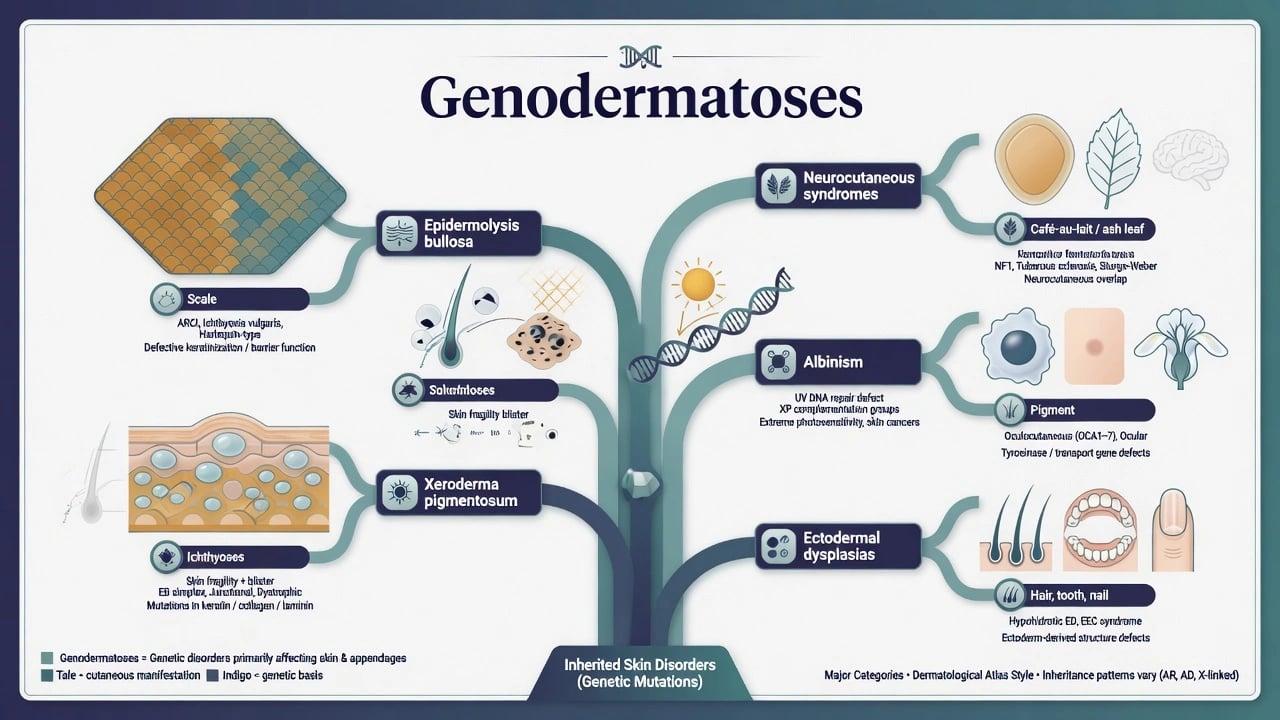
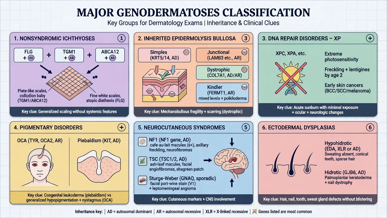
Six exam groups
Ichthyoses / EDD
Epidermolysis bullosa
DNA repair (XP)
Pigmentary
Neurocutaneous
Ectodermal dysplasias
Ichthyosis nomenclature was modernised by the Sorèze consensus and continues to evolve toward pathogenesis-based epidermal differentiation disorder frameworks.[3][4][5] EB was reclassified around molecular and clinical skin-fragility logic, retaining the practical simplex / junctional / dystrophic / Kindler structure for bedside care.[6][7]
Epidemiology and genetic clues
Most genodermatoses are rare. Consanguinity raises autosomal recessive disease (many ichthyoses, junctional and recessive dystrophic EB, XP). De novo mutation is common in autosomal dominant tumour-predisposition syndromes (NF1, TSC). Founder effects create regional hotspots — for example high OCA2 prevalence in parts of sub-Saharan Africa, or XP clusters in some Japanese and North African populations.[1][8][9]
Pathophysiology axes
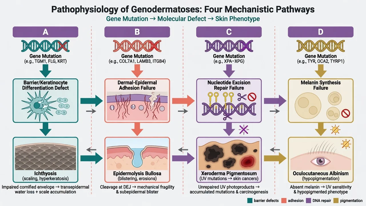
- Barrier / keratinocyte differentiation — defective cornified envelope or lipid processing produces scale, transepidermal water loss, and infection risk (ichthyoses).[3][4]
- Adhesion — keratin intermediate filaments, hemidesmosomes, or anchoring fibrils fail; mechanical trauma opens blisters at defined levels (EB).[6][7]
- DNA repair — NER failure leaves UV photoproducts unrepaired; mutation burden drives early keratinocyte and melanocyte cancers (XP).[8]
- Pigment synthesis / melanosome biology — melanocytes present but under-produce melanin (OCA) versus melanocyte distribution defects (piebaldism).[9]
Neurocutaneous syndromes add signalling pathway logic: Ras-MAPK in NF1, mTOR in TSC. Ectodermal dysplasias reflect developmental pathway defects in hair, tooth, nail, and sweat structures.[10]
Clinical presentation — when to suspect a genodermatosis
Suspect inheritance when you see congenital or early-onset disease, family history or consanguinity, multi-system involvement (eyes, CNS, hearing, teeth), extreme photosensitivity, skin fragility, generalised scale from infancy, or patterned pigmentation (Blaschko lines, mosaic segments). [1]
Differential diagnosis
- Acquired ichthyosis (malignancy, drugs, malnutrition) versus congenital ichthyosis.[4]
- Autoimmune blistering disease versus EB in older children/adults.
- Vitiligo (acquired melanocyte loss) versus albinism/piebaldism.[9]
- Ordinary freckling versus XP pigment network on exposed skin.[8]
- Non-accidental injury versus EB — a critical safeguarding and ethics intersection.[7]
Assessment and investigations
Build a phenotype first: full skin, hair, nails, teeth, mucosa; Wood’s lamp for hypopigmentation; eye and neurodevelopmental screen; three-generation pedigree.[2] Then order phenotype-driven panels (or exome/genome when panels fail), with MLPA or copy-number analysis when deletions are likely. EB may still use immunofluorescence antigen mapping in specialised centres; XP historically used DNA-repair assays but clinical genetics now dominates.[2][6][8]
Stage organs by syndrome: brain MRI and renal imaging in TSC, ophthalmology in albinism and Sturge-Weber, audiology/neurology in some XP groups, cardiac evaluation when rhabdomyoma or other clues appear. [1]
Management principles
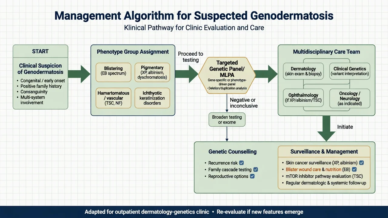
There is rarely a single cure. Care is organised as: [1]
- Skin-directed therapy — emollients and keratolytics for ichthyosis; atraumatic wound care and infection control for EB; absolute photoprotection for XP and albinism.[4][7][8][9]
- Surveillance — skin cancer (XP, albinism), seizures and renal angiomyolipomas (TSC), optic pathway and BP checks (NF1), heat illness risk (anhidrotic ED).
- Targeted systemic therapy where available — e.g. mTOR inhibitors in TSC (entity leaf detail).
- Genetic counselling — recurrence risks, cascade testing, prenatal and preimplantation options when the familial variant is known.[2]
- Psychosocial and educational support — stigma, schooling, career, and heat or UV logistics.
Complications and prognosis
Prognosis tracks genotype and organ involvement: childhood skin-cancer mortality in unprotected XP, squamous carcinoma and failure to thrive risks in severe EB, SUDEP and renal catastrophe risks in TSC, heat stroke risk in hypohidrotic ED, and skin-cancer burden in equatorial albinism.[7][8][9] Early recognition and structured MDT care change trajectories more than any single cream.
Regional notes
Consanguinity patterns in parts of South Asia and the Middle East raise AR disease prevalence. Molecular access is improving but clinical diagnosis plus protective care (sun, wounds, emollients) must not wait for a report. Indian genetics-for-dermatologists guidance emphasises structured evaluation and careful interpretation of sequencing results.[2]
Exam pearls
SCALE-BURN-PIGMENT-NERVE-ECTODERM
- Classify by mechanism, not by alphabet soup of eponyms first.[1]
- Collodion baby is a phenotype, not one disease.[3]
- EB subtype follows level of split.[6]
- XP = NER (or POLH in variant) with extreme cancer risk.[8]
- Always offer counselling when a pathogenic variant is found.[2]
Exam application bank (NEET-PG / INICET)
One-line answer
Genodermatoses are inherited disorders with primary cutaneous phenotypes caused by germline mutations affecting barrier function, dermal-epidermal adhesion, DNA repair, pigmentation, developmental signalling, or ectodermal structures. Exam-facing groups include the ichthyoses and epidermal differentiation disorders, epidermolysis bullosa, xeroderma pigmentosum, albinism, neurocutaneous syndromes (NF1, TSC, Sturge-Weber), and ectodermal dysplasias. The overview teaches a mechanism-first diagnostic approach, when to order genetics, multi-disciplinary surveillance, and how to avoid missing cancer risk, blister crises, and systemic complications.
Worked stems (answer without another resource)
Stem 1 — Classic presentation. Map symptoms to mechanism; name the first investigation and first treatment step with dose/route if drug therapy is standard. [1]
Stem 2 — Unstable / complicated. List red flags that force immediate resuscitation, theatre, ICU, antidote, or reperfusion — and what you do in the first 15 minutes. [1]
Stem 3 — Atypical group. Elderly, pregnancy, child, or immunocompromised: how presentation and thresholds change. [1]
Stem 4 — Differential trap. Name the three closest mimics and one discriminator for each. [1]
Stem 5 — Disposition. Who goes home with safety-netting, who is admitted, who needs HDU/ICU/theatre, and what follow-up is mandatory. [1]
Rapid viva checklist
- Definition + classification
- Pathophysiology chain
- Bedside signs / criteria
- Score with exact components (if any)
- Emergency bundle
- Definitive therapy with doses
- Complications of disease and of treatment
- Special populations
- Guideline/trial name if classic
- Three exam traps
Coverage self-check
If you cannot answer any stem above from this page alone, re-read the matching section — the page is intended to be self-sufficient for final-prof and NEET-PG/INICET questions on Genodermatoses overview.
Expanded exam teaching (depth pass)
Clinical reasoning
For Genodermatoses overview, examiners test whether you can prioritise life threats, choose the right first test, and give specific therapy (agent, dose, route, timing). Generic phrases without numbers score poorly.
Mechanism → feature map
Build a short chain: cause → pathophysiologic intermediate → clinical feature → complication. Every major symptom in the classic vignette should sit on that chain.
Investigation strategy
- Bedside/first-line tests that change immediate management
- Confirmatory or staging tests
- What a normal result does not exclude
- When not to delay treatment for imaging (unstable patient)
Management ladder
- Resuscitation / ABC / sepsis or haemorrhage bundle as relevant
- Specific antidote / procedure / antimicrobial / reperfusion / surgery
- Supportive care and monitoring targets
- Definitive long-term therapy and secondary prevention
- Disposition and safety-net advice
Special populations
Always prepare one line each for children, pregnancy, elderly, renal/hepatic impairment, and immunocompromised patients when the topic allows.
Pitfalls that fail candidates
- Treating the number not the patient
- Missing pregnancy status when relevant
- Imaging before stabilisation
- Wrong empiric cover or wrong antidote timing
- Incomplete counselling on recurrence, adherence, or red-flag return
Genodermatoses are inherited disorders with primary cutaneous phenotypes caused by germline mutations affecting barrier function, dermal-epidermal adhesion, DNA repair, pigmentation, developmental signalling, or ectodermal structures. Exam-facing groups include the ichthyoses and epidermal differentiation disorders, epidermolysis bullosa, xeroderma pigmentosum, albinism, neurocutaneous syndromes (NF1, TSC, Sturge-Weber), and ectodermal dysplasias. The overview teaches a mechanism-first diagnosti [1]
Structured revision sheet
Must-know numbers and names
List every score, size threshold, dose, and time window from this topic on a blank page from memory, then check against the sections above.
Three classic MCQ angles
- Most likely diagnosis given a vignette
- Next best step in management
- Most appropriate investigation
Three classic SAQ angles
- Pathophysiology in five steps
- Management algorithm with doses
- Complications and prevention
Clinical station flow
Greet → focused history → targeted exam → investigations → explain diagnosis → emergency care → definitive plan → safety-net / follow-up → answer examiner questions on mechanism and pitfalls.
References
- [1]Frank J. Selected genodermatoses - Status quo and future prospects J Dtsch Dermatol Ges, 2023.PMID 36976174
- [2]Gupta D, Venugopal R, Sardana K. Genetics for dermatologists. Part 2: Clinical evaluation, sequencing technologies and interpretation Indian J Dermatol Venereol Leprol, 2025.PMID 40357951
- [3]Oji V, Tadini G, Akiyama M, et al. Revised nomenclature and classification of inherited ichthyoses: results of the First Ichthyosis Consensus Conference in Sorèze 2009 J Am Acad Dermatol, 2010.PMID 20643494
- [4]Gutiérrez-Cerrajero C, Sprecher E, Paller AS, et al. [Translated article] ICHTHYOSIS: Clinical and Molecular Update. Part 1: Introduction and Non-Syndromic Ichthyoses Actas Dermosifiliogr, 2025.PMID 40081471
- [5]Gutiérrez-Cerrajero C, Sprecher E, Paller AS, et al. [Translated article] ICHTHYOSIS: Clinical and Molecular Update. Part 2: Syndromic Ichthyosis. Diagnostic and Therapeutic Approach of Ichthyosis Actas Dermosifiliogr, 2025.PMID 40081487
- [6]Has C, Bauer JW, Bodemer C, et al. Consensus reclassification of inherited epidermolysis bullosa and other disorders with skin fragility Br J Dermatol, 2020.PMID 32017015
- [7]Mariath LM, Santin JT, Schuler-Faccini L, et al. Inherited epidermolysis bullosa: update on the clinical and genetic aspects An Bras Dermatol, 2020.PMID 32732072
- [8]Adam MP, Bick S, Mirzaa GM, et al. Xeroderma Pigmentosum 1993.PMID 20301571
- [9]Adam MP, Bick S, Mirzaa GM, et al. Oculocutaneous Albinism and Ocular Albinism Overview 1993.PMID 37053367
- [10]Wright JT, Fete M, Schneider H, et al. Ectodermal dysplasias: Classification and organization by phenotype, genotype and molecular pathway Am J Med Genet A, 2019.PMID 30703280