Dermatology · Medicine
Granuloma annulare
Also known as Granuloma annulare (GA) · Pseudorheumatoid nodule (subcutaneous GA) · Generalized granuloma annulare · Perforating granuloma annulare
Granuloma annulare (GA) is a benign, usually self-limiting granulomatous dermatosis. Histology: palisading granulomas with central necrobiotic collagen + mucin surrounded by histiocytes. Localized (commonest): annular skin-coloured papules on dorsa of hands/feet; self-limiting. Generalized: disseminated; may associate with diabetes, dyslipidaemia, thyroid disease and HIV; refractory. Subcutaneous (deep): firm nodules in children; mimics rheumatoid nodules (but RF negative). Perforating: crusted papules (trans-epidermal elimination). DDx: tinea corporis, annular psoriasis, necrobiosis lipoidica, rheumatoid nodules, sarcoidosis, interstitial granulomatous dermatitis, erythema annulare centrifugum, actinic granuloma. Management: localized — observe or potent topical corticosteroids / intralesional triamcinolone, cryotherapy, tacrolimus; generalized — topical dapsone, hydroxychloroquine, nbUVB, methotrexate, isotretinoin, adalimumab; observation for mild localised. Prognosis is generally excellent.
On this page & tools
Your progress
Saved locally on this device.
Exam tags
Red flags
Overview & Definition
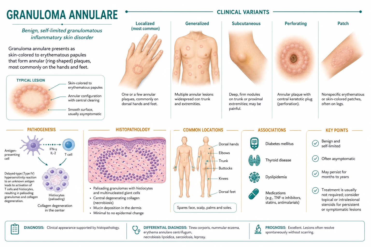
Granuloma annulare (GA) is a benign, usually self-limiting granulomatous dermatosis of the dermis and, in some variants, the subcutaneous fat. The defining histopathological pattern is the palisading granuloma: a central focus of necrobiotic (degenerated) collagen and mucin surrounded by a palisading rim of histiocytes, often with multinucleated giant cells and a variable lymphocytic infiltrate.[1][5] It is not infectious, not contagious and not neoplastic. The disease most often presents as localized annular lesions in children and young adults, but a range of clinical morphologies and anatomical distributions are recognized, each with distinct associations and therapeutic implications.
The name itself describes the lesion: granuloma refers to the histiocytic granulomatous infiltrate, and annulare refers to the annular (ring-shaped) clinical morphology of the common localized form. Despite the name, not all lesions are annular; patch-type and subcutaneous forms may lack a ring-like configuration entirely. [1]
[2]Classification
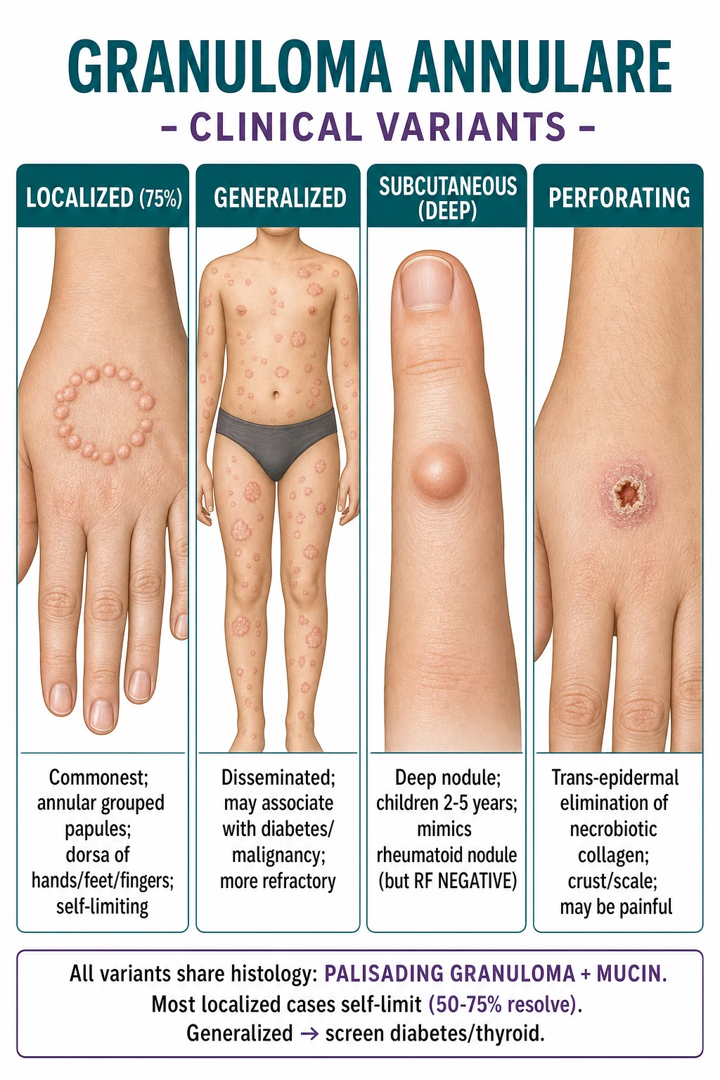
GA is classified primarily by clinical morphology and distribution. The five major variants are not separate diseases but rather presentations along a spectrum of depth and extent. Accurate classification matters because it determines the differential diagnosis, the likelihood of systemic association and the intensity of treatment. [3]
Localized GA
Generalized / disseminated GA
Subcutaneous (deep) GA
Perforating GA
Patch-type GA
Localized GA
Localized GA is the most common form, accounting for roughly 70–75% of cases. It typically affects children and young adults, with a slight female predominance in some series. The dorsal aspects of the hands and feet, fingers, ankles and wrists are the characteristic sites. Lesions are usually asymptomatic, although mild pruritus or tenderness may occur. [1]
Generalized GA
Generalized GA is defined by the presence of 10 or more lesions or involvement of the trunk. It tends to occur in adults, is more persistent and is more strongly associated with systemic disease, particularly diabetes mellitus, dyslipidaemia, thyroid disease and, less commonly, HIV infection and malignancy. Management is more challenging than localized disease. [4]
Subcutaneous GA
Subcutaneous GA, also known as pseudorheumatoid nodule, is the most common deep form and occurs predominantly in children aged 2–5 years. It presents as firm, painless, skin-coloured subcutaneous nodules on the scalp, fingers, shins, pretibial areas and extensor surfaces. The clinical importance of this variant lies in its close resemblance to rheumatoid nodules and, occasionally, to soft tissue sarcoma. [2]
Perforating GA
Perforating GA is characterized by papules that develop a central crust, umbilication or keratotic plug. Histologically, this represents trans-epidermal elimination of necrobiotic collagen through the epidermis. It is often more symptomatic than other forms and may cause discomfort or cosmetic concern. [2]
Patch-type GA
Patch-type GA is the least common morphology, presenting as erythematous or violaceous patches or macules without a raised annular border. It can be mistaken for morphea, parapsoriasis or other patch-form dermatoses, and biopsy is usually required for diagnosis. [1]
Epidemiology & Risk Factors
GA is a common dermatological condition, although precise incidence data are limited because many localized cases are not biopsied and may resolve before medical evaluation. The disease is seen in all ethnic groups and geographic regions, with no strong racial predilection. [1]
Risk factors and precipitants
The aetiology of GA is unknown, but several triggers and associations have been reported in observational studies and case series. These are important because they guide the clinical interview and, in generalized disease, the screening work-up. [4]
Trauma and local insults — Physical trauma, insect bites, sun exposure, tattoo reactions and varicella scars have been reported to precede GA at sites of injury. The Koebner phenomenon (lesions developing at sites of trauma) can occur, particularly in generalized disease.[1]
Infections — Various infections have been reported in temporal association with GA, including human immunodeficiency virus (HIV), hepatitis B and C, Epstein–Barr virus, streptococcal infections and tuberculosis. These associations are rare and probably reflect an immune-mediated reactive process rather than direct infection of the skin.[1]
Drugs — Drug-induced GA has been reported with allopurinol, gold salts, ACE inhibitors, antimalarials, calcium channel blockers, statins, quinidine and diclofenac, among others. The lesions usually resolve after withdrawal of the offending drug, although this can take months.[1]
Systemic disease — The strongest and most clinically relevant associations are with diabetes mellitus, dyslipidaemia, thyroid disease (autoimmune thyroiditis and Graves disease) and, in generalized or atypical cases, malignancy (especially lymphoma and solid tumours). These associations are most relevant for generalized GA, and screening is recommended in this subgroup.[1][6]
Pathophysiology
The exact pathophysiology of GA remains incompletely understood. It is considered a reactive granulomatous process driven by a delayed-type hypersensitivity reaction to an unknown antigen, with subsequent immune-mediated destruction of collagen and deposition of mucin in the dermis. The process is not infectious, and no organism has been consistently identified in GA lesions. [2]
Immunological mechanism
The central event appears to be a T-cell-mediated immune response directed against altered or degenerated dermal collagen. Histiocytes are recruited to the site and organize into a palisading arrangement around the damaged collagen. This pattern is reminiscent of a delayed-type hypersensitivity response, supported by the predominance of T-helper 1 (Th1) cytokines such as interferon-gamma, interleukin-2 and tumour necrosis factor-alpha within the granulomatous infiltrate.[1][2]
Several mechanisms have been proposed to explain why collagen becomes targeted: [1]
- Immune complex deposition with complement activation and vasculitis, leading to collagen damage.
- Direct T-cell recognition of neoantigens on altered collagen after trauma, infection or ultraviolet light exposure.
- Matrix degeneration with altered collagen structure that attracts histiocytes and triggers granuloma formation. [1]
Once histiocytes are activated, they secrete cytokines that recruit additional inflammatory cells and stimulate fibroblasts to produce mucin. This mucin accumulates in the centre of the granuloma, expanding the necrobiotic zone and producing the histological hallmark of the disease. [2]
Histiocyte and collagen interaction
Within the granuloma, histiocytes and multinucleated giant cells are seen in close apposition to necrobiotic collagen. Phagocytosis of collagen fragments by histiocytes has been described, supporting the idea that the granuloma is an attempt to clear damaged extracellular matrix. Elastophagocytosis (fragmentation and ingestion of elastic fibres) may also be seen, particularly in interstitial variants where histiocytes dissect between collagen bundles rather than forming a well-defined palisade. [1]
MUCIN
Why mucin?
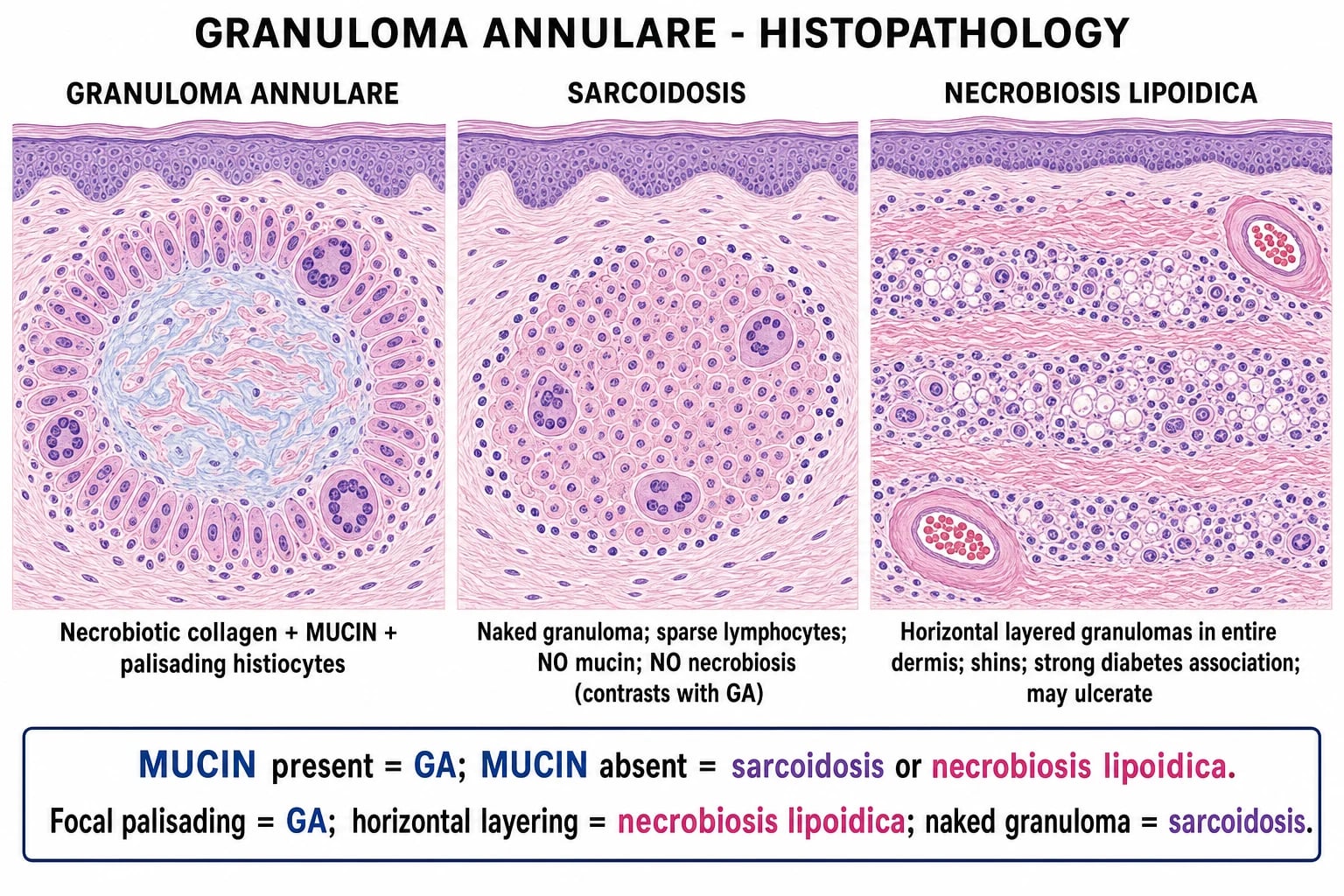
Mucin in GA is a mixture of acid mucopolysaccharides, principally hyaluronic acid, produced by fibroblasts in response to the inflammatory environment. Its accumulation is not a defining pathogenic event but rather a marker of the altered collagen matrix and the inflammatory response. The presence of mucin is the single most useful histological discriminator, as it is absent in the main differential diagnoses of sarcoidosis, necrobiosis lipoidica and rheumatoid nodules. [3]
Clinical Presentation
GA can present in several distinct ways. The clinical history, morphology and distribution usually allow a confident diagnosis, but atypical forms require biopsy. [1]
Localized GA
Localized GA presents as one or a few annular or arcuate plaques composed of small, firm, skin-coloured to erythematous or violaceous papules 1–5 mm in diameter. The papules are grouped tightly together, forming a raised, smooth border with central clearing. The resulting shape is often described as a doughnut ring or a string of beads. [1]
Key clinical features of localized GA include: [1]
- Site: dorsal hands, feet, fingers, toes, ankles, wrists, elbows and knees.
- Surface: smooth, non-scaly, non-crusted surface (unless secondarily traumatized).
- Symptoms: usually asymptomatic; mild pruritus or tenderness in a minority.
- Number: typically one to a few lesions; rarely more than 10.
- Natural history: tends to be self-limiting, with 50–75% resolving within months to a few years.[1][5]
The annular configuration is important because it places GA in the differential diagnosis of any annular skin lesion. However, the absence of scale and the smooth, firm papular surface are the clues that distinguish GA from tinea corporis and annular psoriasis. [3]
Generalized GA
Generalized GA presents with numerous papules, plaques and annular lesions, usually more than 10 and sometimes hundreds. The lesions are distributed over the trunk and proximal and distal extremities, often in a more symmetric pattern than localized disease. The lesions may be skin-coloured, pink, erythematous or violaceous. [4]
Features that distinguish generalized from localized GA include: [4]
- Age: older adults, although it can occur in younger patients.
- Distribution: trunk and extremities, not just acral sites.
- Morphology: may be papular, plaque-type or annular; individual lesions may be smaller and less well-defined than localized GA.
- Symptoms: may be mildly pruritic or asymptomatic.
- Associations: higher likelihood of diabetes mellitus, dyslipidaemia, thyroid disease, HIV and, rarely, malignancy.
- Course: more chronic and more refractory to treatment than localized disease.[1][4]
Because of the association with systemic disease, generalized GA should prompt a targeted screening history and examination, followed by appropriate investigations. [4]
Subcutaneous GA
Subcutaneous GA presents as deep, firm, skin-coloured to mildly erythematous nodules within the subcutaneous tissue. The overlying skin is usually normal. The lesions are typically painless, mobile or fixed, and 1–5 cm in diameter. Common sites include the scalp, fingers, hands, shins, pretibial areas and extensor surfaces of joints. [1]
The clinical importance of subcutaneous GA lies in the differential diagnosis of a child with a deep nodule. The main distinction is from rheumatoid nodule (which occurs over joints in patients with rheumatoid arthritis and is associated with positive rheumatoid factor) and from soft tissue sarcoma (which shows atypical cytology and mitoses on imaging or biopsy). The absence of arthritis and negative rheumatoid factor strongly favour subcutaneous GA. [3]
Perforating GA
Perforating GA presents as papules with a central keratotic plug, crust, scale or umbilication. This central change represents trans-epidermal elimination of necrobiotic collagen through the epidermis. The lesions are usually on the dorsal hands and fingers, and may be more symptomatic than other forms, with pain, pruritus or a tendency to discharge. Trauma and the Koebner phenomenon may be prominent. [1]
Patch-type GA
Patch-type GA is the rarest form, presenting as erythematous, violaceous or brownish patches or macules without a palpable annular border. It often occurs on the extremities and may be mistaken for morphea, parapsoriasis, erythema annulare centrifugum or other patch-form dermatoses. Biopsy is usually required. [3]
Atypical presentations
GA can present in unusual locations or forms: [1]
- Periorificial GA around the eyes or mouth.
- Mucosal GA on the oral mucosa or genitalia (rare).
- Linear GA following Blaschko lines or after trauma.
- GA in pregnancy may flare or remit, and usually resolves postpartum.
- GA associated with HIV is often generalized, atypical and refractory.
- GA in the elderly should prompt consideration of generalized disease and associated malignancy. [4]
Differential Diagnosis
The differential diagnosis of GA depends on the clinical morphology. Annular lesions must be distinguished from tinea corporis, annular psoriasis and erythema annulare centrifugum; deep nodules must be distinguished from rheumatoid nodules and soft tissue tumours; and histologically, the palisading granuloma must be distinguished from necrobiosis lipoidica, sarcoidosis and interstitial granulomatous dermatitis. [3]
Tinea corporis
Annular psoriasis
Necrobiosis lipoidica
Rheumatoid nodule
Sarcoidosis
Interstitial granulomatous dermatitis
Erythema annulare centrifugum
Actinic granuloma (O'Brien)
Clinical clues to distinguish annular lesions
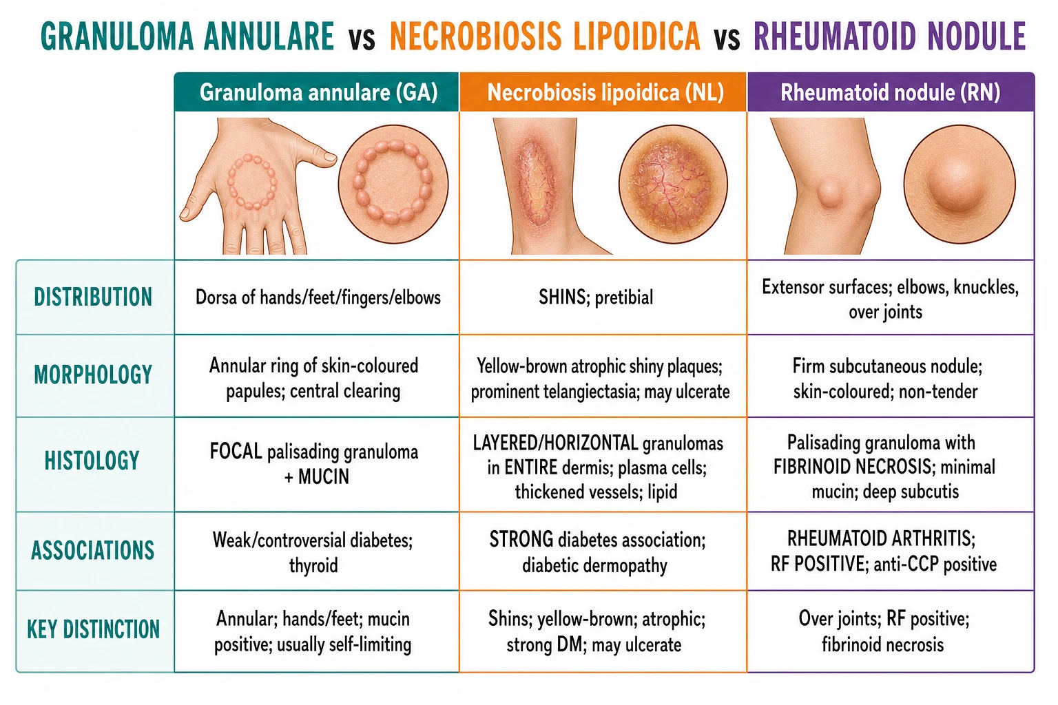
When a patient presents with an annular lesion, the following bedside observations help distinguish GA from the most common mimics: [3]
- Scale: GA has a smooth, non-scaly surface; tinea corporis has a scaly, advancing edge; annular psoriasis has silvery scale.
- Border: GA has a smooth, firm, papular border; tinea has a raised, erythematous, scaly border; erythema annulare centrifugum has a trailing collarette of scale.
- Site: GA favours the dorsal hands and feet; tinea can affect any body surface; necrobiosis lipoidica is almost always on the shins.
- Symptoms: GA is usually asymptomatic; tinea is often pruritic; psoriasis is pruritic and may have a history of plaques elsewhere.
- KOH microscopy: positive for fungal hyphae in tinea; negative in GA. [3]
Histological clues
When biopsy is performed, the pattern of granulomatous inflammation is the key discriminator: [1]
- GA: focal palisading granuloma with central mucin and necrobiotic collagen; no significant plasma cell infiltrate; well-circumscribed.
- Necrobiosis lipoidica: layered, horizontal granulomatous inflammation involving the entire dermis; plasma cells, thickened vessels, lipid deposition; no mucin.
- Sarcoidosis: naked non-caseating granulomas with minimal lymphocytes; no mucin; no necrobiosis.
- Rheumatoid nodule: palisading granuloma with central fibrinoid necrosis; minimal mucin; associated with rheumatoid arthritis.
- Interstitial granulomatous dermatitis: histiocytes dissecting between collagen bundles in an interstitial pattern; often associated with autoimmune disease or medication; less well-formed palisading and less central mucin than GA. [2]
Clinical & Bedside Assessment
The diagnosis of localized GA is usually clinical. The bedside examination focuses on confirming the characteristic morphology, excluding surface scale and deciding whether the presentation is atypical enough to require biopsy or systemic screening. [1]
Focused skin examination
The following features should be assessed: [1]
- Number and distribution of lesions: fewer than 10 lesions in an acral distribution favours localized GA; more than 10 lesions or trunk involvement raises the possibility of generalized GA.
- Morphology: annular or arcuate plaques of smooth, firm papules with central clearing.
- Surface: absence of scale is a key clue; look for central crusting or umbilication in perforating GA.
- Palpation: lesions are firm, non-tender and indurated; subcutaneous lesions are deep and mobile or fixed nodules.
- Diascopy: in GA, diascopy may show an apple-jelly appearance if lesions are erythematous, but this is less reliable than in sarcoidosis. Negative diascopy does not exclude GA.
- Koebner phenomenon: ask about trauma or lesions at sites of injury, particularly in generalized GA. [4]
When to screen for systemic disease
Systemic screening is not required for a typical child or young adult with localized acral GA. Screening is indicated for: [1]
- Generalized GA, especially in adults.
- Atypical, recurrent or refractory GA.
- GA in the setting of HIV risk factors or other systemic symptoms.
- Elderly patients presenting with new-onset generalized GA.
- Subcutaneous GA when the diagnosis is uncertain and rheumatoid arthritis or malignancy must be excluded. [4]
The targeted history should include symptoms of diabetes (polyuria, polydipsia, weight loss), thyroid disease (heat intolerance, weight change, palpitations), HIV risk, malignancy symptoms (B symptoms, weight loss) and drug history. [6]
Investigations
Diagnostic tests
Skin biopsy is the definitive diagnostic test when the diagnosis is uncertain, the lesion is atypical, or the presentation is generalized. A punch biopsy from the active edge of an annular lesion or the centre of a papule is usually adequate. Histology shows the characteristic palisading granuloma with central mucin and necrobiotic collagen. Special stains for mucin, such as Alcian blue, colloidal iron or toluidine blue, may be used to confirm the diagnosis if the histological features are subtle. [4]
Screening tests in generalized or atypical GA
In generalized or atypical GA, the following investigations are reasonable to screen for associated systemic disease: [4]
- Fasting plasma glucose and HbA1c — to screen for diabetes mellitus.
- Lipid profile — dyslipidaemia is associated with generalized GA.
- Thyroid function tests (TSH, free T4) — to screen for thyroid disease.
- HIV test — if risk factors or clinical suspicion.
- Age-appropriate malignancy screening — especially in elderly patients with generalized or refractory disease; consider lymphoma work-up if B symptoms or lymphadenopathy.
- Rheumatoid factor (RF), anti-CCP antibodies — in subcutaneous nodules to distinguish from rheumatoid nodules.
- KOH microscopy — if tinea corporis is suspected clinically. [4]
Histological staining and interpretation
Mucin stains are useful when the diagnosis is uncertain. Alcian blue stains acid mucopolysaccharides blue, and is positive in the central zone of GA granulomas. Colloidal iron also stains mucin. Periodic acid–Schiff (PAS) stain can be used to exclude fungal infection if tinea is in the differential. Fite stain is negative for mycobacteria in GA. Polarized light can identify foreign material in foreign-body granulomas. [3]
Management — Resuscitation
GA is a benign, chronic dermatosis and does not require emergency or life-saving treatment. There is no resuscitation algorithm. The only time-critical consideration is the recognition that an annular lesion may represent an infection or other serious condition rather than GA. If there is rapid expansion, pain, purulence, systemic symptoms or an immunocompromised host, the clinician should reconsider the diagnosis and treat the underlying cause (for example, bacterial cellulitis or necrotizing infection) rather than managing as GA. [3]
Management — Definitive & Stepwise
Management of GA is determined by the subtype, extent, symptoms and patient preference. The approach is stratified into localized disease, generalized disease, subcutaneous disease and perforating disease. [4]
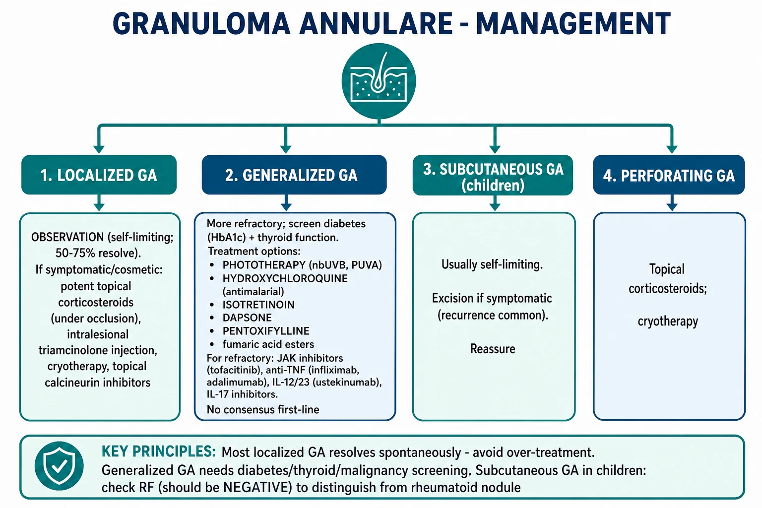
Localized GA
Most localized GA is self-limiting, so observation and reassurance are the first-line approach, especially in children. Active treatment is reserved for symptomatic, cosmetically concerning or persistent lesions. [1]
First-line options [1]
- Potent topical corticosteroids — for example, clobetasol propionate 0.05% ointment applied once or twice daily for 2–4 weeks, then tapered. Occlusion can enhance penetration. Limit duration and potency to avoid skin atrophy, especially on the dorsal hands.[5]
- Intralesional corticosteroids — triamcinolone acetonide 2.5–5 mg/mL injected into the active border of the lesion. Higher concentrations (10–20 mg/mL) may be used for thicker lesions, but increase the risk of atrophy and hypopigmentation. This is particularly useful for a few persistent localized lesions.[5]
Second-line options [1]
- Cryotherapy — liquid nitrogen cryotherapy can be effective, but may cause blistering, dyspigmentation and scarring.
- Topical calcineurin inhibitors — tacrolimus 0.1% ointment or pimecrolimus 1% cream, especially useful on the face and in children to avoid steroid-induced atrophy.
- Topical imiquimod — reported as effective in some cases, presumably by modulating local immune response.
- Phototherapy — narrowband UVB or PUVA can be considered for multiple localized lesions that are refractory to topical therapy. [4]
Generalized GA
Generalized GA is more refractory and there is no universally accepted first-line systemic therapy. Treatment choice depends on comorbidities, patient preference and availability. A stepwise approach is reasonable. [4]
First-line systemic options [1]
- Hydroxychloroquine — 200–400 mg orally daily, depending on body weight. A common regimen is 200 mg twice daily for adults, with ophthalmological monitoring after 5 years of cumulative use. Antimalarials are thought to modulate Th1 cytokine production and granuloma formation. Baseline and periodic eye examination is required because of the risk of retinopathy.[4]
- Dapsone — typically 50–100 mg orally daily, sometimes started at 25 mg daily to reduce haemolysis risk, especially in G6PD-deficient patients. Check G6PD level before starting. Monitor full blood count and liver function regularly. Dapsone is particularly useful when neutrophilic infiltration is prominent.[4]
Second-line options [1]
- Isotretinoin — 0.5–1 mg/kg/day orally for several months, in patients without contraindications. Monitoring includes lipid profile, liver function tests and pregnancy prevention in females of childbearing potential. Isotretinoin may work by altering keratinization and immune modulation.[4]
- Methotrexate — 7.5–25 mg orally once weekly, with folic acid supplementation. Monitor full blood count, liver function and renal function. Methotrexate is useful for severe, refractory generalized GA, particularly when other therapies fail.[4]
- Phototherapy — narrowband UVB (nbUVB) or PUVA can be effective, especially when lesions are widespread. Treatment typically requires 2–3 sessions per week for several weeks to months.
Third-line / refractory options [1]
- Biologics — case reports and small series support adalimumab (anti-TNF), infliximab, ustekinumab (IL-12/23 inhibitor) and secukinumab (IL-17 inhibitor) in refractory generalized GA. Adalimumab is commonly reported as effective, given the Th1/TNF-driven pathogenesis. Dosing follows the approved regimen for psoriasis, for example adalimumab 80 mg subcutaneously at week 0, then 40 mg every other week, with screening for tuberculosis and hepatitis B before initiation.[4]
- Other systemic agents — pentoxifylline, fumaric acid esters, niacinamide, doxycycline, cyclosporine and systemic corticosteroids have all been reported, but evidence is limited and outcomes are variable.
Subcutaneous GA
Subcutaneous GA in children is usually self-limiting and does not require treatment. Reassurance of the family is the mainstay. If the lesion is symptomatic, painful, cosmetically unacceptable or diagnostically uncertain, excisional biopsy can be both diagnostic and therapeutic. Recurrence is common. Systemic therapy is rarely needed. [4]
Perforating GA
Perforating GA is treated with topical corticosteroids and cryotherapy as first-line options. Intralesional corticosteroids may be useful for individual lesions. The central crusting can be managed with gentle debridement and emollients. Phototherapy or systemic therapy may be needed for extensive disease. [4]
Patch-type GA
Patch-type GA is usually treated with topical corticosteroids or topical calcineurin inhibitors. If lesions are extensive or refractory, phototherapy or systemic agents used for generalized GA can be considered. [4]
Specific Subtypes & Scenarios
GA in children
Localized GA is common in children and is usually self-limiting. The main management is reassurance and, if needed, topical corticosteroids or calcineurin inhibitors. Subcutaneous GA is the most common deep nodule in children and must be distinguished from rheumatoid nodules and soft tissue tumours. In children, systemic therapy for generalized GA is rarely needed; phototherapy and topical therapies are preferred. [4]
GA in pregnancy
GA may first present, flare or remit during pregnancy. Treatment is conservative because many systemic agents are contraindicated. Topical corticosteroids and calcineurin inhibitors are generally considered safe in pregnancy. Systemic agents such as hydroxychloroquine, dapsone, methotrexate and isotretinoin are contraindicated or require careful risk–benefit discussion. Most pregnancy-associated GA resolves postpartum. [4]
GA associated with HIV
GA in HIV-positive patients is often generalized, atypical and refractory. Highly active antiretroviral therapy (HAART) may improve the skin condition by restoring immune function. Treatment otherwise follows the principles for generalized GA, with early escalation to systemic or biologic therapy if needed. [4]
GA associated with diabetes and metabolic syndrome
The association between GA and diabetes mellitus is controversial but most relevant for generalized disease. When diabetes is present, optimizing glycaemic control is important for overall health, although glycaemic control alone does not reliably clear GA. Dyslipidaemia and thyroid disease should also be treated if identified. [4]
Drug-induced GA
If a drug is suspected, withdrawal of the offending agent is the first step. Lesions may take weeks to months to resolve after drug cessation. Symptomatic treatment with topical corticosteroids can be used during the resolution phase. [1]
Complications & Pitfalls
Diagnostic pitfalls
The most common diagnostic error is to treat GA as a fungal infection. Because GA is annular and sometimes slightly erythematous, it can be mistaken for tinea corporis. Treating with antifungal agents will not help and delays appropriate management. A KOH preparation is a quick and reliable way to confirm or exclude tinea. [3]
Another pitfall is to assume that a deep nodule in a child is a rheumatoid nodule or malignancy without biopsy. Subcutaneous GA is usually benign and self-limiting, whereas rheumatoid nodules imply systemic rheumatoid arthritis and soft tissue sarcoma requires oncological management. Histology and negative rheumatoid serology distinguish them. [2]
Management pitfalls
- Over-treating localized GA — because most localized GA resolves spontaneously, aggressive systemic therapy is unnecessary and exposes the patient to adverse effects.
- Using systemic corticosteroids as first-line — rebound and adverse effects are common; reserve for exceptional cases.
- Failing to screen for systemic disease — generalized GA in an adult warrants screening for diabetes, dyslipidaemia, thyroid disease and HIV, and malignancy if clinically indicated.
- Missing perforating GA — the central crusting can be mistaken for infection; biopsy may be needed. [4]
Complications of the disease itself
GA is a benign disease and does not cause significant medical complications. The main morbidity is psychological distress or cosmetic concern, particularly when lesions are on the hands or face. Perforating GA can cause discomfort, scarring and secondary infection. Extensive generalized GA can significantly impair quality of life. [4]
Prognosis & Disposition
The prognosis of GA depends on the subtype. Localized GA has an excellent prognosis, with 50–75% of cases resolving spontaneously within months to a few years. Recurrence at the same or different sites is common, affecting roughly 40% of patients. Generalized GA is more persistent and may last for years; complete spontaneous resolution is less common than in localized disease. Subcutaneous GA in children usually resolves spontaneously over months to years. Perforating GA is more chronic and may leave scars or dyspigmentation. [4]
Disposition and referral
Most patients with localized GA can be managed in primary care or general dermatology. Referral to dermatology is appropriate when: [1]
- The diagnosis is uncertain.
- Lesions are generalized, refractory or atypical.
- There is suspicion of perforating, subcutaneous or patch-type GA.
- Systemic disease is suspected and requires coordinated screening.
- First-line topical therapy fails and systemic or phototherapy is being considered. [4]
Special Populations
Children
Localized GA is common in children and is usually managed conservatively. Subcutaneous GA is the most important paediatric variant because it mimics rheumatoid nodules. In children, rheumatoid factor and anti-CCP antibodies are negative, there is no arthritis and the lesions usually resolve spontaneously. Imaging (ultrasound or MRI) may be useful if a soft tissue sarcoma is suspected. Excisional biopsy may be needed for diagnostic confirmation. [2]
Pregnancy
GA may worsen or improve in pregnancy. Treatment is conservative. Topical corticosteroids and calcineurin inhibitors are preferred. Systemic agents are generally avoided; hydroxychloroquine may be continued in selected patients if already established for another indication, but this requires specialist input. Most cases resolve postpartum. [4]
Elderly
New-onset or generalized GA in an elderly patient should prompt screening for diabetes mellitus, dyslipidaemia, thyroid disease, HIV and occult malignancy. The threshold for biopsy should be lower because atypical presentations and mimics are more common in this age group. Phototherapy may be preferable to systemic agents in frail patients. [4]
Immunocompromised patients
In HIV and other immunosuppressed states, GA tends to be more widespread and refractory. Optimize treatment of the underlying condition, screen for opportunistic infections and consider early systemic or biologic therapy. Live vaccines should be avoided in patients on biologics or immunosuppressants. [4]
Evidence, Guidelines & Regional Differences
Evidence for diabetes association
The association between GA and diabetes mellitus has been debated for decades. Early studies suggested a strong association, particularly with generalized GA, while more recent studies have found the association to be weaker or inconsistent. A systematic approach is to screen patients with generalized or atypical GA for diabetes and impaired glucose tolerance, but not to label every patient with GA as diabetic.[1][6]
Evidence for treatment
There are no large randomized controlled trials guiding GA therapy. Management is based on case series, small cohort studies, expert opinion and the 2025 systematic review by Bettolini et al. on generalized GA.[4] The evidence supports observation for localized disease, topical and intralesional corticosteroids for a few lesions, and a range of systemic or phototherapy options for generalized disease. Biologic therapy is supported by case reports and small series, not by controlled trials.
Regional differences
United Kingdom / Europe — NICE does not provide a specific guideline for GA. Management follows British Association of Dermatologists consensus and expert opinion, with emphasis on observation for localized disease and phototherapy or antimalarials for generalized disease. [4]
United States — American Academy of Dermatology guidance supports observation, topical and intralesional corticosteroids, and a broad range of systemic agents for refractory generalized GA. Access to biologics depends on insurance and prior authorization. [4]
Australia / New Zealand — The Australasian Journal of Dermatology systematic review emphasizes phototherapy, antimalarials, dapsone and isotretinoin, with biologics reserved for refractory disease.[4]
India / South Asia — In resource-limited settings, topical and intralesional corticosteroids, dapsone and hydroxychloroquine are the most accessible systemic options. Phototherapy may be available in larger centres. Tuberculosis and fungal infections are more common differential diagnoses in endemic regions, so biopsy and staining are important. [4]
Exam Pearls
[4]Exam application bank (NEET-PG / INICET)
One-line answer
Granuloma annulare (GA) is a benign, usually self-limiting granulomatous dermatosis. Histology: palisading granulomas with central necrobiotic collagen + mucin surrounded by histiocytes. Localized (commonest): annular skin-coloured papules on dorsa of hands/feet; self-limiting. Generalized: disseminated; may associate with diabetes, dyslipidaemia, thyroid disease and HIV; refractory. Subcutaneous (deep): firm nodules in children; mimics rheumatoid nodules (but RF negative). Perforating: crusted papules (trans-epidermal elimination). DDx: tinea corporis, annular psoriasis, necrobiosis lipoidica, rheumatoid nodules, sarcoidosis, interstitial granulomatous dermatitis, erythema annulare centrifugum, actinic granuloma. Management: localized — observe or potent topical corticosteroids / intralesional triamcinolone, cryotherapy, tacrolimus; generalized — topical dapsone, hydroxychloroquine, nbU
Worked stems (answer without another resource)
Stem 1 — Classic presentation. Map symptoms to mechanism; name the first investigation and first treatment step with dose/route if drug therapy is standard. [1]
Stem 2 — Unstable / complicated. List red flags that force immediate resuscitation, theatre, ICU, antidote, or reperfusion — and what you do in the first 15 minutes. [1]
Stem 3 — Atypical group. Elderly, pregnancy, child, or immunocompromised: how presentation and thresholds change. [1]
Stem 4 — Differential trap. Name the three closest mimics and one discriminator for each. [1]
Stem 5 — Disposition. Who goes home with safety-netting, who is admitted, who needs HDU/ICU/theatre, and what follow-up is mandatory. [1]
Rapid viva checklist
- Definition + classification
- Pathophysiology chain
- Bedside signs / criteria
- Score with exact components (if any)
- Emergency bundle
- Definitive therapy with doses
- Complications of disease and of treatment
- Special populations
- Guideline/trial name if classic
- Three exam traps
Coverage self-check
If you cannot answer any stem above from this page alone, re-read the matching section — the page is intended to be self-sufficient for final-prof and NEET-PG/INICET questions on Granuloma annulare.
[4]References
- [1]Joshi TP, Duvic M. Granuloma Annulare: An Updated Review of Epidemiology, Pathogenesis, and Treatment Options Am J Clin Dermatol, 2022.PMID 34495491
- [2]Terziroli Beretta-Piccoli B, Mainetti C, Peeters MA, et al. Cutaneous Granulomatosis: a Comprehensive Review Clin Rev Allergy Immunol, 2018.PMID 29352388
- [3]Trayes KP, Savage K, Studdiford JS. Annular Lesions: Diagnosis and Treatment Am Fam Physician, 2018.PMID 30216021
- [4]Bettolini L, Maione V, Carugno A, et al. Management Strategies for Generalised Granuloma Annulare: A Systematic Review of Current and Emerging Therapies Australas J Dermatol, 2025.PMID 40586485
- [5]Keimig EL. Granuloma Annulare Dermatol Clin, 2015.PMID 26143416
- [6]Lima AL, Illing T, Schliemann S, et al. Cutaneous Manifestations of Diabetes Mellitus: A Review Am J Clin Dermatol, 2017.PMID 28374407