Dermatology · Medicine
Grover's disease (transient acantholytic dermatosis)
Also known as Grover's disease · Transient acantholytic dermatosis (TAD) · Persistent acantholytic dermatosis · Acantholytic dermatosis, transient
Grover's disease (transient acantholytic dermatosis, TAD) is an acquired, usually self-limiting, intensely pruritic papulovesicular eruption on the trunk of middle-aged to elderly men, characterised histologically by focal acantholysis. The eruption is precipitated by heat, sweating, prolonged bed rest and xerosis, and runs a course that ranges from a few weeks to several years. Four histological patterns (Darier-like, pemphigus-like, Hailey-Hailey-like, spongiotic) may coexist in the same biopsy. Diagnosis is confirmed by punch biopsy from a fresh, intact papulovesicle; direct immunofluorescence is characteristically negative. Management is stepwise: trigger avoidance, emollients, topical corticosteroids, antihistamines, then topical calcineurin inhibitors, oral tetracyclines, phototherapy and oral retinoids for refractory disease.
On this page & tools
Your progress
Saved locally on this device.
Exam tags
Red flags
Overview and definition
Grover's disease - first described by Ralph W Grover in 1970 as "transient acantholytic dermatosis" (TAD) - is an acquired, usually self-limiting, intensely pruritic papulovesicular eruption that is defined histologically by focal acantholysis (loss of intercellular cohesion between suprabasal keratinocytes, with the basal layer typically remaining attached like a row of tombstones to the basement membrane).[2][1]
The disease is a sporadic, non-familial disorder of the pilosebaceous unit and adjacent epidermis. It is best conceptualised as a cutaneous reaction pattern, in which a diverse group of precipitants - heat, sweating, occlusion, friction, xerosis, fever, prolonged bed rest, recent surgery and, rarely, drugs or malignancy - triggers acantholysis in a susceptible individual. The word "transient" reflects the original observation that most cases remit within weeks; the term is now used loosely because a substantial minority persist or recur for months to years and have been reclassified as persistent acantholytic dermatosis when disease continues beyond 6-12 months.[4][5]
The diagnosis rests on the triad of (i) typical morphology and distribution, (ii) typical demographic (middle-aged or elderly man), and (iii) confirmatory punch biopsy showing focal acantholysis with one or more of four recognised histological patterns and a negative direct immunofluorescence (DIF). The negative DIF is the single most useful single-investigation discriminator from pemphigus, which is its principal autoimmune mimic.[1]
[1]ICD-11 and classification
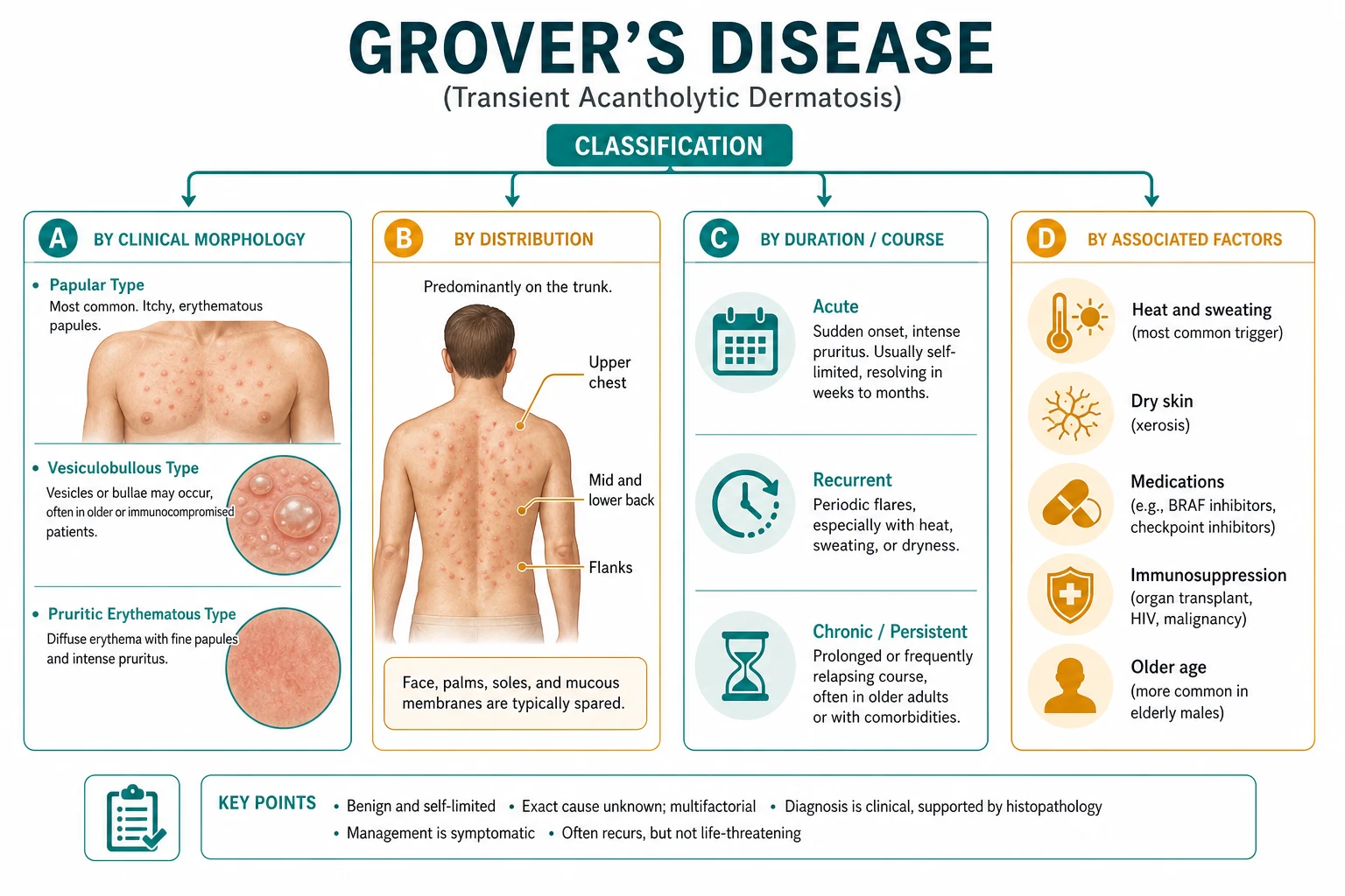
In ICD-11 (WHO, 2024) Grover's disease is classified under Disorders of skin appendages → Other specified disorders of skin as ED7Y (Other specified disorders of skin), with the inclusion term "Transient acantholytic dermatosis". Within the larger family of acantholytic dermatoses, Grover's sits alongside the genetic disorders (Darier's disease, Hailey-Hailey disease) and the autoimmune acantholytic disorders (pemphigus vulgaris, pemphigus foliaceus, paraneoplastic pemphigus, IgA pemphigus). [1]
A clinically useful classification splits Grover's into: [1]
- Classical (transient) Grover's disease - self-limiting eruption resolving in weeks to a few months.
- Persistent (chronic) acantholytic dermatosis - identical histology but a course that extends beyond 6-12 months with frequent relapses, often with continued heat or sweat exposure.
- Subtype variants by predominant histology / clinical pattern: Darier-like, pemphigus-like, Hailey-Hailey-like, spongiotic, bullous/vesiculobullous, eczematous, follicular, photo-distributed. [1]
| Family | Examples | Mechanism | DIF |
|---|---|---|---|
| Inherited | Darier's disease (ATP2A2); Hailey-Hailey disease (ATP2C1) | Loss-of-function mutation in SERCA Ca²⁺ pump → defective desmosomal assembly | Negative |
| Acquired / reactive | Grover's disease (TAD); persistent acantholytic dermatosis | Acquired, transient dysfunction of desmosomal-cytoskeletal complex in heat / sweat | Negative |
| Autoimmune | Pemphigus vulgaris, foliaceus, vegetans, IgA pemphigus, paraneoplastic pemphigus | IgG autoantibody against desmoglein 1 / 3 / plakin | Positive (intercellular IgG/C3) |
Epidemiology and risk factors
Grover's disease is uncommon but not rare, accounting for an estimated 1-2% of dermatology referrals for truncal papular eruptions in temperate climates. The median age of onset is the sixth to seventh decade, and men are affected approximately three times as often as women in most series.[4][2]
Established triggers and associations: [1]
- Heat and sweating - the most consistently reported precipitant. Hot weather, hot baths, occlusive clothing, electric blankets, and febrile illness all reproduce or worsen the eruption.
- Prolonged bed rest and immobility - classically described in patients confined to bed following orthopaedic surgery, stroke, hip fracture, ITU admission, or critical illness. Occlusion of sweat-duct ostia in the supine position is the proposed mechanism.
- Xerosis (dry skin) and winter weather - paradoxically, many European and North American series report a winter peak, attributed to xerotic, itchy skin rather than heat alone. The shared final common pathway is disrupted epidermal barrier.
- Sun exposure - photo-distributed Grover's is a recognised variant, usually on the chest and upper back after intense sun exposure.
- Recent hospitalisation, surgery, general anaesthesia, and ITU admission - acting through a combination of heat, sweating under occlusive drapes, friction, and stress.
- End-stage renal disease / haemodialysis - multiple case series link TAD with uraemic pruritus and the dialysis state.
- HIV / immunosuppression - case reports, but the prevalence is uncertain.
- Haematological malignancy and solid organ cancer - rare but important paraneoplastic association; the label "paraneoplastic acantholytic dermatosis" applies when the eruption heralds or parallels a malignancy, most often haematological (CLL, lymphoma) or, less often, genitourinary.
- Drug triggers - isolated reports implicating recombinant interleukin-4, penicillamine, 5-fluorouracil, and others. Causation is rarely proven. [1]
Pathophysiology
The defining histological event is acantholysis - the loss of intercellular adhesion between suprabasal keratinocytes, leaving a cleft within the epidermis and freeing individual rounded "acantholytic" keratinocytes into the blister cavity. This must be distinguished from spongiosis (intercellular oedema that widens desmosomal gaps but does not rupture them) - the two are separate mechanisms of vesicle formation, and a single biopsy may show elements of both in Grover's disease. [1]
Three observations link Grover's to the genetic acantholytic disorders and to the broader field of desmosomal biology: [1]
- The SERCA / calcium-pump hypothesis. Darier's disease is caused by loss-of-function mutations in ATP2A2 (encoding the SERCA2 calcium pump of the endoplasmic reticulum); Hailey-Hailey disease is caused by mutations in ATP2C1 (encoding the related secretory-pathway Ca²⁺/Mn²⁺-ATPase, SPCA1). Both pumps load Ca²⁺ into the ER, and the resulting high ER Ca²⁺ concentration is required for the conformational maturation and trafficking of desmosomal cadherins (desmogleins, desmocollins) and the linker proteins plakoglobin and plakophilin. Recent work has shown that Grover's disease shares many of the same downstream molecular signatures as Darier and Hailey-Hailey - reduced plakoglobin and desmoglein staining at lesional keratinocyte borders, abnormal keratin filament retraction - even though no germline ATP2A2/ATP2C1 mutation is present. The proposed model is that heat, sweat, friction and ageing acquire a state of functional SERCA insufficiency at the keratinocyte level, producing the same desmosomal destabilisation but in a transient, reversible way.[6]
- Occlusion of the sweat-duct ostium. Sweat retention and the resulting epidermal maceration have been proposed as the local "second hit" that converts a subclinical state of desmosomal fragility into visible acantholysis in a small focus. This explains the predilection for the trunk (high sweat density), the temporal link to bed rest, and the seasonal pattern.
- Xerosis and barrier dysfunction. Winter-precipitated Grover's implicates a defective stratum corneum barrier that allows TEWL-driven inflammation and pruritus, with acantholysis as a secondary phenomenon. This is consistent with the response to aggressive emollient therapy in many patients.
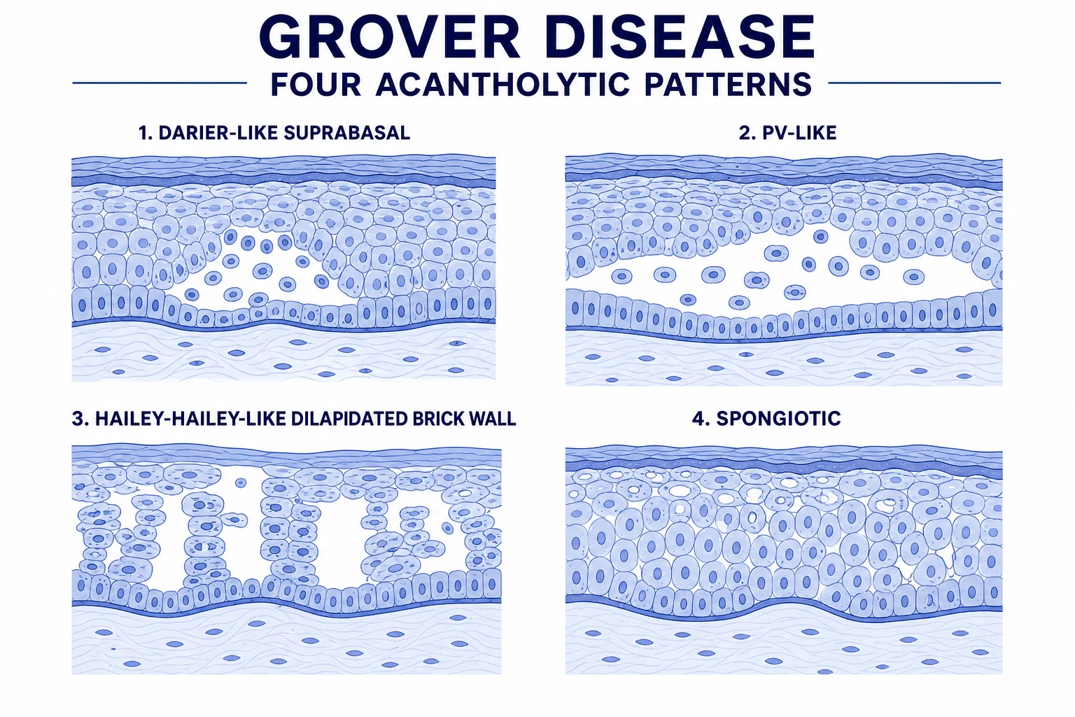
Why four patterns in one biopsy? Each focus of acantholysis begins with a small suprabasal cleft; as it evolves, the surrounding epidermis may develop dyskeratosis (giving a Darier-like pattern with corps ronds and grains), extensive full-thickness acantholysis (giving a Hailey-Hailey-like "dilapidated brick wall" pattern), suprabasal acantholysis without dyskeratosis (giving a pemphigus-like pattern) or spongiotic change with mild acantholysis. The biopsy is therefore a snapshot of acantholysis at multiple time-points and severities, and the four patterns are not four diseases but four morphological stages of one process.[1][3]
| Step | Event | Result |
|---|---|---|
| 1 | Heat, sweat, friction, xerosis in a susceptible individual | Local keratinocyte stress and barrier dysfunction |
| 2 | Functional SERCA2 insufficiency in lesional keratinocytes | Reduced ER Ca²⁺, defective desmosomal cadherin maturation |
| 3 | Loss of desmosomal adhesion | Suprabasal acantholysis, cleft formation |
| 4 | Evolution of the cleft | One of four histological patterns (Darier, pemphigus, Hailey-Hailey, spongiotic) |
| 5 | Pruritus, excoriation, eczematisation | Symptoms and secondary changes (crust, erosion, impetiginisation) |
Clinical presentation
The classical presentation is an acute to subacute onset of intense pruritus with crops of 1-3 mm, firm, discrete, erythematous to red-brown papules and papulovesicles on the central chest, upper back, and clavicular region, sometimes extending to the flanks, mid-back and proximal limbs. The face, scalp, palms, soles and oral mucosa are characteristically spared (a critical distinguishing feature from Darier's and pemphigus). Lesions may become crusted or eroded from scratching, and in darkly pigmented skin the papules may be hyperpigmented rather than erythematous, sometimes mistaken for lichen planus or prurigo nodularis. [1]
Atypical and less common presentations - all recognised: [1]
- Bullous or vesiculobullous Grover's - tense vesicles and small bullae predominate; resembles bullous pemphigoid on first glance but is acantholytic, not subepidermal, and DIF is negative.
- Eczematous / spongiotic-predominant Grover's - erythema, weeping, scaling; the predominant histology is spongiotic; commonly misdiagnosed as eczema until biopsy.
- Follicular / infundibular Grover's - papules centred on hair follicles, mimicking folliculitis.
- Photo-distributed Grover's - crops of papulovesicles on sun-exposed chest and arms after intense sun exposure in summer.
- Herpetiform Grover's - grouped vesicles resembling dermatitis herpetiformis; biopsy is again the discriminator.
- Universal / erythrodermic Grover's - rare; consider paraneoplastic acantholysis in this setting.
- Hailey-Hailey-like widespread eruption - flexural accentuation; mimic of Hailey-Hailey disease. [1]
Temporal course. Onset is typically acute to subacute, with peak severity within days. The phrase "transient" describes the median course of 6 weeks to 6 months; in long-term follow-up series, a substantial minority remain symptomatic beyond 12 months, justifying the term "persistent acantholytic dermatosis" for the chronic form.[1][4]
Differential diagnosis
Grover's disease has a wide differential because the clinical phenotype - itchy truncal papulovesicles in an older adult - is shared by eczema, scabies, drug eruptions, and several primary acantholytic disorders. The histology is the tie-breaker in most cases, and the negative DIF is the single most important laboratory discriminator. [1]
| Condition | How it differs from Grover's | Key test |
|---|---|---|
| Darier's disease (keratosis follicularis) | Onset in adolescence, positive family history, seborrhoeic and flexural greasy crusted papules, V-shaped nail nicks, palmar pits, oral cobblestone mucosa. Chronic. | Clinical + family history; ATP2A2 sequencing |
| Hailey-Hailey disease (familial benign chronic pemphigus) | Onset in second/third decade, AD inheritance, flexural (neck, axillae, groin, inframammary) painful erosions and malodorous macerated plaques. | Clinical; ATP2C1 sequencing |
| Pemphigus vulgaris | Flaccid blisters, painful oral erosions, positive Nikolsky, constitutional symptoms. Anti-Dsg3 (± Dsg1) antibodies. | DIF: intercellular IgG/C3; ELISA Dsg3 |
| Pemphigus foliaceus | Superficial crusted erosions, no mucosa, anti-Dsg1. Older adults, can mimic persistent Grover's. | DIF: intercellular IgG; ELISA Dsg1 |
| Bullous pemphigoid | Tense bullae on urticarial base, pruritic, elderly. Subepidermal split. | DIF: linear IgG/C3 at BMZ; ELISA BP180/BP230 |
| Scabies | Burrows in finger webs, wrists, waistline, genitals. Family contacts. Nocturnal pruritus. | Dermoscopy (delta wing); microscopy for mites/eggs |
| Miliaria rubra (prickly heat) | Tiny erythematous papules in hot/humid environment, no acantholysis, resolves rapidly with cooling. | Clinical; biopsy only if persistent |
| Folliculitis (bacterial, fungal) | Follicular pustules, central hair, no acantholysis. | Swab for culture; KOH |
| Contact dermatitis | Pattern of exposure; vesicular on palmar surfaces; resolves with avoidance. | Patch testing |
| Atopic dermatitis | Flexural lichenification, chronic relapsing, personal or family atopy. | Clinical |
| Dermatitis herpetiformis | Extensor (elbows, knees, buttocks), grouped vesicles, intense burning pruritus, gluten-sensitive enteropathy. | DIF: granular IgA at dermal papillae; anti-tTG, anti-EMA |
| Papular urticaria | Children, exposed sites, recurrent crops, insect hypersensitivity. | Clinical; history |
| Drug eruption (morbilliform) | Widespread, drug-temporally linked, eosinophilia, mucosal involvement. | Clinical; drug dechallenge |
| Prurigo nodularis | Chronic, excoriated nodules, lichenified, often psychogenic. | Clinical |
Clinical and bedside assessment
The diagnosis is reached by combining three lines of evidence: (i) the clinical pattern (typical morphology and distribution in a typical demographic with a typical trigger), (ii) the bedside exclusion of close mimics (scabies, eczema, contact dermatitis, folliculitis), and (iii) the histological confirmation by punch biopsy from a fresh, intact papulovesicle with negative DIF. [1]
Bedside checklist (every suspected case): [1]
- Trunk (central chest, upper back, clavicles, flanks, mid-back, lumbar area) - examine the entire trunk; document lesion count and morphology.
- Face, scalp, oral mucosa, palms, soles, nails - explicitly note the absence of involvement; this is the rule-out for Darier and pemphigus.
- Flexures - axillae, inframammary, groin, neck (for Hailey-Hailey-like variants).
- Finger webs, wrists, waistline, genitals - inspect for scabies burrows; use dermoscopy (the "delta-wing" sign of the mite's pigmented head) to confirm.
- Hair and nails - V-shaped nicks, longitudinal red and white lines, distal subungual hyperkeratosis (Darier); onycholysis and nail dystrophy (eczema, psoriasis).
- Nikolsky sign (gentle lateral pressure on perilesional skin or rubbing of a distant site) - negative in Grover's, positive in pemphigus. Variants include the "shearing" Nikolsky and the "bulla-spread" sign.
- Temperature and ambient - room temperature, recent hot bath, electric blanket, occlusive clothing, recent sun exposure.
- Recent history - hospitalisation, surgery, ITU, hip fracture, stroke, dialysis, new drugs, febrile illness, weight loss, night sweats (paraneoplastic screen if persistent). [1]
Dermoscopy of Grover's disease shows a star-like or "crown" pattern of a brownish-red central area surrounded by a whitish halo and a fine peripheral pigment network, with occasional reddish globules (vascular ectasia) - a non-specific but supportive finding that overlaps with several other acantholytic and spongiotic dermatoses. [1]
Investigations
First-line [1]
- Punch biopsy (4 mm) from a fresh papulovesicle for H&E - the histological cornerstone. Send in formalin.
- Punch biopsy from an adjacent perilesional site for direct immunofluorescence (DIF) - send fresh, in Michel's or Zeus medium, or in saline-soaked gauze if transfer is <2 hours. DIF is negative in Grover's disease; positivity for intercellular IgG/C3 redefines the diagnosis as pemphigus; positivity for linear IgG/C3 at the basement membrane redefines it as bullous pemphigoid; positivity for granular IgA at dermal papillae redefines it as dermatitis herpetiformis.[1][3]
Second-line (selective) [1]
- Serum anti-desmoglein 1 and 3 ELISA - in atypical, persistent or bullous cases, or where DIF is borderline or unavailable. Negative in Grover's.
- Swab for bacterial culture and sensitivity - if impetiginisation is suspected (ooze, crust, surrounding cellulitis).
- Viral PCR for HSV / VZV (swab, vesicular fluid) - if grouped vesicles or umbilicated pustules suggest eczema herpeticum or Kaposi varicelliform eruption.
- Scabies skin scraping (and dermoscopy) - if finger-web or genital burrows are seen.
- KOH preparation - if candidal intertrigo or dermatophyte infection is in the differential.
- Patch testing - if contact dermatitis is suspected and the eruption persists despite treatment. [1]
Selective workup for associated disease (persistent or refractory cases) [1]
- Full blood count, peripheral smear, LDH - paraneoplastic screen for haematological malignancy.
- Urea, creatinine, eGFR - renal failure / dialysis is a recognised association.
- HIV serology - in atypical, widespread or persistent disease in adults.
- Age-appropriate cancer screening (PSA, mammography, CT chest/abdomen/pelvis) - only if paraneoplastic acantholytic dermatosis is suspected, i.e. explosive onset, severe pruritus, older adult, weight loss, B-symptoms, or resistance to multiple therapies.
- Biopsy for ATP2A2 / ATP2C1 sequencing - if Darier's or Hailey-Hailey disease is suspected despite initial clinical picture. [1]
Histopathology
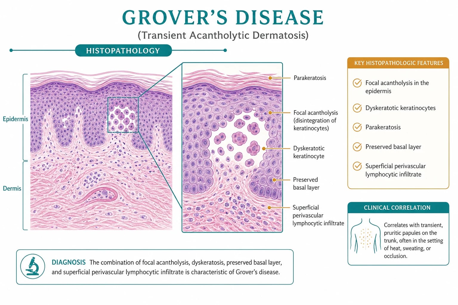
The hallmark is focal acantholysis - small, discrete, well-circumscribed foci in which suprabasal keratinocytes have lost their intercellular cohesion and float free within a cleft. The basal layer remains attached to the basement membrane, often described as a row of "tombstones" (also seen in pemphigus vulgaris, but in Grover's the focus is focal, not confluent). The acantholysis may be accompanied by spongiosis (intercellular oedema) and a perivascular lymphocytic infiltrate in the papillary dermis with scattered eosinophils and neutrophils. [1]
The four classical patterns, often coexisting in the same biopsy:[1][3]
| Pattern | Key histological features | Histological mimic |
|---|---|---|
| 1. Darier-like | Suprabasal cleft; corps ronds (large round dyskeratotic cells with a central pyknotic nucleus surrounded by a clear halo and a basophilic rim of keratohyalin) in the upper spinous / granular layer; grains (small elongated parakeratotic cells) in the stratum corneum | Darier's disease - distinguished by focal (not confluent) lesions and lack of other Darier features (villi, nail changes) |
| 2. Pemphigus vulgaris-like | Suprabasal acantholysis with tombstone basal layer; no dyskeratosis; the cleft is intraepidermal in the lower spinous layer | Pemphigus vulgaris - distinguished by negative DIF |
| 3. Hailey-Hailey-like | Full-thickness acantholysis across the entire spinous layer, producing the classical "dilapidated brick wall" appearance with extensive loss of cell-cell adhesion but preserved keratinocyte viability | Hailey-Hailey disease - distinguished by clinical context (no family history, older patient, trunk only) |
| 4. Spongiotic | Intercellular oedema widening desmosomal gaps; mild focal acantholysis; perivascular lymphocytic infiltrate | Acute eczema / contact dermatitis - distinguished by focal rather than diffuse change, and by clinical context |
Direct immunofluorescence is negative in all four patterns in classical Grover's disease - this is the single most important single test in the workup. Staining of intercellular IgG/C3 redefines the diagnosis as pemphigus; staining of the basement membrane redefines it as bullous pemphigoid; granular IgA at dermal papillae redefines it as dermatitis herpetiformis. [1]
Immunohistochemistry (not routine, used in research and equivocal cases) shows reduced staining of plakoglobin (γ-catenin) and desmoglein 1/3 at lesional keratinocyte borders, with normal staining in perilesional skin, supporting the desmosomal-target model.[6]
Management - resuscitation
Grover's disease is almost never a dermatological emergency. Exceptions are: [1]
- Eczema herpeticum (Kaposi varicelliform eruption) - HSV superinfection of excoriated papules. Presents with painful punched-out erosions, fever, and lymphadenopathy. Admit, start empirical IV aciclovir 5 mg/kg 8-hourly (or 10 mg/kg 8-hourly in immunocompromised) pending PCR, and broad-spectrum antibiotic cover for secondary S. aureus.
- Exfoliative erythroderma - rare; present in ITU with temperature dysregulation, fluid loss, high-output cardiac failure, and protein loss. Admit; resuscitate with IV fluids, active warming/cooling, mid-potency topical steroid (e.g. betamethasone valerate 0.1% cream) to the whole body, and mid-potency antihistamines.
- Secondary bacterial sepsis from extensive impetiginised excoriations - admit for IV flucloxacillin 1-2 g 6-hourly (or local guideline) pending culture. [1]
In all other cases, the immediate symptomatic bundle at first presentation is: [1]
- Cool environment - drop room temperature to 20-22 °C, light cotton clothing, no occlusive layers.
- Emollient (e.g. 50:50 white soft paraffin / liquid paraffin, or ceramide-based moisturiser) at least twice daily and after every wash.
- Sedating antihistamine at night - hydroxyzine 25 mg or doxepin 10-25 mg to break the itch-scratch cycle.
- Topical corticosteroid to inflamed papules - betamethasone valerate 0.1% cream twice daily for 2-4 weeks, stepping down to hydrocortisone 1% once symptoms are controlled.
- Stop / substitute any suspected triggering drug where feasible.
- Address the underlying cause of bed rest where possible (mobilisation, position change 2-hourly, cooling mattress). [1]
Management - definitive and stepwise
Betamethasone valerate 0.1% cream
Dose
Apply thinly to affected papules twice daily
Clobetasol propionate 0.05% cream
Dose
Apply thinly to refractory papules once or twice daily
Tacrolimus 0.1% ointment
Dose
Apply thinly to affected areas twice daily
Doxycycline 100 mg
Dose
100 mg twice daily
Minocycline 100 mg
Dose
100 mg once or twice daily
Isotretinoin 0.25-0.5 mg/kg/day
Dose
10-20 mg daily (typical adult dose)
Acitretin 25-50 mg daily
Dose
0.25-0.5 mg/kg/day
Narrowband UVB (NB-UVB) 311 nm
Dose
Starting dose per skin phototype (e.g. 0.4-0.5 J/cm² for type II), increment 10-20% per session
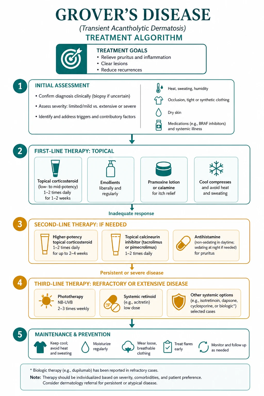
Stepwise ladder: [1]
- Trigger avoidance (cool environment, light clothing, emollient, stop offending drugs, mobilise the bedridden patient).
- Topical corticosteroid (mid-potency betamethasone valerate 0.1% or mometasone furoate 0.1%) twice daily for 2-4 weeks; step down to hydrocortisone 1% as disease settles.
- Antihistamine for pruritus - non-sedating (cetirizine 10 mg, loratadine 10 mg) during the day, sedating (hydroxyzine 25 mg, doxepin 10-25 mg) at night.
- Topical calcineurin inhibitor (tacrolimus 0.1% ointment or pimecrolimus 1% cream) twice daily as a steroid-sparing agent and for face / flexures.
- Oral tetracycline (doxycycline 100 mg BD or minocycline 100 mg BD) for 4-12 weeks as the first systemic option in persistent disease; mechanism is anti-inflammatory rather than antimicrobial.
- Phototherapy (NB-UVB 311 nm 2-3 times weekly for 8-12 weeks) for widespread disease, especially in patients who fail or cannot tolerate tetracyclines.
- Oral retinoid (isotretinoin 10-20 mg daily or acitretin 25 mg daily) for severe or refractory persistent acantholytic dermatosis; monitor LFTs, lipids, pregnancy status.
- Refractory / severe cases - oral corticosteroids (prednisolone 0.5 mg/kg/day, taper over 4-6 weeks) can be considered as a short bridge; biologics (case reports of omalizumab, dupilumab) are experimental. [1]
Step 1 - Environment
Step 2 - Topical steroid
Step 3 - Antihistamine
Step 4 - Topical calcineurin inhibitor
Step 5 - Oral tetracycline
Step 6 - Phototherapy
Step 7 - Oral retinoid
Step 8 - Refractory
Specific subtypes and scenarios
Persistent (chronic) acantholytic dermatosis. By convention, disease continuing beyond 6-12 months with relapses is reclassified as persistent acantholytic dermatosis. The histology is identical. Management is the same stepwise ladder with a lower threshold for systemic therapy (tetracyclines, phototherapy, retinoids). Always re-evaluate the differential in the persistent form. [1]
Bullous / vesiculobullous Grover's. Tense vesicles or small bullae predominate. The diagnostic dilemma is bullous pemphigoid in the elderly; the discriminator is the negative DIF and the suprabasal (not subepidermal) split. Treatment is the same as classical Grover's; tetracyclines are particularly effective. [1]
Eczematous / spongiotic-predominant Grover's. Erythema, weeping and scaling predominate; the dominant histology is spongiotic. Commonly misdiagnosed as eczema. Responds to the same trigger avoidance plus topical steroid and emollient regimen, but the diagnosis is delayed because biopsy is often not performed. [1]
Follicular / infundibular Grover's. Papules are centred on hair follicles and may be mistaken for folliculitis; bacterial culture is negative. Biopsy shows focal acantholysis at the infundibulum. [1]
Photo-distributed Grover's. Crops of papulovesicles on sun-exposed chest, upper back and arms after intense sun exposure; summer seasonality; respond to sun avoidance, broad-spectrum sunscreen, and the same ladder. [1]
Grover's in the ICU / post-operative / bedridden patient. The single most common clinical scenario. The combination of immobility, occlusive dressings, sweating under drapes, xerosis of the elderly skin, and polypharmacy creates the perfect storm. Prevention - cooling mattress, position change 2-hourly, daily emollient, light clothing, judicious review of drugs - is more effective than any active treatment once established. The eruption usually resolves within days to weeks of remobilisation. [1]
Grover's in end-stage renal disease / haemodialysis. Uraemic pruritus may coexist and exacerbate scratching; tetracyclines are best avoided in severe renal impairment (use alternative systemic agents or dose-reduce). Optimise dialysis adequacy and xerosis with aggressive emollient use. [1]
Grover's in HIV / immunosuppression. Biopsy threshold is lower; the differential widens (Kaposi sarcoma, eosinophilic folliculitis, drug eruptions, scabies, papular pruritic eruption of HIV). Anti-Dsg ELISA and DIF are still negative. [1]
Paraneoplastic acantholytic dermatosis. Rare but important. Explosive onset, severe and often generalised pruritus, atypical distribution, and resistance to multiple therapies. Most often associated with haematological malignancy (CLL, lymphoma, myeloma) and, less often, with solid tumours. Workup: full blood count, peripheral smear, LDH, SPEP/immunofixation, CT chest/abdomen/pelvis, age-appropriate cancer screening. Treatment is directed at the underlying malignancy; the eruption often improves when the tumour responds. [1]
Complications and pitfalls
Disease-related [1]
- Pruritus and excoriation - the dominant symptom; disrupts sleep, drives the patient to seek help, and is the gateway to secondary complications.
- Secondary bacterial infection (impetiginisation) - S. aureus or streptococci colonise excoriated papules; treat with topical (fusidic acid 2%) or oral (flucloxacillin 500 mg QID) antibiotics.
- Eczema herpeticum (Kaposi varicelliform eruption) - HSV superinfection; fever, painful punched-out erosions, regional lymphadenopathy. Admit, IV aciclovir, cover for S. aureus.
- Lichenification and prurigo nodularis - chronic scratching produces lichenified plaques and excoriated nodules, which are themselves difficult to reverse.
- Post-inflammatory hyperpigmentation or hypopigmentation - particularly visible in Fitzpatrick IV-VI skin; can persist for months.
- Exfoliative erythroderma - rare; a dermatological emergency with temperature dysregulation and high-output cardiac failure.
- Sleep disturbance, anxiety, depression - chronic pruritus is a significant psychosocial burden. [1]
Diagnostic pitfalls [1]
- Missing pemphigus vulgaris - if the eruption is atypical, persistent, mucosal, or if DIF is not performed.
- Missing Darier's disease - in a young patient, in a patient with nail changes, or in a patient with a positive family history; the eruption may initially look like Grover's.
- Missing scabies - in a hospitalised or institutionalised patient, a truncal papular eruption is more often scabies than Grover's; examine the webs and family contacts.
- Missing drug eruption - new drug within 2-4 weeks; stop the suspect drug and observe.
- Treating eczema as Grover's - chronic topical and oral steroid use in eczema that has not been biopsied may mask an evolving pemphigus.
- Misreading a mixed-pattern biopsy as pemphigus or Darier - a single-section biopsy in a patient with negative DIF and no family history is Grover's, not pemphigus, even if some features "look like" pemphigus. [1]
Therapeutic pitfalls [1]
- Prolonged potent topical steroid in elderly xerotic skin - atrophy, telangiectasia, striae, and the rebound flare on cessation.
- Systemic corticosteroids as first-line - they work but the disease rebounds on taper; reserve as a short bridge.
- Isotretinoin in women of childbearing age without adequate contraception - teratogenicity; iPLEDGE / pregnancy prevention mandatory.
- Phototherapy in patients with a history of skin cancer or photosensitive lupus - carcinogenesis and flare risk.
- Missing the underlying trigger - the disease will recur as long as heat, sweat, occlusion or a drug continues. [1]
Prognosis and disposition
Classical Grover's disease. Approximately 50% of cases resolve spontaneously within 6 weeks to 6 months. A further 30-40% have a relapsing-remitting course that may continue for years. About 10% have truly persistent disease lasting years and require ongoing therapy. The disease does not shorten life expectancy; mortality is from associated conditions (malignancy, ITU admission) rather than Grover's itself. [1]
Persistent acantholytic dermatosis. Once the disease extends beyond 6 months, the probability of complete resolution falls; about 50% of persistent cases continue to relapse over years. Continued exposure to heat / sweat / occlusion, advanced age at onset, xerosis, immunosuppression, and underlying malignancy all worsen the prognosis. [1]
Discharge and safety-netting advice: [1]
- Outpatient management is the rule.
- GP follow-up at 4-6 weeks to review trigger avoidance and treatment response.
- Dermatology review at 8-12 weeks if no improvement; sooner if atypical, persistent, or worsening.
- Safety-net - return if: (i) widespread painful erosions or fever (eczema herpeticum), (ii) facial, mucosal, palmoplantar or nail involvement (re-diagnosis), (iii) persistent disease beyond 6 months (escalation), (iv) new systemic symptoms (paraneoplastic screen). [1]
Special populations
Elderly (>70 years). The dominant demographic. Polypharmacy, xerosis, immobility, and chronic medical conditions (heart failure, renal failure, malignancy) all contribute. Tailor treatment: low-to-mid potency topical steroid (avoid super-potent in the very elderly), emollient-based regimen, sedating antihistamine at night for sleep, tetracyclines in reduced dose if renal function is borderline, phototherapy if mobile. Avoid systemic corticosteroids - falls, infection, glycaemic decompensation. [1]
Renal failure / haemodialysis. Tetracyclines are problematic (doxycycline is partly renally excreted; minocycline is hepatotoxic). Use reduced-dose doxycycline, alternative systemic agents, or aggressive topical / phototherapy. Optimise dialysis adequacy and uraemic pruritus with emollients, gabapentin (off-label), and UV-B. [1]
HIV / immunosuppression. Lower threshold for biopsy; broader differential (Kaposi sarcoma, eosinophilic folliculitis, drug eruption, papular pruritic eruption of HIV, scabies). Anti-Dsg ELISA and DIF remain negative in Grover's. [1]
Post-operative / bedridden. The most common scenario. Prevention is the most effective intervention: cool ambient temperature, light clothing, position change 2-hourly, daily emollient, judicious review of drugs (antibiotics, opioids, antipyretics, anaesthetics), early mobilisation. Once established, the eruption usually resolves within 2-6 weeks of remobilisation and trigger removal. [1]
Pregnancy and breast-feeding. Tetracyclines are contraindicated (foetal teeth and bone). Topical steroids (mild to moderate potency) and tacrolimus 0.1% ointment are first-line; emollient and trigger avoidance are cornerstones. NB-UVB phototherapy is acceptable in pregnancy with appropriate shielding. Isotretinoin and acitretin are absolutely contraindicated. Antihistamines - loratadine and cetirizine are considered safe. [1]
Children. Grover's disease is rare before puberty. Differential widens to include scabies, papular urticaria, atopic dermatitis, Gianotti-Crosti syndrome, pityriasis rosea, and miliaria. Management is conservative - emollient, trigger avoidance, mild topical steroid (hydrocortisone 1%); avoid tetracyclines (tooth discoloration in <12 years) and retinoids. [1]
Evidence, guidelines and regional differences
The evidence base for Grover's disease is dominated by retrospective case series, expert opinion and small uncontrolled trials; there are no large randomised controlled trials of any therapy. Mechanistic understanding has advanced substantially with the 2025 JID review linking the SERCA-calcium-pump axis to Grover's, Darier's and Hailey-Hailey disease, and pointing the way to SERCA-targeted small molecules as a future disease-modifying approach.[6]
American Academy of Dermatology (AAD). No specific Grover's disease guideline. The AAD atopic dermatitis, psoriasis, and acne guidelines provide the framework for the topical and systemic agents used (topical steroids, calcineurin inhibitors, phototherapy, oral retinoids). [1]
British Association of Dermatologists (BAD). No Grover's-specific guideline. The BAD biologic and systemic therapy guidelines cover the agents used off-label (doxycycline, isotretinoin, phototherapy). [1]
European Dermatology Forum (EDF). No Grover's-specific guideline. The EDF position on tetracyclines and isotretinoin for off-label inflammatory dermatoses supports their use in refractory Grover's. [1]
Phototherapy availability varies regionally: NHS and ANZ services have well-developed NB-UVB and PUVA services; in resource-limited settings, phototherapy may be unavailable, and tetracyclines or oral retinoids become the primary systemic options. [1]
Emerging therapies. Case reports of omalizumab (anti-IgE) and dupilumab (anti-IL-4/13) in refractory Grover's have appeared; these remain experimental and should be reserved for tertiary care with informed consent. [1]
ANZ practice follows the standard ladder: trigger avoidance, mid-potency topical steroid, antihistamine, tetracycline (doxycycline 100 mg BD), then NB-UVB phototherapy (311 nm 2-3 x weekly for 8-12 weeks) in the public hospital phototherapy units, and isotretinoin (10-20 mg daily) for refractory disease. PUVA is reserved for NB-UVB failures. Biopsy and DIF are arranged in the public hospital dermatology clinic.
Exam pearls
Four histological patterns of Grover's
DPS-H
Suprabasal cleft, corps ronds, grains
Suprabasal acantholysis, tombstone basal layer, no dyskeratosis
Intraepidermal spongiosis with mild acantholysis
Full-thickness acantholysis, 'dilapidated brick wall'
Triggers of Grover's
HEAT
Hot weather, hot baths, electric blankets, fevers
Sweating
Hospitalisation, ITU, post-op, stroke, hip fracture
IL-4, penicillamine, 5-FU (rare); paraneoplasia
What is the single most important single-investigation discriminator from pemphigus?
Direct immunofluorescence (DIF). It is negative in Grover's disease and positive (intercellular IgG / C3) in pemphigus vulgaris and foliaceus. In any atypical or persistent acantholytic eruption, perform DIF before committing to a long-term treatment plan.
Why do four different histological patterns coexist in the same biopsy?
Because the four patterns are morphological stages of one process, not four different diseases. A focus of acantholysis begins as a small suprabasal cleft (pemphigus-like); as it evolves, dyskeratosis may appear (Darier-like) or the cleft may extend through the full thickness of the spinous layer (Hailey-Hailey-like), or the lesion may be predominantly spongiotic. The biopsy is a snapshot of acantholysis at multiple time-points and severities.
What is the most effective systemic treatment in refractory disease?
Doxycycline 100 mg twice daily for 4-12 weeks is the most useful systemic agent in refractory Grover's. It works as an anti-inflammatory (matrix metalloproteinase inhibitor and neutrophil chemotaxis suppressor), not as an antibiotic. Counsel patients that the effect is slow and not antimicrobial. NB-UVB phototherapy is the alternative second-line option; oral retinoids (isotretinoin, acitretin) are reserved for severe or refractory cases.
Why is DIF always performed alongside the diagnostic H&E biopsy?
Because Grover's disease is, in part, a diagnosis of exclusion of its autoimmune mimics (pemphigus vulgaris, pemphigus foliaceus, paraneoplastic pemphigus) and of its subepidermal mimics (bullous pemphigoid, dermatitis herpetiformis). A single biopsy that includes an adjacent perilesional site for DIF is the cheapest, fastest and most discriminating single investigation in any atypical or persistent acantholytic eruption.
How do you distinguish the paraneoplastic from the classical form?
Paraneoplastic acantholytic dermatosis is suspected when the eruption is explosive in onset, severe and generalised, refractory to multiple therapies, and accompanied by systemic features (weight loss, B-symptoms, lymphadenopathy, organomegaly) in a patient - usually older - with a known or unknown haematological (CLL, lymphoma) or, less often, solid organ malignancy. The histology is identical to classical Grover's, but the disease resolves only with treatment of the underlying malignancy. Always consider this in the persistent or atypical case.
Red flags
Exam application bank (NEET-PG / INICET)
One-line answer
Grover's disease (transient acantholytic dermatosis, TAD) is an acquired, usually self-limiting, intensely pruritic papulovesicular eruption on the trunk of middle-aged to elderly men, characterised histologically by focal acantholysis. The eruption is precipitated by heat, sweating, prolonged bed rest and xerosis, and runs a course that ranges from a few weeks to several years. Four histological patterns (Darier-like, pemphigus-like, Hailey-Hailey-like, spongiotic) may coexist in the same biopsy. Diagnosis is confirmed by punch biopsy from a fresh, intact papulovesicle; direct immunofluorescence is characteristically negative. Management is stepwise: trigger avoidance, emollients, topical corticosteroids, antihistamines, then topical calcineurin inhibitors, oral tetracyclines, phototherapy and oral retinoids for refractory disease.
Worked stems (answer without another resource)
Stem 1 — Classic presentation. Map symptoms to mechanism; name the first investigation and first treatment step with dose/route if drug therapy is standard. [1]
Stem 2 — Unstable / complicated. List red flags that force immediate resuscitation, theatre, ICU, antidote, or reperfusion — and what you do in the first 15 minutes. [1]
Stem 3 — Atypical group. Elderly, pregnancy, child, or immunocompromised: how presentation and thresholds change. [1]
Stem 4 — Differential trap. Name the three closest mimics and one discriminator for each. [1]
Stem 5 — Disposition. Who goes home with safety-netting, who is admitted, who needs HDU/ICU/theatre, and what follow-up is mandatory. [1]
Rapid viva checklist
- Definition + classification
- Pathophysiology chain
- Bedside signs / criteria
- Score with exact components (if any)
- Emergency bundle
- Definitive therapy with doses
- Complications of disease and of treatment
- Special populations
- Guideline/trial name if classic
- Three exam traps
Coverage self-check
If you cannot answer any stem above from this page alone, re-read the matching section — the page is intended to be self-sufficient for final-prof and NEET-PG/INICET questions on Grover's disease (transient acantholytic dermatosis).
[1]Clinical pearl
[1]References
- [1]Weaver J, Bergfeld WF Grover disease (transient acantholytic dermatosis) Arch Pathol Lab Med, 2009.PMID 19722762
- [2]Grover RW Transient acantholytic dermatosis Arch Dermatol, 1970.PMID 5440816
- [3]Heenan PJ, Quirk CJ Transient acantholytic dermatosis Br J Dermatol, 1980.PMID 7387898
- [4]Lockwood RR, Elias PM Transient acantholytic dermatosis Arch Dermatol, 1977.PMID 931408
- [5]Wolff HH Transient acantholytic dermatosis (Grover) Hautarzt, 1977.PMID 845033
- [6]Harmon RM, Ayers JL, McCarthy EF Pumping the Breaks on Acantholytic Skin Disorders: Targeting Calcium Pumps, Desmosomes, and Downstream Signaling in Darier, Hailey-Hailey, and Grover Disease J Invest Dermatol, 2025.PMID 39207315