Dermatology · Medicine
Hidradenitis suppurativa
Also known as Hidradenitis suppurativa · HS · Acne inversa · Verneuil's disease · Apocrinitis
Hidradenitis suppurativa (HS, acne inversa, Verneuil disease) is a chronic, relapsing, immune-mediated inflammatory disease of the folliculopilosebaceous unit in apocrine-bearing intertriginous skin (axillae, groin, buttocks, inframammary, perianal). It presents with painful deep nodules, abscesses, double-headed comedones, sinus tracts ('tombstone' comedones) and rope-like hypertrophic scarring. Pathogenesis centres on follicular hyperkeratosis and occlusion of the infundibulum (NOT primary infection, NOT primary apocrinitis), with secondary follicular rupture, an IL-23/Th17- and TNF-α-dominated inflammatory response, γ-secretase mutations (PSEN1/NCSTN/PSENEN) in familial HS, and dysbiosis. Severity is staged by Hurley (I–III) or the dynamic IHS4 score. Comorbidities are extensive and include the 'follicular occlusion tetrad' (HS, acne conglobata, dissecting cellulitis, pilonidal sinus), obesity/metabolic syndrome, inflammatory bowel disease and depression. Management escalates from lifestyle (smoking cessation, weight loss) → topical clindamycin (Stage I) → oral tetracyclines ± clindamycin–rifampicin, hormonal therapy, acitretin or metformin (Stage II) → biologics (adalimumab anti-TNF, secukinumab/bimekizumab anti-IL-17; JAK inhibitors) ± wide local excision with secondary intention healing, deroofing or CO₂ laser (Stage III).
On this page & tools
Your progress
Saved locally on this device.
Exam tags
Red flags
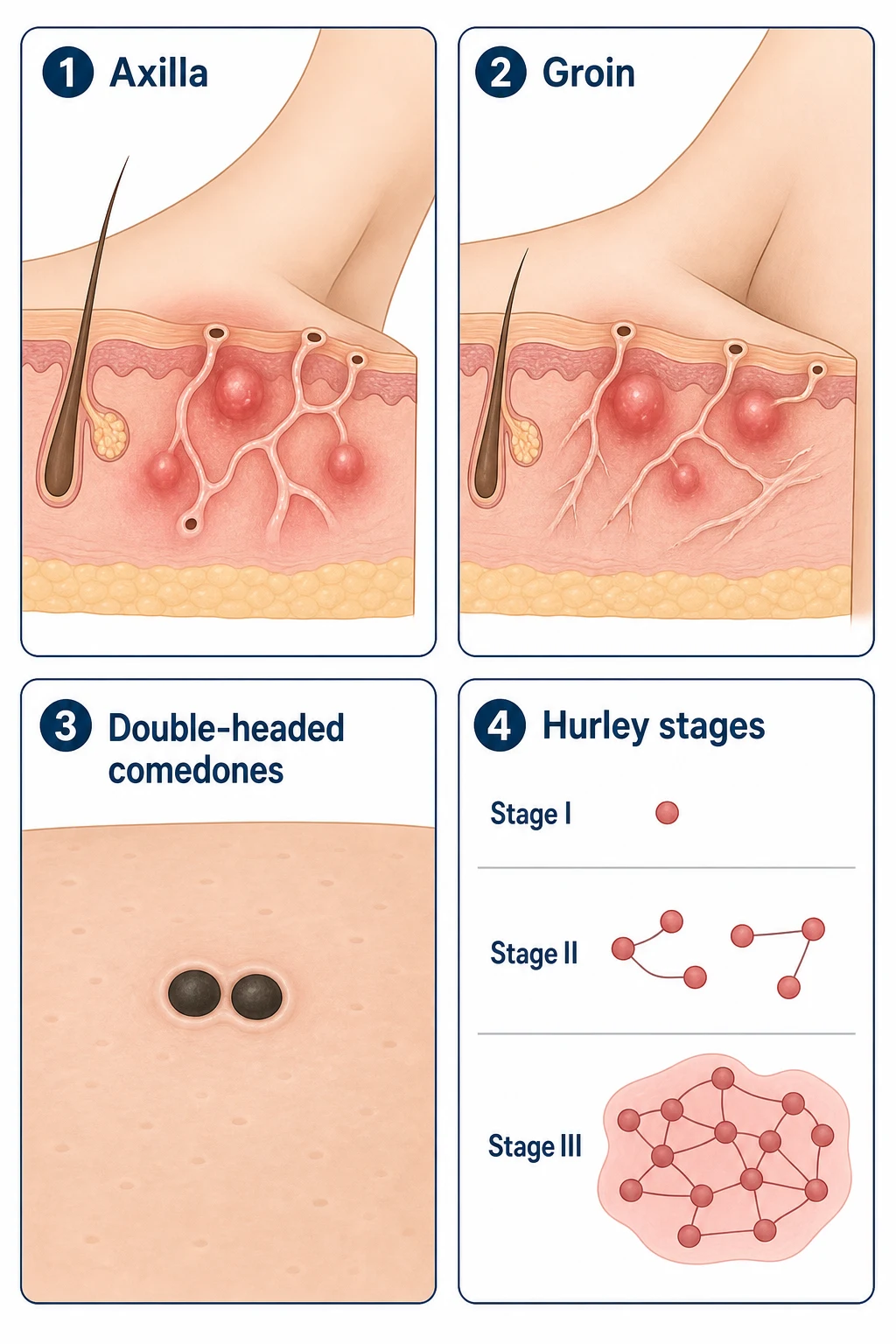
Definition & Classification
Hidradenitis suppurativa (HS) — also known as acne inversa or Verneuil disease (after the French surgeon Aristide Verneuil who described it in 1854) — is a chronic, relapsing, immune-mediated inflammatory dermatosis of the folliculopilosebaceous unit (FPSU) that localises to apocrine-bearing intertriginous skin: the axillae, groin, buttocks, inframammary folds, perianal and genital regions, and (less often) the posterior auricular, occipital scalp and abdominal folds.[1][6]
The naming is historical and partly misleading. The lesions are not primarily an apocrine gland disease (despite "hidro-" referring to sweat) and they are not primarily an infection of the apocrine gland ("apocrinitis"). Modern histology and molecular work confirm that the primary lesion is occlusion of the follicular infundibulum by hyperkeratotic keratin, the so-called "follicular occlusion tetrad" pathogenesis. Once the follicle ruptures, bacteria (typically Cutibacterium acnes, Staphylococcus aureus, mixed anaerobes), keratin and sebum spill into the dermis and trigger an exuberant innate (neutrophil-rich) and adaptive (Th1/Th17-driven) immune response that produces abscesses, sinus tracts and scarring.[1][6]
Three official diagnostic criteria (all required)
The 2019 International Hidradenitis Suppurativa (HS) Alliance / Hidradenitis Suppurativa Foundation diagnostic criteria require all three to be present: [1]
- Typical lesions — deep-seated painful nodules, abscesses, draining sinus tracts, double-headed comedones, bridged scars or rope-like hypertrophic scars.
- Typical distribution — axillae, groin, buttocks, inframammary folds or perianal region (apocrine-bearing intertriginous sites; rarely chest, abdomen, scalp).
- Chronicity / recurrent course — at least two nodules/abscesses in the same area, recurring or persistent, over at least 6 weeks (some guidelines use 6 months). [1]
Atypical or "phenotypic" presentations are increasingly recognised — the follicular HS phenotype (double-headed comedones, cystic acne, severe facial papules), the axillary–genitoperineal distributed disease, and perianal disease (which overlaps with Crohn's fistulation).[6]
Hurley staging — the bedrock classification
The Hurley staging system, described in 1989 by Hurley, remains the universally used bed-side classification and is the primary tool by which management intensity is decided. [1]
| Stage | Description | Practical management implication |
|---|---|---|
| Hurley I — Mild | Single or multiple isolated abscesses; no sinus tracts, no bridging scars | Topical clindamycin 1%, antiseptic washes (chlorhexidine), intralesional triamcinolone for acute flares |
| Hurley II — Moderate | Recurrent abscesses with sinus tracts and scarring; lesions are widely separated within an anatomical region | Oral tetracyclines (doxycycline, lymecycline); clindamycin + rifampicin combination; hormonal therapy; acitretin |
| Hurley III — Severe | Diffuse involvement with confluent sinus tracts and abscesses across an entire anatomical area | Biologics (adalimumab, secukinumab, bimekizumab) ± wide local excision, deroofing, CO₂ laser; reconstructive surgery |
Limitation: Hurley is static (it does not capture disease activity or response to treatment) and is weighted toward late-stage scarring. The dynamic International Hidradenitis Suppurativa Severity Scoring System (IHS4) scores active nodules × 1, abscesses × 1, draining tunnels/fistulae × 2 and inflammatory skin lesions × 4, with totals mild (≤ 3), moderate (4–10), severe (≥ 11). The HS-PGA, HS-IGA and HiSCR (Hidradenitis Suppurativa Clinical Response: ≥ 50 % reduction in abscess and inflammatory nodule count, with no increase in abscesses or draining tunnels) are used in trials.[2][7]
Severity in numbers — what every Grade-7 examiner asks
Epidemiology & Risk Factors
HS is not rare: best estimates are ~1–4 % of the adult population, with the highest reported prevalence in young adults in Western populations (USA, UK, Germany, Denmark) and lower reported prevalence in East Asia and sub-Saharan Africa, possibly reflecting genetic and microbiome differences as much as case-finding.[1][6]
Demographics
Who gets HS
- **Onset** typically post-pubertal — peaks in the late teens to early 30s; a second peak after menopause in women.
- **Sex ratio** is overall **female-predominant (~3:1)**, but **perianal disease** and **severe phenotypes** are more common in men.
- **Ethnicity:** higher prevalence and severity in Black/Afro-Caribbean populations in North American and UK series — partly genetic, partly under-recognition.
- **Delay to diagnosis** is typically **7–12 years** from first symptom; one of the longest diagnostic delays in dermatology.
Modifiable risk factors
What drives severity
- **Smoking** — the strongest and most consistent modifiable factor; 70–90 % of patients smoke; smoking cessation reduces severity and improves biologic response.
- **Obesity / metabolic syndrome** — BMI ≥ 30 kg/m² increases risk and severity; reduces time-to-relapse after treatment.
- **Mechanical friction** — tight clothing, shaving/waxing, mechanical trauma (the 'Koebner' phenomenon).
- **Heat, sweating and occlusion** of intertriginous skin are major precipitants.
Non-modifiable risk factors
What predisposes the patient
- **Genetics:** ~30–40 % have a positive family history; autosomal-dominant inheritance with incomplete penetrance in some kindreds; **γ-secretase mutations (PSEN1, NCSTN, PSENEN)** identified in 5–10 % of familial HS.
- **Hormonal influence:** premenstrual flares are characteristic; hyperandrogenism (PCOS, congenital adrenal hyperplasia) increases severity.
- **Associated genetic syndromes:** Down syndrome (TSCP1/Nicastrin role), Dowling-Degos disease, pachyonychia congenita, KID syndrome.
- **Smoking interacts with the genetics** — γ-secretase mutation carriers who smoke have a markedly elevated penetrance.
Pathogenetically linked comorbidities (the "association triad plus one")
These are associations that exceed mere coincidence and help understand pathogenesis: [1]
| Comorbidity | Approximate prevalence in HS | Direction / mechanism |
|---|---|---|
| Obesity / metabolic syndrome | 50–75 % | Adipokine-driven inflammation, friction, occlusion |
| Smoking | 70–90 % | Nicotine-induced follicular hyperkeratosis, neutrophil activation |
| Inflammatory bowel disease (Crohn's >> UC) | 3–5 % (8–10× general population) | Shared IL-23/Th17 axis, γ-secretase in Paneth cells |
| Spondyloarthropathy / sacroiliitis | 10–20 % | Shared IL-23/IL-17 inflammation |
| Pyoderma gangrenosum / PASH syndrome | Rare but recognised | Autoinflammatory (IL-1) overlap |
| Acne conglobata / dissecting cellulitis / pilonidal sinus | Variable (follicular occlusion tetrad) | Shared FPSU occlusion |
| Depression and anxiety | 30–50 % | Quality-of-life burden, chronic pain |
| Type 2 diabetes mellitus | 20–30 % | Insulin resistance, adipokine inflammation |
| Cardiovascular disease | ↑ 20 % | Systemic chronic inflammation |
| Down syndrome | ↑↑ prevalence | Nicastrin copy-number effect |
A 2025 national multicentre Spanish cohort (GETECCU-ENEID) and a parallel IBD-Prognosis-Study cohort confirm that HS patients have a 3–5-fold increased risk of new-onset IBD — screening with clinical symptoms, faecal calprotectin and gastroenterology referral is now recommended for moderate-to-severe HS.[1][6]
Pathophysiology
HS pathogenesis is best understood as a 4-step cascade on a permissive genetic and immune background. Critically, HS is NOT primarily an infection — bacterial cultures from active lesions are most often sterile or grow only commensal flora, and recurrent antibiotic courses alone rarely induce sustained remission.[1][6]
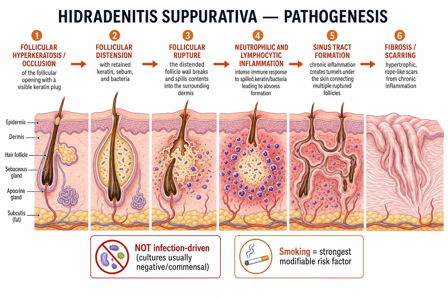
Step 1 — Follicular occlusion (the lesion that starts it all)
Inside the infundibulum (the upper, permanent portion of the hair follicle), hyperkeratosis and retention hyperkeratosis combine with abnormal keratinocyte differentiation (over-expression of K6, K16, K17, proliferation markers Ki-67, and reduced filaggrin) to occlude the follicular opening.[1][6]
The occluded follicle resembles an open comedone and is the histologic hallmark of the disease. In intertriginous sites the combination of occlusion, friction, heat and hormonal stimulation (insulin, IGF-1, androgens) drives this keratin plug. Smoking compounds it: nicotine drives follicular hyperkeratosis and impairs neutrophil function. [1]
Step 2 — Follicular distension
The occluded follicle distends with retained keratin, sebum and commensal bacteria (Cutibacterium acnes, coagulase-negative staphylococci, anaerobic Peptostreptococcus, Porphyromonas, Prevotella). The mass may form a tender, deep, non-pitted nodule. [1]
Step 3 — Follicular rupture (the pivotal event)
As the follicular cyst distends, the follicular wall ruptures — driven by enzymatic breakdown (matrix metalloproteinases, neutrophil elastase) and pressure. The contents (keratin, sebum, hair shafts, bacterial antigens, corneocyte debris) spill into the surrounding dermis and subcutis. [1]
Step 4 — Innate and adaptive immune amplification
Spillage initiates an intense innate response dominated by neutrophils, complement activation (C5a), IL-1β, IL-17, IL-22 and TNF-α, with subsequent activation of an adaptive Th1/Th17 response driven by IL-23 from dendritic cells and a parallel γδ-T-cell/IL-17 axis that is now considered central to HS.[6]
Consequence: [1]
- Abscess (the neutrophil-rich macroscopic lesion) — clinically a tender, deep-seated, fluctuant mass.
- Epithelialised sinus tract ("tunnel" or "burrow") — granulation-tissue-lined, often lined by stratified epithelium (a pseudo-cystic structure), connecting multiple ruptured follicles and acting as a chronic reservoir of inflammation and infection.
- Fibrosis and rope-like scarring — chronic wound-healing-driven, with epithelialised tunnels that are themselves a permanent reservoir of pathogens.[1][4]
The genetic architecture — γ-secretase and beyond
Whole-exome sequencing and candidate-gene studies in familial HS (autosomal-dominant HS with multiple affected generations) have repeatedly identified loss-of-function mutations in subunits of the γ-secretase complex: [1]
| Gene | Subunit | Frequency in familial HS | Functional effect |
|---|---|---|---|
| NCSTN (Nicastrin) | One of four transmembrane subunits | Most common (~5–7 % of familial cases) | Reduces Notch signalling in hair follicle keratinocytes; impairs follicular differentiation |
| PSEN1 (Presenilin-1) | Catalytic subunit | Rare | Loss of γ-secretase activity |
| PSENEN (Presenilin enhancer-2) | Small regulatory subunit | Rare | Same pathway |
| PSTPIP1 (CD2BP1) | Cytoskeletal scaffolding | PAPA / PASH / PAPASH overlap | Constitutive inflammasome activation → IL-1β over-production |
γ-Secretase cleaves Notch receptors — Notch signalling is critical for hair follicle morphogenesis and infundibular keratinocyte differentiation. Loss of γ-secretase → impaired Notch → persistent keratinocyte proliferation in the infundibulum → follicular occlusion.[1][6]
The IL-23 / Th17 axis — the central adaptive driver
HS lesional skin is dominated by IL-17A, IL-17F, IL-22, IL-23 and TNF-α transcripts and infiltrating cells (Th17 cells, Tc17 cells, γδ-T17 cells, innate lymphoid cell 3), with parallel up-regulation of antimicrobial peptides (S100A8/A9, β-defensins) and down-regulation of anti-inflammatory IL-10. This signature is similar to (but distinct from) psoriasis — it is the rationale for the anti-IL-17 (secukinumab, bimekizumab) and anti-IL-23 (risankizumab, guselkumab — under investigation) biologics in HS. TNF-α is highly expressed in perilesional as well as lesional skin, providing the rationale for adalimumab as the first approved biologic.[4][6]
The microbiome — dysbiosis rather than primary infection
16S rRNA sequencing shows HS lesional skin harbours a dysbiotic microbiome dominated by Porphyromonas and Prevotella anaerobes, with reduced Cutibacterium diversity. Bacterial biofilms (especially Staphylococcus aureus and Porphyromonas) line epithelialised sinus tracts and perpetuate inflammation — a key rationale for chronic anti-inflammatory rather than repeated short-course antibiotic therapy.[1][4]
Apocrine gland involvement — a bystander, not the driver
Apocrine glands are anatomically adjacent to the FPSU and are secondarily affected in advanced disease (eosinophilic hidradenitis-like infiltrate, ductal dilatation, ductal rupture). Histologically, apocrine involvement is a consequence of inflammation rather than a primary lesion — supporting the abandonment of "apocrinitis" as the nosological term.[1][6]
Histopathology
| Disease phase | Histology |
|---|---|
| Early | Follicular hyperkeratosis and infundibular plugging; perifollicular lymphocytic infiltrate; cyst formation |
| Established | Ruptured follicle with mixed neutrophilic, lymphocytic and plasmacytic infiltrate; "epidermoid cyst-like" structures lined by stratified squamous epithelium; pseudo-cystic tracts lined by granulation tissue |
| Late | Sinus tracts (epithelialised tunnels), fibrosis with horizontal "bridging" scars, rope-like hypertrophic scarring, foreign-body giant cell reaction to keratin |
A biopsy is not diagnostic but is mandatory to exclude squamous cell carcinoma (SCC) arising in chronic sinus tracts (Marjolin ulcer) when an area becomes acutely proliferative, ulcerated or painful after years of stability.[6]
Hurley Staging in Detail

Hurley I (mild) — typically the early years of HS and the stage at which misdiagnosis as "recurrent boils" or "staphylococcal abscesses" is common. Patients receive multiple courses of incision-and-drainage and short-term antibiotics with partial and transient responses. Topical clindamycin and lifestyle modification are adequate here. [1]
Hurley II (moderate) — sinus tracts appear, lesions are widely separated; single incision-and-drainage no longer clears the region. Tetracyclines, clindamycin–rifampicin combination, hormonal therapy and acitretin are the workhorses; intralesional triamcinolone for acute flares. [1]
Hurley III (severe) — confluent disease, multiple interconnected tunnels bridging across the region; rope-like scarring; chronic purulent and serous discharge with malodour. Topical and oral therapies are inadequate; biologic therapy (adalimumab, secukinumab, bimekizumab) is first-line, combined with wide local excision (to fascia), deroofing of individual tracts or CO₂ laser ablation of tunnels. [1]
Sartorius score, Hidradenitis Suppurativa Clinical Response (HiSCR), IHS4 and HS-PGA complement Hurley in trials and severity assessment; the International HS Severity Scoring System (IHS4) is the preferred dynamic score. [1]
Milestones in hidradenitis suppurativa — a 170-year journey
Clinical Presentation
The clinical manifestations of HS span an extraordinarily wide phenotypic spectrum — from a few isolated perimenstrual nodules in the axilla to grotesquely disfiguring, draining, malodorous disease of axillae, groin, buttocks and perineum. [1]
Cardinal lesions
The 6 cardinal lesions of HS — remember 'ABSCDR'
Anatomical distribution and phenotype
| Site | Phenotype | Sex predominance |
|---|---|---|
| Axillae | Double-headed comedones, nodules, abscesses, sinus tracts; bridging "axillary band" scars | Bilateral, often symmetric, F > M |
| Groin / inguinal / genital (mons pubis, labia majora, scrotum, inner thigh) | Abscesses, sinus tracts; can be unilateral | F > M |
| Perianal / buttocks | Sinus tracts forming horseshoe patterns; perianal disease requiring differentiation from Crohn's fistulation | M > F (reverses the sex ratio) |
| Inframammary / intermammary | Nodules and sinus tracts in skin folds; common in women with large breasts | F (essentially) |
| Less common: waist, abdomen, posterior neck, retroauricular | Isolated lesions, follicular phenotype | Either |
| "Follicular phenotype" | Severe facial papules, comedonal acne, dissecting cellulitis of the scalp; severe cystic acne in HS family | M |
Symptoms
- Pain — the dominant symptom; deep, throbbing, exacerbated by movement and friction. Severity correlates with lesion count and with stage, but isolated nodules can be exquisitely painful.
- Malodorous discharge — the malodour of chronic pus draining from sinus tracts is the second most demoralising symptom and is a major driver of social isolation.
- Pruritus — commoner in early disease and in the 'follicular' phenotype.
- Hyperhidrosis — common in intertriginous regions and compounds friction.
- Sexual dysfunction — common in perineal/genital disease.
- Psychiatric comorbidities — depression, anxiety, social isolation and suicidal ideation are dramatically overrepresented and must be screened for.[4][6]
Clinical course and triggers
- Episodic flares (relapse–remission pattern) in early disease.
- Chronic progressive in late disease with accumulating scarring.
- Triggers: menstruation (premenstrual flares), heat, sweating, friction (tight clothing, exercise), shaving/waxing, smoking, stress, hormonal cycles, corticosteroid withdrawal.
- Seasonal pattern: many patients report improvement in winter and worsening in summer (heat, sweat).[6]
Differential Diagnosis
HS is frequently misdiagnosed as recurrent "boils," "furunculosis," or "staphylococcal skin infection." Distinctive features (double-headed comedones, sinus tracts, scarring, distribution, chronicity) differentiate HS. [1]
HS versus its most frequently confused mimics
Recurrent furunculosis / Staph abscesses
- Responds to appropriate antibiotics; **culture grows *S. aureus***.
- Smaller, more superficial lesions; **no double-headed comedones, no sinus tracts, no bridging scars**.
- **Different distribution:** face, trunk, buttocks — not strictly axillary/groin.
- **Short duration** of individual lesions (days–weeks), not chronic.
Perianal Crohn's disease
- Perianal skin tags, fissures, fistulae and abscesses.
- **Gastrointestinal symptoms** (diarrhoea, abdominal pain, weight loss, rectal bleeding).
- **Endoscopy and biopsy** show granulomatous inflammation.
- Both can COEXIST — HS is 3–5× more common in Crohn's patients; biopsy non-healing sinus tracts to exclude Crohn's.
Acne conglobata
- Coalescing comedones, abscesses, sinus tracts on **trunk (chest, back, shoulders)**.
- **No axillary or groin involvement** typically.
- **No systemic symptoms, no fever**, normal inflammatory markers.
- Both can co-occur as part of the **follicular occlusion tetrad**.
Pilonidal sinus disease
- **Sacrococcygeal midline** primary pit(s) with secondary sinus tracts.
- **Younger males**; hairy sacral fold; often hirsute or with obesity.
- Primary midline pit is pathognomonic; HS in the natal cleft is differentiated by absence of a single primary pit.
Bartholin cyst / abscess
- Solitary labial cyst/abscess at the **Bartholin gland orifice (4 and 8 o'clock on vestibule)**.
- Distinctly **glandular** rather than follicular.
- Surgical marsupialisation rather than adalimumab.
Cutaneous TB, actinomycosis, atypical mycobacterial infection
- Often **culture- or histology-positive** (granulomas, AFB, ray fungi).
- Distinctive distribution or exposure history (atypical mycobacteria: tattoos, fish tanks, hot tubs).
- HS biopsies show **neutrophilic, not granulomatous**, inflammation (unless SCC develops).
Lymphogranuloma venereum (LGV)
- Sexually transmitted **Chlamydia trachomatis L1–L3**.
- **Inguinal buboes, 'groove sign'** and anogenitorectal syndrome.
- **NAAT-positive** for *C. trachomatis*; responds to doxycycline.
Investigations
HS is a clinical diagnosis. The diagnostic criteria — typical lesions in typical sites, with chronicity — are usually sufficient, but investigations play crucial roles in staging, screening for comorbidities, surgical planning and excluding mimics or complications.[1][7]
Clinical assessment tools
- Hurley stage at first assessment and on every visit (notes that stage can advance).
- IHS4 dynamic score — counts nodules × 1, abscesses × 1, draining sinuses/tunnels × 2 and inflammatory skin lesions × 4.
- Sartorius score (older but still used in some research settings).
- HiSCR (Hidradenitis Suppurativa Clinical Response) — ≥ 50 % reduction in abscess + inflammatory nodule count, no increase in abscesses or draining sinuses; primary endpoint in trials.
- Patient-reported outcome measures — HiSQoL, DLQI, PHQ-9 (depression), visual analogue pain score. [1]
Imaging — ultrasound and MRI
When to image — 'I.P.S.S.' for imaging triggers
High-frequency ultrasound (15–20 MHz) is now considered the gold-standard bedside adjunct for counting subclinical lesions, mapping tunnels pre-operatively and confirming the diagnosis in clinically equivocal cases. The "pseudocyst sign," the "keyhole sign" (subepidermal fistulous tract) and the fluid–debris level in tunnels are reproducible ultrasound markers.[4][6]
MRI is reserved for perianal disease (where MRI differentiates HS from Crohn's fistulation), deep gluteal or pelvic disease that cannot be assessed clinically, and suspected SCC (depth and extent). [1]
Microbiology and biopsy
- Swab cultures during acute flares frequently grow polymicrobial flora (S. aureus, Streptococcus, anaerobes), but a positive culture does NOT alter management — HS is not primarily an infection. Cultures are recommended only at first presentation, when atypical features suggest cellulitis or other infection, or when systemic antibiotics are not working to exclude resistant organisms.
- Skin biopsy is generally NOT diagnostic (HS is a clinical diagnosis), but it is MANDATORY when:
- A chronic sinus tract becomes rapidly proliferative, ulcerated, bleeding or painful in long-standing HS (exclude SCC).
- The differential includes Crohn's disease, atypical infection (TB, deep fungal, actinomycosis), or lymphoma.
- Severe or refractory disease is being considered for advanced therapy. [1]
Comorbidity screen at baseline
| Domain | Investigation |
|---|---|
| Metabolic | BMI, fasting glucose / HbA1c, fasting lipid profile, blood pressure |
| IBD | Symptom screen (diarrhoea, abdominal pain, blood per rectum); faecal calprotectin if symptomatic; gastroenterology referral if positive |
| Spondyloarthropathy | Symptom screen (inflammatory back pain, peripheral arthritis); HLA-B27 if symptomatic; MRI of sacroiliac joints if positive |
| Mental health | PHQ-9, GAD-7, suicidal ideation screening |
| Sexual health | Sexual dysfunction screen (DSDS-2 or similar); perineal disease: consider perianal examination under anaesthesia |
| Cardiovascular | ASCVD risk scoring; consider ECG and lipids |
| Pregnancy / contraception | In women of reproductive age, mandatory before acitretin or isotretinoin |
Clinical pearls — what every patient should have at first visit
- Document the Hurley stage and IHS4 score with a dated photograph.
- Screen for smoking and document cessation advice.
- Measure BMI, fasting glucose, lipids, blood pressure — most HS patients have at least one metabolic risk factor.
- Screen for IBD symptoms and depression (PHQ-9); refer appropriately.
- Swab culture only if atypical features or treatment failure.
- Discuss biologic and surgical pathways for Stage II–III disease; do not assume antibiotics will fix everything.
Management
HS management is staged by Hurley stage, personalised for comorbidities (IBD, spondyloarthropathy, pregnancy, PCOS), and almost always combination therapy (medical ± surgical). The European S2k 2025 guideline divides treatment into four pillars: (1) lifestyle and general measures for all, (2) topical and intralesional therapy for Hurley I, (3) oral systemic therapy for Hurley II, (4) biologic therapy ± surgery for Hurley III.[1][7]
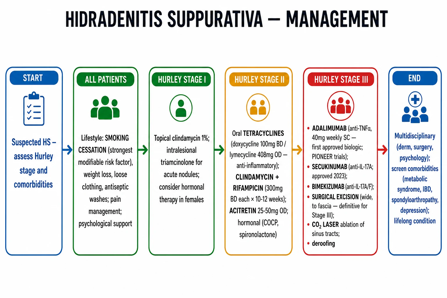
Pillar 1 — Lifestyle and general measures (for ALL patients)
- Smoking cessation is the highest-yield single intervention. Combined with nicotine replacement and counselling, it reduces disease activity within months.
- Weight loss (BMI optimisation). Bariatric surgery and GLP-1 receptor agonists (semaglutide, tirzepatide) are increasingly studied and may produce dramatic improvements; emerging evidence suggests GLP-1 agonists have independent anti-inflammatory effects on HS lesional skin.
- Loose clothing (avoid friction), cotton undergarments, no shaving or waxing affected areas; trimming hair with scissors is preferred.
- Antiseptic washes — chlorhexidine 4 %, benzoyl peroxide 5–10 % washes daily; reduce bacterial load without selecting for resistance.
- Wound care — non-adherent dressings, absorbent foam for draining lesions; absorbent underwear.
- Analgesia — paracetamol, NSAIDs; chronic pain is common and under-recognised.
- Psychological support — actively screen and refer for depression.
- Diet — low-glycaemic-index and anti-inflammatory diets (Mediterranean) help in observational series; dairy and high-glycaemic-index foods can be triggers in some patients. [1]
Pillar 2 — Topical and intralesional therapy (Hurley I; adjunct in Stage II)
| Therapy | Dose | Notes |
|---|---|---|
| Topical clindamycin 1 % solution or cream | BD to affected areas | Only topical with RCT evidence; reduces lesion count |
| Topical resorcinol 15 % peel | Apply to lesion, wash off after 1–2 h | Anti-inflammatory and keratolytic; second-line in some guidelines |
| Benzoyl peroxide 5 % wash | Daily | Reduce bacterial load |
| Intralesional triamcinolone | 2.5–5 mg/mL, 0.1–0.3 mL per nodule | For acute inflamed nodules; rapid analgesia and flattening |
Pillar 3 — Oral systemic therapy (Hurley II; adjunct in Hurley III)
Oral systemic therapies for Hurley II HS
Tetracyclines
- **Doxycycline 100 mg BD** OR **lymecycline 408 mg OD** OR **minocycline 100 mg BD** for **12 weeks**.
- **Anti-inflammatory**, not antimicrobial — they suppress neutrophil chemotaxis, matrix metalloproteinases and T-cell activation at sub-antibiotic doses.
- First-line oral agent per 2025 S2k guidelines.
- Avoid in pregnancy, breastfeeding and in children < 12 y (tooth discoloration).
Clindamycin + rifampicin
- **Clindamycin 300 mg BD** AND **rifampicin 300 mg BD** for **10–12 weeks**.
- More effective than tetracyclines for moderate disease; the Mendola-Sadowski RCT supports combination.
- Caution: rifampicin **induces CYP3A4** — interacts with OCP, methadone, warfarin, biologics; check for drug interactions.
- Caution: clindamycin — *C. difficile* colitis risk; counsel about diarrhoea.
Hormonal therapy (women)
- **Combined oral contraceptive pill** (drospirenone / norgestimate / desogestrel) — reduce androgens.
- **Spironolactone 50–100 mg OD** — anti-androgen; useful in PCOS.
- **Finasteride** 5 mg OD — anti-androgen; useful for hirsute women.
- Effective in women with premenstrual flares.
Acitretin
- **Acitretin 25–50 mg OD** — systemic retinoid; useful when HS coexists with severe acne conglobata or pilonidal disease.
- **Teratogenic** — strict contraception in women (3 years post-stop per EU rules).
- Monitor lipids, LFTs, fasting glucose.
Isotretinoin (controversial)
- **0.5–1 mg/kg/day** for 4–6 months; some patients respond, others worsen.
- Role is controversial — best used for **HS with severe concomitant acne**, low disease activity.
- **NOT** first-line and may aggravate HS in some patients — counsel carefully.
Metformin (PCOS / metabolic syndrome)
- **Metformin 500 mg BD titrated to 1 g BD** in women with PCOS or insulin resistance.
- Reduces androgen-driven follicular hyperkeratosis and is anti-inflammatory via AMPK.
- Useful adjunct for patients with metabolic syndrome or PCOS.
Pillar 4 — Biologics and advanced systemic therapy (Hurley II refractory / Hurley III)
This is the area in which HS management has transformed in the last decade. The biology of HS is dominated by TNF-α, IL-17A/F and IL-23, and these are the established therapeutic targets.[4][7]
[1] [1] [1]| Biologic | Target | Dosing | Trial / evidence base | Approval |
|---|---|---|---|---|
| Adalimumab | TNF-α | 160 mg → 80 mg → 40 mg weekly SC | PIONEER I/II; phase III long-term | FDA 2015, EMA 2016 |
| Secukinumab | IL-17A | 300 mg q2w → q4w SC | SUNSHINE / SUNRISE phase III | FDA / EMA 2023 |
| Bimekizumab | IL-17A + IL-17F | 320 mg q2w × 5 → q4w SC | BE HEARD I/II phase III | FDA / EMA 2024 |
| Ustekinumab (off-label) | IL-12/23 (p40) | 45–90 mg q12w SC | Small case series | Off-label |
| Risankizumab / guselkumab (off-label) | IL-23 (p19) | Case series | Phase II ongoing | Off-label |
| Upadacitinib (case series) | JAK1 | 15–30 mg OD | Refractory HS case series | Off-label / emerging |
| Anakinra (case reports) | IL-1 receptor | 100 mg OD SC | PASH / severe refractory HS | Off-label |
| Apremilast (case reports) | PDE4 | 30 mg BD | Phase II | Off-label |
The 2025 European S2k guidelines recommend adalimumab as first-line biologic, secukinumab as the alternative first-line, bimekizumab as second-line after failure or intolerance, and JAK inhibitors / IL-23 inhibitors as third-line under trial or compassionate-use settings.[7]
Pillar 5 — Surgery (Hurley III, recalcitrant Hurley II)
Surgery is integral to Hurley III and to localised Hurley II disease that has not responded to medical therapy. Four surgical approaches — each with different indications and outcomes.[4][7]
Surgical approaches in HS
Incision and drainage
- For **acute painful abscesses**.
- Provides rapid symptom relief but **does NOT prevent recurrence** of the underlying sinus tract.
- Reserved for symptomatic relief of fluctuant abscesses, not definitive management.
Deroofing
- **Unroofing** of sinus tracts — tangentially excise the roof of a tunnel to expose the granulation-tissue floor.
- Heals by **secondary intention**; minimal morbidity; outpatient procedure under local anaesthetic.
- Ideal for **localised Hurley II disease** or individual symptomatic tunnels in mixed-stage patients.
Wide local excision (WLE)
- **Definitive surgery** — excise all involved tissue **down to deep fascia** with a 1–2 cm clinical margin.
- Recurrence rate 2–5 % vs 50–100 % after incision-and-drainage alone.
- **Reconstruction options:** secondary intention healing (best for axilla, groin); split-thickness skin graft; fasciocutaneous flap; myocutaneous flap for large perineal defects.
Laser and energy-based
- **CO₂ laser ablation** — vaporises sinus tracts and abscess cavities; outpatient; healing by secondary intention.
- **Nd:YAG laser hair reduction** — reduces follicular occlusion by destroying hair follicles; reduces flare frequency.
- **Intense pulsed light + CO₂ fractional laser + triamcinolone** (combined approach) for moderate disease.
Combining medical and surgical therapy
The best outcomes come from medical stabilisation first, surgical extirpation second: [1]
- 3–6 months of biologic therapy (adalimumab or secukinumab) to reduce inflammation and lesion count.
- Wide excision of remaining scarred/confluent areas (continuing biologic perioperatively reduces recurrence).
- Continued biologic therapy postoperatively; consider de-escalation only after 6–12 months of stability.
- Adjuvant laser hair reduction to prevent follicular re-occlusion in the operative field. [1]
Perioperative considerations: HS patients frequently have methicillin-resistant S. aureus (MRSA) colonisation — screen pre-op; bariatric-surgery or weight-management should precede excision for obese patients; smoking cessation 4–6 weeks pre-op reduces wound complications.[4][7]
Special Populations
Paediatric HS (~2 % of all HS; pre-menarche presentation)
- Onset: typically 1–2 years before or around puberty; can be the presenting feature of early PCOS or androgen excess.
- Differential: consider premature adrenarche, androgen-secreting tumour, congenital adrenal hyperplasia if associated with signs of virilisation; in pre-pubertal children, screen for Down syndrome (where HS is markedly over-represented).
- Management: weight-based dosing of topical clindamycin and oral tetracyclines (avoid under 12 y); consider hormonal therapy in adolescents; secukinumab and adalimumab have paediatric data; family education about smoking and weight is pivotal.[5]
Pregnancy
- Topical clindamycin is safe in pregnancy (category B).
- Oral tetracyclines (doxycycline, minocycline) are contraindicated (fetal tooth discoloration, bone growth).
- Clindamycin + rifampicin — both pregnancy category C; not first-line but can be used if benefits outweigh risks.
- Acitretin — teratogenic, contraindicated.
- Isotretinoin — teratogenic, contraindicated.
- Adalimumab — limited safety data but generally considered compatible; discontinue in third trimester if possible to reduce neonatal immunosuppression.
- Secukinumab — limited data; consider on case-by-case.
- Wide surgical excision — best deferred until postpartum if possible.
- Corticosteroid intralesional injections can be used. [1]
HS in association with inflammatory bowel disease
- Co-existing Crohn's — gastroenterology input is essential; adalimumab (anti-TNF-α) treats both HS and Crohn's, so is often first-line. Vedolizumab and ustekinumab are alternatives.
- HS + UC — secukinumab should be used cautiously (can worsen active UC); ustekinumab, vedolizumab or adalimumab are preferred.
- Perianal disease — endoscopy, MRI pelvis, examination under anaesthesia; biopsy to exclude Crohn's vs SCC. [1]
HS with polycystic ovarian syndrome / hyperandrogenism
- Hormonal therapy is central: combined oral contraceptive (avoid norgestrel-only), spironolactone 50–100 mg OD, finasteride 5 mg OD, metformin (if insulin resistance).
- Refer for endocrine / gynaecology evaluation. [1]
Down syndrome and HS
- HS is markedly over-represented in Down syndrome (TSCP1/Nicastrin copy number on HSA21).
- Phenotype is often more severe; challenges with adherence, smoking (which families may not realise is relevant), weight management and surgical wound care.
- Behavioural and communication considerations in management.[5]
Complications
- Squamous cell carcinoma (Marjolin ulcer) in chronic, long-standing sinus tracts — typically after 20–30+ years of disease; incidence ~1–3 % in Hurley III. Biopsy ANY non-healing sinus tract, region of new proliferation or new bleeding. SCC arising in HS sinus tracts is aggressive (high recurrence, metastasis rate up to 50 %).
- Secondary infection — cellulitis, erysipelas, S. aureus septicaemia (rare).
- Chronic pain — often under-treated; consider chronic pain team referral.
- Lymphoedema of the genitalia (penoscrotal, vulval) from chronic scarring of lymphatic channels in Hurley III.
- Fistula to deep structures — urethra, bladder, rectum (very rare; perianal disease).
- Anaemia of chronic disease.
- Amyloidosis (rare; long-standing refractory disease).
- Dystrophic calcification of long-standing sinus tracts.[4][6]
Prognosis & Quality of Life
- HS is chronic and lifelong; spontaneous remission is uncommon (~15 % at 10 years, mostly in mild patients).
- Hurley stage at presentation is the principal prognostic factor: Stage I rarely progresses; Stage III is largely irreversible without surgery.
- Quality of life: HS has one of the lowest Dermatology Life Quality Index (DLQI) scores of any dermatosis, comparable to or worse than psoriasis, atopic dermatitis, chronic kidney disease and active malignancy.
- Depression: 30–50 % of HS patients have clinically significant depression; suicide rates are elevated. Active screening with PHQ-9 at every visit and treatment referral are essential.
- Sexual dysfunction: 50 %+ of patients with perineal disease report sexual dysfunction.
- Workplace impairment: absenteeism rates are high; mean 4–8 days lost per year per patient.[4][6]
Evidence, Guidelines & Regional Differences
- European S2k guidelines 2025 (Zouboulis, JEADV) — the most comprehensive current guideline; covers diagnosis, severity staging and treatment.[7]
- North American (US) guidelines — American Academy of Dermatology (AAD); HS Alliance; HS Foundation.
- PIONEER I/II trials — adalimumab in moderate-to-severe HS (Kimball et al., published 2012 and 2016).[4]
- SUNSHINE/SUNRISE — secukinumab in HS (2023).
- BE HEARD I/II — bimekizumab in HS (2024–25).
- The Lancet 2025 review (Sabat et al.) — comprehensive mechanistic and clinical review; consolidates the IL-17 axis as central.[6]
- Annual Review of Medicine 2025 (McCarthy) — primary-care-facing review.[4]
Australian and New Zealand guidelines align with the European S2k guidelines 2025. Subsidy: Adalimumab and secukinumab are subsidised under PHARMAC for Hurley II/III refractory HS in Aotearoa NZ; bimekizumab pending. Australia — PBS listing for adalimumab (from 2022), secukinumab (from 2023).[7]
Prevention & Lifestyle Modification
- Smoking cessation — single most effective modifiable intervention.
- Weight loss if overweight / obese — modest weight loss (5–10 %) reduces severity.
- Loose clothing, cotton undergarments; avoid friction from tight belts, bras, exercise wear.
- Avoid shaving, waxing, depilation in affected areas; trimming with scissors is preferred.
- Laser hair reduction (Nd:YAG) reduces follicular occlusion in early HS.
- Antiseptic washes (chlorhexidine 4 %, benzoyl peroxide) daily.
- Hormonal management in women — premenstrual flare → consider OCP, spironolactone.
- Diet — observational evidence supports low glycaemic index, dairy reduction, anti-inflammatory Mediterranean diet.[1][6]
Exam Pearls
[1]Red Flags
Exam application bank (NEET-PG / INICET)
One-line answer
Hidradenitis suppurativa (HS, acne inversa, Verneuil disease) is a chronic, relapsing, immune-mediated inflammatory disease of the folliculopilosebaceous unit in apocrine-bearing intertriginous skin (axillae, groin, buttocks, inframammary, perianal). It presents with painful deep nodules, abscesses, double-headed comedones, sinus tracts ('tombstone' comedones) and rope-like hypertrophic scarring. Pathogenesis centres on follicular hyperkeratosis and occlusion of the infundibulum (NOT primary infection, NOT primary apocrinitis), with secondary follicular rupture, an IL-23/Th17- and TNF-α-dominated inflammatory response, γ-secretase mutations (PSEN1/NCSTN/PSENEN) in familial HS, and dysbiosis. Severity is staged by Hurley (I–III) or the dynamic IHS4 score. Comorbidities are extensive and include the 'follicular occlusion tetrad' (HS, acne conglobata, dissecting cellulitis, pilonidal sinus)
Worked stems (answer without another resource)
Stem 1 — Classic presentation. Map symptoms to mechanism; name the first investigation and first treatment step with dose/route if drug therapy is standard. [1]
Stem 2 — Unstable / complicated. List red flags that force immediate resuscitation, theatre, ICU, antidote, or reperfusion — and what you do in the first 15 minutes. [1]
Stem 3 — Atypical group. Elderly, pregnancy, child, or immunocompromised: how presentation and thresholds change. [1]
Stem 4 — Differential trap. Name the three closest mimics and one discriminator for each. [1]
Stem 5 — Disposition. Who goes home with safety-netting, who is admitted, who needs HDU/ICU/theatre, and what follow-up is mandatory. [1]
Rapid viva checklist
- Definition + classification
- Pathophysiology chain
- Bedside signs / criteria
- Score with exact components (if any)
- Emergency bundle
- Definitive therapy with doses
- Complications of disease and of treatment
- Special populations
- Guideline/trial name if classic
- Three exam traps
Coverage self-check
If you cannot answer any stem above from this page alone, re-read the matching section — the page is intended to be self-sufficient for final-prof and NEET-PG/INICET questions on Hidradenitis suppurativa.
[1]References
- [1]Goldburg SR, Strober BE, Payette MJ. Hidradenitis suppurativa: Epidemiology, clinical presentation, and pathogenesis J Am Acad Dermatol, 2020.PMID 31604104
- [2]Jenkins T, Isaac J, Edwards A, et al. Hidradenitis Suppurativa Dermatol Clin, 2023.PMID 37236715
- [3]González-López MA. Hidradenitis suppurativa Med Clin (Barc), 2024.PMID 37968174
- [4]McCarthy S. Hidradenitis Suppurativa Annu Rev Med, 2025.PMID 39869430
- [5]Cotton CH, Chen SX, Hussain SH, et al. Hidradenitis Suppurativa in Pediatric Patients Pediatrics, 2023.PMID 37102307
- [6]Sabat R, Alavi A, Wolk K, et al. Hidradenitis suppurativa Lancet, 2025.PMID 39862870
- [7]Zouboulis CC, Bechara FG, Benhadou F, et al. European S2k guidelines for hidradenitis suppurativa/acne inversa part 2: Treatment J Eur Acad Dermatol Venereol, 2025.PMID 39699926