Dermatology · Medicine
Ichthyosis vulgaris, X-linked ichthyosis, and congenital ichthyoses
Also known as Ichthyosis vulgaris (IV) · X-linked recessive ichthyosis (XLI) · Steroid sulfatase (STS) deficiency · Harlequin ichthyosis (ABCA12) · Collodion baby / lamellar ichthyosis / CIE · Netherton syndrome (SPINK5) · Epidermolytic ichthyosis (KRT1/KRT10) · Sjögren-Larsson syndrome (ALDH3A2)
Ichthyosis vulgaris (IV, 1:250) is the commonest inherited ichthyosis, an autosomal semidominant condition caused by filaggrin (FLG) loss-of-function mutations, presenting with fine white scaling on extensor extremities (sparing flexures), hyperlinearity of palms, and strong association with atopic dermatitis. X-linked recessive ichthyosis (XLI, 1:2000-6000 males) is caused by steroid sulfatase (STS) deficiency at Xp22.3, presenting with large dark-brown scales (including flexures), corneal opacities, cryptorchidism, and contiguous gene syndromes (Kallmann). Congenital ichthyoses — harlequin (ABCA12), lamellar (TGM1), CIE (ALOX12B/ALOXE3), Netherton (SPINK5/LEKTI), epidermolytic (KRT1/KRT10), Sjögren-Larsson (ALDH3A2) — present at birth and require specialist management. Acquired ichthyosis may signal malignancy (Hodgkin lymphoma). Fellowship assessment demands genetic mastery, clinical distinction, and management.
On this page & tools
Your progress
Saved locally on this device.
Exam tags
Red flags
Overview
The ichthyoses are a heterogeneous group of disorders of cornification characterised by generalised scaling of the skin. They are broadly classified into inherited (congenital) forms — further divided into common (ichthyosis vulgaris, X-linked recessive ichthyosis) and rare congenital (harlequin, lamellar, CIE, Netherton, epidermolytic, Sjögren-Larsson) — and acquired forms (secondary to malignancy, drugs, endocrine disease, or nutritional deficiency).[1][4]
Ichthyosis vulgaris (IV), the commonest inherited ichthyosis (1:250), is an autosomal semidominant condition caused by filaggrin (FLG) loss-of-function mutations, presenting with fine white scaling on extensor surfaces (sparing flexures), hyperlinearity of palms, and strong association with atopic dermatitis. X-linked recessive ichthyosis (XLI, 1:2000–6000 males) is caused by steroid sulfatase (STS) deficiency (Xp22.3), presenting with larger dark-brown scales (including flexures), corneal opacities, and cryptorchidism.[1][2][3]
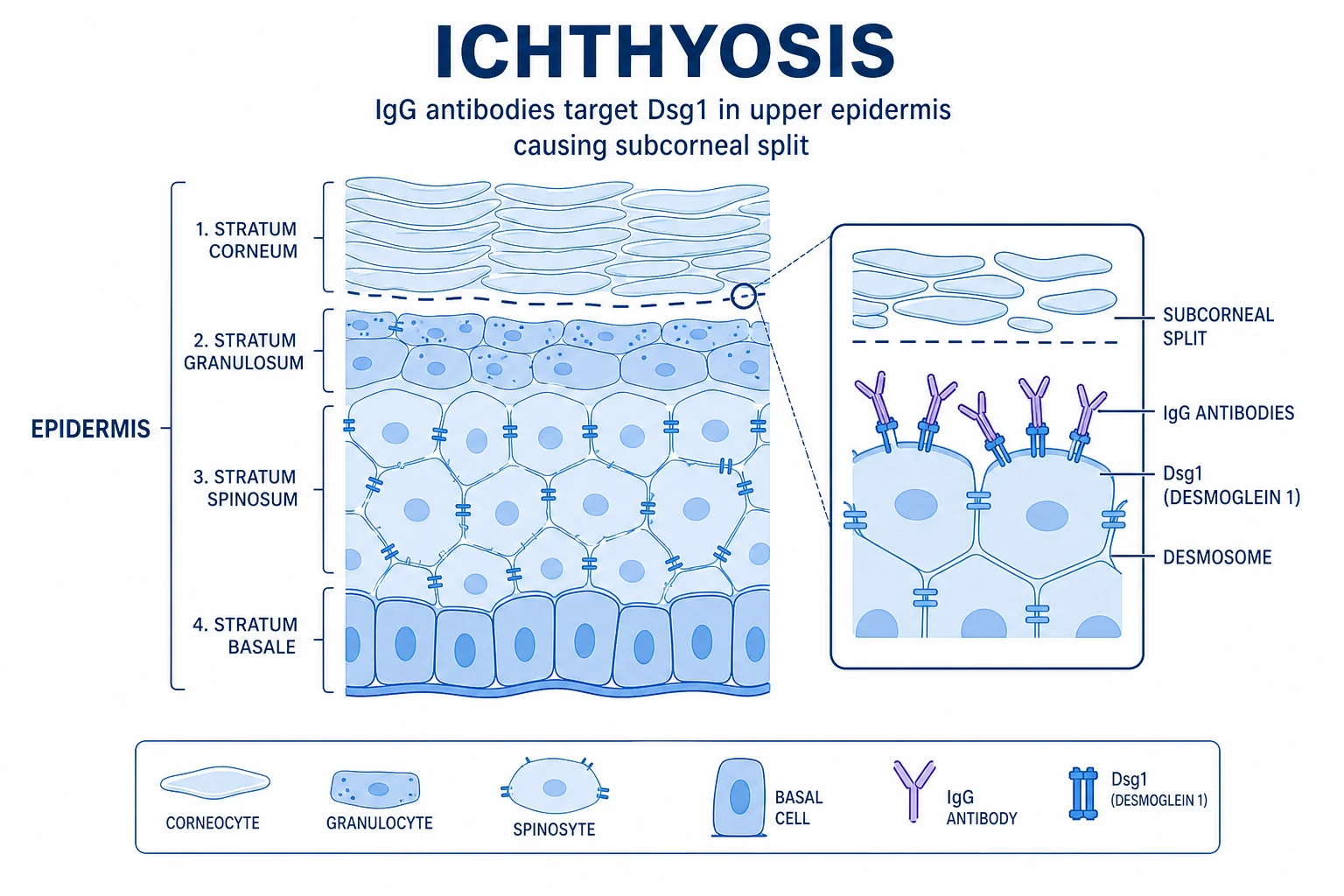
Epidemiology
- Ichthyosis vulgaris: commonest inherited ichthyosis; prevalence 1:250; autosomal semidominant (incomplete penetrance); FLG mutations carried by ~10% of Europeans (founder effect).[2]
- X-linked recessive ichthyosis: 1:2000–6000 males; fully penetrant in hemizygous males; no male-to-male transmission; carrier females usually asymptomatic ( mosaic due to lyonisation).[6]
- Harlequin ichthyosis: ~1:500,000; ABCA12 mutations; autosomal recessive; historically fatal but survival now ~50–70% with early retinoid therapy and intensive NICU care.[7][12]
- Lamellar ichthyosis / CIE (non-bullous CIE): ~1:100,000–200,000; autosomal recessive; TGM1, ALOX12B, ALOXE3 mutations.[3]
- Netherton syndrome: ~1:200,000; autosomal recessive; SPINK5 gene (LEKTI serine protease inhibitor); females more severely affected.[9]
- Acquired ichthyosis: any age; always investigate for underlying cause.[5]
Prevalence and inheritance — the numbers examiners quote
Pathophysiology and genetics
Ichthyosis vulgaris (IV)
Autosomal semidominant (incomplete penetrance); caused by loss-of-function mutations in the FLG gene (1q21.3), encoding profilaggrin → filaggrin. Filaggrin aggregates keratin intermediate filaments to form the cornified envelope and is the source of natural moisturising factor (NMF) — a humectant mixture of amino acids and their derivatives (pyrrolidone carboxylic acid, urocanic acid) that maintains stratum corneum hydration. FLG loss produces a compromised epidermal barrier → transepidermal water loss, xerosis, and scale; it is also the strongest known genetic risk factor for atopic dermatitis (the atopic march — eczema → asthma → allergic rhinitis).[2][13]
X-linked recessive ichthyosis (XLI)
X-linked recessive (Xp22.3); caused by deletions in the STS gene encoding steroid sulfatase — the enzyme that hydrolyses cholesterol sulfate to cholesterol. STS deficiency → accumulation of cholesterol sulfate → inhibits serine proteases and transglutaminases → impaired desquamation → retention of corneocytes → large, adherent scales. Contiguous gene deletions of Xp22.3 may involve adjacent genes: KAL1 (Kallmann syndrome: anosmia + hypogonadotrophic hypogonadism), ARSE (chondrodysplasia punctata), and VCX3A (intellectual disability).[6][3]
Harlequin ichthyosis
Autosomal recessive; ABCA12 mutations (2q35). ABCA12 is a lipid transporter that shuttles glucosylceramides into lamellar granules for the extracellular lipid lamellae of the stratum corneum. ABCA12 deficiency → absent or malformed lipid lamellae → massive hyperkeratosis and barrier failure.[10][7]
Netherton syndrome
Autosomal recessive; SPINK5 gene encoding LEKTI (a serine protease inhibitor). LEKTI deficiency → unregulated serine protease activity (kallikreins 5 and 7) → degradation of desmoglein 1, corneodesmosin, and premature corneocyte desquamation → barrier dysfunction; also drives Th2 inflammation (elevated IgE, atopy).[9]
Other congenital ichthyoses
| Disorder | Gene | Inheritance | Key feature |
|---|---|---|---|
| Lamellar ichthyosis (LI) | TGM1 | AR | Plate-like scales, collodion baby at birth |
| Congenital ichthyosiform erythroderma (CIE) | ALOX12B, ALOXE3 | AR | Fine scaling + generalised erythroderma |
| Epidermolytic ichthyosis (EI) | KRT1, KRT10 | AD | Blistering at birth → hyperkeratosis later |
| Sjögren-Larsson syndrome | ALDH3A2 | AR | Ichthyosis + spastic di-/tetraplegia + macular dystrophy + intellectual disability |
| Refsum disease | PHYH | AR | Ichthyosis + retinitis pigmentosa + ataxia + deafness + anosmia |
| Conradi-Hünermann-Happle | EBP | XLD | Stippled epiphyses + ichthyosiform scaling along Blaschko lines |
| CHILD syndrome | NSDHL | XLD | Unilateral ichthyosiform naevus + ipsilateral limb/hypoplasia |
Subtype deep dive — gene mutations, presentation, prognosis
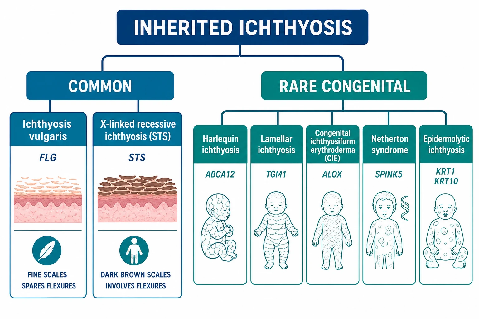
The inherited ichthyoses are an expanding family of Mendelian disorders of cornification, each with a defining molecular lesion. The seven cardinal subtypes examined below are the ones most often tested in fellowship examinations and most often confused at the bedside: X-linked recessive (STS), harlequin (ABCA12), lamellar (TGM1), bullous / epidermolytic (KRT1, KRT10), Netherton (SPINK5), Sjögren-Larsson (ALDH3A2), and Refsum (PHYH). Recognising them is one half of the diagnosis; predicting the systemic trajectory (neurologic, ophthalmic, metabolic) is the other half.[1][3]
X-linked recessive ichthyosis (XLI, STS)
Gene / locus: STS (steroid sulfatase) at Xp22.31. In ~90% of cases the defect is a complete deletion of the STS gene, often together with flanking genes — this is what produces the classic contiguous gene deletion syndromes (Kallmann syndrome from KAL1 loss; chondrodysplasia punctata from ARSE; mental retardation from VCX3A). The remaining 10% carry point mutations or partial deletions. Inheritance is X-linked recessive — fully penetrant in hemizygous males; carrier females are usually asymptomatic but may show mild scaling on shins and may carry corneal opacities on slit-lamp (mosaic lyonisation). [6][1] />
Biochemical hallmark: serum and urinary cholesterol sulfate is markedly elevated (a useful diagnostic surrogate); dehydroepiandrosterone sulfate (DHEA-S) cannot be desulfated in the placenta. Carrier mothers of an XLI fetus therefore show low or absent unconjugated oestriol (uE3) on the triple / quad test and failure to progress in labour requiring caesarean section — a classic perinatal clue.[6]
Clinical course: scales persist lifelong; worsen in winter. Adults are at increased risk of testicular cancer (cryptorchidism, often bilateral; STS deletion also implicates the VCX3A locus), attention deficit, and cardiac arrhythmia (prolonged QTc on ECG has been documented in small series — the STS locus is contiguous with the arylsulfatase locus and nearby cardiac regulators).[1] />
Harlequin ichthyosis (ABCA12)
Gene / locus: ABCA12 at 2q34. ABCA12 is an ATP-binding cassette (ABC) transporter localised to lamellar granules in the upper epidermal keratinocytes; it shuttles glucosylceramides and other lipids into the extracellular space where they form the lipid lamellae of the stratum corneum. Loss-of-function variants (mostly truncating / nonsense / frameshift, with p.Asp1609Glufs*22 and p.Arg2486* the most frequently reported recurrent alleles in Japanese cohorts) abolish transporter activity; lipid lamellae do not form; the cornified envelope is structurally incompetent. Genotype-phenotype correlation is loose — hypomorphic missense variants in ABCA12 produce congenital ichthyosiform erythroderma rather than full harlequin, while biallelic null alleles predict the classic harlequin phenotype.[10][18][17]
Survival: with early systemic retinoid therapy (acitretin or isotretinoin 0.5–1 mg/kg/day started in the first week of life), aggressive NICU care (humidified incubator, fluid/electrolyte replacement, ocular lubrication, emollient bath, prevention of sepsis), survival has improved from near-uniform neonatal death to ~50–80% in modern cohorts, although residual ectropion, eclabium, contractures and poor temperature regulation are lifelong.[7][17]
Lamellar ichthyosis (LI, TGM1) and bathing-suit ichthyosis
Gene / locus: TGM1 (transglutaminase 1) at 14q12. TGM1 catalyses the ε-(γ-glutamyl)lysine cross-linking of cornified envelope proteins (loricrin, small proline-rich proteins, involucrin) — the final covalent step of cornification. Biallelic loss-of-function variants (commonly missense in the catalytic core domain) abolish cross-linking → the cornified envelope is unstable, water and electrolyte barrier is grossly defective, and the neonate is born as a collodion baby. After the membrane sheds (2–4 weeks), the classic LI phenotype emerges: large, dark, plate-like (lamellar) scales over the entire body, with facial tightness and ectropion that improves with age.[3][16]
Temperature-sensitive variant — bathing-suit ichthyosis (BSI): certain TGM1 missense variants (e.g. p.Arg315His, p.Arg315Leu) produce a chaperone-rescuable enzyme that functions normally at the lower skin temperature of the trunk and limbs but folds incorrectly at warmer temperatures; thus scaling is confined to the warmer areas (scalp is spared, bathing-suit distribution of the trunk is preserved).[3]
Self-healing collodion baby — a further minority (TGM1, ALOX12B) shed the collodion membrane and are left with near-normal skin. [1]
Congenital ichthyosiform erythroderma (CIE, ALOX12B / ALOXE3 / NIPAL4)
Genes / locus: ALOX12B (17p13.1), ALOXE3 (17p13.1), NIPAL4 (5q33.3). All encode lipoxygenase / ichthyin pathway components that synthesise the hepoxilin pathway lipids essential for epidermal barrier formation. The infant is born as a collodion baby; after shedding, the phenotype is fine, white scaling overlying generalised erythroderma — erythema is the distinguishing feature from lamellar ichthyosis. CIE generally has milder scaling than LI but more persistent erythema.[3][1]
Bullous (epidermolytic) ichthyosis — KRT1 / KRT10
Genes / locus: KRT1 (12q13.13) and KRT10 (17q21.2) — autosomal dominant keratin mutations. Approximately 50% of cases arise de novo. KRT1 and KRT10 are the type II and type I keratins that co-assemble into the intermediate filament cytoskeleton of suprabasal keratinocytes; dominant-negative missense variants in the helix initiation / termination motifs collapse the cytoskeleton → cytolysis and blistering at birth (the bullous phase) and then, as blistering remits in early childhood, verrucous (wart-like) hyperkeratosis in flexural areas (the ichthyotic phase).[3]
Genotype-phenotype correlation is unusually strong in EI: KRT1 mutations → severe palmoplantar keratoderma (often with thick "keratin heels" that fissure); KRT10 mutations → sparing of palms and soles. Histology shows epidermolytic hyperkeratosis (vacuolar degeneration of upper spinous and granular layers with eosinophilic clumped keratin aggregates).[3]
Netherton syndrome (SPINK5)
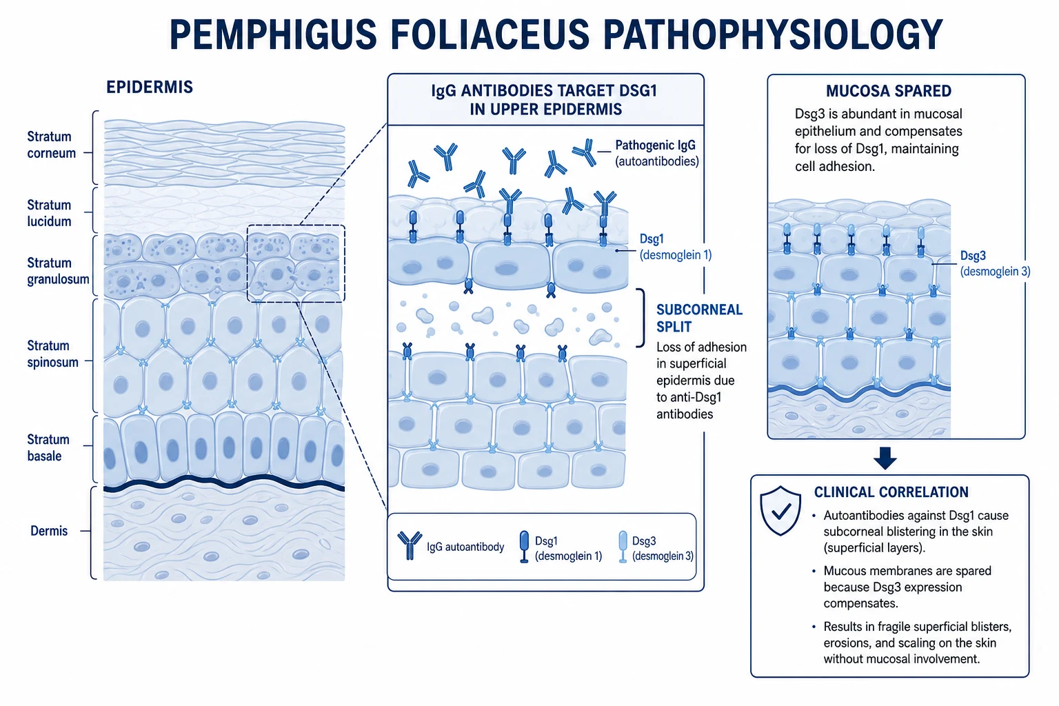
Gene / locus: SPINK5 at 5q33.1, encoding LEKTI (lympho-epithelial Kazal-type-related inhibitor), a serine protease inhibitor expressed in stratified epithelia. LEKTI restrains kallikrein 5 (KLK5) and kallikrein 7 (KLK7) in the stratum corneum. Loss of LEKTI → unopposed KLK5 / KLK7 activity → premature degradation of desmoglein 1 and corneodesmosin → excessive corneodesmosomal shedding (ichthyosis linearis circumflexa) and defective epidermal barrier driving Th2-skewed inflammation (high IgE, food allergy, eosinophilia, asthma).[9]
Genotype-phenotype correlation: variants in the 5′ half of SPINK5 (N-terminal, affecting more inhibitor domains) cause more severe disease with failure to thrive and higher infant mortality; homozygous "fatal" variants (c.153delT, c.1431-12G>A, c.995delT) are statistically enriched in lethal cases. The c.1258A>G (rs2303067) polymorphism modulates the atopic phenotype.[9]
Sjögren-Larsson syndrome (SLS, ALDH3A2)
Gene / locus: ALDH3A2 at 17p11.2, encoding fatty aldehyde dehydrogenase (FALDH), a peroxisomal enzyme that oxidises long-chain fatty aldehydes (derived from fatty alcohol metabolism and from leukotriene / sphingolipid catabolism) into fatty acids. Loss of FALDH → accumulation of fatty alcohols and aldehydes → disruption of epidermal lipid lamellae (ichthyosis) and leukoencephalopathy with dysmyelination (spasticity).[14]
Diagnostic triad: (1) congenital ichthyosis (often collodion-baby presentation; pruritic — unusual among ichthyoses); (2) spastic diplegia or tetraplegia with intellectual disability (usually evident after 1 year of age); (3) crystalline maculopathy ("glistening white dots" in the retina) — present in >95% of cases by age 3–4 years and diagnostic on ophthalmoscopy. Brain MRI shows leukoencephalopathy; MR spectroscopy reveals a characteristic abnormal lipid peak (1.3 ppm). Diagnosis is confirmed by deficient FALDH activity in cultured skin fibroblasts or by biallelic ALDH3A2 sequencing. Management is supportive (topical keratolytics, antispasticity agents, anticonvulsants); survival to adulthood is the rule but disability is lifelong.[14]
Refsum disease (PHYH / PEX7)
Genes / locus: PHYH (phytanoyl-CoA hydroxylase) at 10p13 in >90% of cases; PEX7 (peroxin 7, peroxisomal targeting signal receptor) at 6q23.3 in the remainder. PHYH catalyses the first step of α-oxidation of phytanic acid — a branched-chain fatty acid obtained exclusively from the diet (ruminant fat, dairy, certain fish). Loss of PHYH → phytanic acid accumulates in plasma and tissues (normal < 10 µmol/L; Refsum often > 200 µmol/L). The accumulating phytanic acid disrupts myelin and photoreceptor outer segments.[15]
Clinical tetrad: retinitis pigmentosa (night blindness is the earliest symptom, often beginning in childhood), peripheral polyneuropathy, cerebellar ataxia, and anosmia; sensorineural deafness, ichthyosis, and cardiac arrhythmia / cardiomyopathy complete the multisystem phenotype. CSF shows elevated protein with no pleocytosis. Treatment: lifelong dietary restriction of phytanic acid (avoid ruminant meats, dairy, certain fish); acute deteriorations respond to plasmapheresis or lipid apheresis.[15]
Subtype key numbers — the high-yield figures for viva
Clinical features
Ichthyosis vulgaris
- Onset: after 3 months of age (not present at birth); improves in summer/humidity, worsens in winter.
- Scaling: fine, white, semi-adherent, polygonal scales on extensor extremities (shins, forearms, thighs) and trunk; flexures spared (antecubital and popliteal fossae) — the key clinical distinction from XLI.
- Palms: hyperlinearity (increased palmar creases) and mild keratosis pilaris on the upper arms, thighs, and cheeks.
- Atopic association: strong link to atopic dermatitis (FLG loss → barrier dysfunction → allergen penetration), asthma, and allergic rhinitis.[2][13]
X-linked recessive ichthyosis
- Onset: first few months of life (earlier than IV); persists lifelong.
- Scaling: larger, dark-brown, firmly adherent scales on extensor surfaces AND flexures (popliteal and antecubital fossae involved — the opposite of IV), neck, and trunk; sparing of the face, palms, and soles.
- Corneal opacities: stromal corneal dots/opacities (comma-shaped) on slit-lamp — present in ~50% of patients and in carrier females; does not affect vision.
- Cryptorchidism: undescended testes in ~20% of patients (STS deficiency affects testicular descent); increased risk of testicular cancer and infertility.
- Pregnancy in carrier mothers: STS deficiency in the placenta → decreased oestriol on maternal serum screening (triple/quad test) and failure to progress in labour / prolonged labour requiring caesarean section.[6]
Key clinical distinction: IV vs XLI
| Feature | Ichthyosis vulgaris | X-linked recessive ichthyosis |
|---|---|---|
| Inheritance | Autosomal semidominant | X-linked recessive (males only) |
| Gene | FLG (1q21) | STS (Xp22.3) |
| Scale | Fine, white, small | Large, dark-brown, adherent |
| Flexures | Spared | Involved |
| Palms/soles | Hyperlinear | Normal |
| Corneal opacities | Absent | Present (~50%) |
| Cryptorchidism | Absent | Present (~20%) |
| Atopic association | Strong (eczema, asthma) | Absent |
| Histology | Reduced/absent granular layer | Normal granular layer |
| Onset | >3 months | <3 months |
Collodion baby
- A presentation at birth (not a diagnosis): the neonate is encased in a tight, shiny, translucent membrane (the collodion membrane) resembling parchment or cellophane, causing ectropion (eversion of eyelids), eclabium (eversion of lips), pseudo-clawing of hands/feet, and restricted movement.
- The membrane desquamates over 2–4 weeks, revealing the underlying ichthyosis (usually lamellar ichthyosis or CIE, occasionally normal skin — "lamellar ichthyosis in a hurry" self-healing collodion baby).
- Complications: temperature instability (evaporative heat/water loss), electrolyte imbalance, infection (skin barrier compromised), nursing difficulties (eclabium), and corneal exposure (ectropion).[8]
Harlequin ichthyosis
- The most severe form: at birth, massive, thick, armour-like hyperkeratotic plates separated by deep, erythematous fissures, producing a grotesque appearance; severe ectropion (eyes extruded), eclabium, flattened ears, hypoplasia of nose and digits, and respiratory compromise (restricted chest wall movement).
- Historically fatal in the first days of life; survival now ~50–70% with early systemic retinoids (acitretin/isotretinoin within days of birth), intensive NICU support, ocular lubrication, infection prophylaxis, and a dedicated multidisciplinary team.[7][10][12]
Netherton syndrome
- Ichthyosis linearis circumflexa (migratory, polycyclic, serpiginous, double-edged scaly plaques with a characteristic double border) OR atopic-like dermatitis or congenital ichthyosiform erythroderma.
- Hair abnormality: trichorrhexis invaginata ("bamboo hair" / "golf-tee hair") — the pathognomonic feature; fragile, spiky, brittle hair.
- Atopy: elevated serum IgE, food allergies (especially peanuts), asthma, and urticaria.
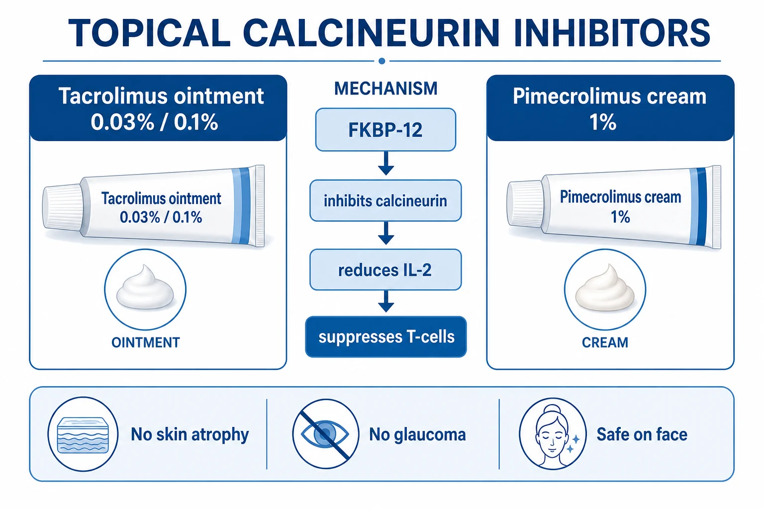
- Skin barrier: severe Netherton patients have a profound skin barrier defect — topical calcineurin inhibitors (tacrolimus/pimecrolimus) are often used but carry risk of significant systemic absorption.[9]
Epidermolytic ichthyosis
- Bullous at birth (blistering, widespread erosions, risk of sepsis and electrolyte loss) — may mimic staphylococcal scalded skin syndrome or epidermolysis bullosa; skin swabs to exclude infection.
- Later in childhood, blistering ceases and is replaced by verrucous hyperkeratosis, especially in flexural areas.[3]
Acquired ichthyosis
- Adult-onset ichthyosis not present since childhood — always investigate for underlying cause.[5]
- Malignancy: classically Hodgkin lymphoma (most common); also non-Hodgkin lymphoma, mycosis fungoides, multiple myeloma; may precede the diagnosis of malignancy by months.
- Endocrine: hypothyroidism, diabetes.
- Drugs: cholesterol-lowering agents (triparanol, niacin), cimetidine, clofazimine, hydroxyurea, targeted therapies (EGFR inhibitors — cetuximab, erlotinib).
- Nutritional: essential fatty acid deficiency, vitamin A deficiency, malabsorption.
- Infectious: HIV/AIDS (xerosis and ichthyosis-like scaling).[5]
Causes of acquired ichthyosis — MAIDEN
MAIDEN
Hodgkin lymphoma (classic association); also non-Hodgkin lymphoma, mycosis fungoides, multiple myeloma, Kaposi sarcoma, leiomyosarcoma
Older adults; autoimmune connective tissue disease (SLE, dermatomyositis), sarcoidosis, graft-versus-host disease
HIV/AIDS (xerosis and ichthyosis-like scaling), leprosy, tuberculosis
EGFR inhibitors (cetuximab, erlotinib, gefitinib), niacin, triparanol, cimetidine, clofazimine, hydroxyurea, statins, allopurinol
Hypothyroidism, hyperthyroidism, diabetes mellitus, panhypopituitarism
Essential fatty acid deficiency, vitamin A deficiency, malabsorption, chronic alcoholism
Histopathology
- Ichthyosis vulgaris: reduced or absent granular layer (stratum granulosum) with compact hyperkeratosis — the hallmark; normal or slightly reduced epidermis.
- X-linked ichthyosis: normal or thickened granular layer with hyperkeratosis — the key histological distinction from IV.
- Harlequin/lamellar ichthyosis: massive orthokeratotic hyperkeratosis; lipid droplets in the stratum corneum (ABCA12/TGM1 deficiency).
- Netherton syndrome: may show parakeratosis and eosinophilic spongiosis; reduced or abnormal LEKTI expression on immunohistochemistry.
- Epidermolytic ichthyosis: epidermolytic hyperkeratosis — vacuolar degeneration of the upper spinous and granular layers with clumped keratin intermediate filaments (eosinophilic clumps); the hallmark.[3][10]
Investigations
- Diagnosis is primarily clinical based on family history, distribution, morphology, and onset.
- Skin biopsy (H&E): confirms the characteristic histology (absent granular layer in IV; epidermolytic hyperkeratosis in EI).
- Genetic testing: FLG sequencing (IV), STS deletion analysis (XLI — usually a contiguous gene deletion detectable by MLPA or array CGH), ABCA12 (harlequin), TGM1 (lamellar), SPINK5 (Netherton), KRT1/KRT10 (epidermolytic).
- Lipoprotein electrophoresis: increased cholesterol sulfate in XLI (a rapid surrogate for STS deficiency).
- Maternal serum screening: low or absent unconjugated oestriol (uE3) on triple/quad test in a carrier mother carrying an XLI fetus (placental STS deficiency prevents conversion of DHEA-S to oestriol).
- Slit-lamp examination: corneal opacities in XLI (and in carrier females).
- Hair microscopy: trichorrhexis invaginata (bamboo hair) in Netherton syndrome.
- ** Serum IgE**: markedly elevated in Netherton syndrome.[4][9]
Management
General principles (all ichthyoses)
- Emollients generously and frequently — the mainstay of therapy; ceramide-dominant formulations, petrolatum-based ointments, and creams containing urea (5–10%), lactic acid, or glycerol.[4]
- Keratolytics: salicylic acid (2–6%), urea (10–20%), lactic acid (12%), propylene glycol (50% under occlusion for severe scaling).
- Bath oils and soap substitutes: avoid alkaline soaps (maintain acidic skin pH); use emollient wash products.[4]
Specific therapy by disease
- IV: emollients + keratolytics; the condition is mild and manageable with topical therapy alone; treat associated atopic dermatitis appropriately.[2]
- XLI: as for IV; topical keratolytics are usually sufficient; no specific systemic treatment needed.
- Lamellar/CIE/harlequin: oral retinoids — acitretin (0.5 mg/kg/day) or isotretinoin — to reduce hyperkeratosis and improve mobility; essential for harlequin ichthyosis survival (start within days of birth).[7][10]
- Netherton syndrome: topical calcineurin inhibitors (tacrolimus, pimecrolimus) reduce inflammation but monitor for systemic absorption (the barrier defect is profound); IVIG for severe cases; biologic therapy (anti-IL-17, anti-IL-4Rα dupilumab) under investigation.[11]
- Epidermolytic ichthyosis: oral retinoids improve hyperkeratosis but may increase blistering (use low doses); topical antibacterials during blistering phase.[3]
Emerging therapies
- Biologics: dupilumab (anti-IL-4Rα) for Netherton (off-label); anti-IL-17 (secukinumab) for lamellar/CIE; leradelimumab (anti-kallikrein 5) under investigation for Netherton.[11]
- Gene therapy: experimental — not yet clinical.
- ABCA12 gene replacement: preclinical.
Differential diagnosis
- Atopic dermatitis (xerosis mimics mild IV; FLG mutations link the two).
- Xerosis (dry skin without ichthyotic scaling; age-related, climate-related).
- Refsum disease (ichthyosis + retinitis pigmentosa + cerebellar ataxia + polyneuropathy + anosmia + deafness; elevated phytanic acid).
- Psoriasis (well-demarcated erythematous plaques with silvery scale, not ichthyotic).
- Pityriasis rotunda (perfectly circular, scaly, hyperpigmented patches; associated with underlying malignancy in some populations).[5]
Exam pearls
[1]Red flags
Exam application bank (NEET-PG / INICET)
One-line answer
Ichthyosis vulgaris (IV, 1:250) is the commonest inherited ichthyosis, an autosomal semidominant condition caused by filaggrin (FLG) loss-of-function mutations, presenting with fine white scaling on extensor extremities (sparing flexures), hyperlinearity of palms, and strong association with atopic dermatitis. X-linked recessive ichthyosis (XLI, 1:2000-6000 males) is caused by steroid sulfatase (STS) deficiency at Xp22.3, presenting with large dark-brown scales (including flexures), corneal opacities, cryptorchidism, and contiguous gene syndromes (Kallmann). Congenital ichthyoses — harlequin (ABCA12), lamellar (TGM1), CIE (ALOX12B/ALOXE3), Netherton (SPINK5/LEKTI), epidermolytic (KRT1/KRT10), Sjögren-Larsson (ALDH3A2) — present at birth and require specialist management. Acquired ichthyosis may signal malignancy (Hodgkin lymphoma). Fellowship assessment demands genetic mastery, clinical
Worked stems (answer without another resource)
Stem 1 — Classic presentation. Map symptoms to mechanism; name the first investigation and first treatment step with dose/route if drug therapy is standard. [1]
Stem 2 — Unstable / complicated. List red flags that force immediate resuscitation, theatre, ICU, antidote, or reperfusion — and what you do in the first 15 minutes. [1]
Stem 3 — Atypical group. Elderly, pregnancy, child, or immunocompromised: how presentation and thresholds change. [1]
Stem 4 — Differential trap. Name the three closest mimics and one discriminator for each. [1]
Stem 5 — Disposition. Who goes home with safety-netting, who is admitted, who needs HDU/ICU/theatre, and what follow-up is mandatory. [1]
Rapid viva checklist
- Definition + classification
- Pathophysiology chain
- Bedside signs / criteria
- Score with exact components (if any)
- Emergency bundle
- Definitive therapy with doses
- Complications of disease and of treatment
- Special populations
- Guideline/trial name if classic
- Three exam traps
Coverage self-check
If you cannot answer any stem above from this page alone, re-read the matching section — the page is intended to be self-sufficient for final-prof and NEET-PG/INICET questions on Ichthyosis vulgaris, X-linked ichthyosis, and congenital ichthyoses.
Expanded exam teaching (depth pass)
Clinical reasoning
For Ichthyosis vulgaris, X-linked ichthyosis, and congenital ichthyoses, examiners test whether you can prioritise life threats, choose the right first test, and give specific therapy (agent, dose, route, timing). Generic phrases without numbers score poorly.
Mechanism → feature map
Build a short chain: cause → pathophysiologic intermediate → clinical feature → complication. Every major symptom in the classic vignette should sit on that chain.
Investigation strategy
- Bedside/first-line tests that change immediate management
- Confirmatory or staging tests
- What a normal result does not exclude
- When not to delay treatment for imaging (unstable patient)
Management ladder
- Resuscitation / ABC / sepsis or haemorrhage bundle as relevant
- Specific antidote / procedure / antimicrobial / reperfusion / surgery
- Supportive care and monitoring targets
- Definitive long-term therapy and secondary prevention
- Disposition and safety-net advice
Special populations
Always prepare one line each for children, pregnancy, elderly, renal/hepatic impairment, and immunocompromised patients when the topic allows.
Pitfalls that fail candidates
- Treating the number not the patient
- Missing pregnancy status when relevant
- Imaging before stabilisation
- Wrong empiric cover or wrong antidote timing
- Incomplete counselling on recurrence, adherence, or red-flag return
Ichthyosis vulgaris (IV, 1:250) is the commonest inherited ichthyosis, an autosomal semidominant condition caused by filaggrin (FLG) loss-of-function mutations, presenting with fine white scaling on extensor extremities (sparing flexures), hyperlinearity of palms, and strong association with atopic dermatitis. X-linked recessive ichthyosis (XLI, 1:2000-6000 males) is caused by steroid sulfatase (STS) deficiency at Xp22.3, presenting with large dark-brown scales (including flexures), corneal opac [1]
[1]References
- [1]Gutiérrez-Cerrajero C, Sprecher E, Paller AS, et al. Ichthyosis Nat Rev Dis Primers, 2023.PMID 36658199
- [2]Jaffar H, Shakir Z, Kumar G, et al. Ichthyosis vulgaris: An updated review Skin Health Dis, 2023.PMID 36751330
- [3]Takeichi T, Akiyama M. Inherited ichthyosis: Non-syndromic forms J Dermatol, 2016.PMID 26945532
- [4]Butala S, Mazereeuw-Hautier J, Paller AS, et al. Ichthyosis: presentation and management Curr Opin Pediatr, 2023.PMID 37345742
- [5]Haber R, Feghali J, Nadir U, et al. Acquired ichthyosis: a clinical review Arch Dermatol Res, 2023.PMID 37422878
- [6]Hazan C, Orlow SJ, Schaffer JV, et al. X-linked recessive ichthyosis Dermatol Online J, 2005.PMID 16403384
- [7]Tsivilika M, Kavvadas D, Karachrysafi S, et al. Management of Harlequin Ichthyosis: A Brief Review of the Recent Literature Children (Basel), 2022.PMID 35740830
- [8]Perumal VK, Baalann KP, et al. Collodion baby Pan Afr Med J, 2021.PMID 34804344
- [9]Sarri CA, Roussaki-Schulze A, Vasilopoulos Y, et al. Netherton Syndrome: A Genotype-Phenotype Review Mol Diagn Ther, 2017.PMID 27905021
- [10]Akiyama M. Harlequin ichthyosis and other autosomal recessive congenital ichthyoses: the underlying genetic defects and pathomechanisms J Dermatol Sci, 2006.PMID 16481150
- [11]Mazereeuw-Hautier J, Granier Tournier C, Hernandez-Martin A, et al. Biologics in congenital ichthyosis: are they effective? Br J Dermatol, 2025.PMID 39470394
- [12]Shibata A, Akiyama M. Epidemiology, medical genetics, diagnosis and treatment of harlequin ichthyosis in Japan Pediatr Int, 2015.PMID 25857373
- [13]Afshari M, Kolackova M, Rosecka M, et al. Unraveling the skin; a comprehensive review of atopic dermatitis, current understanding, and approaches Front Immunol, 2024.PMID 38500882
- [14]Bindu PS. Sjogren-Larsson Syndrome: Mechanisms and Management Appl Clin Genet, 2020.PMID 32021380
- [15]Waterham HR, Wanders RJA, Leroy BP. Adult Refsum Disease 1993.PMID 20301527
- [16]Hotz A, Kopp J, Bourrat E, et al. Meta-Analysis of Mutations in ALOX12B or ALOXE3 Identified in a Large Cohort of 224 Patients Genes (Basel), 2021.PMID 33435499
- [17]Kurdi A, Alahmadi M, Alghamdi S, et al. Epidemiology and Clinical Characteristics of Harlequin Ichthyosis: A Systematic Review and Meta-Analysis of Case Reports Dermatol Pract Concept, 2026.PMID 41912165
- [18]Liu JW, Guo K, Zhang R, et al. Compound heterozygous ABCA12 variants identified in a Chinese patient with congenital ichthyosiform erythroderma: Advancing genotype-phenotype correlations and literature review Mol Genet Genomic Med, 2024.PMID 38702946