Dermatology · Medicine
IgA vasculitis (Henoch-Schönlein purpura)
Also known as IgA vasculitis · Henoch-Schönlein purpura · HSP · IgAV · Anaphylactoid purpura
IgA vasculitis is an IgA1 immune-complex small-vessel leukocytoclastic vasculitis, classically presenting with lower-limb palpable purpura, arthralgia or arthritis, colicky abdominal pain or GI bleeding, and renal involvement with haematuria or proteinuria. Diagnosis is clinical when purpura is typical and platelets are normal; early skin biopsy shows leukocytoclastic vasculitis with IgA-dominant direct immunofluorescence. Management is supportive for most children, with urinalysis, blood pressure, proteinuria, and renal function surveillance; corticosteroids are reserved for severe abdominal or renal disease, and persistent proteinuria or renal impairment needs nephrology-led care.
On this page & tools
Your progress
Saved locally on this device.
Practise this topic
Exam tags
Red flags
Overview & Definition
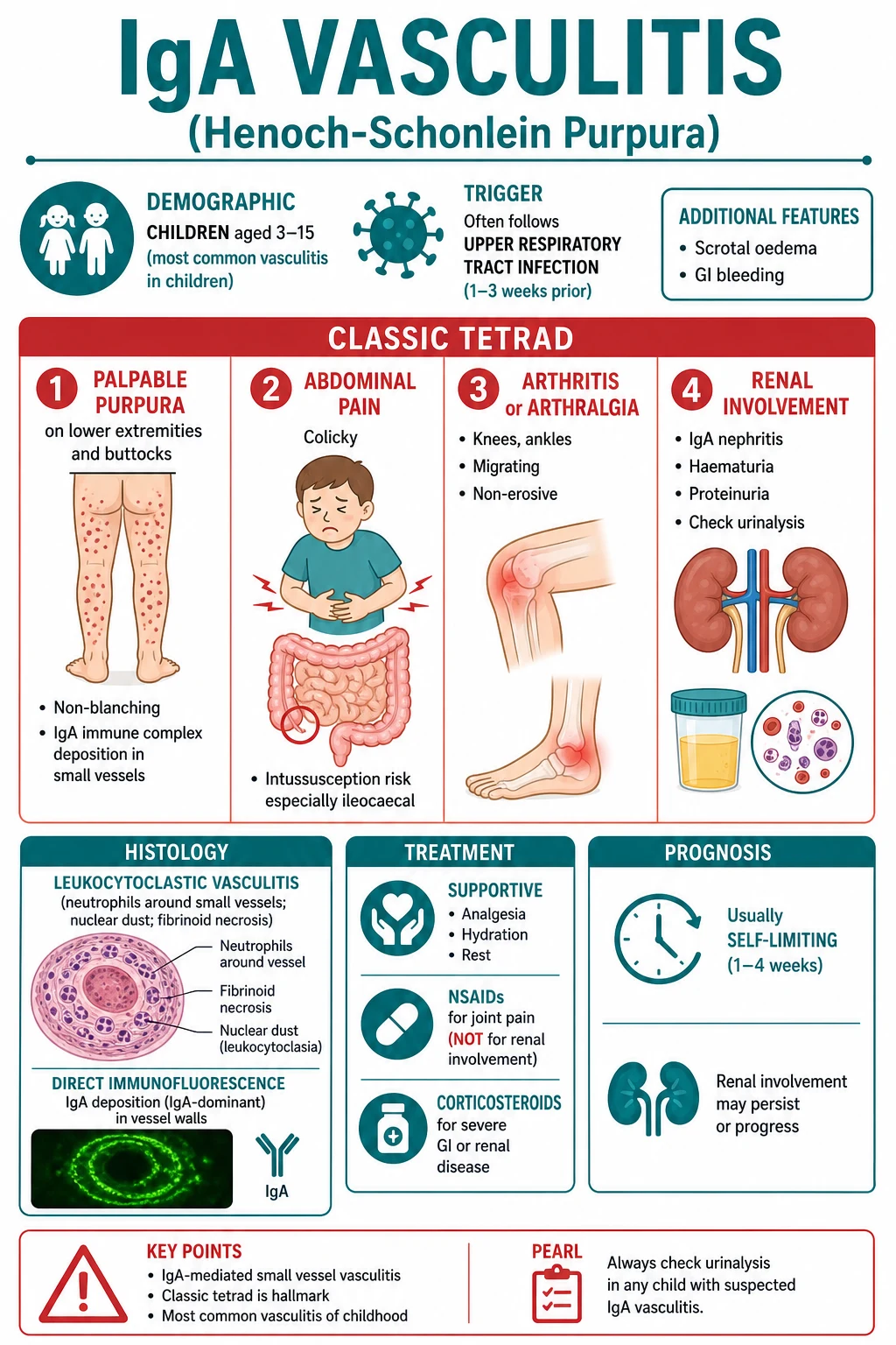
IgA vasculitis is the prototype small-vessel immune-complex vasculitis of childhood. The pathology is not a platelet problem and not a coagulation problem: the purpura is palpable because inflamed dermal post-capillary venules leak red cells and plasma into the skin. The same IgA-dominant immune complexes can involve synovium, gut submucosal vessels, and glomerular capillaries, which explains the exam tetrad of palpable purpura, arthralgia or arthritis, abdominal pain or gastrointestinal bleeding, and renal disease.[1][2][4]
For MBBS and postgraduate entrance exams, the first safe move is to separate IgA vasculitis from the dangerous purpuric syndromes. A well child with lower-limb palpable purpura, ankle pain, colicky abdominal pain, normal platelet count, and microscopic haematuria has the classic pattern. A febrile toxic child with rapidly spreading purpura, shock, neck stiffness, altered consciousness, or disseminated intravascular coagulation has meningococcaemia or purpura fulminans until proven otherwise. Do not use the benign reputation of childhood IgA vasculitis to miss sepsis.[5][7]
Classification
IgA vasculitis can be classified by vessel size, diagnostic certainty, organ involvement, age group, and renal severity. It is a small-vessel vasculitis because the main targets are capillaries, venules, and arterioles; it is immune-complex mediated because direct immunofluorescence shows IgA-dominant vascular deposition; and it becomes clinically serious when renal, gastrointestinal, or rarely pulmonary disease is severe.[2][4]
Skin-limited or mild systemic IgA vasculitis
- Palpable purpura with minimal arthralgia or mild abdominal discomfort
- Normal blood pressure, normal creatinine, absent or trace proteinuria
- Supportive care, safety-netting, and scheduled urine/BP surveillance are the key interventions
- Common in children and usually self-limiting
GI-predominant IgA vasculitis
- Colicky abdominal pain, vomiting, melaena, haematochezia, or occult blood
- Purpura can appear after abdominal pain, so appendicitis and intussusception may be considered first
- Severe pain, bleeding, or obstructive symptoms need urgent imaging and senior review
- Corticosteroids are used for severe pain after surgical emergencies are considered
IgA vasculitis nephritis
- Microscopic or macroscopic haematuria, proteinuria, hypertension, reduced eGFR, nephritic syndrome, or nephrotic syndrome
- Long-term outcome is determined mainly by the kidney, not by the rash
- ACE inhibitor or ARB is used for persistent proteinuria when clinically appropriate
- Moderate or severe disease needs nephrology, renal biopsy, and guideline-led immunosuppression
Adult-onset IgA vasculitis
- Less common than paediatric disease but more likely to be severe
- Higher rates of renal impairment, relapse, and chronic kidney disease
- Triggers include infection, drugs, autoimmune disease, and occasionally malignancy
- Requires lower threshold for biopsy, renal referral, and evaluation for secondary causes
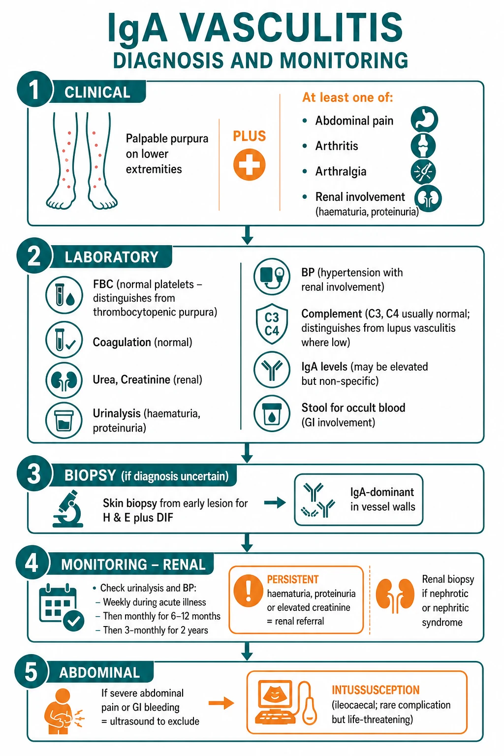
The EULAR/PRINTO/PRES paediatric classification framework is the exam-friendly structure: purpura or petechiae with lower-limb predominance and not due to thrombocytopenia is mandatory, and at least one of abdominal pain, arthritis or arthralgia, renal involvement, or histopathology with IgA deposition supports the diagnosis. In adults the same clinical logic is used, but biopsy is more often needed because adult palpable purpura has a wider differential and adult IgA vasculitis carries more renal risk.[8][2]
Epidemiology & Risk Factors
IgA vasculitis is the most common systemic vasculitis in children, with a typical age range of about three to fifteen years and a peak in the younger school-age years. It is more common in boys, more frequent in autumn and winter in many series, and often follows an upper respiratory tract infection by one to three weeks. Adult disease is uncommon but important because it is less benign.[1][6][7]
Epidemiology and clinical frequency anchors
The strongest practical trigger is infection, especially streptococcal or viral upper respiratory infection, but the trigger list is wider: gastrointestinal infection, vaccination, insect bites, foods, and drugs such as antibiotics or non-steroidal anti-inflammatory drugs have been reported. In many children no single trigger is proven, so the history is used to identify danger and recurrence rather than to force a cause. In adults, a careful drug, infection, autoimmune, and age-appropriate malignancy screen is more important because adult-onset IgA vasculitis can be secondary.[4][5][6]
Risk factors for worse outcome are not the number of purpuric spots or the severity of ankle pain; they are renal. Macroscopic haematuria, heavy or persistent proteinuria, hypertension, reduced kidney function at presentation, nephritic syndrome, nephrotic syndrome, and adverse renal biopsy findings predict closer follow-up and specialist treatment. Older age also matters: adult IgA vasculitis has more severe renal involvement and a less predictable course than the usual paediatric illness.[2][3]
Pathophysiology
The pathophysiology begins with an abnormal IgA1 response. Many patients produce increased amounts of galactose-deficient IgA1 after a mucosal trigger. Anti-glycan antibodies recognise this altered IgA1, producing circulating immune complexes that are too large or too poorly cleared. These complexes deposit in small-vessel walls and glomerular mesangium, activate complement pathways, recruit neutrophils, and injure the endothelium. The skin result is leukocytoclastic vasculitis; the renal result resembles IgA nephropathy in mechanism but occurs as part of a systemic vasculitis.[4][2]
From mucosal trigger to palpable purpura and nephritis
Mucosal trigger
Upper respiratory or gastrointestinal infection stimulates IgA production in a genetically and immunologically susceptible host.
Galactose-deficient IgA1
Abnormal IgA1 hinge-region glycosylation creates a neoepitope that is less easily cleared and more likely to form immune complexes.
Anti-glycan antibody and immune complex
IgG or IgA antibodies bind galactose-deficient IgA1, creating circulating complexes that deposit in small vessels and mesangium.
Complement and neutrophil recruitment
Alternative and lectin pathway activation plus endothelial injury recruit neutrophils, producing leukocytoclasia, fibrinoid necrosis, and red-cell extravasation.
Organ phenotype
Dermal venules produce palpable purpura, gut vessels produce colicky pain or bleeding, synovium produces arthralgia, and glomeruli produce haematuria, proteinuria, and sometimes renal failure.
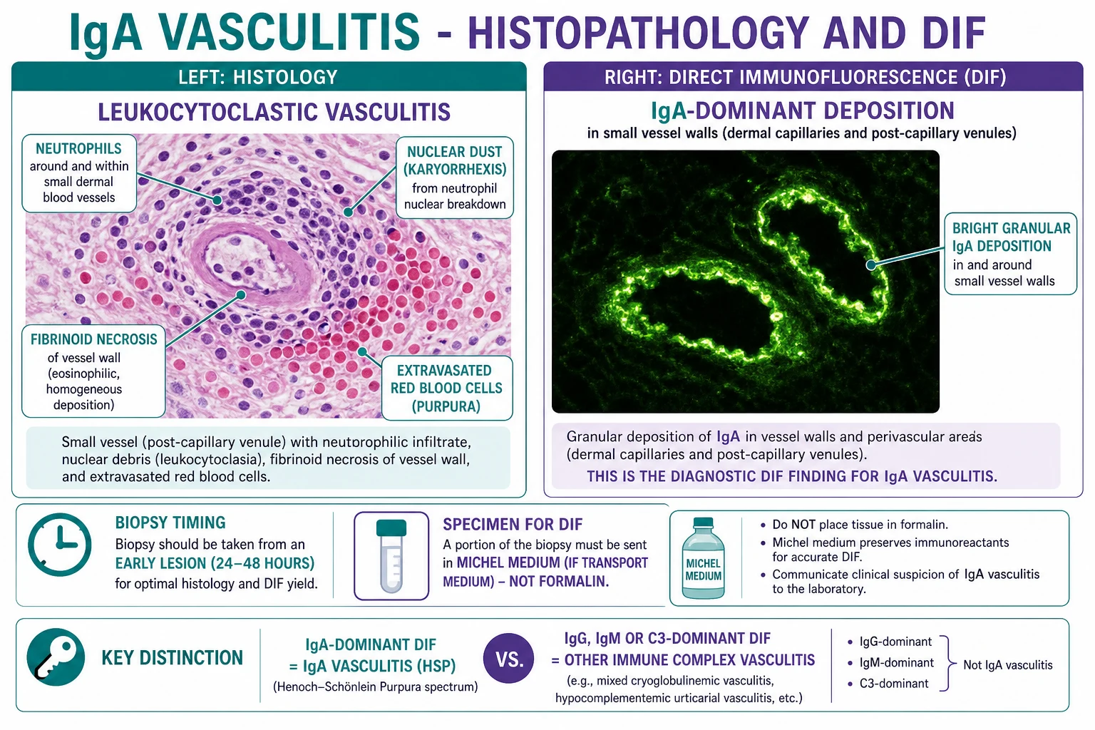
The word leukocytoclastic means broken neutrophils. On routine histology, an early purpuric papule shows neutrophils around superficial dermal vessels, nuclear dust from karyorrhexis, fibrinoid necrosis of the vessel wall, endothelial swelling, oedema, and extravasated red blood cells. On direct immunofluorescence, IgA is deposited in vessel walls, often with C3 and fibrin. Because immune deposits fade with lesion age, the best skin biopsy is from a fresh lesion, ideally within the first twenty-four to forty-eight hours, and the DIF specimen must be placed in appropriate transport medium rather than formalin.[2][5]
The same mechanism explains the clinical timing. Purpura may recur in crops because new waves of immune-complex deposition occur as the mucosal immune response evolves. Joint symptoms are typically periarticular and non-erosive because the inflammation is vascular and soft-tissue dominant rather than a destructive synovitis. Gut pain is colicky because submucosal oedema, haemorrhage, and transient ischaemia irritate the bowel wall. Renal disease may appear after the rash because glomerular immune injury evolves over weeks, which is why a normal urine dip at the first visit does not finish the assessment.[4][6]
Clinical Presentation
The rash is the anchor. It begins as erythematous macules, urticarial papules, or petechiae, then becomes non-blanching palpable purpura. The typical distribution is gravity-dependent: lower legs, ankles, feet, buttocks, and extensor surfaces; in younger children it may involve the face, ears, or upper limbs more than in adults. Lesions can be tender or itchy, may coalesce into ecchymoses, and can occasionally blister or ulcerate. The exam clue is not simply "purpura" but palpable purpura with normal platelets.[5][7]
Classic tetrad
PAIR
Palpable, non-blanching, lower-limb or buttock predominant; platelets are normal.
Migratory, non-erosive, often knees and ankles; periarticular swelling is common.
Colicky abdominal pain, vomiting, occult blood, melaena, haematochezia, or intussusception.
Microscopic haematuria, proteinuria, hypertension, nephritic or nephrotic syndrome.
Joint involvement is common and often dramatic but usually not destructive. Knees and ankles are the classic joints; elbows and wrists can be affected. Children may refuse to walk because of ankle swelling and pain, but erosions and chronic deformity are not expected. This helps separate IgA vasculitis from juvenile idiopathic arthritis, septic arthritis, rheumatic fever, and connective-tissue disease.[5][7]
Gastrointestinal disease ranges from mild abdominal pain to life-threatening complications. Typical pain is colicky, periumbilical or diffuse, and may be associated with vomiting, diarrhoea, occult blood, melaena, or haematochezia. Serious complications include intussusception, bowel wall haemorrhage, ischaemia, perforation, pancreatitis, and rarely massive GI bleeding. In IgA vasculitis, intussusception is often ileo-ileal rather than the classic idiopathic ileocolic pattern, so ultrasound interpretation and surgical discussion must be alert to that possibility.[5][6]
Renal disease is the prognostic organ. It may be detected only by dipstick or microscopy, so asking about dark urine is not enough. The spectrum includes microscopic haematuria, macroscopic haematuria, mild proteinuria, persistent proteinuria, hypertension, nephritic syndrome, nephrotic syndrome, acute kidney injury, rapidly progressive glomerulonephritis, and chronic kidney disease. Renal involvement often appears within the first weeks but can emerge after the rash has improved, which is why follow-up must be scheduled rather than left to symptoms.[3][7]
Other features are examinable because they create traps. Boys can develop scrotal pain or swelling from vasculitic oedema; it may mimic torsion, so Doppler ultrasound and surgical assessment are needed if torsion cannot be excluded. Oedema of the hands, feet, scalp, or periorbital tissues is common in younger children. Neurological features, pulmonary haemorrhage, myocarditis, and ocular inflammation are rare but serious; they should prompt specialist involvement and reconsideration of alternative systemic vasculitides.[6][7]
Why abdominal pain before rash is dangerous
IgA vasculitis can start with abdominal pain before purpura appears. A child may therefore be worked up as appendicitis, mesenteric adenitis, gastroenteritis, or intussusception. The safe approach is to treat severe abdominal signs on their merits, re-examine the skin repeatedly, dip the urine, and avoid anchoring on IgA vasculitis until surgical emergencies and sepsis have been considered.[5]
Differential Diagnosis
The differential diagnosis is the heart of safe practice. The first split is purpura with normal platelets versus purpura with thrombocytopenia or coagulopathy. IgA vasculitis usually has normal platelets and normal coagulation tests. Immune thrombocytopenia has petechiae and bruising that are not classically palpable, with an isolated low platelet count and no colicky abdominal pain or nephritic urine. Disseminated intravascular coagulation has abnormal coagulation, systemic illness, and bleeding from multiple sites.[5][7]
Immune thrombocytopenia
- Non-palpable petechiae, bruising, mucosal bleeding
- Platelets low; coagulation usually normal
- No IgA tetrad; abdominal pain and haematuria are not expected as a linked syndrome
- Do FBC before diagnosing IgA vasculitis
Meningococcaemia / purpura fulminans
- Fever, toxicity, shock, neck stiffness, altered mental state, rapidly spreading purpura
- May have thrombocytopenia, DIC, acidosis, high lactate
- Immediate antibiotics and resuscitation; do not wait for rash evolution
- This is the must-not-miss mimic
Haemolytic uraemic syndrome
- Triad of microangiopathic haemolytic anaemia, thrombocytopenia, and acute kidney injury
- Often follows bloody diarrhoea; schistocytes and high LDH
- Purpura is not the classic palpable lower-limb vasculitic rash
- Renal failure is central and urgent
ANCA-associated vasculitis
- Older child or adult; systemic features, sinus/lung disease, neuropathy
- Purpura can occur but renal disease is often rapidly progressive
- ANCA may be positive; biopsy shows pauci-immune necrotising vasculitis
- Requires urgent immunosuppression
Other mimics include hypersensitivity or drug-induced cutaneous small-vessel vasculitis, systemic lupus erythematosus vasculitis, cryoglobulinaemic vasculitis, septic emboli from infective endocarditis, rickettsial illness, viral exanthems with purpura, acute leukaemia, non-accidental injury, erythema multiforme, urticaria, and scurvy. A skin-limited drug vasculitis may be indistinguishable clinically from IgA vasculitis until history, systemic review, urine testing, and biopsy clarify the pattern. Lupus is suggested by photosensitivity, oral ulcers, arthritis beyond the typical ankle-knee pattern, cytopenias, low complement, ANA or anti-dsDNA positivity, and multisystem features. Cryoglobulinaemia points to hepatitis C, cold sensitivity, neuropathy, low complement, and serum cryoglobulins.[2][5]
The abdominal differential includes appendicitis, gastroenteritis, mesenteric adenitis, inflammatory bowel disease, Meckel diverticulum, intussusception, pancreatitis, and testicular torsion when pain is scrotal or lower abdominal. The renal differential includes post-streptococcal glomerulonephritis, IgA nephropathy without systemic vasculitis, lupus nephritis, ANCA glomerulonephritis, HUS, and hereditary nephritis. The rash differential narrows the list, but the urine and vital signs determine urgency.[3][5]
Clinical & Bedside Assessment
The bedside assessment is structured around the four organs and the dangerous mimics. Start with ABCDE if the child looks ill: airway, breathing, circulation, disability, exposure, capillary refill, temperature, pulse, blood pressure, oxygen saturation, hydration, and sepsis features. Fever, tachycardia out of proportion, hypotension, confusion, meningism, rapidly spreading purpura, or severe limb pain are not routine IgA vasculitis; they trigger emergency sepsis and purpura fulminans management while investigations proceed.[5][7]
History must ask about timing of rash, preceding respiratory or gastrointestinal infection, sore throat, new medicines, vaccination, food exposure, insect bite, previous episodes, family renal disease, haematuria, foamy urine, reduced urine output, abdominal pain, vomiting, stool colour, joint pain, scrotal pain, headache, cough or haemoptysis, and weight loss or constitutional symptoms. In adults add a deliberate screen for drug triggers, hepatitis risk, autoimmune disease, and malignancy symptoms such as weight loss, night sweats, altered bowel habit, haemoptysis, or lymphadenopathy.[2][6]
Examination begins with the rash. Describe morphology, palpability, blanching, distribution, necrosis, bullae, ulceration, mucosal bleeding, and oedema. Then examine joints for swelling, range of movement, warmth, and ability to bear weight. Abdominal examination checks tenderness, guarding, distension, bowel sounds, mass, hepatosplenomegaly, and rectal bleeding where appropriate. Genital examination is needed in boys with scrotal pain. Renal assessment means blood pressure measured correctly for age, oedema, urine dipstick, hydration, and signs of nephritic or nephrotic syndrome.[5][7]
Investigations
IgA vasculitis is usually a clinical diagnosis, but investigations are mandatory because they exclude mimics and stage organ involvement. The baseline set is FBC with platelet count, blood film if haemolysis or HUS is considered, CRP or ESR, urea and electrolytes, creatinine and eGFR, liver tests if systemic disease or drug reaction is considered, coagulation profile if bruising or bleeding is prominent, urinalysis, urine microscopy if available, urine protein-to-creatinine ratio or albumin-to-creatinine ratio, and blood pressure. Stool occult blood is useful if GI symptoms are present.[5][8]
Must do at first contact
- FBC with platelets to exclude thrombocytopenic purpura
- Urea, creatinine, eGFR, electrolytes to stage renal involvement
- Urinalysis for blood and protein, plus urine protein quantification if protein positive
- Blood pressure with age-appropriate cuff; hypertension is renal involvement until proven otherwise
Do when diagnosis is uncertain or severe
- Skin biopsy: fresh lesion for H and E and DIF
- Abdominal ultrasound for severe pain, vomiting, mass, or GI bleeding
- Scrotal Doppler ultrasound for scrotal pain or swelling
- Complement, ANA, ANCA, hepatitis serology, blood cultures, or cryoglobulins when mimics are plausible
Renal specialist tests
- Serial urine protein-to-creatinine ratio or albumin-to-creatinine ratio
- Renal ultrasound when renal disease is significant or atypical
- Renal biopsy for nephritic or nephrotic syndrome, impaired kidney function, or persistent significant proteinuria
- Histological grading guides nephrology-led immunosuppression
The expected simple pattern is normal or raised white cells, normal or raised platelets, normal coagulation, normal complement, and urinalysis showing no abnormality or microscopic haematuria with variable protein. Serum IgA can be elevated, but it is neither sensitive nor specific enough to confirm or exclude IgA vasculitis. A normal complement helps distinguish IgA vasculitis from lupus nephritis or hypocomplementaemic urticarial vasculitis, but it is supportive rather than diagnostic.[5][8]
Skin biopsy is indicated when the rash is atypical, adult-onset, necrotic or ulcerative, diagnosis is uncertain, systemic disease is severe, or mimics remain plausible. Send one specimen in formalin for routine histology and a second fresh specimen in appropriate medium for DIF. Renal biopsy is not required for isolated microscopic haematuria but is considered for nephritic syndrome, nephrotic syndrome, impaired kidney function, persistent significant proteinuria, or rapidly progressive features. The kidney biopsy can show mesangial IgA deposition and crescents in severe disease; the decision belongs with nephrology.[2][3]
Abdominal ultrasound is the first-line imaging for suspected intussusception, bowel wall oedema, or surgical abdomen. In IgA vasculitis, the involved segment may be small bowel, so a negative limited scan must be interpreted with the clinical picture. CT abdomen is reserved for uncertain severe disease, complications, or adult presentations where alternative pathology is likely. Scrotal ultrasound is used when testicular torsion cannot be confidently excluded.[5][7]
Management — Resuscitation
Most IgA vasculitis is not a resuscitation illness, but the first duty is to identify the minority that is. If the patient is toxic or shocked, manage as sepsis with senior help, IV access, blood cultures if this does not delay treatment, broad-spectrum antibiotics according to local sepsis or meningococcal policy, crystalloid boluses when indicated, glucose correction, oxygen if hypoxic, and early paediatric or emergency physician review. A purpuric child with shock is not an outpatient dermatology case.[5][7]
Immediate triage algorithm
Is the patient toxic or unstable?
Fever with shock, altered consciousness, meningism, rapidly spreading purpura, DIC features, or severe limb pain means sepsis or purpura fulminans until proven otherwise.
Is the abdomen surgical?
Severe colicky pain, guarding, persistent vomiting, bilious vomiting, distension, melaena, haematochezia, or mass requires urgent imaging and surgical or paediatric review.
Is the kidney threatened?
Hypertension, oliguria, macroscopic haematuria, rising creatinine, nephritic syndrome, nephrotic syndrome, or heavy proteinuria requires urgent nephrology discussion.
If stable, stage disease
Confirm typical rash and normal platelets, check urine and blood pressure, record baseline renal function, treat symptoms, educate, and schedule follow-up.
Severe abdominal pain needs analgesia and surgical thinking before steroids are allowed to blur the picture. Use paracetamol first, add opioid analgesia when pain is severe, keep the patient nil by mouth if obstruction or surgery is possible, arrange ultrasound, correct dehydration, and involve surgery for intussusception, perforation, peritonism, or ongoing bleeding. Corticosteroids can reduce severe vasculitic gut pain, but they are not a substitute for imaging when intussusception is plausible.[5][8]
Renal emergency management is supportive plus specialist. Hypertensive emergency, rapidly rising creatinine, oliguria, pulmonary oedema, nephritic syndrome, or nephrotic syndrome needs same-day nephrology or paediatric nephrology review. Stop nephrotoxins, avoid NSAIDs, quantify proteinuria, monitor fluid balance, and manage blood pressure with nephrology guidance. Immunosuppression for severe nephritis is not started casually in an outpatient rash clinic; it is guided by renal severity, biopsy, and specialist protocol.[3][8]
Management — Definitive & Stepwise
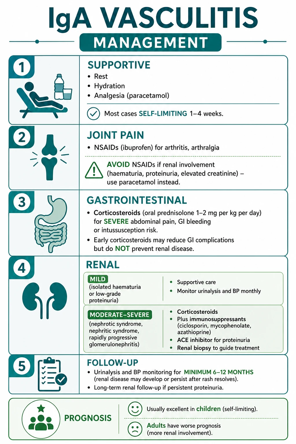
The default treatment is supportive care plus renal surveillance. Explain that the rash and joint symptoms usually settle, but the urine may become abnormal after the skin looks better. Provide rest during acute pain, oral fluids if tolerated, paracetamol for pain or fever, antiemetic if needed, and practical skin care for tender purpura. Avoid unnecessary antibiotics unless there is a proven bacterial infection. Avoid intramuscular injections when purpura or coagulation uncertainty is present.[5][7]
For joint pain, paracetamol is safe. NSAIDs such as ibuprofen may be used for significant arthralgia or arthritis only when renal function, hydration, blood pressure, and urine protein are reassuring and there is no GI bleeding. Avoid NSAIDs in renal involvement, dehydration, GI bleeding, peptic symptoms, or significant abdominal pain. This is a common exam trap: NSAIDs help ankles but can worsen the kidney or gut context in the very patients who need caution.[5][8]
Corticosteroids are not required for every rash. They are used for severe abdominal pain, significant GI bleeding after surgical emergencies are considered, severe joint symptoms impairing function, orchitis-like scrotal inflammation after torsion is excluded, and renal disease under specialist direction. A commonly taught regimen for severe symptomatic disease is oral prednisolone about 1 to 2 mg/kg/day, usually for a short course with taper according to response; severe renal disease may require higher-dose or pulsed corticosteroids and additional immunosuppression through nephrology. Steroids may shorten abdominal pain duration, but they do not reliably prevent nephritis, so follow-up remains mandatory.[5][8][3]
Mild rash plus mild arthralgia
- Confirm normal platelets and no sepsis features
- Paracetamol, hydration, rest, reassurance
- Urine dipstick and BP at baseline and follow-up
- No routine steroid
Severe abdominal or joint symptoms
- Assess for surgical abdomen, GI bleeding, and dehydration
- Analgesia, antiemetic, fluids, imaging if severe
- Prednisolone 1-2 mg/kg/day may be used for severe vasculitic pain after emergencies are addressed
- Review early and safety-net return symptoms
Proteinuria or nephritis
- Quantify urine protein and check BP, creatinine, albumin
- Avoid NSAIDs and nephrotoxins
- ACE inhibitor or ARB for persistent proteinuria when appropriate and supervised
- Nephrology for biopsy and immunosuppression decisions
Proteinuria management matters because it is modifiable. Children or adults with persistent proteinuria should be assessed for renin-angiotensin system blockade, usually an ACE inhibitor or ARB, if blood pressure, renal function, potassium, age, pregnancy status, and local guidance allow. Monitor creatinine and potassium after initiation or dose changes. ACE inhibitor or ARB therapy does not treat the rash; it reduces glomerular pressure and proteinuria, which are linked to renal outcome.[3][8]
Immunosuppression beyond corticosteroids is specialist territory. Options reported or recommended in selected severe IgA vasculitis nephritis include mycophenolate mofetil, azathioprine, cyclophosphamide, calcineurin inhibitors, rituximab, or plasma exchange in exceptional situations, but the evidence base is heterogeneous and decisions depend on biopsy, renal trajectory, age, and local nephrology protocol. The MBBS answer is not to list every drug as routine; it is to identify severe nephritis early and refer.[3][2]
Follow-up should be explicit on the discharge plan. Check urinalysis and blood pressure frequently during the first weeks, then at intervals for at least six to twelve months; continue longer if haematuria, proteinuria, hypertension, or impaired renal function persists. Many centres use weekly checks early, then monthly checks through six months, then spaced checks to twelve months, but local paediatric or renal guidance should be followed. Tell parents to return urgently for reduced urine output, dark urine, swelling, severe headache, severe abdominal pain, blood in stool, vomiting, scrotal pain, breathlessness, or lethargy.[3][8]
ANZ practice follows the same principles: stable children can often be managed with supportive care and general-practice or paediatric follow-up, while significant abdominal disease, renal abnormalities, hypertension, or diagnostic uncertainty require paediatric and nephrology input. Use local paediatric renal thresholds for urine protein quantification, ACE inhibitor or ARB initiation, and biopsy discussion.[3][8]
Specific Subtypes & Scenarios
Skin-limited disease presents with classic purpura and little else. It still needs FBC, urine dip, BP, and renal follow-up because "skin-limited today" can become renal disease later. Management is rest, analgesia, reassurance, avoidance of trauma to painful lesions, and review if lesions ulcerate, necrose, become infected, or recur unusually.[5][7]
Articular-predominant disease may look alarming because a child refuses to walk, but the joints are usually knees and ankles, migratory, periarticular, non-erosive, and self-limiting. Septic arthritis is suggested by a single hot joint, fever, inability to move the joint through passive range, marked systemic illness, or high inflammatory markers. Treat pain safely and reassess; do not label a single septic-looking joint as IgA vasculitis simply because purpura is present.[5][7]
GI-predominant disease is the dangerous diagnostic chameleon. Abdominal pain can precede the rash, and GI bleeding may be occult or obvious. Severe pain needs imaging and surgical thought. Steroids are useful for vasculitic bowel wall oedema and pain, but intussusception, perforation, pancreatitis, and appendicitis need their own management. The patient with worsening pain despite analgesia, bilious vomiting, guarding, or blood per rectum is not suitable for remote reassurance.[5][6]
IgA vasculitis nephritis spans mild urinary abnormalities to rapidly progressive glomerulonephritis. Isolated microscopic haematuria may only need surveillance. Persistent proteinuria, hypertension, reduced eGFR, nephritic syndrome, nephrotic syndrome, or macroscopic haematuria pushes the patient toward nephrology. Because renal outcome determines long-term prognosis, the safest exam phrase is: urinalysis and BP for every patient, quantify protein if positive, and refer if proteinuria persists or renal function/BP is abnormal.[3][8]
Recurrent disease occurs in a minority and usually repeats skin, joint, or abdominal symptoms rather than causing sudden new organ failure. Each recurrence still requires urine and BP checks because renal involvement can emerge later. Review triggers, medication exposures, infection, and in adults secondary causes. Recurrent severe abdominal disease or recurrent nephritis deserves specialist review.[6][7]
Adult-onset disease deserves respect. Adults are more likely to have renal disease, severe skin necrosis, relapse, and chronic kidney disease. They also have a broader purpura differential, so skin biopsy and systemic evaluation are more often appropriate. Ask specifically about new drugs, infection, inflammatory disease, hepatitis risk, and malignancy symptoms. Age-appropriate cancer screening should not be forgotten when disease is unexplained or atypical.[2][6]
Complications & Pitfalls
Acute complications include severe abdominal pain, GI haemorrhage, intussusception, bowel ischaemia or perforation, pancreatitis, scrotal oedema that mimics torsion, acute kidney injury, hypertension, nephritic syndrome, nephrotic syndrome, and very rare pulmonary haemorrhage or neurological disease. Late complications include recurrent IgA vasculitis, persistent haematuria, persistent proteinuria, chronic kidney disease, and rarely end-stage kidney disease. The rash is visible; the kidney is the silent organ that decides the long-term story.[3][5][6]
Pitfall: diagnosing by rash alone
- Meningococcaemia, DIC, leukaemia, ITP, HUS, and ANCA vasculitis can all have purpura
- Always check vitals, platelets, renal function, urine, and clinical toxicity
- A toxic purpuric child needs emergency care, not dermatology reassurance
Pitfall: forgetting renal surveillance
- The first urine can be normal
- Nephritis can appear after rash improvement
- No discharge is complete without a BP and urine follow-up plan
- Persistent proteinuria needs quantification and referral
Pitfall: NSAIDs in the wrong patient
- NSAIDs may help arthritis
- Avoid them in renal involvement, dehydration, GI bleeding, significant abdominal pain, or impaired kidney function
- Use paracetamol and senior review instead
Pitfall: overpromising steroids
- Steroids can reduce severe abdominal or joint symptoms
- They do not reliably prevent nephritis
- Renal monitoring continues even if steroids are given
The most common documentation failure is a vague discharge instruction such as "return if worse". A safe discharge note names the diagnosis, records platelet count, creatinine/eGFR, urinalysis, urine protein quantification if indicated, blood pressure, abdominal warning signs, renal warning signs, analgesia plan, who will repeat urine and BP, and when. Parents need to understand that the child may look better before the kidney risk window is closed.[3][8]
Prognosis & Disposition
In children, prognosis is usually excellent. Rash, joint pain, and abdominal symptoms often settle within weeks, although crops of purpura and relapse can occur. Mortality is very low in uncomplicated paediatric disease. The main determinant of long-term outcome is renal involvement, especially persistent proteinuria, hypertension, impaired kidney function, nephritic or nephrotic syndrome, and severe biopsy findings.[1][7][3]
Prognosis anchors
Disposition depends on severity. A stable child with typical rash, normal platelets, normal BP, normal renal function, absent or minimal urine abnormality, no severe abdominal pain, and reliable follow-up can often be managed as an outpatient. Admit or observe if there is diagnostic uncertainty, severe pain, dehydration, GI bleeding, vomiting, inability to maintain oral intake, suspected intussusception, scrotal pain requiring torsion exclusion, hypertension, proteinuria requiring quantification, macroscopic haematuria, rising creatinine, nephritic or nephrotic syndrome, social concern, or inability to ensure follow-up.[5][8]
Adult disposition is more conservative because renal and secondary-cause risks are higher. Adults with new IgA vasculitis usually need baseline biopsy consideration when the rash is atypical, renal workup, review of triggers and medications, and follow-up that does not assume childhood benignity. Persistent proteinuria or reduced eGFR in adults warrants nephrology input early.[2][3]
Special Populations
Children are the typical population, so most teaching describes them. Use weight-based analgesia and corticosteroid dosing when steroids are indicated. Explain the disease to caregivers in concrete terms: the rash is from inflamed small vessels; the joint and abdominal symptoms usually settle; the urine and blood pressure checks are not optional; and severe abdominal pain, blood in stool, reduced urine, swelling, headache, breathlessness, or drowsiness requires urgent review.[5][7]
Adults have a higher risk of renal disease and a wider differential. Do not overuse the childhood script. Consider skin biopsy, renal workup, medication review, infection testing, autoimmune testing when indicated, and age-appropriate malignancy assessment in unexplained or atypical cases. Adult purpura with renal disease could be IgA vasculitis, ANCA vasculitis, lupus, cryoglobulinaemia, drug vasculitis, infection-related vasculitis, or haematological disease.[2][6]
Pregnancy is uncommon but important because hypertension, proteinuria, haematuria, and renal impairment overlap with pre-eclampsia and glomerular disease. Coordinate dermatology, obstetrics, nephrology, and rheumatology as needed. Avoid teratogenic drugs; ACE inhibitors and ARBs are contraindicated in pregnancy. Corticosteroids may be used when benefits justify risk, but renal and fetal monitoring are specialist decisions.[2][3]
Elderly and immunocompromised patients need a broader differential and lower threshold for infection and malignancy assessment. Purpura in an immunocompromised patient may be sepsis, disseminated infection, drug reaction, thrombocytopenia, vasculitis, or embolic disease. Immunosuppression for presumed vasculitis is unsafe until infection has been considered.[2][5]
Patients with chronic kidney disease, dehydration, anticoagulation, or bleeding risk require medication caution. Avoid NSAIDs in kidney disease and dehydration. Anticoagulated patients can have more dramatic purpura or GI bleeding, but anticoagulation does not explain palpable vasculitis by itself. If renal disease is already present, involve nephrology early and interpret proteinuria against baseline.[3][5]
Evidence, Guidelines & Regional Differences
The SHARE initiative provides consensus-based paediatric recommendations for diagnosis and treatment, reinforcing the clinical classification pattern, the role of biopsy in uncertain cases, and the need to stratify treatment by organ severity. The IPNA recommendations focus on IgA vasculitis nephritis and IgA nephropathy in children, emphasising proteinuria quantification, blood pressure control, kidney function assessment, indications for biopsy, and renoprotective therapy such as ACE inhibitor or ARB when persistent proteinuria is present and clinically appropriate.[8][3]
The evidence controversy most often tested is corticosteroids. Steroids can reduce severe abdominal pain and may be used for serious GI or renal disease, but routine early corticosteroids for every patient have not reliably prevented later nephritis. Therefore, a candidate who says "give steroids to prevent renal disease" loses marks. The better answer is: treat severe symptoms and nephritis appropriately, but monitor urine and blood pressure regardless of steroid use.[5][8]
Renal immunosuppression remains an area where evidence and practice vary. Severe IgA vasculitis nephritis can resemble severe IgA nephropathy, but paediatric evidence is less robust than exam answers sometimes imply. Guidelines therefore use risk stratification: supportive and renoprotective care for mild disease, corticosteroids and specialist therapy for significant nephritis, and biopsy-guided escalation for crescentic or rapidly progressive disease.[3][2]
Exam Pearls
The classic stem is a child aged three to fifteen who develops palpable purpura over the buttocks and legs after a recent upper respiratory infection, with ankle or knee pain, colicky abdominal pain, and microscopic haematuria. The platelet count is normal. The answer is IgA vasculitis. If the stem asks for histology, write leukocytoclastic vasculitis; if it asks for immunofluorescence, write IgA-dominant deposition in vessel walls; if it asks for prognosis, write renal involvement determines long-term outcome.[5][7]
Diagnostic one-liners
- Most common vasculitis in children
- Palpable purpura with lower-limb predominance and normal platelets
- Tetrad: purpura, arthralgia or arthritis, abdominal pain or GI bleeding, renal involvement
- Leukocytoclastic vasculitis with IgA-dominant DIF
Investigation one-liners
- Do FBC to prove platelets are not low
- Do urinalysis, urine protein quantification if positive, creatinine/eGFR, and blood pressure in every patient
- Biopsy atypical or adult disease; fresh lesion for DIF
- Ultrasound severe abdominal pain to exclude intussusception
Management one-liners
- Supportive care for most children
- NSAIDs may help joints but avoid if renal/GI disease
- Steroids for severe abdominal symptoms or renal disease; they do not reliably prevent nephritis
- ACE inhibitor or ARB for persistent proteinuria under renal guidance
Prognosis one-liners
- Children usually recover; adults are higher risk
- The kidney determines long-term outcome
- Follow urine and BP for at least six to twelve months
- Persistent proteinuria, hypertension, or reduced eGFR needs nephrology
Exam application bank (NEET-PG / INICET)
One-line answer
IgA vasculitis is an IgA1 immune-complex small-vessel leukocytoclastic vasculitis, classically presenting with lower-limb palpable purpura, arthralgia or arthritis, colicky abdominal pain or GI bleeding, and renal involvement with haematuria or proteinuria. Diagnosis is clinical when purpura is typical and platelets are normal; early skin biopsy shows leukocytoclastic vasculitis with IgA-dominant direct immunofluorescence. Management is supportive for most children, with urinalysis, blood pressure, proteinuria, and renal function surveillance; corticosteroids are reserved for severe abdominal or renal disease, and persistent proteinuria or renal impairment needs nephrology-led care.
Worked stems (answer without another resource)
Stem 1 — Classic presentation. Map symptoms to mechanism; name the first investigation and first treatment step with dose/route if drug therapy is standard. [1]
Stem 2 — Unstable / complicated. List red flags that force immediate resuscitation, theatre, ICU, antidote, or reperfusion — and what you do in the first 15 minutes. [1]
Stem 3 — Atypical group. Elderly, pregnancy, child, or immunocompromised: how presentation and thresholds change. [1]
Stem 4 — Differential trap. Name the three closest mimics and one discriminator for each. [1]
Stem 5 — Disposition. Who goes home with safety-netting, who is admitted, who needs HDU/ICU/theatre, and what follow-up is mandatory. [1]
Rapid viva checklist
- Definition + classification
- Pathophysiology chain
- Bedside signs / criteria
- Score with exact components (if any)
- Emergency bundle
- Definitive therapy with doses
- Complications of disease and of treatment
- Special populations
- Guideline/trial name if classic
- Three exam traps
Coverage self-check
If you cannot answer any stem above from this page alone, re-read the matching section — the page is intended to be self-sufficient for final-prof and NEET-PG/INICET questions on IgA vasculitis (Henoch-Schönlein purpura).
“”
SAQ — IgA vasculitis in a child
10 minutes · 10 marks
A seven-year-old boy develops palpable purpura on the legs and buttocks, ankle swelling, colicky abdominal pain, and dipstick haematuria ten days after a sore throat. Platelets are normal.
Model answer: The diagnosis is IgA vasculitis. Supporting features are lower-limb palpable purpura with normal platelets, ankle arthritis or arthralgia, colicky abdominal pain, and haematuria. Initial investigations are FBC with platelets, renal function and electrolytes, urinalysis, urine protein quantification if protein is present, blood pressure, stool occult blood if GI symptoms are present, and biopsy only if the diagnosis is uncertain. Management is supportive with rest, hydration, paracetamol, avoidance of NSAIDs if renal or GI disease exists, and corticosteroids for severe abdominal symptoms or nephrology-directed renal disease. Escalate for toxicity, severe abdominal pain or GI bleeding, suspected intussusception, hypertension, proteinuria, macroscopic haematuria, rising creatinine, nephritic syndrome, or nephrotic syndrome. The kidney determines prognosis, so urine and blood pressure surveillance must continue for at least six to twelve months.[3][5][8]
References
- [1]Parums DV. A Review of IgA Vasculitis (Henoch-Schönlein Purpura) Past, Present, and Future Med Sci Monit, 2024.PMID 38281080
- [2]Pillebout E, Sunderkötter C. IgA vasculitis Semin Immunopathol, 2021.PMID 34170395
- [3]Vivarelli M, Samuel S, Coppo R, et al. IPNA clinical practice recommendations for the diagnosis and management of children with IgA nephropathy and IgA vasculitis nephritis Pediatr Nephrol, 2025.PMID 39331079
- [4]Song Y, Huang X, Yu G, et al. Pathogenesis of IgA Vasculitis: An Up-To-Date Review Front Immunol, 2021.PMID 34858429
- [5]Reamy BV, Servey JT, Williams PM. Henoch-Schönlein Purpura (IgA Vasculitis): Rapid Evidence Review Am Fam Physician, 2020.PMID 32803924
- [6]Hetland LE, Susrud KS, Lindahl KH, et al. Henoch-Schönlein Purpura: A Literature Review Acta Derm Venereol, 2017.PMID 28654132
- [7]Leung AKC, Barankin B, Leong KF. Henoch-Schönlein Purpura in Children: An Updated Review Curr Pediatr Rev, 2020.PMID 32384035
- [8]Ozen S, Marks SD, Brogan P, et al. European consensus-based recommendations for diagnosis and treatment of immunoglobulin A vasculitis-the SHARE initiative Rheumatology (Oxford), 2019.PMID 30879080