Dermatology · Medicine
Impetigo
Also known as Non-bullous impetigo · Bullous impetigo · Impetigo contagiosa · Ecthyma · Staphylococcal scalded skin syndrome (SSSS)
Impetigo is a highly contagious superficial bacterial skin infection, most common in children, caused predominantly by Staphylococcus aureus and group A streptococci. Fellowship-level assessment demands mastery of the non-bullous, bullous (exfoliative-toxin-mediated), and ecthyma subtypes, the staphylococcal scalded skin syndrome spectrum, the distinction between localized and toxin-mediated disease, first-line topical (mupirocin, retapamulin, fusidic acid) versus oral antibiotic therapy (flucloxacillin, cephalexin, clindamycin) guided by local resistance and severity, the role of MRSA and PVL-positive strains, recurrent disease and decolonisation, and the serious non-suppurative complications of group A streptococcal impetigo — post-streptococcal glomerulonephritis and the epidemiological link to acute rheumatic fever.
On this page & tools
Your progress
Saved locally on this device.
Exam tags
Red flags
Overview
Impetigo is a highly contagious, superficial bacterial skin infection confined to the epidermis, and is the commonest bacterial skin infection in children worldwide. It is caused predominantly by Staphylococcus aureus and group A β-haemolytic streptococci (Streptococcus pyogenes, GAS), alone or in combination. Three clinical subtypes are recognised: non-bullous impetigo (the commonest — about 70% of cases), bullous impetigo (staphylococcal exfoliative-toxin-mediated), and ecthyma (a deeper ulcerative form). Staphylococcal scalded skin syndrome (SSSS) is the related generalised toxin-mediated desquamative disorder of infants and young children. Fellowship-level competence requires precise phenotyping, an understanding of the exfoliative-toxin pathophysiology, rational first-line topical-versus-oral antibiotic selection guided by extent, severity, and local resistance, awareness of MRSA and Panton-Valentine leucocidin (PVL)-positive strains, and recognition of the serious non-suppurative complications of GAS impetigo — post-streptococcal glomerulonephritis (PSGN) and the epidemiological link to acute rheumatic fever (ARF).[1][3][4]
Epidemiology
Impetigo is global in distribution but disproportionately affects children aged 2–5 years, with a peak incidence in preschool and early school-age children. Population estimates suggest a global point prevalence of impetigo of around 1–5% in paediatric populations, with the highest rates in tropical, warm-humid climates and in settings of crowding, poverty, and poor hygiene. Impetigo is one of the commonest skin diseases worldwide and a leading cause of skin-related primary-care presentations in children, accounting for roughly 10% of all childhood skin problems seen in general practice.[15]
The disease is seasonal — peaks in late summer and autumn in temperate regions and persists year-round in tropical climates. Risk factors for acquisition include poor skin barrier integrity (atopic dermatitis, scabies, tinea, insect bites, abrasions), warm humid environments, crowding, low socioeconomic status, recent antibiotic use, and recurrent contact with young children (nurseries, schools). Institutional outbreaks are well-recognised in nurseries, schools, military barracks, prisons, Indigenous communities, refugee camps, and contact-sport settings (wrestling, rugby) — sharing of towels, sports equipment, and skin-to-skin contact drive transmission. Hospital nurseries and neonatal units are particularly high-risk environments where index cases require urgent infection-control input.[10][11]
The global burden of group A streptococcal disease — of which impetigo (also called pyoderma in the public-health literature) is the commonest cutaneous manifestation — is substantial. Impetigo is the principal source of GAS exposure in many Indigenous and resource-limited populations (Aboriginal Australian, Maori and Pacific Islander, South Asian, sub-Saharan African, and some Native American communities), where it contributes disproportionately to the pool of GAS strains that drive both PSGN and ARF.[10][11][14]
[1]Microbiology and pathophysiology
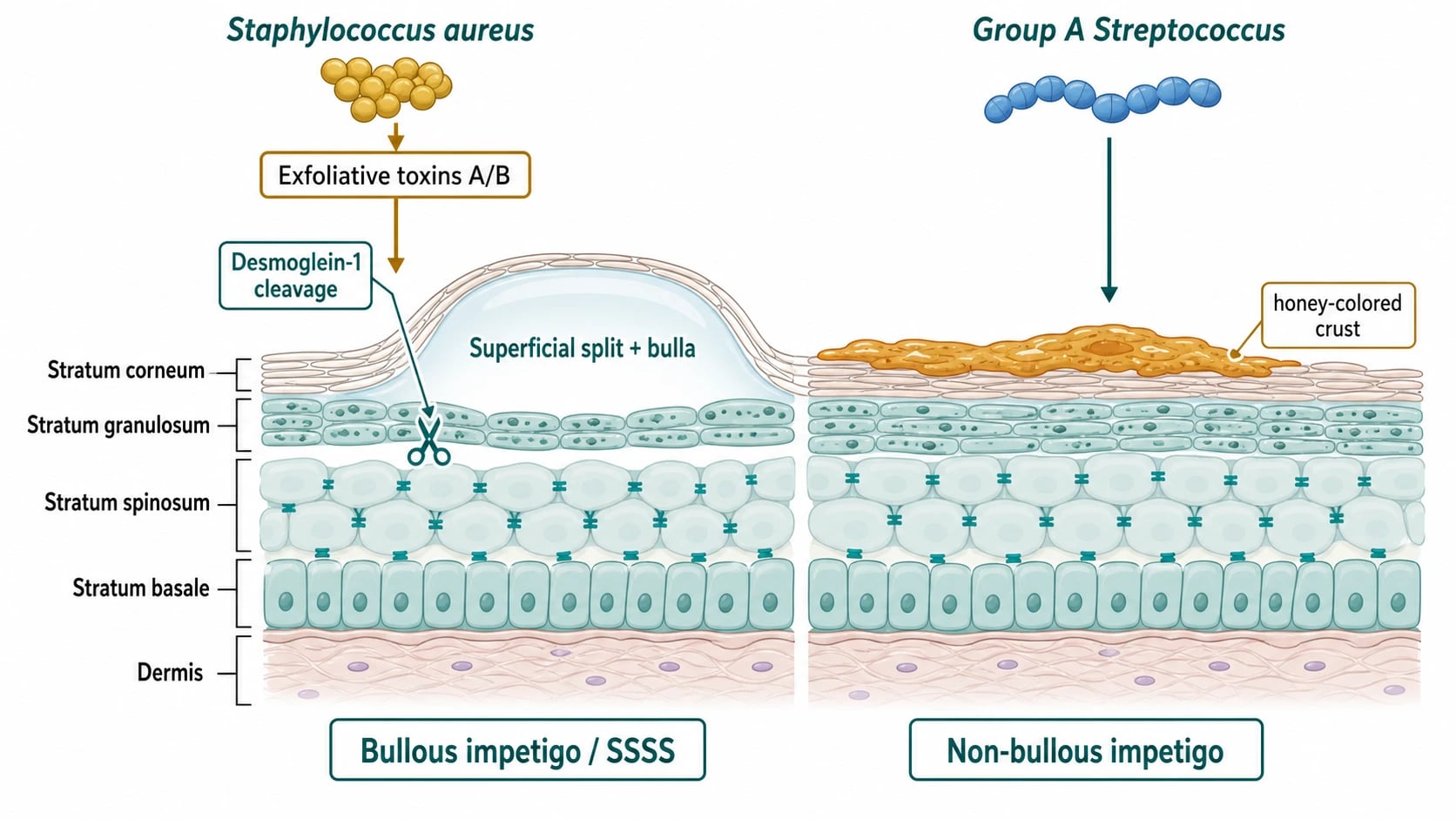
- Staphylococcus aureus is the dominant pathogen in most settings and the cause of essentially all bullous impetigo. It produces exfoliative toxins A and B (ETA / ETB) — serine proteases encoded by the eta and etb genes carried on phage or plasmid — that cleave desmoglein-1, a desmosomal cadherin in the superficial epidermis. Loss of keratinocyte–keratinocyte adhesion produces the intra/subcorneal splitting that gives rise to the flaccid bullae of bullous impetigo and the widespread superficial desquamation of SSSS.
- Group A β-haemolytic streptococcus (S. pyogenes, GAS) causes non-bullous impetigo and ecthyma, often as a secondary invader of broken skin. Distinct M-protein serotypes — M49, M2, M55, M60, M57 — are nephritogenic and underlie the link between impetigo and post-streptococcal glomerulonephritis.
- Co-infection with both organisms is common in non-bullous disease. MRSA is an increasing cause, particularly in endemic regions and in recurrent or treatment-resistant disease, and influences antibiotic choice; Panton-Valentine leucocidin (PVL)-positive strains drive recurrent necrotic lesions and family clusters.
- The toxin mechanism is central: in bullous impetigo, toxin is produced in the local lesion; in SSSS the exfoliative toxin disseminates systemically from a distant focus (such as conjunctivitis, otitis media, nasopharyngitis, or umbilical staphylococcal infection in a neonate), producing widespread epidermal desquamation at sites remote from the infection. Skin cultures of the bullae are negative in SSSS — the disease is toxin-mediated.[7][8][9][11][12]
Pre-existing skin barrier dysfunction (atopic dermatitis, scabies, tinea pedis, insect bites, abrasions, IV cannulation, surgical wounds) is the usual portal; the term "impetiginisation" refers to secondary bacterial infection of an underlying dermatosis, most often atopic eczema. [1]
Classification
Impetigo is classified by clinical morphology, depth, and pathogenesis.[1][3]
Impetigo contagiosa — commonest
- ~70% of impetigo cases; predominantly GAS with or without S. aureus
- Begins as a small red papule or vesicopustule that rapidly ruptures
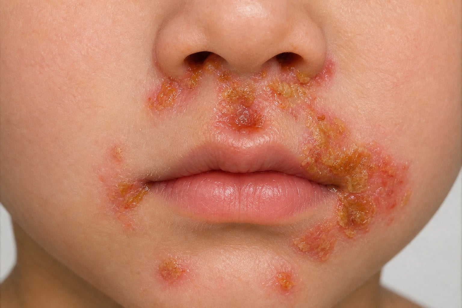
- Hallmark golden-yellow (honey-coloured) crust overlying a moist erythematous base
- Face (perinasal, perioral), limbs, areas of broken skin; spreads by autoinoculation
- Mildly pruritic rather than painful; regional lymphadenopathy common; no scarring
Exfoliative-toxin-mediated
- ~30% of cases; essentially always staphylococcal (eta/etb genes)
- Flaccid, transparent bullae with clear or yellow fluid on erythematous / intact skin
- Trunk, axillae, neck, intertriginous areas; common in neonates and infants
- Bullae rupture easily leaving a thin brown crust with a peripheral collarette of scale
- Caused by superficial epidermal split (granular layer) — heals without scarring
Deeper ulcerative form
- Uncommon; typically GAS (often with S. aureus); extends into the dermis
- Punched-out ulcer with necrotic (grey-yellow) eschar and surrounding erythema
- Predominantly on the legs; often follows minor trauma or insect bites
- Regional lymphadenopathy common; heals with scarring
- Do NOT confuse with ecthyma gangrenosum (Pseudomonas, immunocompromised host)
Generalised toxin-mediated
- Infants and young children (< 5 years); rarely adults with renal impairment
- Constitutional upset → tender diffuse erythema → flaccid bullae and sheet-like desquamation
- Nikolsky sign positive; mucous membranes are SPARED (key discriminator vs TEN)
- Distant focus (conjunctiva, umbilicus, nasopharynx, wound); skin cultures of bullae are negative
- Skin biopsy shows a superficial split at the granular layer; prognosis in children is excellent
Clinical features
Non-bullous impetigo (~70% of cases)
Begins as a small papule or vesicopustule that rapidly ruptures and forms the hallmark golden-yellow (honey-coloured) crust overlying a moist erythematous base. Lesions are typically on the face (perinasal, perioral), limbs, or areas of broken skin, spread by autoinoculation (scratching, picking), and are mildly itchy rather than painful. Regional lymphadenopathy is common; constitutional upset is mild or absent. Healing occurs without scarring over 2–3 weeks if untreated and within 7–10 days with topical antibiotics.[1][4]
Bullous impetigo (~30%)
Caused by toxin-producing S. aureus. Presents with flaccid, transparent bullae containing clear or yellow fluid on erythematous (and often intact) skin, most often on the trunk, axillae, neck, and intertriginous areas. The bullae rupture easily, leaving a thin, shiny, brownish crust with a peripheral collarette of scale — a more superficial crust than the thick honey crust of non-bullous disease. Common in neonates and infants; tends to occur in small outbreaks in nurseries.[7][2]
Ecthyma
A deeper form producing punched-out, ulcerative lesions with necrotic (grey-yellow) eschar and surrounding erythema, healing with scarring. Typically on the legs, caused by GAS (often with S. aureus). Ecthyma gangrenosum is a distinct entity (Pseudomonas aeruginosa, usually in immunocompromised/haematology patients with neutropenia) and must not be confused with streptococcal ecthyma.[3][16]
Staphylococcal scalded skin syndrome (SSSS)
A generalised toxin-mediated disorder of infants and young children (rare in adults, and then usually with renal impairment or immunosuppression). Begins with constitutional upset, conjunctivitis, or a focal staphylococcal infection, then tender, diffuse erythema progresses within 24–48 hours to large superficial flaccid bullae and sheet-like desquamation, with a positive Nikolsky sign. Mucous membranes are spared — the cardinal discriminator from toxic epidermal necrolysis. Management is supportive (fluid, temperature, analgesia, wound care) plus anti-staphylococcal antibiotics; prognosis in children is generally good with appropriate care, but SSSS can be life-threatening in neonates.[7][9]
Quick numbers for the examiner
Diagnosis
The diagnosis is clinical. Useful bedside adjuncts include Gram stain of pus or crust (Gram-positive cocci in clusters = S. aureus; chains = GAS — though most clinicians proceed without this), KOH preparation of peripheral scale to exclude tinea, Tzanck smear or HSV PCR of vesicular fluid to exclude herpes simplex, and dermoscopy for scabies mites when secondary impetiginisation of scabies is suspected. [1]
Swab for bacterial culture and sensitivity is indicated for: extensive or atypical lesions, treatment failure or recurrence within 2 weeks, suspected MRSA, immunocompromised host, institutional outbreaks, and neonatal disease. Pus swabs are plated onto blood agar and mannitol salt agar (or chromogenic MRSA agar in endemic settings); consider PVL PCR in recurrent or family-cluster disease. [1]
In SSSS, the causative focus (conjunctiva, umbilicus, wound, nasopharynx) is cultured — skin cultures of the bullae are negative (toxin-mediated, not locally infected). Skin biopsy is reserved for diagnostic uncertainty (e.g., distinguishing SSSS from TEN: SSSS shows a superficial split at the granular layer with normal dermis; TEN shows full-thickness epidermal necrosis with mucosal involvement).[1][7]
Laboratory workup for the non-suppurative complications of GAS impetigo includes urinalysis (haematuria, RBC casts, mild proteinuria), U&E (raised urea and creatinine), complement studies (transiently low C3), ASO and anti-DNase B titres (the latter rises higher after skin infection), and blood pressure monitoring — typically checked at presentation and 3 weeks after the rash.[13]
For recurrent disease, S. aureus nasal carriage swabs (anterior nares, ± perineum, axilla, umbilicus) guide decolonisation. Screening for underlying atopic dermatitis, scabies, tinea, immunodeficiency (HIV, immunoglobulins), iron deficiency, and diabetes is appropriate.[1][4][17]
Differential diagnosis
The differential depends on the lesion morphology. Two practical axes — bullous vs non-bullous, and superficial vs generalised toxin-mediated — anchor the workup.[3][16]
- Atopic dermatitis / contact dermatitis (secondarily infected) — bilateral, pruritic, eczematous background
- Herpes simplex — grouped vesicles on an erythematous base (often perioral); Tzanck / HSV PCR positive
- Scabies — burrows in finger webs, wrists, axillae; contact dermatitis to scabicides can mimic
- Insect bites and papular urticaria — pruritic papules, central punctum, no honey crust
- Tinea (corporis, faciei) — annular, scaly, advancing edge; KOH positive
- Bullous pemphigoid — older adults, tense bullae on urticarial plaques, trunk and flexures
- Pemphigus vulgaris — adults, mucosal involvement, positive Nikolsky on uninvolved skin
- Erythema multiforme / SJS — target lesions, mucosal involvement, drug or infection trigger
- Friction or suction bullae — history of trauma, single site, no systemic features
- Epidermolysis bullosa — congenital, recurrent bullae from minor trauma, scarring
- Toxic epidermal necrolysis (TEN) — MUCOSAE involved, FULL-THICKNESS epidermal necrosis, drug trigger
- Kawasaki disease — fever ≥ 5 days, conjunctivitis, cervical lymphadenopathy, mucositis, extremity changes
- Scarlet fever — sandpaper rash, strawberry tongue, Pastia's lines, GAS pharyngitis
- Drug eruptions — morbilliform / DRESS / acute generalised exanthematous pustulosis (AGEP)
- Burn (chemical or thermal) — history of exposure; SSSS misdiagnosed as scald in some series
- Ecthyma gangrenosum — Pseudomonas aeruginosa in neutropenic / immunocompromised host; central necrotic black eschar with surrounding erythema, sepsis
- Pyoderma gangrenosum — violaceous undermined edge, pathergy, IBD / haematological malignancy
- Cutaneous anthrax — painless black eschar with massive surrounding oedema; occupational exposure
- Cutaneous leishmaniasis — chronic ulcer at sandfly-bite site, travel to endemic area
- Venous or arterial leg ulcer — chronic, associated venous disease or peripheral arterial disease
Management
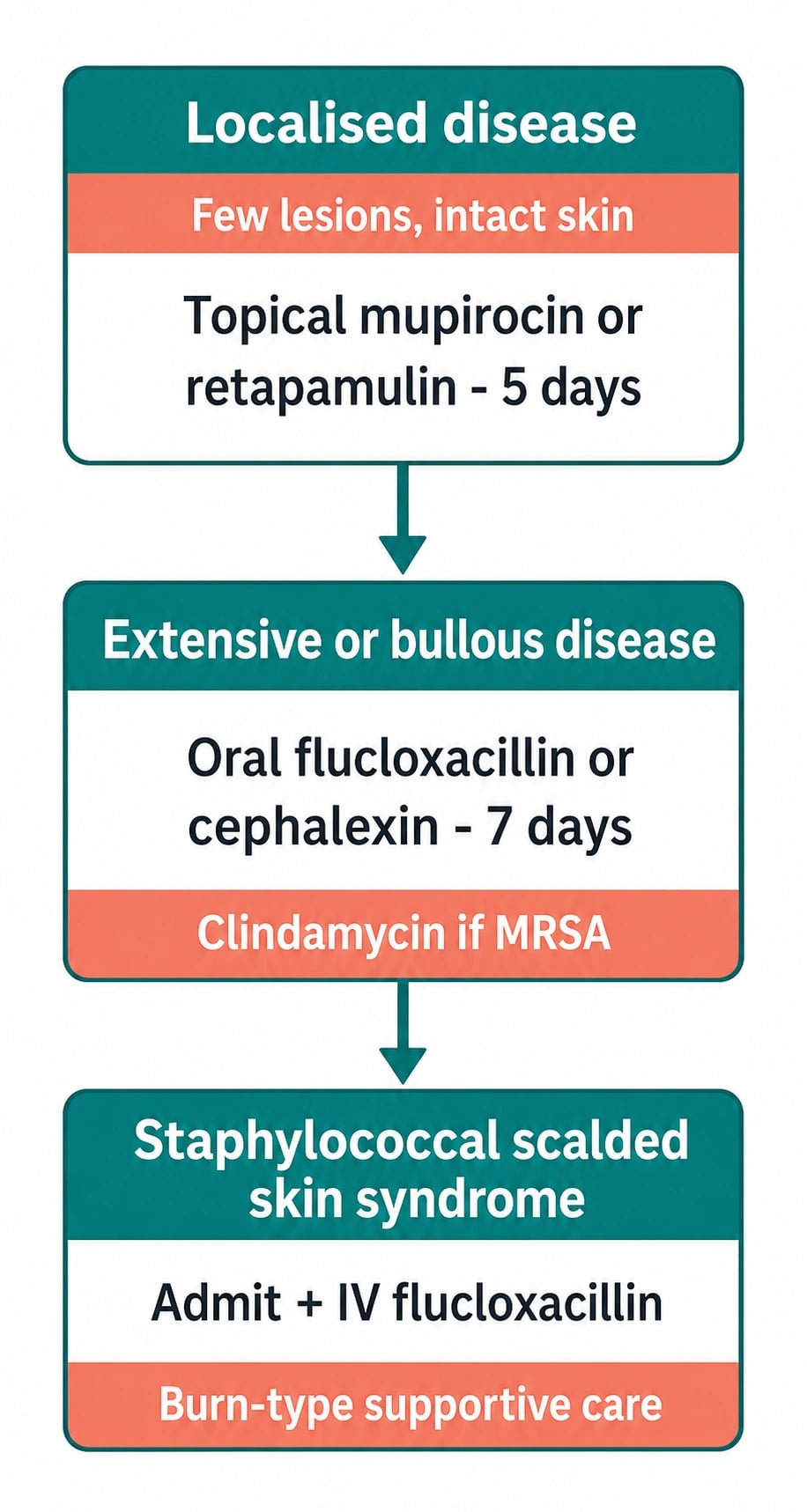
Treatment aims to resolve infection, reduce transmission, and prevent complications. Choice depends on the extent, severity, local resistance patterns, and host factors.[1][5][2][17]
Localised disease (few lesions, intact host)
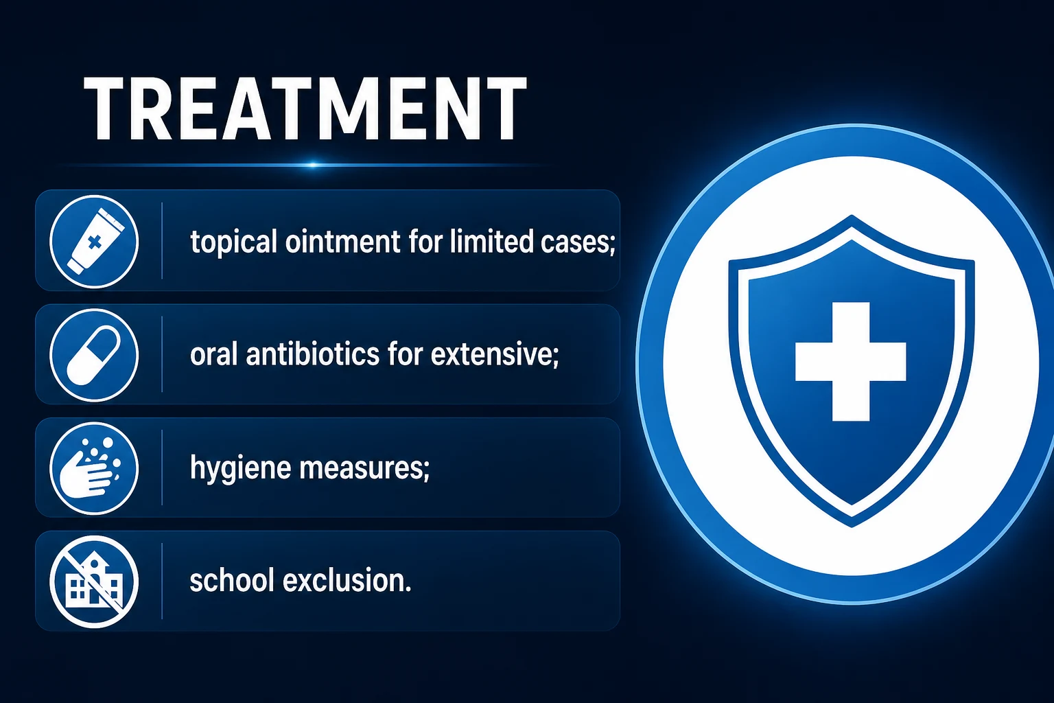
- Topical antibiotics first-line — fusidic acid 2% ointment or cream TDS for 5–7 days (favoured in continental Europe because of low resistance) OR mupirocin 2% ointment TDS for 5 days OR retapamulin 1% ointment BD for 5 days. Apply to the lesion after gentle crust removal (soak in warm soapy water or use a moist dressing for 10–15 minutes); wash hands before and after.
- Topical hydrogen peroxide 1% cream is an effective steroid- and antibiotic-free alternative endorsed by NICE CKS — particularly relevant where antibiotic stewardship is a concern (UK BAD advice to curb rising fusidic acid resistance).
- Cover the lesion with a loose dressing to limit spread and contact-spread within the household; exclude from school until 24 hours of effective treatment has been given (UK CKS) or until the lesions are crusted (US AAD). [1]
Extensive, bullous, or systemic disease; SSSS
- Oral antibiotics for 7 days (5–7 for uncomplicated extensive disease): a β-lactamase-resistant anti-staphylococcal agent — flucloxacillin 500 mg QDS (or dicloxacillin), or a first-generation cephalosporin cephalexin 500 mg BD. Erythromycin 500 mg QDS or clarithromycin 500 mg BD for true penicillin allergy.
- Recurrent, treatment-failure, or MRSA-suspected disease — clindamycin 300 mg QDS, doxycycline 100 mg BD (over 12 years), or TMP-SMX 960 mg BD (where local resistance is low). PVL-positive strains — clindamycin or linezolid (toxin suppression) ± rifampicin 300 mg BD for the last 4 days of an anti-staphylococcal course.
- SSSS — admit, fluid resuscitation, temperature control, analgesia, IV anti-staphylococcal therapy (IV flucloxacillin 1–2 g 6-hourly, or vancomycin / linezolid if MRSA); manage wound care as for a superficial scald (non-adherent dressings, bland emollients, avoid shearing forces). Most children recover within 1–2 weeks without scarring. [1]
Recurrent disease — decolonisation
- 5-day decolonisation cycle — intranasal mupirocin 2% BD to anterior nares PLUS chlorhexidine 4% body wash daily PLUS hot laundering of towels and bedlinen. Treat household contacts if recurrent within the family. Address underlying atopic dermatitis, scabies, tinea, or chronic skin disease. [1]
General and preventive measures
- Gently remove crusts with warm soapy water; keep lesions covered; wash hands and linen frequently; avoid sharing towels, flannels, and toys.
- Manage predisposing skin disease (atopic dermatitis, scabies, tinea); bleach baths 1:100 2x/week if recurrent staphylococcal skin infection is associated with atopic dermatitis (Huang 2009; tolerability debated).
- Treat household contacts and screen staff in nursery outbreaks. [1]
Regional deltas
[1] [1] [1]Complications
Suppurative complications
- Cellulitis and lymphangitis — extension of the local infection; tender erythema, red streaking, regional lymphadenopathy; treat with systemic antibiotics.
- Ecthyma — progression to deeper ulceration; heals with scarring.
- Abscess formation — requires incision and drainage.
- Staphylococcal bacteraemia and sepsis — uncommon in immunocompetent children; high risk in neonates, immunocompromised, and those with indwelling devices.
- Osteomyelitis and septic arthritis — rare haematogenous seeding in children with bacteraemia. [1]
Non-suppurative (GAS) — the high-yield exam points
-
Post-streptococcal glomerulonephritis (PSGN) — the most important non-suppurative complication. Occurs 1–3 weeks after impetigo (or 1–2 weeks after pharyngitis), caused by nephritogenic GAS strains (M49, M2, M55, M60, M57). Presents with haematuria (smoky or tea-coloured urine), oedema (periorbital and peripheral), hypertension, and oliguria; biopsy shows proliferative glomerulonephritis with subepithelial humps; complement C3 is transiently low. Treatment is supportive (fluid balance, antihypertensives, diuretics, occasionally dialysis); the disease usually resolves in 2–4 weeks in children. Antibiotics do NOT prevent PSGN once infection is established — the role of antibiotic therapy is to limit transmission.[13]
-
Acute rheumatic fever (ARF) — primarily a complication of GAS pharyngitis, but skin streptococci contribute to the GAS reservoir in high-risk populations (Indigenous Australian, Maori, Pacific, South Asian, resource-limited tropical settings) and skin-derived GAS strains can be rheumatogenic. Recurrent impetigo is therefore an important target for public-health intervention in those communities. ARF typically occurs 2–4 weeks after pharyngitis with migratory polyarthritis, carditis, chorea, erythema marginatum, and subcutaneous nodules (revised Jones criteria).[14]
Toxin-mediated complications
- SSSS — see above; generalised toxin-mediated disease in infants.
- Streptococcal toxic shock syndrome — rare, invasive GAS with hypotension and multi-organ failure; clindamycin suppresses toxin production. [1]
Recurrence
- Recurrent impetigo — S. aureus nasal carriage + barrier dysfunction (atopic dermatitis, scabies, tinea); managed by decolonisation, household contact screening, and treatment of underlying skin disease.[1][4]
Special populations
- Neonates — high sepsis risk; admit, IV anti-staphylococcal therapy, paediatric and infection-control input; umbilicus and perineum are common portals; nursery outbreaks require screening of staff and parents.
- Children — the principal population; cephalexin 25 mg/kg/dose BD or flucloxacillin elixir 62.5–125 mg QDS for under 6 years; gentle washing; short school exclusion (24 h after starting antibiotics or until lesions are crusted).
- Pregnancy / breastfeeding — topical fusidic acid is safe; oral flucloxacillin, cephalexin, and erythromycin are safe; avoid tetracyclines and TMP-SMX near term.
- Atopic dermatitis patients — recurrent impetigo is common; aggressive emollients, treat flares early, bleach baths 1:100 twice weekly, decolonisation if recurrent.
- Diabetes — lower threshold for oral therapy and culture; screen for nasal carriage if recurrent; review glycaemic control.
- Elderly — atypical presentation common; lower threshold for systemic therapy and admission; review renal function for antibiotic dose.
- Immunocompromised host — broader differential, low threshold for culture and admission, consider MRSA and PVL; biopsy for atypical lesions.
- Household contacts and healthcare workers — outbreak screening, decolonisation before return to high-risk work, occupational health referral.[1][17]
Mnemonic
The IMPETIGO bedside checklist
Key facts
[1] [1]Clinical pearl
[1]Red flags
Exam application bank (NEET-PG / INICET)
One-line answer
Impetigo is a highly contagious superficial bacterial skin infection, most common in children, caused predominantly by Staphylococcus aureus and group A streptococci. Fellowship-level assessment demands mastery of the non-bullous, bullous (exfoliative-toxin-mediated), and ecthyma subtypes, the staphylococcal scalded skin syndrome spectrum, the distinction between localized and toxin-mediated disease, first-line topical (mupirocin, retapamulin, fusidic acid) versus oral antibiotic therapy (flucloxacillin, cephalexin, clindamycin) guided by local resistance and severity, the role of MRSA and PVL-positive strains, recurrent disease and decolonisation, and the serious non-suppurative complications of group A streptococcal impetigo — post-streptococcal glomerulonephritis and the epidemiological link to acute rheumatic fever.
Worked stems (answer without another resource)
Stem 1 — Classic presentation. Map symptoms to mechanism; name the first investigation and first treatment step with dose/route if drug therapy is standard. [1]
Stem 2 — Unstable / complicated. List red flags that force immediate resuscitation, theatre, ICU, antidote, or reperfusion — and what you do in the first 15 minutes. [1]
Stem 3 — Atypical group. Elderly, pregnancy, child, or immunocompromised: how presentation and thresholds change. [1]
Stem 4 — Differential trap. Name the three closest mimics and one discriminator for each. [1]
Stem 5 — Disposition. Who goes home with safety-netting, who is admitted, who needs HDU/ICU/theatre, and what follow-up is mandatory. [1]
Rapid viva checklist
- Definition + classification
- Pathophysiology chain
- Bedside signs / criteria
- Score with exact components (if any)
- Emergency bundle
- Definitive therapy with doses
- Complications of disease and of treatment
- Special populations
- Guideline/trial name if classic
- Three exam traps
Coverage self-check
If you cannot answer any stem above from this page alone, re-read the matching section — the page is intended to be self-sufficient for final-prof and NEET-PG/INICET questions on Impetigo.
Expanded exam teaching (depth pass)
Clinical reasoning
For Impetigo, examiners test whether you can prioritise life threats, choose the right first test, and give specific therapy (agent, dose, route, timing). Generic phrases without numbers score poorly.
Mechanism → feature map
Build a short chain: cause → pathophysiologic intermediate → clinical feature → complication. Every major symptom in the classic vignette should sit on that chain.
Investigation strategy
- Bedside/first-line tests that change immediate management
- Confirmatory or staging tests
- What a normal result does not exclude
- When not to delay treatment for imaging (unstable patient)
Management ladder
- Resuscitation / ABC / sepsis or haemorrhage bundle as relevant
- Specific antidote / procedure / antimicrobial / reperfusion / surgery
- Supportive care and monitoring targets
- Definitive long-term therapy and secondary prevention
- Disposition and safety-net advice
Special populations
Always prepare one line each for children, pregnancy, elderly, renal/hepatic impairment, and immunocompromised patients when the topic allows.
Pitfalls that fail candidates
- Treating the number not the patient
- Missing pregnancy status when relevant
- Imaging before stabilisation
- Wrong empiric cover or wrong antidote timing
- Incomplete counselling on recurrence, adherence, or red-flag return
Impetigo is a highly contagious superficial bacterial skin infection, most common in children, caused predominantly by Staphylococcus aureus and group A streptococci. Fellowship-level assessment demands mastery of the non-bullous, bullous (exfoliative-toxin-mediated), and ecthyma subtypes, the staphylococcal scalded skin syndrome spectrum, the distinction between localized and toxin-mediated disease, first-line topical (mupirocin, retapamulin, fusidic acid) versus oral antibiotic therapy (fluclo [1]
[1]References
- [1]Hartman-Adams H, Banvard C, Juckett G. Impetigo: diagnosis and treatment Am Fam Physician, 2014.PMID 25250996
- [2]Trang DT, Buck EC, Schoenherr DT. Impetigo: Rapid Evidence Review Am Fam Physician, 2026.PMID 41839109
- [3]Pereira LB. Impetigo - review An Bras Dermatol, 2014.PMID 24770507
- [4]Johnson MK. Impetigo Adv Emerg Nurs J, 2020.PMID 33105179
- [5]Koning S, van der Sande R, Verhagen AP, et al. Interventions for impetigo Cochrane Database Syst Rev, 2012.PMID 22258953
- [6]Yang LP, Keam SJ. Retapamulin: a review of its use in the management of impetigo and other uncomplicated superficial skin infections Drugs, 2008.PMID 18416589
- [7]Brazel M, Desai A, Are A, et al. Staphylococcal Scalded Skin Syndrome and Bullous Impetigo Medicina (Kaunas), 2021.PMID 34833375
- [8]Dollani LC, Marathe KS. Impetigo/Staphylococcal Scalded Skin Disease Pediatr Rev, 2020.PMID 32238552
- [9]Saleh HM, Ross A, Sathe NC. Staphylococcal Scalded Skin Syndrome 2026.PMID 28846262
- [10]Carapetis JR, Steer AC, Mulholland EK, et al. The global burden of group A streptococcal diseases Lancet Infect Dis, 2005.PMID 16253886
- [11]Brouwer S, Rivera-Hernandez T, Curren BF, et al. Pathogenesis, epidemiology and control of Group A Streptococcus infection Nat Rev Microbiol, 2023.PMID 36894668
- [12]Stevens DL, Bryant AE. Streptococcus pyogenes Impetigo, Erysipelas, and Cellulitis 2022.PMID 36479753
- [13]Alhamoud MA, Salloot IZ, Mohiuddin SS, et al. A Comprehensive Review Study on Glomerulonephritis Associated With Post-streptococcal Infection Cureus, 2021.PMID 35004032
- [14]Hirani K, Rwebembera J, Webb R, et al. Acute rheumatic fever Lancet, 2025.PMID 40484016
- [15]Hay RJ, Johns NE, Williams HC, et al. The global burden of skin disease in 2010: an analysis of the prevalence and impact of skin conditions J Invest Dermatol, 2014.PMID 24166134
- [16]Empinotti JC, Uyeda H, Ruaro RT, et al. Pyodermitis An Bras Dermatol, 2012.PMID 22570033
- [17]Zha M, Usatine R. Common Skin Conditions in Children and Adolescents: Bacterial Infections FP Essent, 2024.PMID 38896826
- [18]Clebak KT, Malone MA. Skin Infections Prim Care, 2018.PMID 30115333