Dermatology · Medicine
Infantile haemangioma
Also known as Infantile haemangioma · IH · Strawberry naevus · Strawberry mark
Infantile haemangioma (IH) is the most common TUMOUR of infancy — a benign vascular neoplasm (GLUT1-positive) that undergoes a characteristic natural history of proliferation (rapid growth in the first 3-5 months) followed by slow involution (50% resolved by age 5, 90% by 9). It is distinct from congenital haemangiomas (RICH/NICH/PICH — GLUT1-negative, fully grown at birth). ISSVA classifies IH as a vascular tumour, separate from vascular malformations (PWS, venous, lymphatic). Most IH require no treatment (active non-intervention); for high-risk lesions (ulceration, obstruction, disfigurement, segmental distribution), oral propranolol (2-3 mg/kg/day) has revolutionised management as the first-line systemic agent (FDA-approved as Hemangeol). Segmental facial lesions 5 cm warrant screening for PHACES syndrome (MRI brain/MRA + echocardiogram + ophthalmology). Fellowship-level assessment demands mastery of the natural history, the GLUT1+/IH vs GLUT1-/congenital haemangioma distinction, the ISSVA classification, propranolol mechanism and monitoring, PHACES/LUMBAR syndromes, the Kasabach-Merritt trap (caused by KHE/tufted angioma, NOT IH), and the management ladder.
On this page & tools
Your progress
Saved locally on this device.
Exam tags
Red flags
Quick numbers for the examiner
Definition & Classification [1]
Infantile haemangioma (IH) is the most common TUMOUR of infancy — a benign vascular neoplasm that undergoes a characteristic cycle of proliferation and spontaneous involution. It is GLUT1-positive (glucose transporter-1), which distinguishes it from congenital haemangiomas and vascular malformations.[1][4][5]
ISSVA classification (International Society for the Study of Vascular Anomalies):[5]
| Category | Examples | Key feature |
|---|---|---|
| Vascular tumours | Infantile haemangioma (GLUT1+); congenital haemangiomas (RICH, NICH, PICH — GLUT1-); tufted angioma; kaposiform haemangioendothelioma (KHE) | Neoplastic; proliferate |
| Vascular malformations | Capillary (port-wine stain); venous; lymphatic; arteriovenous | Developmental error; grow with child; never involute |
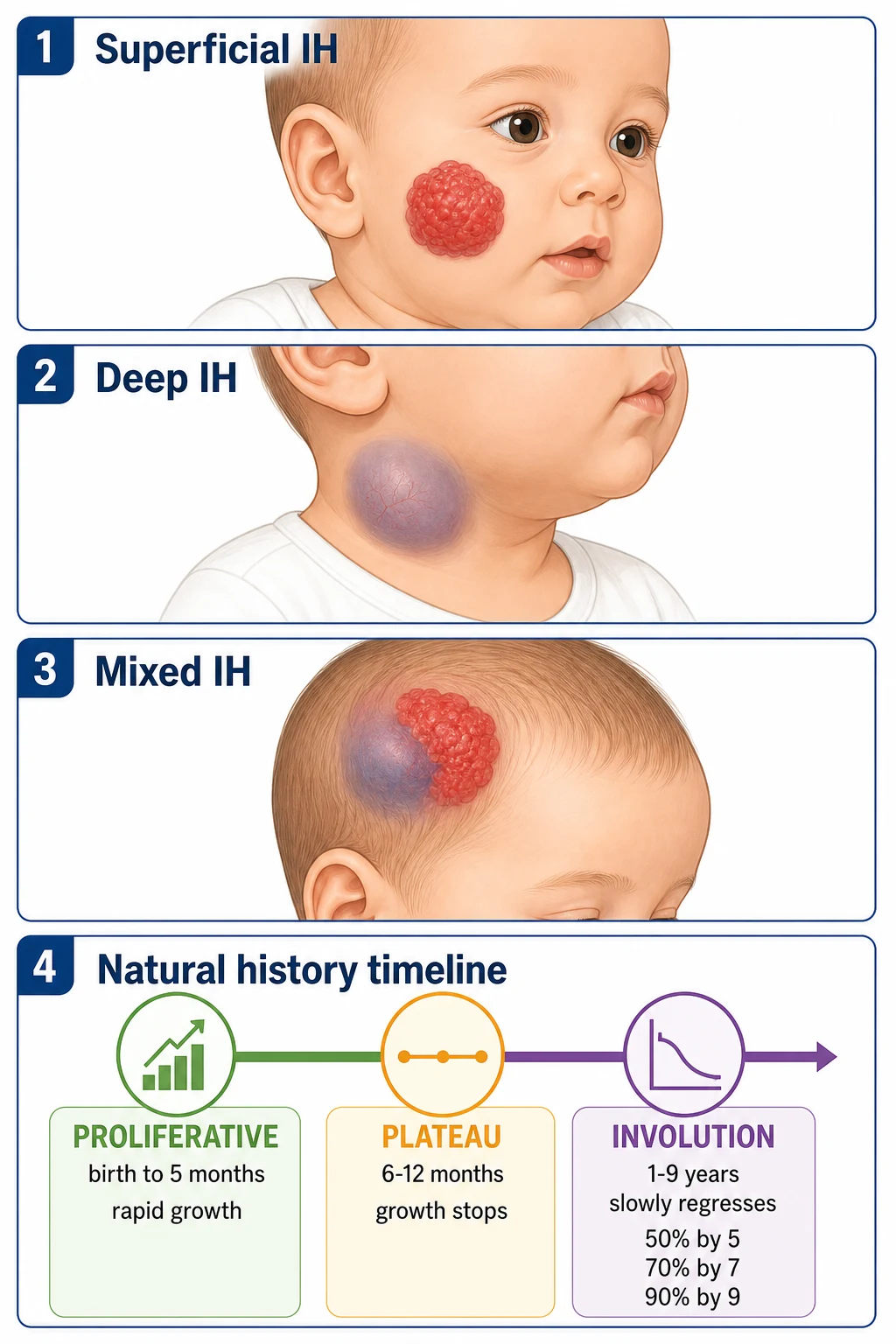
Clinical types:[1]
- Superficial — bright red, raised, lobulated "strawberry" plaque.
- Deep — subcutaneous blue-purple mass with overlying telangiectasia or bluish hue.
- Mixed — both superficial and deep components.
Epidemiology
- Most common tumour of infancy; ~4-5% of infants; female:male 2-3:1; higher in premature/low-birth-weight infants, multiple gestations, white non-Hispanic.[1][2]
- ~80% are solitary; ~20% are multiple.[2]
Natural History (the defining feature)
[1]Quick numbers for the examiner
The natural history has three phases:[1][6]
- Proliferative phase (0-5/9 months): rapid growth; 80% of final size reached by 5 months; most growth in the first 3 months.
- Plateau phase (6-12 months): growth stops; lesion stabilises.
- Involution phase (1-9+ years): slow spontaneous regression; ~50% resolved by age 5, ~70% by 7, ~90% by 9; may leave residual skin changes (atrophy, telangiectasia, fibrofatty tissue, anetoderma, hypopigmentation).[1]
IH is usually absent at birth or presents as a precursor lesion (pale telangiectatic or bruised macule). The growth after birth distinguishes IH from congenital haemangiomas (RICH/NICH/PICH), which are fully formed at birth and either rapidly involute (RICH — Rapidly Involuting Congenital Haemangioma), never involute (NICH — Non-Involuting), or partially involute (PICH).[4][5]
Pathophysiology
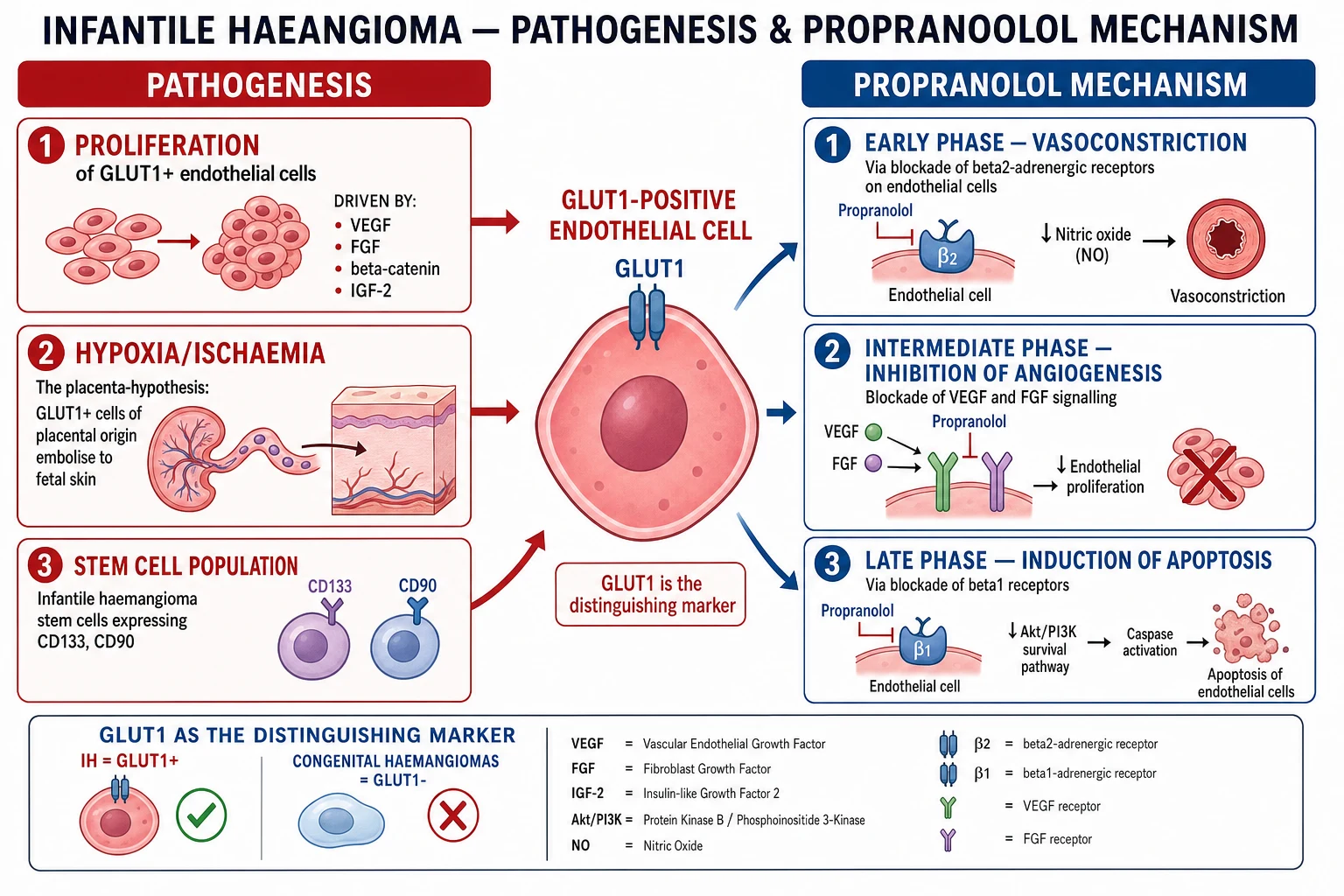
- IH arises from GLUT1-positive endothelial cells — likely of placental origin (the "placental hypoxia hypothesis").[4]
- During proliferation, endothelial cells are driven by VEGF, FGF-2, β-catenin, IGF-2 and an infantile haemangioma stem cell population (CD133+, CD90+).[4]
- During involution, endothelial cells are replaced by fibrofatty tissue (apoptosis-mediated).[1]
Propranolol mechanism (three phases):[3]
- Early (hours-days): vasoconstriction — blockade of β2-adrenergic receptors on endothelial cells → reduced nitric oxide → vasoconstriction (visible flattening/softening within 24-48 hours).
- Intermediate (weeks): inhibition of angiogenesis — blockade of VEGF and FGF signalling → reduced endothelial proliferation.
- Late (months): induction of apoptosis — blockade of β1 receptors → reduced Akt/PI3K survival signalling → caspase-mediated apoptosis of endothelial cells.
Clinical Presentation & Complications
Most IH are uncomplicated and located on the head and neck (~60%), trunk, or limbs. The key task is identifying high-risk features:[1][3]
| Complication | Clinical scenario |
|---|---|
| Ulceration (MOST COMMON ~16%) | Painful; sites: perineum, lip, neck, intertriginous; risk of infection, bleeding, scarring |
| Visual obstruction | Periocular IH → amblyopia, astigmatism (urgent ophthalmology + propranolol) |
| Airway obstruction | Subglottic/beard distribution IH → biphasic stridor at 4-8 weeks (urgent propranolol ± bronchoscopy) |
| High-output cardiac failure | Hepatic haemangiomatosis (multiple cutaneous → screen liver with ultrasound) |
| Disfigurement | Facial IH — psychosocial impact |
| Bleeding | Usually minor self-limiting |
Associated syndromes
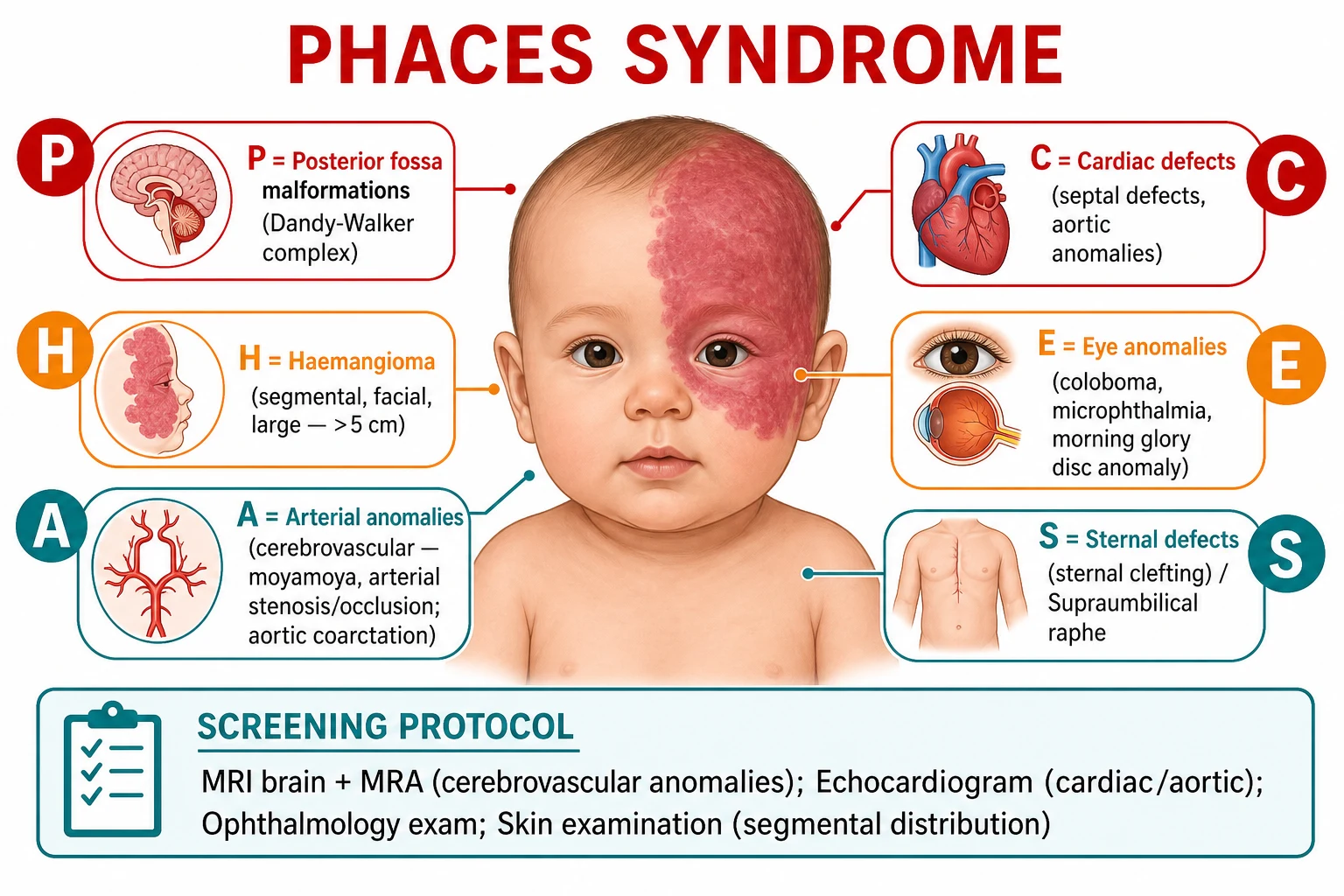
- PHACES syndrome: large segmental facial haemangioma (>5 cm) + Posterior fossa malformations (Dandy-Walker), Haemangioma, Arterial anomalies (cerebrovascular — moyamoya, stenosis; aortic coarctation), Cardiac defects, Eye anomalies (coloboma, microphthalmia), Sternal defects/supraumbilical raphe. Screen: MRI brain + MRA + echocardiogram + ophthalmology.[1][2]
- LUMBAR/PELVIS syndrome: lumbosacral haemangioma + urogenital/myelopathy/bony/anorectal/renal anomalies. Screen: spinal MRI (tethered cord).[1]
The Kasabach-Merritt trap
Kasabach-Merritt phenomenon (thrombocytopaenia + consumption coagulopathy + enlarging vascular lesion) is caused by kaposiform haemangioendothelioma (KHE) or tufted angioma — NOT by infantile haemangioma. This is a classic exam trap. IH does NOT cause Kasabach-Merritt.[4][5]
Differential Diagnosis
| Mimic | Distinguishing features |
|---|---|
| Congenital haemangioma (RICH/NICH/PICH) | Fully grown at birth; GLUT1-negative; RICH involutes rapidly, NICH never involutes |
| Vascular malformation (PWS, venous, lymphatic) | Present at birth; grows with child; never involutes; no proliferative phase |
| Pyogenic granuloma | Rapidly growing friable bleeding papule (children/adults); not present at birth |
| Tufted angioma / KHE | GLUT1-negative; may cause Kasabach-Merritt |
| Spitz naevus | Pink-red hairless papule; melanocytic; not vascular |
Investigations
- Clinical diagnosis — based on history (absent at birth → proliferates) and morphology.[1]
- Ultrasound (Doppler) — for deep/subcutaneous lesions or to distinguish from malformations.
- MRI — for large/deep lesions, suspected PHACES (brain/MRA), or LUMBAR (spine).
- GLUT1 immunostaining (biopsy) — confirms IH vs congenital haemangioma/malformation (rarely needed clinically).[4]
- Abdominal ultrasound — if >5 cutaneous IH (screen liver for hepatic haemangiomatosis).[3]
- FBC, coagulation — if Kasabach-Merritt suspected (though NOT caused by IH).
Management
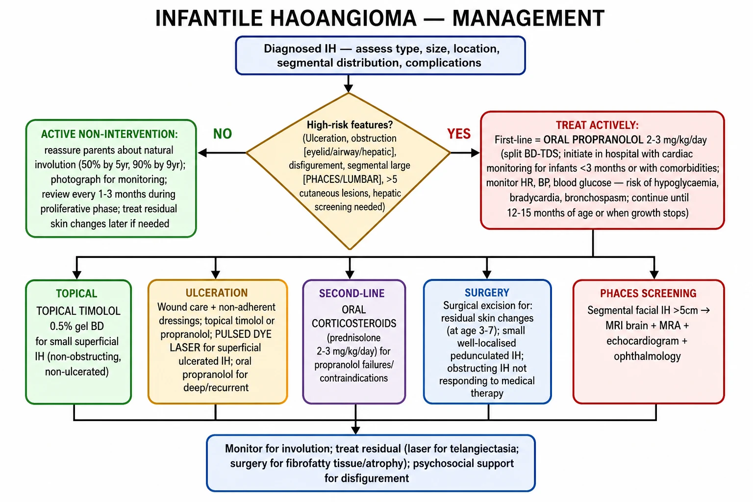
Active non-intervention (most IH)
- Reassure parents about natural involution (50% by age 5; 90% by 9).[1][6]
- Photograph for monitoring; review every 1-3 months during the proliferative phase.
- Treat residual skin changes (telangiectasia, fibrofatty tissue) later if needed.[3]
Oral propranolol (first-line systemic for high-risk IH)
- 2-3 mg/kg/day in divided doses (BD or TDS), continued until 12-15 months of age (or when growth stops).[3][6][15]
- Dose titration (AAP 2019): start at 0.5-1 mg/kg/day, increase by 0.5 mg/kg every 1-2 days up to target 2-3 mg/kg/day; PHACES infants titrate more slowly because of stroke risk during hypotension.[15]
- Pretreatment ECG is recommended (strong recommendation, AAP 2019); echocardiogram if history or examination suggest cardiac disease.[15]
- Initiate in hospital with cardiac monitoring for infants less than 3 months, with cardiac disease, or with airway involvement.[3]
- Monitoring: heart rate, blood pressure, blood glucose (risk of hypoglycaemia, especially with poor feeding), bronchospasm (especially in infants with respiratory conditions).[3]
- FDA-approved as Hemangeol (the first FDA-approved drug for IH; 4.28 mg/mL grape-flavoured oral solution, 2014).[6][15]
- Contraindications: severe asthma/bronchospasm, heart block, severe heart failure, hypoglycaemia risk.[3]
Topical timolol 0.5% gel
- For small, superficial, non-obstructing IH; applied BD.[3][15]
- Less effective than oral propranolol for deep or large lesions.[3]
- AAP 2019 weak recommendation; 47-88% improvement in published series; avoid on ulcerated IH or in preterm infants.[15][10]
Atenolol (off-label alternative)
- β1-selective non-selective β-blocker; starting dose 0.5 mg/kg/day PO once daily, titrated to 1 mg/kg/day OD.[16]
- Equivalent efficacy to propranolol in the 2014 RCT (Ábarzúa-Araya et al., JAAD); fewer sleep and bronchospasm events; useful in propranolol-intolerant infants.[16]
Corticosteroids (second-line / historical first-line)
- Prednisolone 2-3 mg/kg/day — historical first-line; now second-line for propranolol failures/contraindications.[3]
- Side effects: growth retardation, hypertension, immunosuppression, irritability.[3]
- AAP 2019 explicitly deprecates systemic corticosteroids as first-line for uncomplicated IH.[15]
Captopril and other investigational therapies
- Captopril 0.1 mg/kg/12h → up-titrate to 2 mg/kg/12h is a candidate third-line agent for infants intolerant of beta-blockers (severe asthma, bradycardia, prior propranolol failure).[17]
- Monitor renal function and serum potassium before and during therapy; check BP at each dose increase.[17]
- Sirolimus and vincristine are reserved for kaposiform haemangioendothelioma (KHE) and complicated vascular lesions — NOT used for uncomplicated IH.[15]
Surgery and laser
- Surgical excision for: residual skin changes (age 3-7); small pedunculated IH; obstructing IH not responding to medical therapy.[3]
- Pulsed dye laser (PDL) for ulcerated superficial IH and residual telangiectasia.[3]
- In infancy, surgery is uncommon because 70-90% of IH involute without scarring; PDL is the most common intervention for residual skin changes after involution.[1][3]
PHACES screening
Propranolol monitoring quick numbers
PHACE(L) - high-risk IH screening
Dandy-Walker, cerebellar hypoplasia; MRI brain + MRA needed for segmental facial IH
Segmental >5 cm facial IH (S3 distribution); 90% have PHACE if facial
Cerebellar, internal carotid, aortic; dysgenesis, aneurysms, stenosis; MRA needed
Coarctation, aortic arch abnormalities, VSD; echocardiogram needed
Persistent fetal vasculature, glaucoma, retinal vascular; ophthalmology
Lower body segmental IH; LUMBAR = Lower body IH + Urogenital anomalies + Myelopathy + Bony deformities + Anorectal malformations + Renal anomalies
Prognosis [1]
- Excellent for most IH — spontaneous involution; near-zero mortality in uncomplicated cases.[1]
- Residual skin changes (telangiectasia, atrophy, fibrofatty tissue, anetoderma) in ~20-40% even after complete involution; these may need laser or surgical correction.[1]
- Psychosocial impact of facial IH is significant — early treatment for disfigurement is justified.[2]
Evidence, Guidelines
- Léauté-Labrèze et al. (NEJM 2015) — randomized placebo-controlled trial (n=460) that established oral propranolol (3 mg/kg/day for 6 months) as standard of care; 60% of treated infants had complete/near-complete resolution vs 4% placebo.[7][8]
- Léauté-Labrèze et al. (NEJM 2008) — original serendipitous observation at Bordeaux Children's Hospital: two infants with IH and hypertrophic cardiomyopathy given propranolol showed dramatic IH regression within 24 hours.[8]
- AAP Clinical Practice Guideline (Krowchuk et al., Pediatrics 2019) — first US evidence-based guideline; recommends oral propranolol 2-3 mg/kg/day as first-line for high-risk IH; published jointly with AAP Section on Dermatology, Otolaryngology, and Plastic Surgery.[15]
- Ábarzúa-Araya et al. (JAAD 2014) — first randomized controlled trial comparing atenolol vs propranolol; equivalent efficacy, better sleep, less bronchospasm.[16]
- Gupta et al. (J Indian Assoc Pediatr Surg 2021) — captopril pilot study (n=18, dose 0.1→2 mg/kg/12h); "excellent response" in ~50%, candidate for propranolol-intolerant patients.[17]
- JAAD Part 1 (2021) and Part 2 (2021) (Rodríguez Bandera / Sebaratnam) — comprehensive two-part review covering assessment and management.[1][3]
- ISSVA classification (updated 2018) — the international standard for vascular anomaly classification.[5]
Clinical Trial Evidence & Pharmacology
The 2008 NEJM serendipitous discovery — Bordeaux, France

The propranolol era of IH management began not by design but by clinical serendipity. In 2007, Léauté-Labrèze and colleagues at Bordeaux University Hospital prescribed propranolol to two infants being treated for hypertrophic cardiomyopathy caused by a high-output pericardial lesion incidentally co-existing with severe facial IH. Within 24 hours of the first dose the haemangiomas visibly softened and darkened; at one week they had flattened substantially and changed colour from bright red to a dull purple. This observation led to a 2008 NEJM letter describing 11 additional infants treated with the same serendipitous regimen — all improved, none required corticosteroids, and surgery was avoided in every case.[8] The discovery was so remarkable because at that time systemic corticosteroids were the only evidence-based option, with notoriously poor side-effect profiles (irritability, adrenal suppression, immunosuppression, growth failure). Propranolol rapidly displaced them and has remained first-line ever since.
The 2015 NEJM randomized placebo-controlled trial
The evidence that converted propranolol from anecdotal to standard-of-care came from the Léauté-Labrèze 2015 NEJM trial (NCT01445741), a multicentre European randomized placebo-controlled study of 460 infants aged 1-5 months at treatment initiation. Infants were randomized to one of four arms:[7]
- Placebo (n=55)
- Propranolol 1 mg/kg/day (n=100)
- Propranolol 3 mg/kg/day (n=100)
- Propranolol 0.5 mg/kg/day (not used in pooled efficacy analysis) [1]
Primary outcome: complete or near-complete resolution of the target IH at week 24 (end of treatment). [1]
Key results: [1]
| Endpoint | Placebo | Propranolol 1 mg/kg/day | Propranolol 3 mg/kg/day |
|---|---|---|---|
| Complete or near-complete resolution | 4% | 38% | 60% |
| Any improvement (any level) | 38% | 92% | 96% |
| Required rescue therapy | 24% | 6% | 2% |
| Treatment-related serious AE | 0% | 0% | 1% (hypoglycaemia) |
The 3 mg/kg/day dose was superior on every efficacy endpoint and had a comparable safety profile to 1 mg/kg/day — most adverse events (sleep disturbance, diarrhoea, bronchiolitis) were mild and self-limiting. The single serious adverse event (hypoglycaemia) reinforced the first-dose-in-clinic recommendation that has become standard. The trial led to FDA approval of propranolol hydrochloride oral solution (Hemangeol, Pierre Fabre Dermatologie) in March 2014, and to NICE guideline approval in the UK. The dose tested (3 mg/kg/day) remains the registered target dose globally.[7][6]
AAP 2019 Clinical Practice Guideline
In January 2019, the American Academy of Pediatrics published the first formal US Clinical Practice Guideline for IH management, developed by a 15-member multidisciplinary subcommittee led by Krowchuk and Frieden.[15] Key recommendations:
- High-risk IH must be treated early — propranolol initiation is recommended as soon as possible after recognition, ideally during the early proliferative phase (1 month of age), when response is greatest.
- Oral propranolol 2-3 mg/kg/day is the recommended first-line systemic therapy (strong recommendation; high-quality evidence).
- Pretreatment screening: history, physical exam; ECG prior to initiation (strong recommendation); echocardiogram if history/physical suggests cardiac disease; consider outpatient initiation in infants younger than eight weeks corrected age, those with comorbidities, or any infant whose home monitoring is uncertain.
- Dose escalation: start at 0.5-1 mg/kg/day divided BID or TDS, increase by 0.5 mg/kg/day every few days up to a target of 2-3 mg/kg/day, monitoring heart rate and blood pressure at each titration step.
- Treatment duration: continue through the proliferative phase — typically until 8-12 months of age, or longer if rebound occurs.
- Topical timolol 0.5% gel-forming solution: recommended as first-line for small, superficial, non-complicated IH (weak recommendation; moderate-quality evidence).
- Special situations:
- PHACES syndrome: neuroimaging (MRI/MRA) and echocardiogram before propranolol initiation; some authorities recommend lower starting dose (0.5 mg/kg/day) and slower titration because of stroke risk during periods of hypotension.
- Airway IH: escalate to 3 mg/kg/day quickly; combine with ENT/anaesthesia consult.
- Periocular IH: urgent ophthalmology + propranolol; consider intralesional or systemic corticosteroid bridge while propranolol ramps up (less common now that propranolol is fast-acting). [1]
The guideline explicitly deprecates systemic corticosteroids as first-line for uncomplicated IH and reserves vincristine and sirolimus for kaposiform haemangioendothelioma (KHE) and other complicated vascular lesions. It does not yet recommend atenolol or captopril as first-line, reflecting the lower level of evidence — see below.[15]
Propranolol trial-evidence quick numbers
Propranolol pharmacology and dosing
Mechanism of action (three sequential phases — vasoconstriction, anti-angiogenesis, apoptosis):[3][4]
- Early (hours-days): β2-adrenergic blockade → reduced nitric oxide → vasoconstriction. This causes the visible flattening and softening seen within 24-48 hours of the first dose.
- Intermediate (days-weeks): inhibition of VEGF-A, FGF-2, and MMP-2/9 secretion → reduced endothelial proliferation and angiogenesis. This accounts for the slowed growth during the proliferative phase.
- Late (weeks-months): β1-adrenergic blockade → inhibition of the Akt/PI3K/mTOR survival pathway → caspase-mediated apoptosis of capillary endothelial cells. This produces the long-term regression. [1]
Pharmacokinetics in infants: oral bioavailability ~25-35% (lower than adults because of first-pass hepatic metabolism); peak plasma levels 1-4 hours after dosing; elimination half-life 3-6 hours in infants under 6 months of age, increasing to ~10 hours by age 12 months. Hemangeol oral solution 4.28 mg/mL is the FDA-approved pediatric formulation (grape-flavoured; given BD or TDS with feeds).[6]
Dose regimens in clinical practice (consistent across AAP 2019 and NEJM 2015):[3][7][15]
| Patient profile | Starting dose | Titration | Target dose |
|---|---|---|---|
| Standard infant ≥1 month, healthy | 0.5-1 mg/kg/day | Increase by 0.5 mg/kg every 1-2 days | 2-3 mg/kg/day |
| Preterm (corrected age younger than one month) | 0.25-0.5 mg/kg/day | Slow titration over 2-4 weeks | 2 mg/kg/day |
| PHACES syndrome | 0.5 mg/kg/day | Increase over 2-4 weeks; MRI/A first | 1-2 mg/kg/day |
| History of hypoglycaemia / poor feeding | 0.5 mg/kg/day | Slower titration; give with feeds | 2 mg/kg/day |
Monitoring protocol: pre-treatment ECG (baseline) ± echocardiogram; first dose in clinical setting with heart-rate and blood-pressure monitoring for 2 hours; subsequent outpatient titration as tolerated. Treat for 6-12 months minimum, usually until 12-15 months chronological age. Monitor for sleep disturbance, cold extremities, irritability, hypoglycaemia (especially during intercurrent illness or poor feeding — hold dose if vomiting or fasting for any reason).[9][14][15]
Atenolol — the β1-selective alternative
Because non-selective β-blockade (propranolol) carries a small risk of bronchospasm, hypoglycaemia, and sleep disturbance (due to β2 cross-reactivity), β1-selective blockers have been investigated as alternatives. The seminal trial was Ábarzúa-Araya et al. (JAAD 2014) — a randomized controlled study of atenolol (n=13) vs propranolol (n=13) at 1 mg/kg/day for 6 months in infants aged 4-8 months with proliferating IH.[16]
Key findings: [1]
- Equivalent efficacy: response was "good or excellent" in 100% of both groups by week 24.
- Better side-effect profile: atenolol-treated infants had statistically fewer sleep disturbances (p less than 0.05) and no bronchospasm episodes.
- No rebound in either group at 12-week follow-up after cessation. [1]
Subsequent systematic reviews and network meta-analyses (e.g., Fei et al., EClinicalMedicine 2020) have confirmed atenolol as non-inferior to propranolol for superficial and mixed IH, with a more favourable respiratory and sleep side-effect profile. Atenolol dosing mirrors propranolol: start at 0.5 mg/kg/day PO once daily (atenolol is dosed once daily in children due to longer half-life), titrate to 1 mg/kg/day. Atenolol is not FDA-approved for IH and remains off-label, but is widely used in Europe and some US centres for propranolol-intolerant infants.[16]
Captopril — the ACE-inhibitor alternative
Building on laboratory evidence that the renin-angiotensin system drives IH angiogenesis (angiotensin II up-regulates VEGF; ACE inhibition down-regulates VEGF), pediatric surgeons at King George's Medical University, Lucknow, India conducted a prospective pilot study of captopril monotherapy in 18 children with IH (Gupta et al., J Indian Assoc Pediatr Surg 2021).[17]
Regimen: oral captopril starting at 0.1 mg/kg every 12 hours, gradually up-titrated every 1-2 days to a maximum of 2 mg/kg every 12 hours (i.e., target 4 mg/kg/day), with BP, electrolytes and renal function monitored at each dose increase. Treatment continued for 16-18 months (until plateau/involution). [1]
Outcomes: [1]
- 9/18 (50%) — excellent response (near-complete regression).
- 4/18 (22%) — good response (>50% reduction).
- 5/18 required conversion to propranolol or corticosteroids (one propranolol allergy).
- No serious adverse events (no symptomatic hypotension, no hyperkalaemia, no acute kidney injury). [1]
Captopril is not currently recommended as first-line by AAP 2019 or NICE, but remains a candidate for infants who cannot tolerate any beta-blocker (severe asthma, bradycardia, prior propranolol failure). The pharmacologic rationale — partial overlap with propranolol's anti-angiogenic effect via VEGF down-regulation — supports further randomized trials. Lisinopril and enalapril are not yet studied in IH. Always check baseline and follow-up renal function and serum potassium before and during ACE-inhibitor therapy.[17]
Topical timolol — for small superficial IH
Topical timolol 0.5% gel-forming solution (1 drop, twice daily, applied to the lesion — not surrounding skin) is a non-systemic option for small (typically under one centimetre), thin, superficial IH that are not causing functional or cosmetic concern. Pharmacokinetic studies show systemic absorption is negligible (peak plasma timolol below one ng/mL), making it attractive for clinic-first treatment in suitable patients. A 2016 review of seventy-three reports (n greater than one thousand patients) found 47-88% improvement with topical timolol; the strongest effect was in superficial IH under five millimetres in depth.[10] Avoid in ulcerated IH (rapid systemic absorption through denuded surface), in preterm infants, and in any infant with asthma/heart block. Usually avoid in infants under one month corrected age.[3][15]
[1]IH-EMBOLIC - complications of IH
Common in ulcerated IH; S. aureus, Strep; oral flucloxacillin
Most IH do NOT bleed significantly; treat with pressure; cautery rarely needed
Periorbital IH threatens vision; propranolol mandatory; urgent ophthalmology
Biphasic stridor in infant; biphasic stridor; emergency ENT; propranolol + airway management
Pulsed dye laser; topical timolol; propranolol; consider tranexamic acid
Subglottic IH - emergency; subglottic stenosis risk; tracheostomy if severe
5+ cutaneous IH = screen for hepatic; diffuse IH causes high-output cardiac failure
Ulcerated perioral IH prevents feeding; propranolol + topical timolol + dressing
Facial IH causes psychosocial impact; treat early with propranolol for best cosmetic outcome
Prevention [1]
- No primary prevention. Early identification of high-risk features (obstruction, ulceration, PHACES) is the key to preventing complications.[1]
Exam Pearls
[1]Red Flags
Exam application bank (NEET-PG / INICET)
One-line answer
Infantile haemangioma (IH) is the most common TUMOUR of infancy — a benign vascular neoplasm (GLUT1-positive) that undergoes a characteristic natural history of proliferation (rapid growth in the first 3-5 months) followed by slow involution (50% resolved by age 5, 90% by 9). It is distinct from congenital haemangiomas (RICH/NICH/PICH — GLUT1-negative, fully grown at birth). ISSVA classifies IH as a vascular tumour, separate from vascular malformations (PWS, venous, lymphatic). Most IH require no treatment (active non-intervention); for high-risk lesions (ulceration, obstruction, disfigurement, segmental distribution), oral propranolol (2-3 mg/kg/day) has revolutionised management as the first-line systemic agent (FDA-approved as Hemangeol). Segmental facial lesions >5 cm warrant screening for PHACES syndrome (MRI brain/MRA + echocardiogram + ophthalmology). Fellowship-level assessment d [1]
Worked stems (answer without another resource)
Stem 1 — Classic presentation. Map symptoms to mechanism; name the first investigation and first treatment step with dose/route if drug therapy is standard. [1]
Stem 2 — Unstable / complicated. List red flags that force immediate resuscitation, theatre, ICU, antidote, or reperfusion — and what you do in the first 15 minutes. [1]
Stem 3 — Atypical group. Elderly, pregnancy, child, or immunocompromised: how presentation and thresholds change. [1]
Stem 4 — Differential trap. Name the three closest mimics and one discriminator for each. [1]
Stem 5 — Disposition. Who goes home with safety-netting, who is admitted, who needs HDU/ICU/theatre, and what follow-up is mandatory. [1]
Rapid viva checklist
- Definition + classification
- Pathophysiology chain
- Bedside signs / criteria
- Score with exact components (if any)
- Emergency bundle
- Definitive therapy with doses
- Complications of disease and of treatment
- Special populations
- Guideline/trial name if classic
- Three exam traps
Coverage self-check
If you cannot answer any stem above from this page alone, re-read the matching section — the page is intended to be self-sufficient for final-prof and NEET-PG/INICET questions on Infantile haemangioma.
[1]References
- [1]Rodríguez Bandera AI, Sebaratnam DF, Wargon O, et al. Infantile hemangioma. Part 1: Epidemiology, pathogenesis, clinical presentation and assessment J Am Acad Dermatol, 2021.PMID 34419524
- [2]Leung AKC, Lam JM, Leong KF, et al. Infantile Hemangioma: An Updated Review Curr Pediatr Rev, 2021.PMID 32384034
- [3]Sebaratnam DF, Rodríguez Bandera AL, Wong LF, et al. Infantile hemangioma. Part 2: Management J Am Acad Dermatol, 2021.PMID 34419523
- [4]Holm A, Mulliken JB, Bischoff J. Infantile hemangioma: the common and enigmatic vascular tumor J Clin Invest, 2024.PMID 38618963
- [5]Kunimoto K, Yamamoto Y, Jinnin M. ISSVA Classification of Vascular Anomalies and Molecular Biology Int J Mol Sci, 2022.PMID 35216474
- [6]Léauté-Labrèze C, Harper JI, Hoeger PH. Infantile haemangioma Lancet, 2017.PMID 28089471
- [7]Léauté-Labrèze C, Hoeger P, Mazereeuw-Hautier J, et al. Oral Propranolol for Infantile Hemangioma N Engl J Med, 2015.PMID 26176392
- [8]Léauté-Labrèze C, Dumas de la Roque E, Hubiche T, et al. Propranolol for severe hemangiomas of infancy N Engl J Med, 2008.PMID 18550886
- [9]Starkey E, Shahidullah H. Propranolol for infantile haemangiomas: review of report of a consensus conference Arch Dis Child Educ Pract Ed, 2014.PMID 24242336
- [10]Painter SL, Hildebrand GD. Review of topical beta blockers as treatment for infantile hemangiomas Surv Ophthalmol, 2016.PMID 26408055
- [11]Cheng J, et al. Clinical characteristics, treatment outcomes and prognostic factors of ulcerated infantile hemangioma: 15 years of experience from a pediatric dermatology center in Hong Kong J Dermatolog Treat, 2025.PMID 41221590
- [12]Maguiness SM, Hoffman WY, McCalmont TH, Frieden IJ. Early white discoloration of infantile hemangioma: a sign of impending ulceration Arch Dermatol, 2010.PMID 21079059
- [13]American College of Radiology ACR Appropriateness Criteria Committee. ACR Appropriateness Criteria® Soft Tissue Vascular Anomalies: Vascular Malformations and Infantile Vascular Tumors (Non-CNS)-Child J Am Coll Radiol, 2024.PMID 38823953
- [14]Püttgen KB, et al. Outpatient use of oral propranolol and topical timolol for infantile hemangiomas: survey results and comparison with propranolol consensus statement guidelines Pediatr Dermatol, 2015.PMID 25556828
- [15]Krowchuk DP, Frieden IJ, Mancini AJ, et al. (Subcommittee on the Management of Infantile Hemangiomas). Clinical Practice Guideline for the Management of Infantile Hemangiomas Pediatrics, 2019.PMID 30584062
- [16]Ábarzúa-Araya A, Navarrete-Dechent CP, Heusser F, Retamal J, Zegpi-Trueba MS. Atenolol versus propranolol for the treatment of infantile hemangiomas: a randomized controlled study J Am Acad Dermatol, 2014.PMID 24656727
- [17]Gupta A, Kureel SN, Pandey A, et al. Angiotensin-converting Enzyme Inhibitors: Can it be a Potential Treatment of Infantile Hemangioma J Indian Assoc Pediatr Surg, 2021.PMID 34385766