Dermatology · Medicine
Infantile seborrhoeic dermatitis / cradle cap
Also known as Cradle cap · Infantile seborrheic dermatitis · Pityriasis capitis infantum
Infantile seborrhoeic dermatitis (cradle cap) is a common early-life inflammatory dermatosis of sebum-rich sites with greasy yellow scale, minimal itch, and usual spontaneous improvement by 6–12 months. Exams test differentiation from infantile atopic and nappy dermatitis, Malassezia–sebum pathophysiology, emollient/scale-softening care, cautious low-potency steroid or topical imidazole use, Cochrane evidence limits, and red flags for immunodeficiency or Langerhans cell histiocytosis.
On this page & tools
Your progress
Saved locally on this device.
Exam tags
Red flags
Definition & Classification
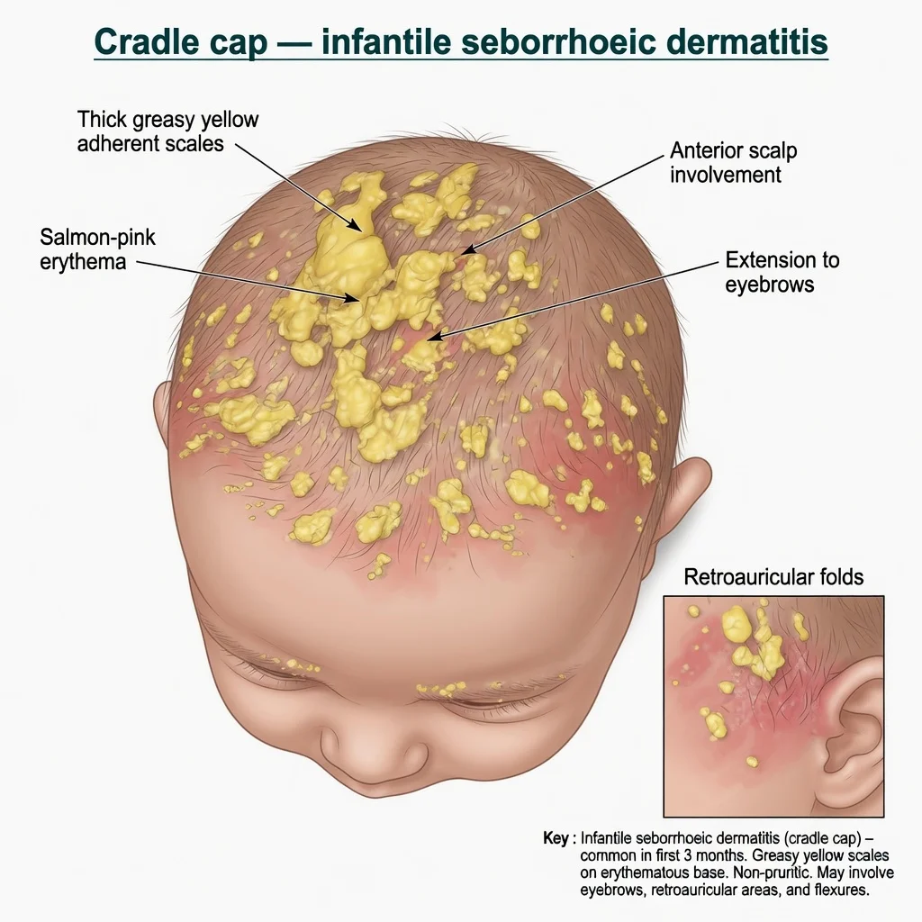
Infantile seborrhoeic dermatitis (ISD) is the early-life phenotype of seborrhoeic dermatitis, classically presenting as cradle cap — greasy yellow scale on the scalp of young infants — with or without involvement of other seborrhoeic and intertriginous sites.[3][5][6] It is related to adult seborrhoeic dermatitis but is typically more self-limited as infantile sebum production declines.[5][6]
Spectrum:
- Mild pityriasiform scalp scale
- Classic cradle cap
- Intertriginous/napkin seborrhoeic pattern with fold involvement [1]
Epidemiology
Onset is usually within the first 3 months of life; most infants improve by 6–12 months.[6][7][8] Global seborrhoeic dermatitis meta-analysis shows meaningful population prevalence across ages, with lower rates in neonates than older groups overall — ISD remains one of the common rashes of early infancy in clinical practice.[9] Infants are typically systemically well.
Pathophysiology
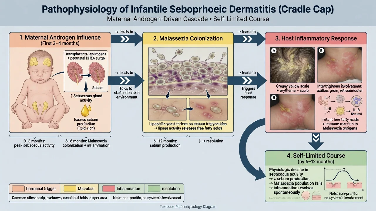
Infantile sebum production is influenced by maternal androgens and early postnatal endocrine milieu, creating a lipid-rich habitat for Malassezia yeasts.[5][10] Yeast lipase activity and host inflammatory responses to fungal products and free fatty acids generate erythema with greasy scale on scalp and flexures.[10] As sebaceous activity falls later in infancy, the ecological drive often wanes and disease remits.[6]
Clinical Presentation
- Scalp: thick, greasy, yellow, adherent scales; salmon-pink base; vertex and anterior scalp common.[6][7]
- Face: eyebrows, glabella, nasolabial folds, retroauricular creases.
- Body: neck folds, axillae, inguinal/napkin folds involved (contrast irritant nappy dermatitis).
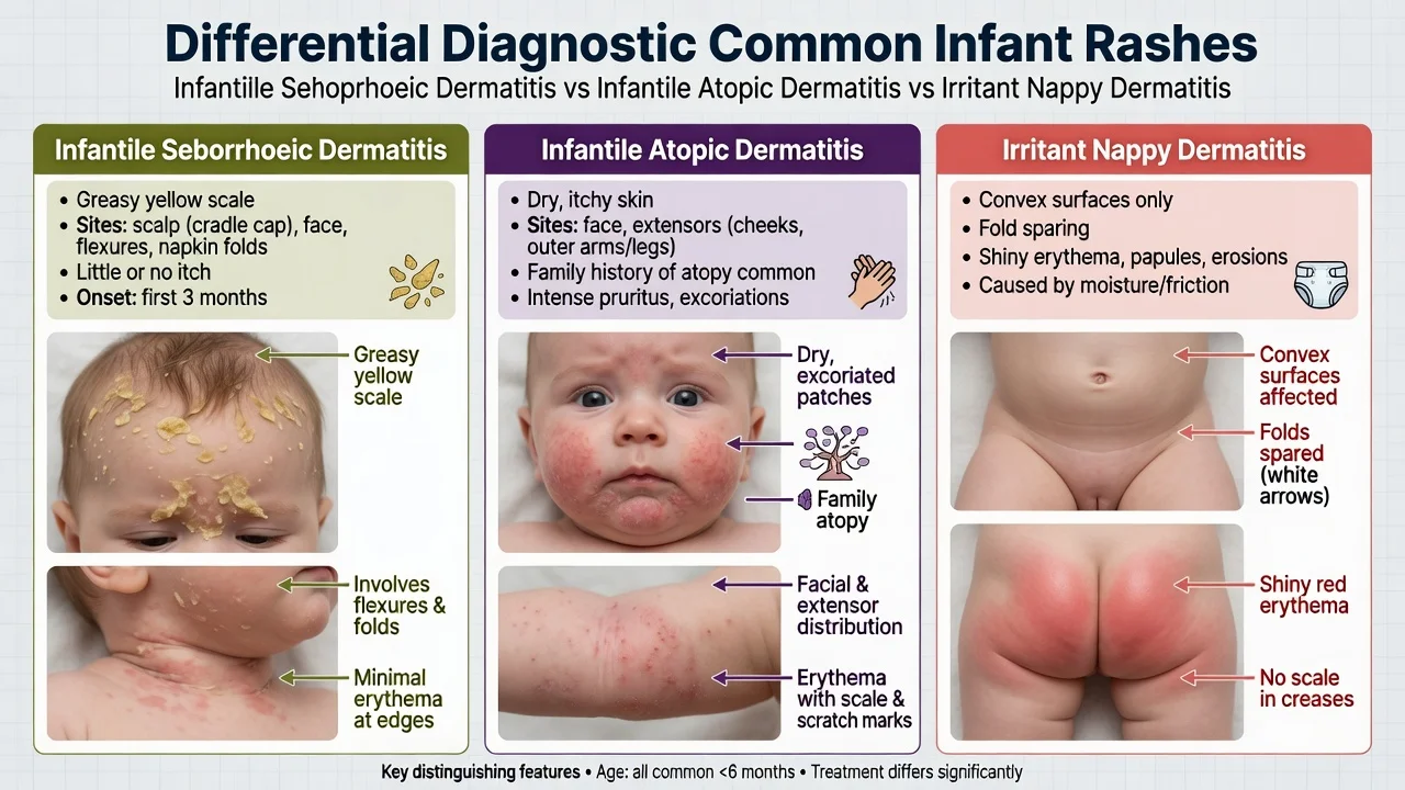
- Itch minimal or absent — infant usually comfortable relative to AD.[6][8]
- Feeding, growth and behaviour typically normal.
Differential Diagnosis
| Diagnosis | Distinguishing feature |
|---|---|
| Infantile AD | Itch, dry scale, facial/extensor, sleep loss, family atopy |
| Irritant nappy dermatitis | Convex surfaces; folds spared |
| Candidal napkin rash | Bright red folds + satellite pustules |
| Psoriasis | Sharper plaques; may persist; family history |
| Tinea | Uncommon scalp pattern in neonates; KOH if atypical |
| Langerhans cell histiocytosis | Persistent erosive/purpuric seborrhoeic-like rash; systemic signs |
| Immunodeficiency-associated rash | FTT, diarrhoea, infections, refractory severe SD |
Bedside Assessment & Investigations
Diagnosis is clinical in typical well infants.[3][6] Examine all seborrhoeic sites, growth chart, and infection history. Investigations are selective: swab if superinfection; broader work-up if red flags for immunodeficiency or LCH.[7][8]
Management — First Line
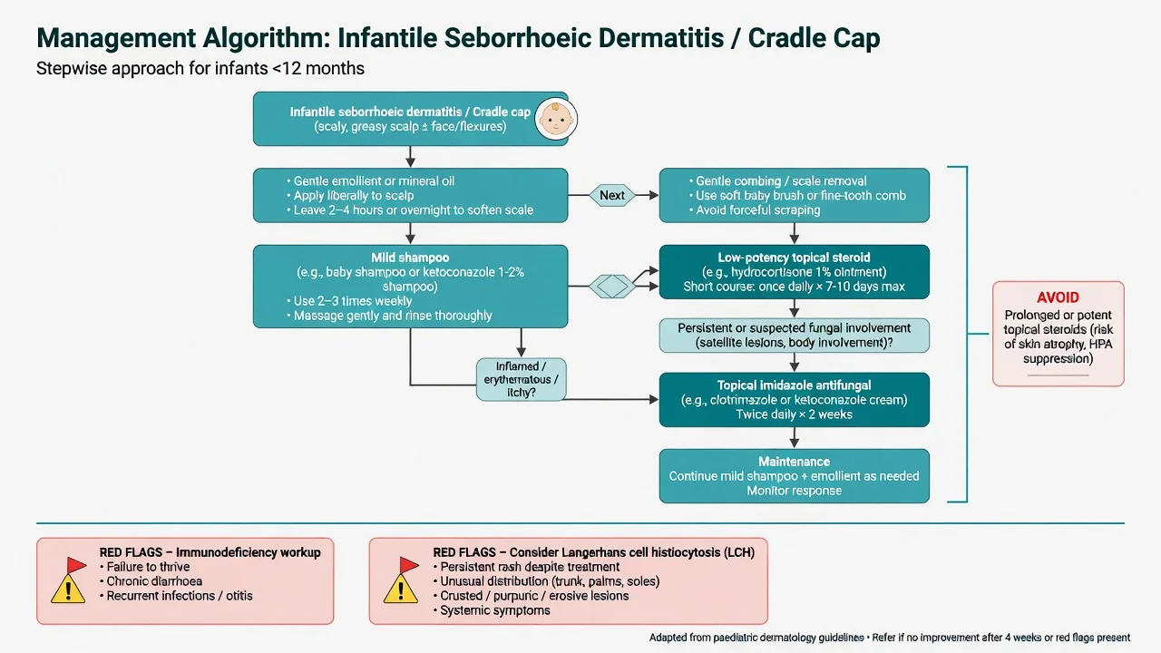
Practical ladder used internationally: [1]
- Reassure — usually self-limited.[6]
- Soften scale with emollient or mineral/vegetable oil; leave on, then gently comb; never force scrape.[6][7]
- Mild shampoo regimens for scalp.
- Maintain skin hygiene in folds; barrier care if napkin area involved.
Cochrane 2019 found limited high-quality evidence for specific active interventions in ISD — counselling and gentle care remain foundational; treat inflammation/yeast when clinically needed without overstating certainty.[1][2]
Management — Medicated Topicals
- Short course low-potency topical corticosteroid (e.g. hydrocortisone class teaching) for inflamed scalp/skin when scale care alone is insufficient.[3][4]
- Topical imidazole antifungals (e.g. clotrimazole/ketoconazole cream teaching) when yeast-driven or persistent intertriginous disease is suspected.[3][4]
- Avoid prolonged potent steroids on infant scalp/face — atrophy and HPA suppression risk.[3]
- Adult innovations (e.g. roflumilast foam) are not the standard cradle-cap pathway for young infants.
Complications & Pitfalls
- Secondary bacterial or candidal infection.
- Steroid misuse by caregivers.
- Misdiagnosis of AD leading to under-treatment of itch, or mislabel of LCH as stubborn cradle cap.
- Parental distress out of proportion to medical risk — address with clear prognosis. [1]
Special Populations & Red Flags
Immunodeficiency: severe, widespread, refractory seborrhoeic-like dermatitis with failure to thrive, diarrhoea or recurrent infections warrants immunology/paediatric work-up (exam classic includes entities such as leucocyte adhesion deficiency in broader differentials of severe infantile dermatitis).[5][8]
LCH: seborrhoeic-like eruption that is erosive, purpuric, or treatment-resistant with systemic features — biopsy pathway.
Overlap: some infants evolve toward atopic phenotypes; reassess if itch dominates.
Prognosis & Follow-Up
Most improve within the first year.[6] Infantile disease does not reliably predict adult seborrhoeic dermatitis. Re-review if not improving, if itch escalates (consider AD), or if red flags appear.
Evidence, Guidelines & Regional Notes
Exam anchors: Cochrane ISD review,[1] JAAD Cochrane summary,[2] AFP diagnosis/treatment overviews,[3][7] comprehensive treatment review,[4] classic JEADV SD review,[5] cradle cap clinical reviews,[6][8] global prevalence meta-analysis,[9] and Malassezia biology update.[10] ANZ/UK/India practice converges on gentle scale care first; medicated topicals second; systemic therapy almost never for uncomplicated cradle cap.
Exam Pearls
Red Flags
Exam application bank (NEET-PG / INICET)
One-line answer
Infantile seborrhoeic dermatitis (cradle cap) is a common early-life inflammatory dermatosis of sebum-rich sites with greasy yellow scale, minimal itch, and usual spontaneous improvement by 6–12 months. Exams test differentiation from infantile atopic and nappy dermatitis, Malassezia–sebum pathophysiology, emollient/scale-softening care, cautious low-potency steroid or topical imidazole use, Cochrane evidence limits, and red flags for immunodeficiency or Langerhans cell histiocytosis.
Worked stems (answer without another resource)
Stem 1 — Classic presentation. Map symptoms to mechanism; name the first investigation and first treatment step with dose/route if drug therapy is standard. [1]
Stem 2 — Unstable / complicated. List red flags that force immediate resuscitation, theatre, ICU, antidote, or reperfusion — and what you do in the first 15 minutes. [1]
Stem 3 — Atypical group. Elderly, pregnancy, child, or immunocompromised: how presentation and thresholds change. [1]
Stem 4 — Differential trap. Name the three closest mimics and one discriminator for each. [1]
Stem 5 — Disposition. Who goes home with safety-netting, who is admitted, who needs HDU/ICU/theatre, and what follow-up is mandatory. [1]
Rapid viva checklist
- Definition + classification
- Pathophysiology chain
- Bedside signs / criteria
- Score with exact components (if any)
- Emergency bundle
- Definitive therapy with doses
- Complications of disease and of treatment
- Special populations
- Guideline/trial name if classic
- Three exam traps
Coverage self-check
If you cannot answer any stem above from this page alone, re-read the matching section — the page is intended to be self-sufficient for final-prof and NEET-PG/INICET questions on Infantile seborrhoeic dermatitis / cradle cap.
Expanded exam teaching (depth pass)
Clinical reasoning
For Infantile seborrhoeic dermatitis / cradle cap, examiners test whether you can prioritise life threats, choose the right first test, and give specific therapy (agent, dose, route, timing). Generic phrases without numbers score poorly.
Mechanism → feature map
Build a short chain: cause → pathophysiologic intermediate → clinical feature → complication. Every major symptom in the classic vignette should sit on that chain.
Investigation strategy
- Bedside/first-line tests that change immediate management
- Confirmatory or staging tests
- What a normal result does not exclude
- When not to delay treatment for imaging (unstable patient)
Management ladder
- Resuscitation / ABC / sepsis or haemorrhage bundle as relevant
- Specific antidote / procedure / antimicrobial / reperfusion / surgery
- Supportive care and monitoring targets
- Definitive long-term therapy and secondary prevention
- Disposition and safety-net advice
Special populations
Always prepare one line each for children, pregnancy, elderly, renal/hepatic impairment, and immunocompromised patients when the topic allows.
Pitfalls that fail candidates
- Treating the number not the patient
- Missing pregnancy status when relevant
- Imaging before stabilisation
- Wrong empiric cover or wrong antidote timing
- Incomplete counselling on recurrence, adherence, or red-flag return
Infantile seborrhoeic dermatitis (cradle cap) is a common early-life inflammatory dermatosis of sebum-rich sites with greasy yellow scale, minimal itch, and usual spontaneous improvement by 6–12 months. Exams test differentiation from infantile atopic and nappy dermatitis, Malassezia–sebum pathophysiology, emollient/scale-softening care, cautious low-potency steroid or topical imidazole use, Cochrane evidence limits, and red flags for immunodeficiency or Langerhans cell histiocytosis. [1]
Structured revision sheet
Must-know numbers and names
List every score, size threshold, dose, and time window from this topic on a blank page from memory, then check against the sections above.
Three classic MCQ angles
- Most likely diagnosis given a vignette
- Next best step in management
- Most appropriate investigation
Three classic SAQ angles
- Pathophysiology in five steps
- Management algorithm with doses
- Complications and prevention
Clinical station flow
Greet → focused history → targeted exam → investigations → explain diagnosis → emergency care → definitive plan → safety-net / follow-up → answer examiner questions on mechanism and pitfalls.
[1]Exam triad
ACT
Define the problem and red flags
Highest-yield test or bedside clue
First-line management and follow-up
References
- [1]Victoire A, Magin P, Coughlan J, et al. Interventions for infantile seborrhoeic dermatitis (including cradle cap) Cochrane Database Syst Rev, 2019.PMID 30828791
- [2]Hassan S, Szeto MD, Sivesind TE, et al. From the Cochrane Library: Interventions for infantile seborrheic dermatitis (including cradle cap) J Am Acad Dermatol, 2022.PMID 34571061
- [3]Clark GW, Pope SM, Jaboori KA. Diagnosis and treatment of seborrheic dermatitis Am Fam Physician, 2015.PMID 25822272
- [4]Borda LJ, Perper M, Keri JE. Treatment of seborrheic dermatitis: a comprehensive review J Dermatolog Treat, 2019.PMID 29737895
- [5]Gupta AK, Bluhm R. Seborrheic dermatitis J Eur Acad Dermatol Venereol, 2004.PMID 14678527
- [6]Nobles T, Harberger S, Krishnamurthy K. Cradle Cap(Archived) 2026.PMID 30285358
- [7]O'Connor NR, McLaughlin MR, Ham P. Newborn skin: Part I. Common rashes Am Fam Physician, 2008.PMID 18236822
- [8]Chadha A, Jahnke M. Common Neonatal Rashes Pediatr Ann, 2019.PMID 30653638
- [9]Polaskey MT, Chang CH, Daftary K, et al. The Global Prevalence of Seborrheic Dermatitis: A Systematic Review and Meta-Analysis JAMA Dermatol, 2024.PMID 38958996
- [10]Adalsteinsson JA, Kaushik S, Muzumdar S, et al. An update on the microbiology, immunology and genetics of seborrheic dermatitis Exp Dermatol, 2020.PMID 32125725