Dermatology · Medicine
Inflammatory dermatopathology patterns
Also known as Reaction patterns skin pathology · Ackerman pattern analysis · Spongiotic interface lichenoid pathology · Inflammatory skin biopsy interpretation
Board-level pattern-based approach to inflammatory skin disease histopathology. Covers Ackerman-style algorithmic diagnosis, major reaction patterns (spongiotic, psoriasiform, interface/lichenoid, bullous, vasculopathic, granulomatous, folliculitic, panniculitic), biopsy and DIF strategy, special stains, clinicopathologic correlation, and how reports change management without overcalling lymphoma or missing infection.
On this page & tools
Your progress
Saved locally on this device.
Exam tags
Red flags
Definition & Method
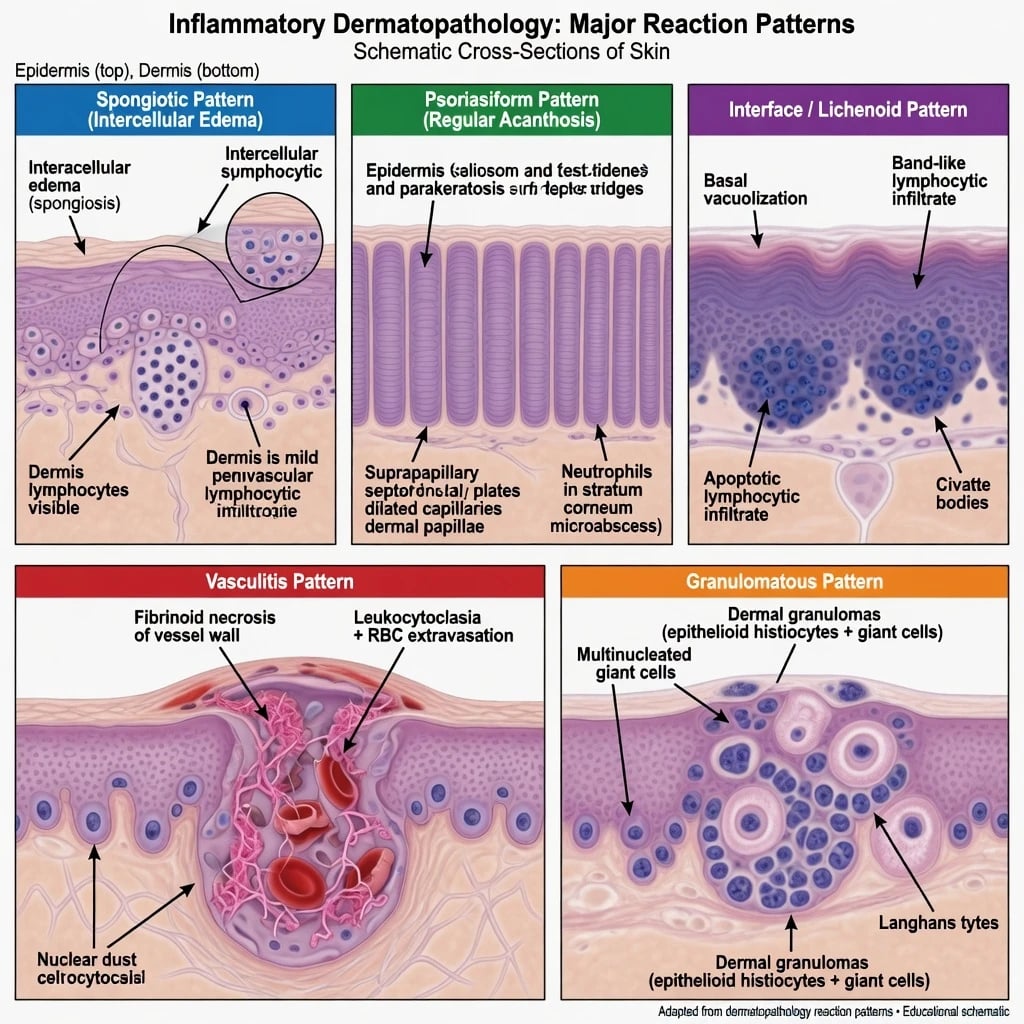
Inflammatory dermatopathology interprets non-neoplastic skin disease by reaction pattern rather than by guessing a clinical name from a single microscopic clue.[1][3] Ackerman’s algorithmic method — scan scanning magnification for the dominant pattern, then refine with infiltrate composition, epidermal change, and special studies — remains the teaching backbone decades later.[1][2]
Why pattern diagnosis exists
Many diseases share one pattern (e.g. spongiosis = eczema, dermatophyte, drug, PR). Conversely, one disease evolves through patterns over time (acute vs chronic spongiotic dermatitis). Clinicopathologic correlation is mandatory.[3][4]
The Pattern Map
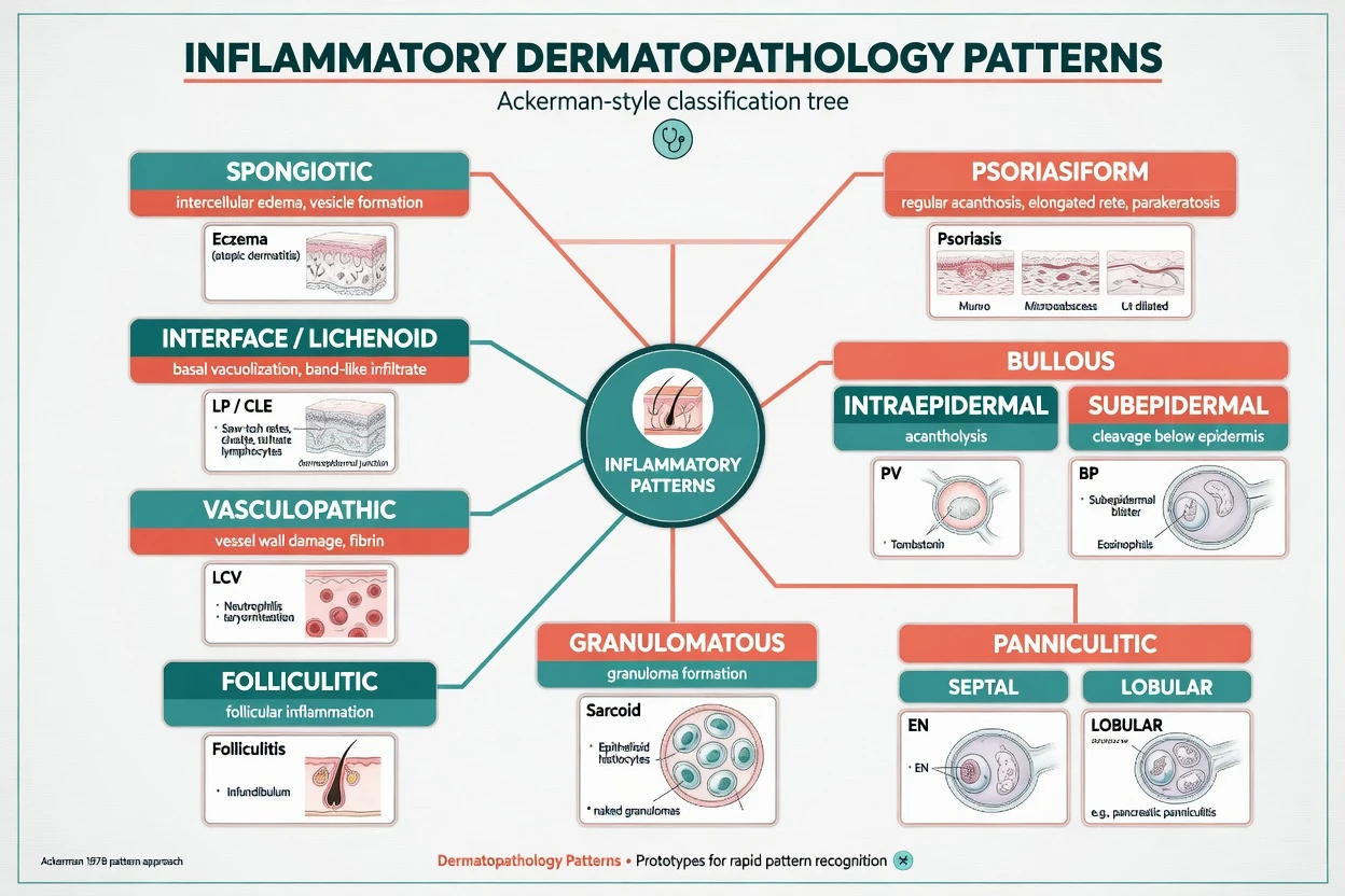
| Pattern | Core microscopic idea | Prototype diseases (not exhaustive) |
|---|---|---|
| Spongiotic | Intercellular epidermal oedema ± vesicles | Acute/subacute eczema, id reaction, dermatophyte, early drug |
| Psoriasiform | Regular acanthosis, clubbed rete | Psoriasis, chronic eczema, PRP, early MF mimic |
| Interface / lichenoid | Basal vacuolisation ± band-like infiltrate | LP, CLE, EM/SJS, lichenoid drug, GVHD |
| Bullous | Cleft level + acantholysis or intact roof | PV/PF, BP, DH, linear IgA, porphyria |
| Vasculopathic | Vessel injury ± fibrinoid necrosis | LCV, IgA vasculitis, cryoglobulinaemic |
| Granulomatous | Histiocytic aggregates ± necrosis | Sarcoid, GA, infection, FB |
| Folliculitic | Follicle-centred inflammation | Infectious folliculitis, eosinophilic folliculitis |
| Panniculitic | Septal vs lobular fat inflammation | EN (septal), lupus panniculitis (lobular) |
Biopsy Strategy for Inflammatory Disease
Technique determines whether the pathologist can help you.[6][7]
| Clinical question | Preferred sample | Notes |
|---|---|---|
| Most rashes | 4–6 mm punch to subcutis | Include active edge |
| Deep process / panniculitis | Deep punch or incisional to fat | Shave is useless |
| Blistering + DIF | Lesional H&E and perilesional DIF (Michel’s/saline per lab) | Not ulcer base alone |
| Vasculitis | Early palpable purpura punch | Old pigmented lesion may show only debris |
| Scalp / alopecia | Punch(es) with orientation protocol | Horizontal sections often needed |
Always write on the form: duration, distribution, drugs, immunosuppression, differential, and whether infection is possible. [1]
Spongiotic Pattern
Acute: marked spongiosis, lymphocyte exocytosis, papillary dermal oedema.
Subacute: less oedema, early acanthosis.
Chronic: psoriasiform hyperplasia with residual spongiosis — overlaps psoriasis clinically and histologically. [1]
Always exclude: dermatophyte (PAS), contactant/drug history, scabies (if acral/burrows). Paediatric spongiotic disease has a broader infectious and atopic context.[4]
Psoriasiform Pattern
Classic psoriasis teaching set: regular acanthosis, thinned suprapapillary plates, dilated tortuous papillary vessels, Munro microabscesses / spongiform pustules of Kogoj, confluent parakeratosis with diminished granular layer. Partial treatment and rubbing distort the picture — correlate clinically. [1]
Differential of psoriasiform hyperplasia: chronic spongiotic dermatitis, pityriasis rubra pilaris, secondary syphilis (always special stains/serology when fitting), early mycosis fungoides (requires multiple biopsies + immunophenotype over time — do not overcall on one mildly atypical spongiotic specimen). [1]
Interface & Lichenoid Patterns
Vacuolar interface
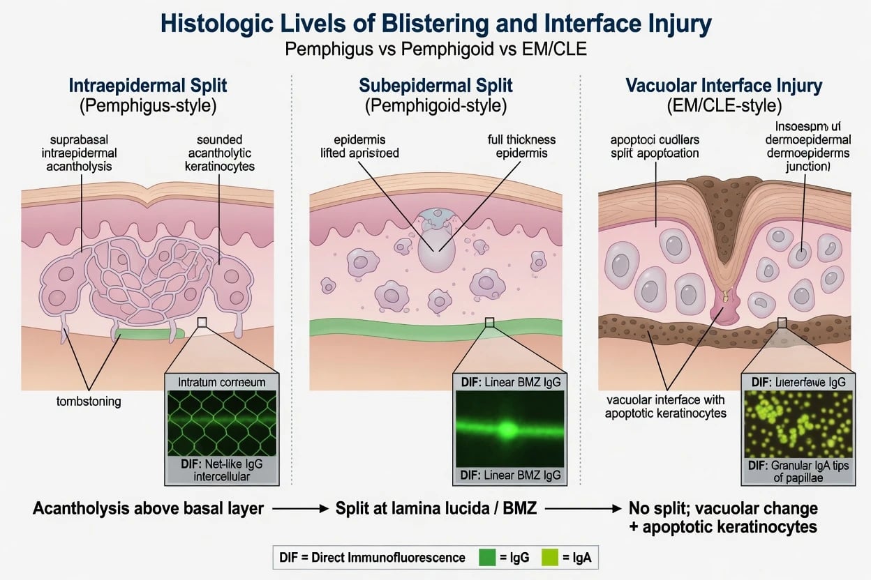
Basal keratinocyte damage, apoptotic (Civatte-type) bodies, pigment incontinence — classic for cutaneous lupus and many drug/viral exanthems.[5] CLE histology pairs with clinical subtype (ACLE, SCLE, DLE) and often mucin in DLE.[5]
Band-like lichenoid
Dense band-like lymphocytic infiltrate hugging the DEJ with saw-tooth rete — prototype lichen planus.[10][11] Lichenoid drug eruption and lichenoid GVHD enter the differential; drug charts and transplant history are decisive.[10][12]
EM / SJS-TEN spectrum
Interface damage with conspicuous necrotic keratinocytes, often out of proportion, sparse infiltrate relative to epidermal death — clinical severity staging drives management more than a fancy histologic subtype name. [1]
Exam application bank (NEET-PG / INICET)
One-line answer
Board-level pattern-based approach to inflammatory skin disease histopathology. Covers Ackerman-style algorithmic diagnosis, major reaction patterns (spongiotic, psoriasiform, interface/lichenoid, bullous, vasculopathic, granulomatous, folliculitic, panniculitic), biopsy and DIF strategy, special stains, clinicopathologic correlation, and how reports change management without overcalling lymphoma or missing infection.
Worked stems (answer without another resource)
Stem 1 — Classic presentation. Map symptoms to mechanism; name the first investigation and first treatment step with dose/route if drug therapy is standard. [1]
Stem 2 — Unstable / complicated. List red flags that force immediate resuscitation, theatre, ICU, antidote, or reperfusion — and what you do in the first 15 minutes. [1]
Stem 3 — Atypical group. Elderly, pregnancy, child, or immunocompromised: how presentation and thresholds change. [1]
Stem 4 — Differential trap. Name the three closest mimics and one discriminator for each. [1]
Stem 5 — Disposition. Who goes home with safety-netting, who is admitted, who needs HDU/ICU/theatre, and what follow-up is mandatory. [1]
Rapid viva checklist
- Definition + classification
- Pathophysiology chain
- Bedside signs / criteria
- Score with exact components (if any)
- Emergency bundle
- Definitive therapy with doses
- Complications of disease and of treatment
- Special populations
- Guideline/trial name if classic
- Three exam traps
Coverage self-check
If you cannot answer any stem above from this page alone, re-read the matching section — the page is intended to be self-sufficient for final-prof and NEET-PG/INICET questions on Inflammatory dermatopathology patterns.
Expanded exam teaching (depth pass)
Clinical reasoning
For Inflammatory dermatopathology patterns, examiners test whether you can prioritise life threats, choose the right first test, and give specific therapy (agent, dose, route, timing). Generic phrases without numbers score poorly.
Mechanism → feature map
Build a short chain: cause → pathophysiologic intermediate → clinical feature → complication. Every major symptom in the classic vignette should sit on that chain.
Investigation strategy
- Bedside/first-line tests that change immediate management
- Confirmatory or staging tests
- What a normal result does not exclude
- When not to delay treatment for imaging (unstable patient)
Management ladder
- Resuscitation / ABC / sepsis or haemorrhage bundle as relevant
- Specific antidote / procedure / antimicrobial / reperfusion / surgery
- Supportive care and monitoring targets
- Definitive long-term therapy and secondary prevention
- Disposition and safety-net advice
Special populations
Always prepare one line each for children, pregnancy, elderly, renal/hepatic impairment, and immunocompromised patients when the topic allows.
Pitfalls that fail candidates
- Treating the number not the patient
- Missing pregnancy status when relevant
- Imaging before stabilisation
- Wrong empiric cover or wrong antidote timing
- Incomplete counselling on recurrence, adherence, or red-flag return
Board-level pattern-based approach to inflammatory skin disease histopathology. Covers Ackerman-style algorithmic diagnosis, major reaction patterns (spongiotic, psoriasiform, interface/lichenoid, bullous, vasculopathic, granulomatous, folliculitic, panniculitic), biopsy and DIF strategy, special stains, clinicopathologic correlation, and how reports change management without overcalling lymphoma or missing infection. [1]
Structured revision sheet
Must-know numbers and names
List every score, size threshold, dose, and time window from this topic on a blank page from memory, then check against the sections above.
Three classic MCQ angles
- Most likely diagnosis given a vignette
- Next best step in management
- Most appropriate investigation
Three classic SAQ angles
- Pathophysiology in five steps
- Management algorithm with doses
- Complications and prevention
Clinical station flow
Greet → focused history → targeted exam → investigations → explain diagnosis → emergency care → definitive plan → safety-net / follow-up → answer examiner questions on mechanism and pitfalls.
[1]Bullous Patterns & DIF
| Level of split | H&E clue | DIF teaching pattern | Disease prototypes |
|---|---|---|---|
| Intraepidermal (suprabasal) | Acantholysis | Intercellular IgG/C3 net | Pemphigus vulgaris |
| Intraepidermal (subcorneal) | Superficial acantholysis | Intercellular IgG (often higher) | Pemphigus foliaceus |
| Subepidermal | Intact epidermis roof | Linear IgG/C3 BMZ | Bullous pemphigoid, EBA (salt-split refines) |
| Subepidermal | Neutrophilic papillae | Granular IgA tips of papillae | Dermatitis herpetiformis |
| Subepidermal | Neutrophils along BMZ | Linear IgA BMZ | Linear IgA disease |
Pemphigus biology and diagnosis rest on clinic, histopathology, DIF, and autoantibody tests as complementary pillars.[8][9]
Vasculopathic Pattern
Leukocytoclastic vasculitis (LCV) teaching criteria: neutrophilic infiltration of post-capillary venules, fibrinoid necrosis of walls, leukocytoclasia (nuclear dust), extravasated erythrocytes — in the right clinical setting of palpable purpura. Not every perivascular lymphocyte collection is vasculitis. [1]
Work-up triggered by confirmed LCV: drugs, infection, autoimmune serologies, urine, when indicated ANCA/cryoglobulins — disease-specific leaves cover systemic algorithms. [1]
Granulomatous, Folliculitic, Panniculitic
- Sarcoidal naked granulomas vs tuberculoid vs palisading (GA, NLD) vs suppurative (infection, rupture) — culture/special stains before pure idiopathic labels in immunocompromised hosts.[3]
- Folliculitis: neutrophils in follicle ± Demodex/bacteria; eosinophilic folliculitis in HIV/other contexts.
- Panniculitis: septal (erythema nodosum prototype) vs lobular (lupus panniculitis, pancreatic, infection) — requires fat in the specimen.[6]
Special Stains & Ancillary Tests (High-Yield Menu)
| Test | When |
|---|---|
| PAS / GMS | Spongiotic or neutrophilic epidermis; any “eczema” not responding |
| Gram, AFB, silver | Suppurative granulomas, ulcers, immunocompromised |
| Colloidal iron / Alcian blue | Dermal mucin (CLE, REM) |
| Elastic stains | Anetoderma, mid-dermal elastolysis context |
| DIF | Blisters, vasculitis subsets, DH, porphyria patterns |
| IHC (CD3/4/8, CD30) | Only when lymphoma truly in differential across time |
Algorithm: From Slide to Action
Reading order (board habit)
- Scanning power — which pattern dominates?
- Epidermis — spongiosis, acanthosis, necrosis, acantholysis, organisms?
- DEJ — vacuolar damage, band infiltrate, split level?
- Dermis — infiltrate type (lymphocyte, neutrophil, eosinophil, histiocyte), vessels, mucin.
- Fat / follicle / nerve — if present and relevant.
- Special studies — PAS, DIF, cultures as pre-planned.
- Clinical synthesis — write a diagnosis or focused differential, not a novel.
Good report
- States pattern
- Gives favoured diagnosis + 2 mimics
- Notes limits (partial Rx, age of lesion)
- Suggests DIF/PAS if not done
- Uses plain language for action
Weak report
- Only 'chronic dermatitis'
- No clinical correlation note
- Misses PAS on feet/hands
- Calls MF on first mild biopsy
- No depth for panniculitis
Management Implications of Common Reports
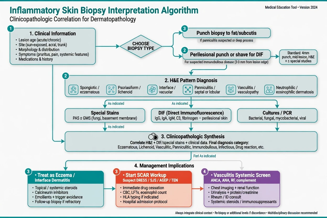
| Report gist | Clinical move |
|---|---|
| Spongiotic dermatitis, PAS neg | Eczema pathway; reconsider contact/drug |
| Spongiotic + fungus | Antifungal; stop chronic steroid alone |
| Consistent with LP | LP guidelines pathway; check drugs/hep C context as indicated[10][11] |
| Interface necrosis, EM-like | Drug chart + severity staging |
| LCV | Systemic screen + remove triggers |
| Subepidermal blister, DIF linear IgG | BP work-up / treatment ladder |
| Non-specific chronic dermatitis | Treat clinically; re-biopsy if evolves |
Special Populations
- Children: infection, atopic, and genodermatosis patterns feature more; sampling and differentials differ from adult boards.[4]
- Pregnancy: DIF distinguishes pemphigoid gestationis from polymorphic eruption when blisters confuse.
- Elderly: BP vs scabies vs drug — eosinophils do not equal BP without the full picture.
- Immunosuppressed: infection until proven otherwise in granulomatous/neutrophilic patterns.[3]
Pitfalls
- Biopsying treated/burned-out centre of a plaque.
- Calling every perivascular infiltrate “vasculitis.”
- Missing fungus, scabies, or herpes in “inflammatory” skin.
- Overcalling mycosis fungoides on early spongiotic dermatitis.
- No fat in a panniculitis work-up.
- DIF from necrotic centre instead of perilesional skin.[8][9]
Regional Notes
Pattern language is universal; access to IF laboratories, molecular tests for infection, and dermatopathology second opinions varies. European LP guidance and international pemphigus primers still depend on correct first-pass histology plus DIF when available.[8][10]
Exam Pearls
PATTERN
PATTERN
Scanning magnification
Early vs late changes
Site and distribution on form
PAS, DIF, culture before fixative mistakes
Eos, neuts, lymphs, histiocytes
Especially spongiotic & granulomatous
Clinical photo beats guesswork
- Ackerman pattern analysis is still the exam operating system.[1][2]
- PAS the spongiotic foot/hand before calling pure eczema.[3]
- Interface + necrosis + sick patient = SCAR thinking.
- True vasculitis needs vessel wall damage, not just lymphocytes near vessels.
- Pemphigus/pemphigoid diagnosis is multimodal — H&E + DIF + serology.[8][9]
References
- [1]Ackerman AB. An algorithmic method for histologic diagnosis of inflammatory and neoplastic skin diseases by analysis of their patterns Am J Dermatopathol, 1985.PMID 4025726
- [2]Paredes BE. [Pattern analysis of inflammatory skin diseases according to A. B. Ackerman-always up to date] Pathologe, 2020.PMID 32377832
- [3]Smith EH, Chan MP. Inflammatory Dermatopathology for General Surgical Pathologists Clin Lab Med, 2017.PMID 28802506
- [4]Hsi AC, Rosman IS. Histopathology of Cutaneous Inflammatory Disorders in Children Pediatr Dev Pathol, 2018.PMID 29607753
- [5]Fetter T, Braegelmann C, de Vos L, et al. Current Concepts on Pathogenic Mechanisms and Histopathology in Cutaneous Lupus Erythematosus Front Med (Lausanne), 2022.PMID 35712102
- [6]Greenwood JD, Merry SP, Boswell CL. Skin Biopsy Techniques Prim Care, 2022.PMID 35125151
- [7]Pickett H. Shave and punch biopsy for skin lesions Am Fam Physician, 2011.PMID 22046939
- [8]Schmidt E, Kasperkiewicz M, Joly P. Pemphigus Lancet, 2019.PMID 31498102
- [9]Kasperkiewicz M, Ellebrecht CT, Takahashi H, et al. Pemphigus Nat Rev Dis Primers, 2017.PMID 28492232
- [10]Ioannides D, Vakirlis E, Kemeny L, et al. European S1 guidelines on the management of lichen planus: a cooperation of the European Dermatology Forum with the European Academy of Dermatology and Venereology J Eur Acad Dermatol Venereol, 2020.PMID 32678513
- [11]Le Cleach L, Chosidow O. Clinical practice. Lichen planus N Engl J Med, 2012.PMID 22356325
- [12]Atzmony L, Reiter O, Hodak E, et al. Treatments for Cutaneous Lichen Planus: A Systematic Review and Meta-Analysis Am J Clin Dermatol, 2016.PMID 26507510