Dermatology · Medicine
Intertrigo
Also known as Intertrigo · Intertriginous dermatitis · Skin-fold dermatitis · Fold moisture-associated skin damage
Intertrigo is a common inflammatory dermatosis of opposed skin surfaces (skin folds) caused by the triad of moisture, friction, and warmth, presenting as well-demarcated, glistening, macerated erythema with fissuring in intertriginous areas (inframammary, axillary, inguinal, abdominal pannus, intergluteal cleft, neck, interdigital webs, peristomal). It is a clinical descriptor rather than a microbiological diagnosis: secondary colonisation with Candida albicans, Staphylococcus aureus, beta-haemolytic streptococci, Corynebacterium minutissimum (erythrasma), or dermatophytes is layered on an irritant, macero-inflammatory base. Risk factors include obesity, diabetes mellitus, hot humid climate, incontinence, immobility, occlusive clothing, and infancy. Management is built on four pillars: keep folds dry, apply barrier creams, treat the secondary organism, and correct the predisposing cause. The critical differentials are candidal intertrigo (satellite pustules, potassium-hydroxide positive), erythrasma (coral-red Wood's lamp fluorescence), tinea cruris (annular scaly border), and inverse psoriasis (smooth, non-scaly plaques).
On this page & tools
Your progress
Saved locally on this device.
Exam tags
Red flags
Overview and definition
Intertrigo (from the Latin intertrere, "to rub between") is an acute or chronic inflammatory dermatosis of opposed skin surfaces, the intertriginous or skin-fold areas where two surfaces touch, rub, and trap moisture. It is set in motion whenever friction, moisture, and warmth converge on a fold, and it presents clinically as well-demarcated, glistening, macerated erythema that may fissure, weep, and become malodorous. The characteristic sites are the inframammary and submammary folds, axillae, inguinal and crural folds, scrotum, abdominal pannus, intergluteal cleft, neck (especially in infants and the obese), retroauricular folds, interdigital webs of toes and fingers, and peristomal skin.[1][2]
A point examiners press is that intertrigo is a clinical descriptor, not a microbiological diagnosis. The bare term denotes the irritant, macero-inflammatory base; on top of that base, a secondary coloniser — most often Candida albicans, but also Staphylococcus aureus, beta-haemolytic streptococci, Corynebacterium minutissimum (erythrasma), or a dermatophyte (tinea) — establishes itself and colours the clinical picture. Naming the coloniser is what shifts treatment from "barrier only" to "barrier plus targeted antimicrobial". [1]
Classification
Intertrigo is classified along two axes that together direct treatment: [1]
- By site — inframammary or submammary, axillary, inguinal or crural, scrotal, abdominal pannus, intergluteal cleft, neck, retroauricular, interdigital toe-web, finger-web, and peristomal.
- By the dominant coloniser layered onto the irritant base — simple (irritant) intertrigo, candidal intertrigo, bacterial intertrigo (staphylococcal or streptococcal), erythrasma (corynebacterial), and dermatophyte (tinea) intertrigo. The inflammatory/inverse-psoriasis overlap and rare causes (Hailey-Hailey, granuloma gluteale, Marjolin ulcer) are considered separately. [1]
This dual classification is more than taxonomic housekeeping. A submammary plaque that is coral-red under Wood's lamp is erythrasma, not "intertrigo plus eczema", and is treated with clindamycin or oral clarithromycin rather than a steroid. The classification drives the bedside test (Wood's lamp, potassium hydroxide) and the drug. [1]
Epidemiology and risk factors
Intertrigo is among the commonest of all skin complaints and, because it is often self-managed or mis-attributed to "chafing" or "heat rash", its true incidence is under-reported. It concentrates wherever moisture and occlusion persist. [1]
The unifying mechanism is skin-fold depth and duration of wetness. Obesity creates deep folds that trap sweat and resist drying; diabetes raises tissue glucose and impairs local immunity, favouring Candida; infancy produces short necks, chubbiness, and occlusive nappies; immobility and incontinence in the elderly create a continuously wet perineal environment; and tropical climates defeat evaporation. Occlusive or synthetic clothing, occupational heat and personal protective equipment, poor hygiene, ostomy effluent, and hyperhidrosis all amplify the same mechanism.[1][2][7]
Pathophysiology
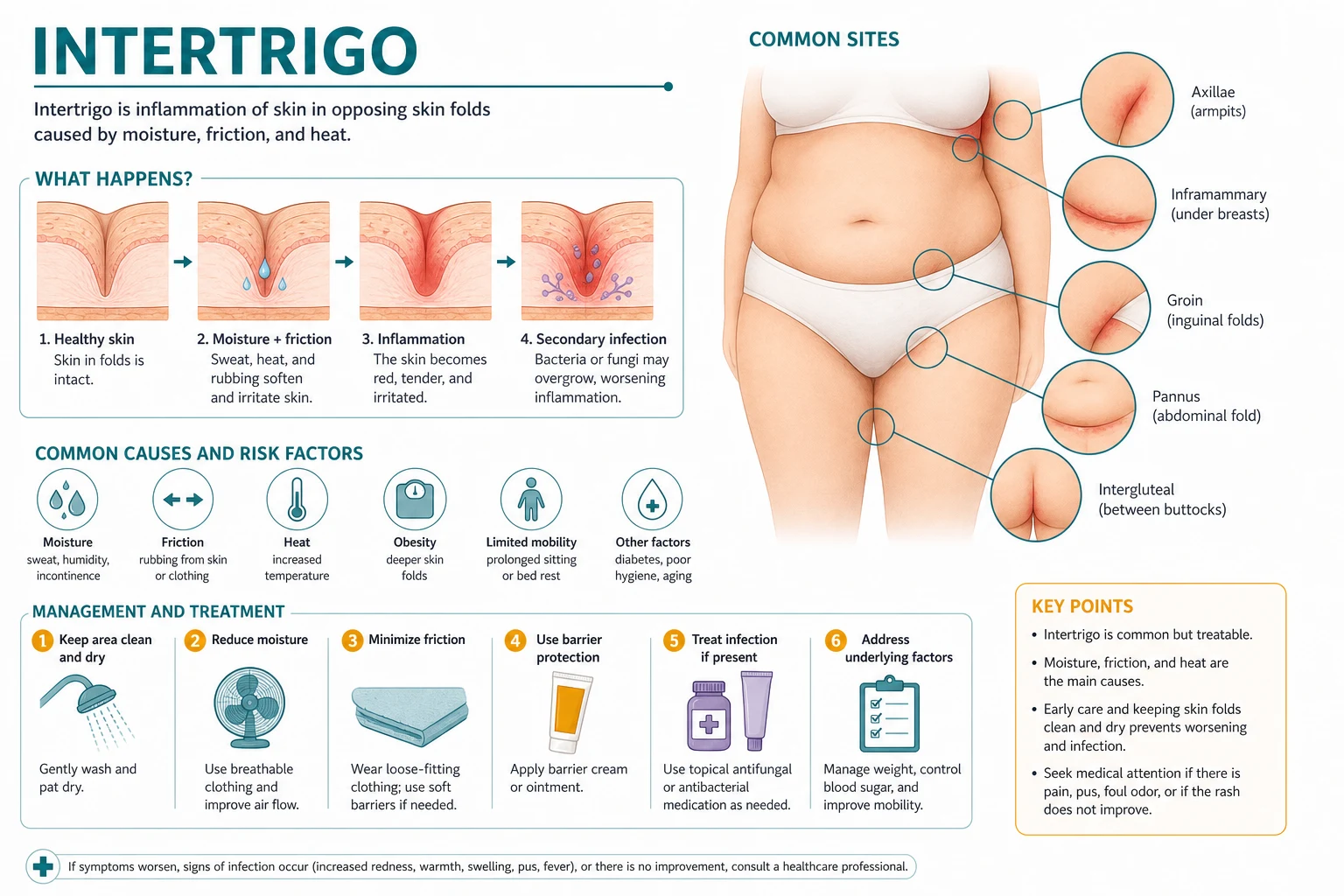
The lesion is the predictable consequence of three forces acting on a fold. Friction between the two opposed surfaces chronically abrades and shears the stratum corneum. Moisture — sweat, urine, faecal enzymes, wound or stoma exudate — over-hydrates the stratum corneum, raising its permeability and solubilising intercellular lipid so that corneocytes lose cohesion; the skin turns white, soggy, and easily torn (maceration). Warmth and occlusion then prevent evaporation and, by raising local pH towards the alkaline, create the incubator that selects for colonisation.[1][3]
The pathophysiology is best read as a self-reinforcing cascade: [1]
Opposed surfaces trap a warm, wet micro-environment; evaporation fails.
Over-hydration and friction denude the stratum corneum; corneocyte cohesion collapses; the skin turns white and soggy.
Fissures open at the deepest crease; irritants (sweat, faecal enzymes, detergents) penetrate.
Moisture and a raised pH select for Candida (yeast, thrives in moisture), Staphylococcus aureus, beta-haemolytic streptococci, Corynebacterium minutissimum (erythrasma), or a dermatophyte.
Infection and irritation drive erythema, exudate, and itch; more scratching and rubbing deepen the maceration.
Inflammation begets more exudate, which begets more maceration, which begets more colonisation — the itch-scratch-maceration cycle that makes the disease chronic.
The clinical appearance of each coloniser reflects its biology. Candida is a dimorphic yeast that favours a moist, slightly acidic fold; it produces bright-red, glistening erythema with satellite pustules and a collarette of scale. C. minutissimum digests keratin and elaborates a porphyrin that fluoresces coral-red under Wood's lamp, the single most useful bedside discriminator in fold disease. Dermatophytes (Trichophyton species) invade keratin and produce the classic annular, scaling, centrifugally advancing border of tinea. Recognising the organism by its pattern — and confirming with Wood's lamp or potassium-hydroxide microscopy — is what turns a generic "fold rash" into a treatable diagnosis.[3][4]
Cellular and molecular mechanisms
At the molecular level, a healthy stratum corneum is held together by intercellular lipids (ceramides, cholesterol, free fatty acids) and corneodesmosomes, and it is kept mildly acidic at a pH of roughly 4.5 to 5.5 by the so-called acid mantle. Three things happen when a fold is occluded and wet. First, prolonged hydration swells the corneocytes and leaches out water-soluble natural moisturising factor, weakening the bricks-and-mortar of the barrier. Second, the trapped moisture and urea raise the local pH towards the alkaline, which activates serine proteases that cleave corneodesmosomes and strip the corneocytes apart — the histological correlate of maceration. Third, the now-permeable, alkaline surface is a better substrate for microbial adhesion: Candida albicans switches from blastospore to the adherent pseudohyphal form, Corynebacterium minutissimum digests keratin and sheds coral-red porphyrin, and Staphylococcus aureus binds exposed fibronectin. The result is a surface that is simultaneously structurally weakened, immunologically exposed, and microbially primed. [1]
This molecular sequence explains the clinical cascade and, more importantly, explains why drying alone can interrupt the disease at any stage: removing moisture restores the acid mantle, halts protease activation, and removes the substrate for microbial adhesion. It also explains why potent topical steroids are uniquely hazardous in folds — occlusion multiplies their absorption precisely at the site of an already-thinned, protease-damaged barrier. [1]
Clinical presentation
Distribution
Intertrigo is confined to folds and is characteristically symmetrical. The high-yield sites are the inframammary and submammary creases, axillae, inguinal and crural folds, scrotum, abdominal pannus, intergluteal cleft, neck (infants and the obese), retroauricular folds, interdigital toe-webs and finger-webs, and peristomal skin. Involvement of the depth of the fold with sparing of surrounding flat skin is the geographical signature that separates intertrigo from a generalised eczema. [1]
Morphology and symptoms
The lesional skin shows well-demarcated, moist, glistening erythema lying in the depth of the fold. With persistence, maceration gives a white, sodden surface; fissuring produces painful cracks at the deepest crease (classically the inframammary fold, gluteal cleft, and ano-genital crease); and a collarette of scale rims the advancing edge. Satellite pustules or small erythematous papules scattered beyond the main margin are the hallmark of candidal intertrigo and should be sought deliberately, because their presence rewrites the prescription. Malodour reflects bacterial colonisation of macerated skin.[1][2]
Patients complain of burning, itching, and pain, made worse by any movement that brings the two surfaces together — walking, bending, sitting, and exercise. Discomfort on movement is so characteristic that a flexural rash made worse by ambulation is intertrigo until proved otherwise. [1]
Site-specific clinical patterns
The fold that is involved often predicts the coloniser and the precipitant, so the site is itself a diagnostic clue. Inframammary and submammary intertrigo in a large-breasted or obese woman is the classic canvas for Candida and for erythrasma — a Wood's lamp is mandatory here. Axillary intertrigo tracks with hyperhidrosis, occlusive deodorants, and shaving-related micro-trauma, and it is the favoured site of Fox-Fordyce disease and hailey-hailey when chronic. Inguinal and crural intertrigo raises tinea cruris and erythrasma, and the scrotum is classically spared in tinea but involved in candidal and irritant disease — a high-yield discriminator at viva. Abdominal pannus (apron) intertrigo in the obese can reach large surface areas and resist drying, and it is a strong driver of recurrent candidal colonisation. Intergluteal cleft intertrigo fissures painfully and must be distinguished from inverse psoriasis, hidradenitis suppurativa, and a pilonidal sinus. Neck and retroauricular intertrigo dominates infancy (short neck, chubbiness) and the obese; in infants the bright-red, satellite-studded creases of candidal intertrigo contrast with the fold-sparing pattern of simple irritant nappy rash. Interdigital toe-web intertrigo is the common portal for erythrasma, pitted keratolysis, tinea pedis, and — critically — streptococcal toe-web infection, a recognised gateway to lower-limb cellulitis that must be treated. Finger-web intertrigo is usually irritant (wet work, detergents) or candidal (chronic paronychia overlap). Peristomal intertrigo reflects appliance leakage and effluent, demanding refit rather than just cream. [1]
The tempo of presentation is also informative. Acute, unilateral, rapidly spreading erythema with pain and systemic upset is cellulitis or a necrotising infection, not intertrigo, and must not be dismissed as a "fold rash". Chronic, relapsing, symmetrical disease points back to the fold environment and the predisposing cause. A single, non-healing, indurated area within a chronic fold is a red flag for squamous cell carcinoma (Marjolin) and warrants biopsy regardless of how benign it looks. [1]
Atypical and special-population presentations
Examiners test the corners. Several presentations reward the candidate who looks beyond the textbook: [1]
- Infant and neonate: a bright-red, glistening, folded rash in the neck, axillae, and thigh creases with satellite pustules is candidal intertrigo; crucially, candidal napkin dermatitis involves the creases, whereas simple irritant nappy rash spares them.
- Diabetic: recurrent or severe candidal intertrigo that is unusually bright red, extensive, or resistant should trigger screening — it may be the presenting clue to undiagnosed diabetes.
- Immunocompromised (HIV, transplant, chemotherapy): intertrigo that is persistent, atypical, or caused by unusual organisms, and that may need systemic therapy.
- Elderly, frail, bed-bound: immobility plus incontinence produce perineal and gluteal intertrigo that must be distinguished from incontinence-associated dermatitis and from pressure injury; the three overlap and coexist.
- Peristomal: moisture and effluent around a colostomy or ileostomy produce a weeping, eroded intertrigo driven by appliance leakage. [1]
Differential diagnosis
The differential is the heart of this topic at viva. Every fold rash must be run through the same set of competitors, distinguished by three features each, because each carries a different treatment. [1]
A short list of can't-miss mimics deserves emphasis. Hailey-Hailey disease (benign familial chronic pemphigus) is a recurrent, relapsing flexural erosive dermatosis of the neck, axillae, and groin, autosomal dominant via ATP2C1, whose biopsy shows suprabasal acantholysis likened to a "dilapidated brick wall"; it masquerades as chronic intertrigo and is refractory to antifungals.[6] A non-healing ulcer within a chronic fold should be biopsied to exclude squamous cell carcinoma (a Marjolin-type change in chronically inflamed or scarred skin). And a fold rash in a patient who is systemically unwell, with spreading erythema, is cellulitis or a necrotising infection — not intertrigo — and needs urgent antibiotics and surgical review. A second tier of less common but exam-rewarded mimics completes the differential. Granuloma gluteale infantum produces reddish-purple nodules in the nappy area of infants, paradoxically linked to fluorinated steroids and candidal overgrowth under occlusion. Acrodermatitis enteropathica (zinc deficiency) gives a sharply marginated periorificial and flexural dermatitis with diarrhoea and alopecia in infants. Pitted keratolysis, caused by Kytococcus (formerly Micrococcus) species in macerated toe-webs, shows discrete superficial pits and a malodorous surface, distinguishable from erythrasma by the absence of coral-red fluorescence. Fox-Fordyce disease is an apocrine miliaria of the axilla and anogenital folds with chronic itchy papules in young women. Scabies can present with burrows and nodules in the interdigital webs, genitalia, and axillae; a careful search for burrows and household contacts, rather than Wood's lamp, makes the call. Acanthosis nigricans — a velvety, hyperpigmented thickening of the folds associated with insulin resistance — is a "fold dermatosis" that is not intertrigo and should not be treated as such, though it can coexist.[1][2] The unifying rule is that any fold eruption resistant to a barrier-plus-azole regimen after a reasonable trial deserves a Wood's lamp, a potassium-hydroxide preparation, and — if still unexplained — a biopsy.
Clinical and bedside assessment
The focused assessment is built around three bedside tools and a deliberate inspection of every fold. [1]
- Inspect all folds systematically: submammary, axillary, inguinal and crural, scrotal, abdominal pannus, intergluteal, neck, retroauricular, interdigital webs of hands and feet, and peristomal. Note symmetry, the confinement to the fold depth, maceration, fissuring, scale, and — specifically — satellite lesions. Look for and grade any cellulitis, lymphangitis, or abscess.
- Wood's lamp examination: shine a Wood's (ultraviolet) lamp in a darkened room across the fold. Coral-red fluorescence is diagnostic of erythrasma (the porphyrin of Corynebacterium minutissimum) and instantly distinguishes it from candidal and dermatophyte intertrigo, which are Wood's lamp negative. This is the highest-yield single test in fold disease.
- Potassium-hydroxide (KOH) microscopy of a skin scraping: budding yeast and pseudohyphae confirm Candida; septate hyphae confirm a dermatophyte. It is rapid and confirms the organism when the clinical pattern is ambiguous or recurrent. [1]
DRY-FOLD
Investigations
Intertrigo is a clinical diagnosis in the great majority of cases, and over-investigation is a recognised error. Investigations are reserved for when the diagnosis is uncertain, the disease is recurrent or resistant, or a complication or underlying cause must be defined. [1]
Step 1 — Clinical
Confident pattern recognition: symmetrical, well-demarcated, glistening erythema confined to a fold. Treat empirically and review.
Step 2 — Bedside tests
Wood's lamp (coral-red = erythrasma); KOH microscopy (pseudohyphae + budding yeast = Candida; septate hyphae = dermatophyte).
Step 3 — Microbiology
Bacterial swab for culture and sensitivity if purulent, cellulitic, or resistant (S. aureus, Strep, MRSA); fungal culture on Sabouraud agar if ambiguous or recurrent.
Step 4 — Biopsy
Skin biopsy if recalcitrant, to exclude inverse psoriasis, tinea incognito, Hailey-Hailey disease, or squamous cell carcinoma in a chronic fold ulcer.
Step 5 — Systemic screen
When recurrent or severe: fasting glucose and HbA1c (diabetes), HIV test, urinalysis for glucosuria, full blood count, iron and zinc if deficiency suspected.
Sensitivities and specificities are not quoted in single figures for intertrigo because the disease is a syndrome, but the operating point is clear: Wood's lamp and KOH microscopy are the high-yield, low-cost tests that should be used liberally in any non-straightforward fold eruption; bacterial and fungal cultures add resistance data when empirical topical therapy fails; and biopsy is reserved for the chronic, recalcitrant, or ulcerated case where a mimic is plausible.[1][4] Three practical interpretation rules sharpen the bedside use of these tests. First, a negative Wood's lamp does not exclude erythrasma if the lesion has been washed recently — the water-soluble porphyrin is easily removed, so examine before any cleansing. Second, potassium-hydroxide microscopy can be false-negative if the scraping is too superficial or the loading is light; repeat a suspiciously negative result before concluding there is no yeast. Third, a bacterial swab is not a first-line test in uncomplicated intertrigo — it is reserved for purulence, cellulitis, suspected MRSA, or failure of empirical therapy, because routine swabs often grow colonising flora of no clinical relevance and invite over-treatment. A fungal culture on Sabouraud agar with cycloheximide and chloramphenicol adds species identification and, increasingly, susceptibility data when Candida species other than albicans or emerging resistance are suspected. When a fold eruption is genuinely chronic and recalcitrant, a 4 mm punch biopsy for haematoxylin-and-eosin (and a separate sample for periodic-acid-Schiff staining when fungal elements are sought) settles the question of inverse psoriasis, tinea incognito, Hailey-Hailey disease, or squamous cell carcinoma.
Management — resuscitation and red-flag escalation
Intertrigo is rarely a dermatological emergency, and a single red-flag question should be answered first: is this just a fold rash, or is there spreading infection or systemic toxicity? Spreading erythema with warmth, pain out of proportion, fever, chills, or malaise escalates the diagnosis from intertrigo to cellulitis or, with rapid progression and systemic upset, to necrotising soft-tissue infection. These patients need urgent systemic antibiotics (intravenous flucloxacillin, with MRSA-guided agents such as vancomycin where appropriate), blood cultures, surgical review, and admission — not topical therapy and reassurance. [1]
[1]Management — definitive and stepwise
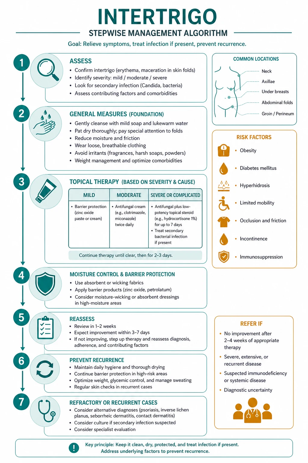
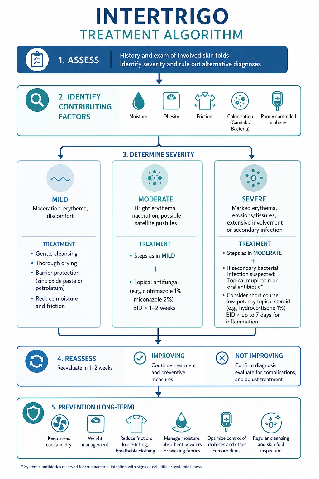
The definitive plan is built on four pillars that operate together: reduce moisture and friction (the cornerstone), apply a barrier, treat the secondary organism, and correct the predisposing cause. No single agent works if the fold stays wet, which is why the general measures precede every drug. [1]
Pillar 1 — Reduce moisture and friction
Dryness breaks the pathophysiological cycle. Dry every fold thoroughly after bathing, using a hair-dryer on a cool setting to reach the depth of deep pannus and submammary folds that towels cannot. Apply a barrier cream — zinc oxide, white soft paraffin (petrolatum), or dimethicone-based preparations — to create a water-repellent film between the opposed surfaces. Talc-free absorbent powders (cornstarch-based; talc is avoided because of inhalation risk) wick moisture. Replace occlusive or synthetic clothing with loose, breathable cotton, manage incontinence with containment products and scheduled toileting, and treat the cause: weight loss for obesity, glycaemic control for diabetes, stoma-appliance refitting for peristomal disease, and hyperhidrosis control for sweating-driven disease.[2][7][8]
Pillar 2 — Treat the secondary organism
Selecting the antimicrobial against the identified coloniser is the decisive therapeutic move. [1]
Topical azole (candidal intertrigo — first line)
Pillar 3 — Reduce inflammation (safely)
When inflammation is severe, a mild topical corticosteroid — hydrocortisone 1 percent — for a short course of 5 to 7 days is acceptable and often rapidly symptom-relieving. Potent steroids must never be used in folds: occlusion multiplies percutaneous absorption many-fold, producing epidermal atrophy, striae, telangiectasia, and hypothalamic-pituitary-adrenal-axis suppression, sometimes within weeks. For chronic, recurrent, or inflammatory/inverse-psoriasis-overlap disease, topical calcineurin inhibitors — tacrolimus 0.1 percent ointment or pimecrolimus 1 percent cream — are the steroid-sparing agents of choice, supported by a systematic review of inverse psoriasis treatments that found calcineurin inhibitors effective in flexural disease without the atrophy of corticosteroids.[5] Combined antifungal plus mild-steroid preparations (for example miconazole with hydrocortisone) suit mixed irritant-plus-candidal disease but should be time-limited to avoid the same steroid pitfalls.
Pillar 4 — Correct the predisposing cause
No antimicrobial prevents recurrence if the fold environment persists. Weight loss (including bariatric or metabolic surgery for morbid obesity) removes the deep folds that sustain disease; diabetes control lowers candidal colonisation; incontinence management dries the perineum; hyperhidrosis treatment (topical aluminium chloride, glycopyrrolate, or botulinum toxin for refractory axillary/groin sweating) reduces the moisture load; and stoma-appliance refitting addresses peristomal disease. Debridement or drainage is reserved for abscess or necrotising infection.[1][2]
Prevention
Because intertrigo is regenerated by the fold environment whenever it is left wet, prevention is indistinguishable from the first pillar of treatment and is the only reliable way to stop recurrence. The preventive bundle is deliberately simple and applies to every at-risk patient: dry every fold completely after bathing (a hair-dryer on a cool setting reaches deep folds towels cannot), apply a barrier of zinc oxide, white soft paraffin, or a dimethicone-based film daily, wear loose, breathable cotton clothing and avoid occlusive synthetics, use talc-free absorbent powders (cornstarch-based — talc is avoided because of inhalation and ovarian-cancer concerns), and reduce weight in the obese to remove the deep folds that trap moisture. For the incontinent, combine containment products, scheduled toileting, and a barrier film; for the bed-bound, reposition every two hours and protect the perineal and gluteal skin; for the diabetic, achieve glycaemic control and inspect the toe-webs and folds daily; for the peristomal patient, refit the appliance to stop effluent leakage; and for hyperhidrosis, consider topical aluminium chloride or botulinum toxin.[2][7][8]
Two prevention pitfalls deserve emphasis. First, cornstarch powders can feed Candida in an already-colonised fold — use a barrier cream rather than powder when candidal intertrigo is active, and reserve powder for prevention once the skin is healed. Second, over-washing with harsh soaps strips the lipid barrier and worsens maceration; use a gentle, non-soap cleanser and pat (never rub) the folds dry. Patient education on these measures is the single highest-yield intervention at follow-up, because recurrence tracks almost perfectly with whether the fold environment has actually changed. [1]
Specific subtypes and scenarios
Regional guideline deltas are small for intertrigo because it is a clinical syndrome rather than a single disease with a global registry. European (BAD/EuroDERM, NICE clinical knowledge summaries), American (AAD), and Australian (RANZCD) guidance converge on the same sequence — barrier and dryness first, topical azole for candidal disease, topical or oral antibacterial for bacterial disease, mild-only corticosteroids, and calcineurin inhibitors for chronic disease. Where they differ is in formulary availability (clotrimazole versus miconazole versus econazole as first-line azole), preference for fusidic acid (widely used in the UK/Australasia, restricted in some markets for stewardship reasons), and the avoidance of talc (now universal because of inhalation and ovarian-cancer concerns). Wood's lamp examination is emphasised equally across regions. Always name the guideline you are quoting.
Each major subtype deserves its own handling, because the treatment follows the coloniser. [1]
- Candidal intertrigo is the single commonest infective overlay. The triad of bright-red glistening erythema, satellite pustules, and a collarette of scale, confirmed by potassium-hydroxide microscopy showing pseudohyphae and budding yeast, points to a topical azole (clotrimazole 1 percent or miconazole 2 percent cream twice daily for 2 to 4 weeks). Oral fluconazole is reserved for extensive, refractory, or immunocompromised disease.[4]
- Bacterial intertrigo (staphylococcal or streptococcal) presents with purulence, crusting, or spreading erythema; localised disease responds to topical mupirocin 2 percent, while cellulitis needs oral or intravenous flucloxacillin with MRSA-guided agents when resistant. Streptococcal toe-web intertrigo is a recognised portal of entry for lower-limb cellulitis and must be treated.
- Erythrasma is corynebacterial (C. minutissimum), reddish-brown and finely wrinkled, with coral-red Wood's lamp fluorescence. It responds to topical clindamycin 2 percent or clotrimazole, with oral clarithromycin (a single 1 g dose or a short course) or erythromycin for extensive or refractory disease.
- Dermatophyte (tinea) intertrigo mimics tinea cruris — annular, with an active scaly advancing border and classically sparing the scrotum — and is treated with topical terbinafine 1 percent or clotrimazole 1 percent for 2 to 4 weeks, checking the feet and nails as a reservoir.
- Inverse-psoriasis overlap and chronic inflammatory intertrigo favour topical calcineurin inhibitors (tacrolimus, pimecrolimus) over corticosteroids, with systemic agents reserved for severe, widespread disease.[5]
- Infant and nappy-area intertrigo distinguishes candidal napkin dermatitis, which involves the creases and shows satellite pustules, from irritant nappy rash, which spares the creases. Treatment is a topical azole plus a zinc-oxide barrier, with no potent steroids.
- Peristomal and ostomy intertrigo is driven by effluent leakage and appliance misfit; management is appliance refitting, barrier films and wafers, and treatment of any secondary candidal colonisation.
Complications and pitfalls
The complications of intertrigo fall into three groups. Disease-related complications include secondary bacterial infection (impetiginisation, cellulitis, lymphangitis, and abscess), fungal spread with treatment-resistant or recurrent candidal disease and, in the immunocompromised, the rare progression to systemic candidiasis; painful fissuring of deep creases with bleeding and delayed healing; post-inflammatory pigment change (hyper- or hypopigmentation) that can persist long after the inflammation settles; and, in the bed-bound elderly, a vicious cycle of pain, reduced mobility, and worsening moisture that deepens the disease. Iatrogenic complications follow inappropriate treatment: potent-steroid-induced atrophy, striae, and hypothalamic-pituitary-adrenal-axis suppression from using strong corticosteroids under occlusion, and tinea incognito when a steroid masks a dermatophyte. The greatest diagnostic pitfall is masking serious disease — a squamous cell carcinoma (Marjolin ulcer) in a chronic fold ulcer, undiagnosed diabetes or immunodeficiency behind recurrent disease, or Hailey-Hailey disease mistaken for treatment-resistant intertrigo.[1][6] A quality-of-life and functional layer sits on top of these complications. The disease is socially embarrassing because of its site and malodour, and the pain on movement restricts walking, exercise, and personal hygiene — which in turn deepens the fold environment and worsens the lesion, a vicious cycle that is especially destructive in the obese and the bed-bound. In infants, chronic candidal napkin and fold intertrigo unsettles feeding and sleeping and distresses carers. For each complication there is a defined response: impetiginisation and cellulitis need antibacterials (oral flucloxacillin first, MRSA-guided agents if resistant); abscess needs incision and drainage; deep fissures benefit from a barrier paste, rest, and analgesia; post-inflammatory pigment change fades over months but is helped by sun protection; and the patient trapped in the pain-immobility-moisture cycle needs active repositioning, continence care, and weight-loss support to break it. The iatrogenic complications are prevented by a single rule — never apply a potent corticosteroid to a fold — and by recognising that a "steroid-responsive" fold eruption that relapses immediately on stopping is far more likely to be unrecognised tinea (tinea incognito) than a steroid-responsive eczema.
[1]Prognosis and disposition
The prognosis is excellent when treatment and correction of the predisposing cause are combined, and the disease usually resolves without scarring. Recurrence is the rule, not the exception, if the underlying fold environment persists — obesity, diabetes, incontinence, occlusion, and climate will regenerate the lesion unless they are addressed. Post-inflammatory pigment change may linger. Chronic or recalcitrant disease should prompt reassessment of the diagnosis (biopsy to exclude a mimic), screening for systemic causes (diabetes, HIV), and consideration of calcineurin inhibitors or specialist referral. Most patients are managed in the community or primary care; dermatology referral is reserved for recurrent, resistant, diagnostically uncertain, or complicated disease. Follow-up should be framed around the two questions that decide outcome: has the lesion settled, and has the fold environment changed? A sensible review is at two to four weeks for a straightforward case — long enough for a topical azole to clear candidal disease and for the patient to demonstrate that they can keep the folds dry. If the lesion has resolved, the visit pivots entirely to prevention education (drying technique, barrier use, weight, incontinence, clothing); if it persists, the visit triggers the diagnostic escalation pathway — a Wood's lamp, a potassium-hydroxide preparation, a swab, and ultimately a biopsy. Patients who relapse repeatedly despite good technique almost always have an unaddressed systemic driver (diabetes, immunodeficiency) or an unrecognised mimic (Hailey-Hailey disease, inverse psoriasis, tinea incognito from a misapplied steroid). Setting this expectation at the first visit — that cure depends on changing the fold, not on a stronger cream — is the single most useful piece of patient education and the best insurance against the dispiriting cycle of recurrence. [1]
Special populations
- Infants and neonates: candidal fold and napkin intertrigo involves the creases and shows satellite pustules; treatment is a topical azole plus a zinc-oxide barrier, avoiding potent steroids and talc. Distinguish from irritant nappy rash (creases spared) and from rare blistering disorders.
- Pregnancy: candidal intertrigo and vulvovaginal candidiasis are common (oestrogen-driven); topical azoles are first-line and safe. Oral azoles are avoided, particularly in the first trimester.
- Elderly, frail, and bed-bound: immobility and incontinence drive perineal and gluteal disease; the priority is prevention — repositioning, continence care, and skin barriers — and the discrimination from pressure injury and incontinence-associated dermatitis, all of which coexist.
- Diabetic: recurrent or severe candidal intertrigo is a screening trigger for HbA1c and fasting glucose; glycaemic control reduces recurrence, and foot and toe-web intertrigo must be treated to protect against cellulitis.
- Immunocompromised (HIV, transplant, chemotherapy): intertrigo may be persistent, atypical, or caused by unusual organisms, and may require systemic antifungals; screen for chronic mucocutaneous candidiasis of immune dysregulation when disease begins in childhood.
- Stoma and ostomy patients: peristomal moisture damage is addressed by appliance refitting, barrier films and wafers, and treatment of secondary candidal colonisation.
- Bariatric and morbidly obese patients: deep folds resist drying and self-care; assist with hygiene, use barrier pastes, and consider metabolic surgery to remove the fold environment. [1]
Evidence, guidelines, and regional differences
The contemporary evidence base is anchored by the Romanelli/Voegeli 2023 review on the diagnosis, management, and prevention of intertrigo in adults, the Voegeli 2020 narrative review on causes, prevention, and management, and the Kottner 2021 Wund-D.A.CH best-practice recommendation on Moisture-Associated Skin Damage, which provides the unifying MASD framework that places intertrigo alongside incontinence-associated and peristomal moisture damage.[1][2][3] The antifungal ladder for candidal intertrigo rests on the Taudorf 2019 evidence-based review of cutaneous candidiasis, which supports topical azoles as first-line with systemic fluconazole reserved for extensive, refractory, or immunocompromised disease.[4] The Reynolds/Wu 2020 systematic review of inverse psoriasis treatments underpins the use of topical calcineurin inhibitors and the avoidance of potent corticosteroids in chronic flexural disease.[5] The Hailey-Hailey disease update (2024) guides recognition and management of the principal genetic mimic.[6] Skin-integrity maintenance in the aged and in nursing practice, including prevention of fold moisture damage, is supported by two nursing-studies reviews.[7][8]
Test yourself — the highest-yield single fact in intertrigo
What is the one bedside test that most changes management in a fold eruption, and what does a coral-red result mean?
A coral-red result under the Wood's lamp is diagnostic of erythrasma (Corynebacterium minutissimum), and it redirects treatment from an azole or steroid to topical clindamycin or clotrimazole, or oral clarithromycin for extensive disease. No other fold test carries the same decision-changing weight, which is why Wood's lamp examination belongs in the assessment of every non-straightforward intertrigo. [1]
Controversies include the choice between antiseptic washes and soap, the relative place of antibacterial versus antifungal therapy in mixed infection, and the emerging role of botulinum toxin for refractory axillary or groin hyperhidrosis-driven intertrigo. The evidence in these areas is limited and consensus is regional. [1]
Exam pearls
[1]Definition and mechanism
- Inflammatory dermatosis of opposed skin surfaces from moisture, friction, and warmth
- A clinical descriptor, not a microbiological diagnosis — name the coloniser
- Fold-occlusion member of the MASD family (alongside IAD and peristomal moisture)
- Cascade: occlusion, maceration, barrier breach, colonisation, inflammation, the itch-scratch-maceration loop
Risk factors (the six to name)
- Obesity (leading risk factor — deep, hard-to-dry folds)
- Diabetes mellitus (glycosuria, impaired immunity, candidal colonisation)
- Hot humid climate and summer
- Infancy (neck folds and nappy area)
- Bed-bound elderly (immobility plus incontinence)
- Occlusive clothing, ostomy effluent, hyperhidrosis
Differential — one test each
- Candidal intertrigo: satellite pustules, collarette scale; KOH shows budding yeast and pseudohyphae
- Erythrasma: coral-red Wood's lamp fluorescence (C. minutissimum porphyrin)
- Tinea cruris: annular, active scaly border, spares scrotum; KOH shows septate hyphae
- Inverse psoriasis: well-demarcated salmon-pink, smooth, non-scaly; psoriasis at scalp, nails, extensors
- Hailey-Hailey: recurrent crusted flexural erosions; AD ATP2C1; biopsy suprabasal acantholysis ('dilapidated brick wall')
- Cellulitis: spreading, warm, purulent, systemic symptoms
Treatment by coloniser
- Candida: clotrimazole 1 percent or miconazole 2 percent cream twice daily for 2 to 4 weeks; fluconazole if extensive
- Staph or strep: mupirocin 2 percent three times daily; flucloxacillin for cellulitis
- Erythrasma: topical clindamycin 2 percent or clotrimazole; oral clarithromycin if extensive
- Dermatophyte: terbinafine 1 percent or clotrimazole 1 percent for 2 to 4 weeks
Steroid safety in folds
- Mild hydrocortisone 1 percent for a short 5 to 7 day course only, if inflammation is severe
- Never potent steroids (clobetasol, betamethasone) in folds — occlusion multiplies absorption
- Complications: epidermal atrophy, striae, telangiectasia, HPA-axis suppression
- Steroid-sparing: tacrolimus 0.1 percent or pimecrolimus 1 percent for chronic disease
Red flags to escalate
- Persistent or recurrent disease: screen diabetes and HIV, culture for MRSA, biopsy to exclude mimic
- Spreading erythema with fever: cellulitis or necrotising infection — intravenous antibiotics
- Chronic non-healing fold ulcer: biopsy to exclude squamous cell carcinoma (Marjolin)
- Recurrent flexural crusting unresponsive to therapy: think Hailey-Hailey disease
“Fold dermatosis driven by moisture, friction, and warmth; name the coloniser; dryness is the cure; never potent steroids in folds.”
Exam application bank (NEET-PG / INICET)
One-line answer
Intertrigo is a common inflammatory dermatosis of opposed skin surfaces (skin folds) caused by the triad of moisture, friction, and warmth, presenting as well-demarcated, glistening, macerated erythema with fissuring in intertriginous areas (inframammary, axillary, inguinal, abdominal pannus, intergluteal cleft, neck, interdigital webs, peristomal). It is a clinical descriptor rather than a microbiological diagnosis: secondary colonisation with Candida albicans, Staphylococcus aureus, beta-haemolytic streptococci, Corynebacterium minutissimum (erythrasma), or dermatophytes is layered on an irritant, macero-inflammatory base. Risk factors include obesity, diabetes mellitus, hot humid climate, incontinence, immobility, occlusive clothing, and infancy. Management is built on four pillars: keep folds dry, apply barrier creams, treat the secondary organism, and correct the predisposing cause.
Worked stems (answer without another resource)
Stem 1 — Classic presentation. Map symptoms to mechanism; name the first investigation and first treatment step with dose/route if drug therapy is standard. [1]
Stem 2 — Unstable / complicated. List red flags that force immediate resuscitation, theatre, ICU, antidote, or reperfusion — and what you do in the first 15 minutes. [1]
Stem 3 — Atypical group. Elderly, pregnancy, child, or immunocompromised: how presentation and thresholds change. [1]
Stem 4 — Differential trap. Name the three closest mimics and one discriminator for each. [1]
Stem 5 — Disposition. Who goes home with safety-netting, who is admitted, who needs HDU/ICU/theatre, and what follow-up is mandatory. [1]
Rapid viva checklist
- Definition + classification
- Pathophysiology chain
- Bedside signs / criteria
- Score with exact components (if any)
- Emergency bundle
- Definitive therapy with doses
- Complications of disease and of treatment
- Special populations
- Guideline/trial name if classic
- Three exam traps
Coverage self-check
If you cannot answer any stem above from this page alone, re-read the matching section — the page is intended to be self-sufficient for final-prof and NEET-PG/INICET questions on Intertrigo.
[1]References
- [1]Romanelli M, Voegeli D, Colboc H, et al. The diagnosis, management and prevention of intertrigo in adults: a review J Wound Care, 2023.PMID 37405940
- [2]Voegeli D. Intertrigo: causes, prevention and management Br J Nurs, 2020.PMID 32579453
- [3]Kottner J, Banerjee R, Bui C, et al. Moisture-associated skin damage (MASD): A best practice recommendation from Wund-D.A.CH J Dtsch Dermatol Ges, 2021.PMID 33942514
- [4]Taudorf EH, Jemec GBE, Hay RJ, Saunte DM. Cutaneous candidiasis - an evidence-based review of topical and systemic treatments to inform clinical practice J Eur Acad Dermatol Venereol, 2019.PMID 31287594
- [5]Reynolds KA, Pithadia DJ, Lee EB, Wu JJ. Treatments for inverse psoriasis: a systematic review J Dermatolog Treat, 2020.PMID 31100992
- [6]Ralden DA, Perillo NL, Marinho CES, et al. Hailey-Hailey disease: clinical, diagnostic and therapeutic update An Bras Dermatol, 2024.PMID 38789364
- [7]Beldon P, Gorecki C, Nixon J, et al. Maintaining skin integrity in the aged: A systematic review Int J Nurs Stud, 2020.PMID 31945604
- [8]Hahnel E, Farth K, Blume-Peytavi U, Kottner J. Skin assessments and interventions for maintaining skin integrity in nursing practice: An umbrella review Int J Nurs Stud, 2023.PMID 37099847