Dermatology · Medicine
Kaposi sarcoma
Also known as Kaposi sarcoma (KS) · KS · Kaposi's sarcoma · HHV-8-associated vascular tumour
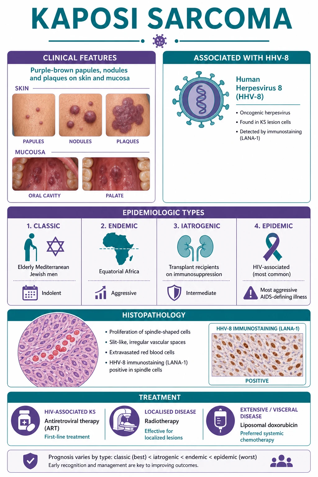
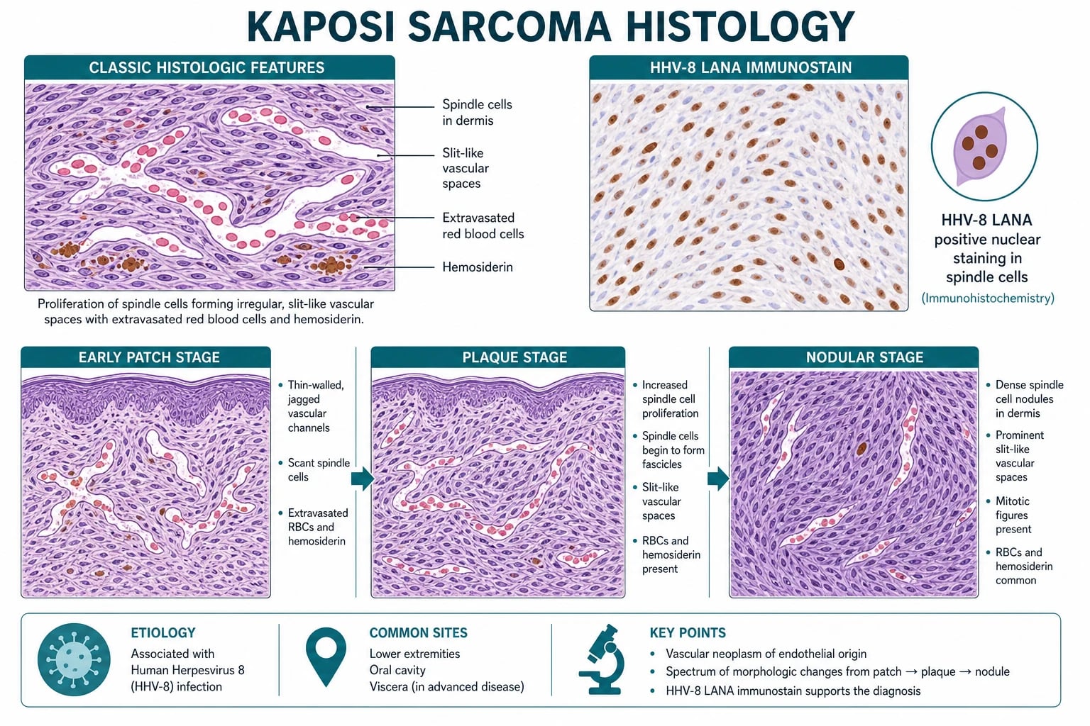
Kaposi sarcoma (KS) = a vascular tumour of lymphatic endothelial origin caused by human herpesvirus 8 (HHV-8 / KSHV). Presents as purple/red/brown macules, patches, plaques, and nodules on skin and mucosa (the hard palate is the classic oral site). Four epidemiologic variants share identical HHV-8 aetiology and histology: classic (elderly Mediterranean/Eastern European/Jewish men; lower legs; indolent), endemic/African (sub-Saharan Africa; aggressive; lymphadenopathic paediatric form), iatrogenic (solid-organ transplant / chronic immunosuppression), and epidemic/HIV-associated (an AIDS-defining illness; the commonest and most aggressive form). Histology shows spindle cells forming slit-like vascular spaces with extravasated RBCs and hemosiderin, confirmed by HHV-8 LANA immunostain. Management pivots on the cause: ART for HIV-associated KS (immune reconstitution may regress lesions), reduce immunosuppression and switch to an mTOR inhibitor (sirolimus) for transplant-associated KS, radiotherapy/cryotherapy for localised disease, and liposomal doxorubicin 20 mg/m2 every 3 weeks for extensive or visceral disease.
On this page & tools
Your progress
Saved locally on this device.
Exam tags
Red flags
Overview & Definition
Kaposi sarcoma (KS) is a locally aggressive angioproliferative tumour of endothelial (predominantly lymphatic) origin caused by infection of endothelial cells with human herpesvirus 8 (HHV-8), also known as Kaposi sarcoma-associated herpesvirus (KSHV).[1][5] First described by the Hungarian dermatologist Moritz Kaposi in 1872 as an "idiopathic multiple pigmented sarcoma of the skin," the disease was, for a century, a curiosity of elderly Mediterranean and Eastern European men. The HIV epidemic transformed it into a global clinical priority: in 1994 Chang and Moore identified HHV-8 in KS lesions, and in its epidemic (HIV-associated) form KS became an AIDS-defining illness and the most common tumour arising in people living with HIV.[1]
Despite the name "sarcoma," KS is best understood as a low-grade angioproliferative neoplasm rather than a true soft-tissue sarcoma: the proliferating spindle cell is an endothelial cell (often of lymphatic lineage, expressing markers such as PROX1, LYVE-1, and D2-40/podoplanin) that has been reprogrammed by latent HHV-8 infection.[1] The tumour is multifocal at onset — lesions appear independently in widely separated skin and visceral sites, a feature reflected in its staging and management. A single defining fact frames the whole chapter: HHV-8 is necessary but not sufficient for KS — the virus infects the majority of infected individuals silently, and clinical KS emerges only when host immunity fails to control it, whether through HIV, pharmacologic immunosuppression, or the waning immunity of age.[1][4]
Classification
The four epidemiologic variants of KS are clinically and prognostically distinct but histologically indistinguishable and share a single viral cause. Mastering the four-type framework is the single most frequently tested fact in this topic.[1]
Classic
Endemic (African)
Iatrogenic
Epidemic (HIV-associated)

Beyond the epidemiologic variants, cutaneous KS evolves through morphologic stages that mirror histologic progression — a fact examiners use to test recognition of early disease:[6]
Patch (early)
Plaque
Nodular (late)
Epidemiology & Risk Factors
KS is globally uncommon outside sub-Saharan Africa and populations living with HIV, but its distribution is sharply driven by two factors: HHV-8 seroprevalence in the population and the prevalence of immunosuppression.[1][5]
The principal risk factors cluster around viral exposure and immune compromise:[1][4]
- HHV-8 infection — the indispensable precursor. Transmitted sexually (predominant in HIV-associated KS, particularly among men who have sex with men), via saliva (the main route in endemic African children, with mother-to-child spread), and rarely through blood or transplanted organs.
- HIV infection — the dominant cofactor. HHV-8-positive men with untreated HIV have a 30 to 50 percent lifetime risk of KS, falling dramatically with effective ART. The risk is highest in those infected with HIV sexually and at low CD4 counts.
- Pharmacologic immunosuppression — calcineurin inhibitors (cyclosporine, tacrolimus) carry the highest KS risk in transplant recipients; the risk is lower with mTOR inhibitors (sirolimus, everolimus), which have intrinsic anti-KS activity.[9]
- Increasing age — waning cell-mediated immunity explains the classic form in elderly men.
- Geographic ancestry — Mediterranean, Eastern European, and Ashkenazi Jewish heritage for classic KS; equatorial African heritage for endemic KS.
- Male sex — all variants show male predominance, most striking in classic KS; hormonal or behavioural factors are invoked.
Pathophysiology
The pathogenesis of KS is a paradigm of virus-driven oncogenesis gated by host immunity, and it pays to learn the cascade in order, because each step is both a viva question and a therapeutic target.[1][4]
HHV-8 (KSHV) is a gamma-2-herpesvirus (Rhadinovirus genus) with a double-stranded DNA genome that has pirated a remarkable array of human cellular homologue genes. After infecting endothelial cells (via heparan sulfate, integrins, and other receptors), the virus establishes latency in a fraction of cells, while a smaller population supports lytic replication. The latent infection drives the proliferative phenotype of the spindle cell; the lytic cycle supplies paracrine angiogenic and inflammatory signals.[1][5]
The viral latency programme expresses a small set of oncoproteins and immune-evasion molecules that together convert a normal endothelial cell into a KS spindle cell: [1]
**HHV-8 infection of endothelial cells** (lymphatic lineage preferred) — establishes latency; LANA tethers the viral episome to host chromatin and blocks p53/Rb tumour-suppressor pathways.
**viral IL-6 (vIL-6)** — a viral homologue of human IL-6 that signals through gp130, driving proliferation, angiogenesis (via VEGF), and inflammation in an autocrine/paracrine loop.
**viral G-protein-coupled receptor (vGPCR)** — a constitutively active receptor (homologous to human IL-8 receptor) that activates MAPK, PI3K/Akt, and NF-kB, producing VEGF, angiopoietin, and inflammatory cytokines; the principal oncogenic driver of the lytic cascade.
**viral cyclin (vCyclin)** and **viral FLIP (vFLIP)** — push the cell cycle (resist cyclin-dependent kinase inhibitors) and block apoptosis (FLICE inhibitory protein).
**Angiogenesis and lymphangiogenesis** — VEGF, VEGF-C/D (lymphangiogenic), angiopoietin-2, and basic FGF secreted by infected spindle cells generate the vascular slits and leaky channels characteristic of KS.
**Extravasation of RBCs and hemosiderin deposition** — fragile vascular channels leak red cells; hemosiderin breakdown produces the purple-brown pigmentation of clinical lesions.
**Immunosuppression gates progression** — HIV-driven CD4 depletion, calcineurin-inhibitor therapy, or ageing immunity allow HHV-8 to evade cytotoxic T-cell surveillance; cytokines from co-infections (oncostatin M, IFN-gamma, TNF-alpha) further fuel lytic reactivation.
The same virus causes two other HHV-8-associated lymphoproliferative disorders that examiners may pair with KS in a viva: primary effusion lymphoma (PEL) — a B-cell lymphoma presenting as a malignant effusion in body cavities — and multicentric Castleman disease (MCD), especially its plasma-cell variant in HIV.[2] A patient may carry more than one HHV-8-associated diagnosis simultaneously, particularly in advanced HIV.
Clinical Presentation
The clinical picture of KS is dominated by cutaneous and mucosal vascular lesions, but its four epidemiologic variants differ in distribution, tempo, and visceral burden. The lesions themselves are stereotyped; what changes is where they appear and how fast they progress. [1]
Morphology of an individual lesion. KS begins as a pink, red, purple, or brown macule or patch, oval and typically aligned with skin lines, that evolves into a raised plaque and eventually a dome-shaped nodule that may ulcerate or bleed.[1][6] Lesions are usually non-pruritic and initially painless, though lymphoedematous or ulcerated lesions may become tender. A helpful clinical pearl: the colour darkens as hemosiderin accumulates, and lesions can mimic bruises that do not resolve. Multiple lesions at different stages coexist, reflecting the multifocal nature of the tumour.
Distribution by variant.[1][3]
- Classic KS — slowly progressive purple-brown patches, plaques, and nodules on the lower legs, ankles, and feet, often asymmetric, frequently complicated by lymphoedema. New lesions appear proximally over years; visceral involvement is uncommon and late.
- Endemic (African) KS — more aggressive cutaneous disease with visceral and nodal involvement common even in HIV-negative patients. The lymphadenopathic variant affects prepubertal children, presenting with generalised lymphadenopathy and little or no skin disease, and carries a poor prognosis.
- Iatrogenic KS — appears months to years after transplantation; distribution is variable (skin plus viscera). Often the first sign that immunosuppression is excessive.
- Epidemic (HIV-associated) KS — widespread cutaneous lesions on the face (nose, ears), trunk, genitals, and oral mucosa, often rapidly progressive, with visceral involvement frequent. [1]
Mucosal involvement is most characteristic of the epidemic form. The hard palate is the single most common oral site and is virtually pathognomonic in an HIV-positive patient — a purple, red, or bluish patch, plaque, or nodule on the hard palate that does not blanch.[1] Other oral sites include the gums, soft palate, and tongue. Lesions may ulcerate, bleed, and cause dysphagia or odynophagia.
Visceral KS affects the gastrointestinal tract, respiratory tract, and lymph nodes, and less commonly the liver, spleen, and other organs:[3][6]
- Gastrointestinal — the commonest visceral site; may cause occult or overt bleeding, abdominal pain, diarrhoea, or obstruction, even when asymptomatic endoscopically.
- Pulmonary — dyspnoea, cough, wheeze, and haemoptysis; imaging shows lower-lobe-predominant interstitial or nodular infiltrates, pleural effusions, or endobronchial lesions. Pulmonary KS carries a worse prognosis.
- Lymph nodes and lymphatics — lymphadenopathy and lymphoedema disproportionate to the visible cutaneous burden (lymphatic infiltration impairs drainage); the lymphoedema can be painful, disfiguring, and refractory. [1]
Atypical presentations examiners test deliberately.[3][10]
- KS-IRIS — paradoxical worsening or new appearance of KS within weeks to months of ART initiation, in the setting of immune recovery. More common when ART is started at very low CD4 counts and high viral loads; high fever and lymphadenopathy may accompany the flare.
- Isolated visceral KS without skin lesions — GI or pulmonary KS as the first manifestation, especially in iatrogenic disease.
- Classical KS in a young non-Mediterranean patient — raises the question of unrecognised HIV or immunosuppression.
- KS presenting as painful lymphoedema alone — lymphangitic KS with minimal skin signs. [1]
Dermoscopy of cutaneous KS. Dermoscopy is a useful adjunct at the bedside but is not diagnostic — biopsy remains mandatory. The classical pattern is violaceous to bluish-reddish homogeneous areas corresponding to the dense vascular proliferation, traversed by small brownish globules and structureless zones reflecting hemosiderin and extravasated erythrocytes.[6] A characteristic finding is a "rainbow pattern" — multicoloured (blue, red, white, yellow) areas seen under polarised dermoscopy — that is suggestive though not pathognomonic (it is also reported in melanoma and other vascular lesions). Early patch-stage KS may show only a fine, irregular vascular network with dotted/glomerular vessels, easily mistaken for an inflammatory dermatitis; nodular-stage disease shows a denser homogeneous violaceous background with whitish veil. The dermatoscopic finding that should always prompt biopsy in an at-risk patient is any new violaceous vascular-appearing lesion on the hard palate, nose, or lower leg.
[1]Differential Diagnosis
The KS differential centres on vascular and pigmented lesions of skin and mucosa. The discriminator examiners want is the recognition that bacillary angiomatosis is the great mimic — and is treatable with antibiotics.[1][6]
Bacillary angiomatosis
Pyogenic granuloma
Angiosarcoma
Stasis dermatitis / purpura
Dermatofibroma
Acroangiodermatitis (Mali)
Other considerations include microvenular haemangioma, targetoid haemosiderotic haemangioma, arteriovenous malformation, cutaneous lymphoma, and amelanotic melanoma. The decisive discriminator is always the skin biopsy with HHV-8 LANA immunostain: a LANA-positive spindle-cell vascular tumour with slit-like spaces and extravasated RBCs is KS. [1]
Clinical & Bedside Assessment
A focused KS examination is systematic because the tumour is multifocal and visceral disease changes management.[1]
- Full skin examination, including scalp, retroauricular skin, ears and nose (commonly involved in HIV-KS), oral cavity (lift the tongue; inspect the hard palate — the single highest-yield site), genitalia, palms, soles, and web spaces. Document the number, morphology (patch/plaque/nodule), colour, size, and distribution of lesions, and whether any are ulcerated or bleeding.
- Lymph node examination — cervical, axillary, epitrochlear, inguinal. Lymphadenopathy may indicate KS, a second HHV-8-associated disorder (MCD), or an unrelated process.
- Lymphoedema assessment — measure limb circumference; grade severity; KS-related lymphoedema is often disproportionate to visible cutaneous disease.
- Abdominal examination — hepatosplenomegaly, masses, tenderness (GI KS).
- Respiratory examination — crackles, wheeze, effusion, signs of pulmonary KS (a respiratory flare in an HIV patient with cutaneous KS is pulmonary KS until excluded).
- Symptom review: GI bleeding or melaena, abdominal pain, dysphagia, dyspnoea, cough, haemoptysis, fevers, night sweats, weight loss (B symptoms). [1]
Investigations
The skin biopsy is the diagnostic gold standard; serology and imaging stage the disease and characterise the immunosuppressive context.[1][6]
Skin biopsy (H&E)
HHV-8 LANA immunostain
HIV test and CD4 count
Baseline bloods
Imaging — extent
Endoscopy / bronchoscopy
HHV-8 blood PCR (selected)
Management — Resuscitation
KS is rarely a time-critical resuscitation problem in itself; the lesions evolve over days to weeks. The genuine emergencies relate to visceral KS complications and to its immunosuppressive context:[3][10]
- Massive GI bleeding or melaena from GI KS — resuscitate with IV access, fluids, and blood transfusion; arrange urgent upper and lower GI endoscopy; consider systemic chemotherapy for diffuse disease.
- Pulmonary KS with respiratory failure — oxygen, ventilatory support as needed; urgent bronchoscopy/CT; systemic chemotherapy (liposomal doxorubicin) is first-line; manage co-existing opportunistic infection.
- KS-associated IRIS with organ compromise — do not stop ART; systemic steroids are used with caution and specialist input; systemic chemotherapy may be required for progressive disease.
- Newly diagnosed HIV — confirm with CD4 and viral load, start ART promptly (immune restoration is itself treatment for KS), screen for and treat opportunistic infections, and arrange oncology/dermatology follow-up. [1]
The overarching resuscitative principle is to identify and treat the immunosuppressive driver: in HIV this means ART; in transplant recipients it means reviewing and adjusting immunosuppression. Without addressing the driver, local therapy alone rarely controls KS for long. [1]
Management — Definitive & Stepwise
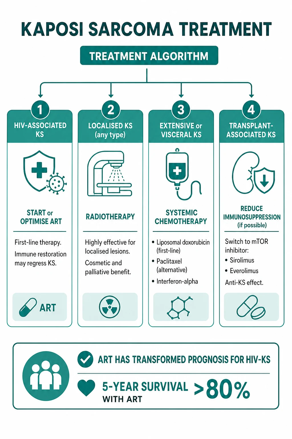
Definitive management is stratified by variant, extent, and immune context. The unifying principle: restore immune control of HHV-8 wherever possible, then layer on local or systemic therapy for symptomatic, cosmetic, or visceral disease.[1][3]
Epidemic (HIV-associated) KS
**Start or optimise ART** — first-line for all stages of HIV-associated KS. Immune restoration (rising CD4, suppressed viral load) leads to **partial or complete KS regression in 60 to 80 percent** of patients within months. ART is continued regardless of KS response.
**Assess extent and symptom burden** — limited cutaneous disease may need only ART plus local therapy; bulky, painful, cosmetically distressing, or visceral disease requires additional treatment.
**Local therapy** — **radiotherapy** (highly effective for localised cutaneous/oral lesions, including palliation of painful or ulcerated nodules), **cryotherapy** (small superficial lesions), **intralesional vinblastine** (small number of lesions), **topical alitretinoin gel** (limited cutaneous disease), or **surgical excision** (diagnostic or solitary symptomatic lesions).
**Systemic chemotherapy** for extensive (T1), rapidly progressive, symptomatic visceral, or ART-unresponsive disease — **pegylated liposomal doxorubicin 20 mg/m2 IV every 3 weeks** is first-line (4 to 6 cycles typical); alternatives include **paclitaxel 100 mg/m2 every 2 weeks** for refractory disease.
**Investigational/emerging** — pomalidomide, immunomodulatory agents, and mTOR inhibitors in selected cases; consult HIV-oncology.
Pegylated liposomal doxorubicin
Dose
20 mg/m2
Iatrogenic (Transplant-associated) KS
The decisive move is to modify the immunosuppressive regimen rather than reach for chemotherapy first. The aim is to regain immune control of HHV-8 while protecting the graft.[1][9]
**Reduce immunosuppression** to the lowest level compatible with graft survival — reduction alone produces regression in many cases, particularly early post-transplant KS.
**Switch the calcineurin inhibitor (cyclosporine or tacrolimus) to an mTOR inhibitor (sirolimus or everolimus)** — mTOR inhibitors have **intrinsic anti-KS and anti-angiogenic activity** (they block the PI3K/Akt/mTOR axis that HHV-8 exploits) and may produce KS regression while maintaining immunosuppression.<Cite id='9' />
**Local therapy** (radiotherapy, cryotherapy, excision) for residual or symptomatic cutaneous lesions.
**Systemic chemotherapy** (liposomal doxorubicin) only for extensive, progressive, or visceral disease not controlled by regimen modification — coordinate with the transplant team to balance oncologic and graft outcomes.
Classic KS
Classic KS is indolent and rarely life-threatening; treatment is shaped by symptoms, cosmesis, and patient preference.[1]
- Observation is reasonable for asymptomatic indolent disease.
- Radiotherapy — the workhorse for localised symptomatic or cosmetic disease; highly effective on the lower legs.
- Cryotherapy and intralesional chemotherapy (vinblastine) — useful for a small number of lesions.
- Surgical excision — for biopsy or a solitary symptomatic nodule; KS is multifocal, so excision is not curative.
- Systemic therapy (liposomal doxorubicin) reserved for extensive, painful, lymphoedematous, or progressive disease. [1]
Endemic (African) KS
Treated with the same modalities but more aggressive course warrants earlier systemic therapy. In HIV-positive patients, ART is first-line (the commonest scenario in sub-Saharan Africa). For HIV-negative endemic KS with extensive or visceral disease, liposomal doxorubicin is standard, with radiotherapy for local control. Resource constraints and access to ART/chemotherapy shape regional pathways (see Regional Differences). [1]
Local therapy across variants
RICE for localised KS
Specific Subtypes & Scenarios
KS-associated immune reconstitution inflammatory syndrome (KS-IRIS)
A paradoxical worsening or new onset of KS within weeks to months of starting ART, KS-IRIS is a T-cell-driven inflammatory reaction against HHV-8-infected cells as immunity recovers.[10] Risk is highest in patients starting ART with very low CD4 counts (often below 50 to 100), high viral load, pre-existing subclinical KS, and rapid CD4 rise. Presentation is explosive cutaneous and nodal KS (sometimes visceral) with fever and systemic inflammation. Management is conservative: continue ART, treat opportunistic infections, and reserve systemic steroids and chemotherapy for severe or organ-threatening disease. The prognosis is generally favourable if ART is sustained, but visceral IRIS-KS can be fatal.[10]
Paediatric endemic KS
The lymphadenopathic variant of endemic KS affects prepubertal children in equatorial Africa, presenting with generalised lymphadenopathy and minimal or no skin disease, often with visceral involvement and a rapidly progressive course. Diagnosis rests on lymph node biopsy (spindle-cell vascular tumour, LANA-positive). Management combines ART where HIV-positive with systemic chemotherapy.[1]
Isolated visceral KS
GI, pulmonary, or nodal KS may occur without visible cutaneous lesions, particularly in iatrogenic disease. A high index of suspicion is required in immunosuppressed patients with unexplained GI bleeding, unexplained pulmonary infiltrates, or lymphadenopathy — biopsy is diagnostic.[3][6]
Coincident HHV-8-associated disorders
A patient with KS may simultaneously develop multicentric Castleman disease (MCD) (B symptoms, anaemia, hypoalbuminaemia, diffuse lymphadenopathy, polyclonal inflammatory markers) or primary effusion lymphoma (PEL) (lymphomatous effusions in body cavities). Recognising MCD is critical because it requires its own treatment (rituximab-based, siltuximab).[2]
Complications & Pitfalls
KS complications arise from the tumour, its immunosuppressive context, and its treatment.[1][3]
Lymphoedema
Visceral involvement
KS-IRIS
Secondary malignancy
Treatment toxicity
Classic pitfalls to avoid include failing to test HIV in any new KS; biopsying a lesion without HHV-8 LANA staining and missing the diagnosis in a morphologically borderline vascular tumour; confusing bacillary angiomatosis for KS and omitting antibiotics; stopping ART during KS-IRIS (it must be continued); and escalating to chemotherapy before optimising ART or modifying transplant immunosuppression, when the cause is treatable. [1]
Prognosis & Disposition
Prognosis is dominated by the variant and the immune context, not by the tumour bulk alone.[1][3]
Poor prognostic factors include visceral (T1) disease, low CD4 (below 150 to 200 cells/microL), high HIV viral load, systemic illness/opportunistic infection (S1), poor performance status (Karnofsky below 70), and failure to achieve immune restoration.[8] The strongest single favourable factor in epidemic KS is sustained ART with viral suppression and CD4 recovery.[3]
Disposition depends on disease extent. Limited cutaneous KS can be managed outpatient with ART (HIV) or regimen modification (transplant) plus local therapy. Extensive, rapidly progressive, or visceral KS requires specialist oncology/haematology input, often with inpatient systemic chemotherapy. Newly diagnosed HIV requires linkage to HIV care and prompt ART initiation. Transplant-KS requires close coordination between oncology and the transplant team. [1]
Special Populations
- Pregnancy — KS is uncommon in pregnancy. The principle is to continue or initiate ART for HIV-positive mothers (ART is safe and effective), avoid systemic chemotherapy where possible, defer cosmetic local therapy to the postpartum period, and involve maternal-fetal medicine and HIV specialists. Vertical transmission of HHV-8 is uncommon.
- Paediatrics — the endemic lymphadenopathic variant (see Specific Subtypes) is the dominant paediatric form; ART if HIV-positive and systemic chemotherapy for extensive disease.
- Transplant recipients — see Management; the mTOR switch is the cornerstone, balanced against graft risk. KS may regress with adjustment alone.[9]
- Non-HIV, non-transplant immunosuppression — long-term high-dose corticosteroids, immunosuppressive biologics, and autoimmune disease have all been associated with KS; reducing immunosuppression where feasible is the first therapeutic lever.
- Elderly with classic KS — emphasis is on quality of life, cosmesis, and avoiding overtreatment of indolent disease; radiotherapy is well tolerated.
Evidence, Guidelines & Regional Differences
Landmark studies and references that anchor KS management:[1][3]
Northfelt 1997 — pegylated liposomal doxorubicin in AIDS-related KS
Phase II/III study of pegylated liposomal doxorubicin in AIDS-related KS after failure of standard chemotherapy (doxorubicin, bleomycin, vincristine).
Key finding
Significant tumour response (overall response rates around 50 to 60 percent) with favourable tolerability compared with conventional anthracycline regimens; established pegylated liposomal doxorubicin as the first-line systemic agent for advanced KS.<Cite id='7' />
Krown 1997 — ACTG TIS staging validation
Prospective validation of the AIDS Clinical Trials Group (Tumour/Immune/Systemic illness) staging classification in AIDS-related KS.
Key finding
Demonstrated that the **TIS axes independently predict survival**, with poor-risk patients (any of T1, I1, S1) doing substantially worse.<Cite id='8' />
The disease is global; the drugs and pathways are regional.[3]
[1] [1]WHO sub-Saharan Africa pathways — endemic and epidemic KS common; ART scale-up has transformed prognosis; radiotherapy and liposomal doxorubicin access vary by region; paclitaxel and investigational agents where available. EACS (European AIDS Clinical Society) guidelines align ART-first principles with NCCN. Where resources are limited, ART plus palliative radiotherapy remains the backbone.
Controversies and emerging evidence. Whether to give empiric chemotherapy with ART versus ART-alone first remains debated for moderate-burden disease; the optimal management of KS-IRIS (role of steroids, timing of chemo) is evolving; pomalidomide and other immunomodulators show promise; and mTOR inhibitors are being explored beyond transplant-KS.[3]
Exam Pearls
Exam application bank (NEET-PG / INICET)
One-line answer
Kaposi sarcoma (KS) = a vascular tumour of lymphatic endothelial origin caused by human herpesvirus 8 (HHV-8 / KSHV). Presents as purple/red/brown macules, patches, plaques, and nodules on skin and mucosa (the hard palate is the classic oral site). Four epidemiologic variants share identical HHV-8 aetiology and histology: classic (elderly Mediterranean/Eastern European/Jewish men; lower legs; indolent), endemic/African (sub-Saharan Africa; aggressive; lymphadenopathic paediatric form), iatrogenic (solid-organ transplant / chronic immunosuppression), and epidemic/HIV-associated (an AIDS-defining illness; the commonest and most aggressive form). Histology shows spindle cells forming slit-like vascular spaces with extravasated RBCs and hemosiderin, confirmed by HHV-8 LANA immunostain. Management pivots on the cause: ART for HIV-associated KS (immune reconstitution may regress lesions), redu
Worked stems (answer without another resource)
Stem 1 — Classic presentation. Map symptoms to mechanism; name the first investigation and first treatment step with dose/route if drug therapy is standard. [1]
Stem 2 — Unstable / complicated. List red flags that force immediate resuscitation, theatre, ICU, antidote, or reperfusion — and what you do in the first 15 minutes. [1]
Stem 3 — Atypical group. Elderly, pregnancy, child, or immunocompromised: how presentation and thresholds change. [1]
Stem 4 — Differential trap. Name the three closest mimics and one discriminator for each. [1]
Stem 5 — Disposition. Who goes home with safety-netting, who is admitted, who needs HDU/ICU/theatre, and what follow-up is mandatory. [1]
Rapid viva checklist
- Definition + classification
- Pathophysiology chain
- Bedside signs / criteria
- Score with exact components (if any)
- Emergency bundle
- Definitive therapy with doses
- Complications of disease and of treatment
- Special populations
- Guideline/trial name if classic
- Three exam traps
Coverage self-check
If you cannot answer any stem above from this page alone, re-read the matching section — the page is intended to be self-sufficient for final-prof and NEET-PG/INICET questions on Kaposi sarcoma.
Rapid Self-Test
What is the causative virus of Kaposi sarcoma, and why is the virus 'necessary but not sufficient'?
Human herpesvirus 8 (HHV-8 / KSHV), a gamma-2-herpesvirus. HHV-8 infection is necessary, but clinical KS emerges only when host T-cell immunity fails (HIV, transplant immunosuppression, ageing) — hence the four epidemiologic variants all involve some form of immune compromise.[1]
Name the four epidemiologic variants and the single feature that distinguishes the epidemic form.
Classic (elderly Mediterranean/Jewish; lower legs; indolent), endemic/African (sub-Saharan Africa; aggressive; paediatric lymphadenopathic variant), iatrogenic (transplant/chronic immunosuppression), and epidemic/HIV-associated (an AIDS-defining illness and the commonest form globally). The epidemic form is distinguished by being an AIDS-defining illness.[1]
Describe the histology of KS and the confirmatory immunostain.
Spindle cells forming slit-like vascular spaces, with extravasated RBCs and hemosiderin; the early patch stage shows the promontory sign (a pre-existing vessel protruding into a new vascular channel). The HHV-8 LANA (latency-associated nuclear antigen) immunostain shows characteristic speckled nuclear positivity in spindle cells and confirms the diagnosis.[1][6]
First-line management of (a) HIV-associated KS, (b) transplant-associated KS, (c) extensive visceral KS.
(a) Start or optimise ART — immune reconstitution produces partial/complete KS regression in 60 to 80 percent. (b) Reduce immunosuppression and switch the calcineurin inhibitor to sirolimus (mTOR inhibitor), which has intrinsic anti-KS activity. (c) Pegylated liposomal doxorubicin 20 mg/m2 IV every 3 weeks is first-line systemic chemotherapy.[1][3][9]
What is the great mimic of KS and how do you distinguish it?
Bacillary angiomatosis (Bartonella henselae/quintana) — also in HIV. It resembles KS clinically but is antibiotic-responsive (erythromycin or doxycycline). On histology it shows a lobular capillary proliferation with neutrophils and Warthin-Starry-positive bacilli, and the HHV-8 LANA stain is negative. Failing to consider it and omitting antibiotics is a classic exam error.[6]
What is KS-IRIS and what must you NOT do?
KS-associated immune reconstitution inflammatory syndrome — a paradoxical flare or new-onset KS within weeks to months of starting ART, as T-cell immunity recovers against HHV-8-infected cells. Risk is highest with very low baseline CD4 and high viral load. The critical rule: do NOT stop ART — continue it, treat opportunistic infections, and reserve systemic steroids/chemotherapy for severe or organ-threatening disease.[10]
References
- [1]Cesarman E, Damania B, Krown SE, et al. Kaposi sarcoma Nat Rev Dis Primers, 2019.PMID 30705286
- [2]Carbone A, Borok M, Damania B, et al. Castleman disease Nat Rev Dis Primers, 2021.PMID 34824298
- [3]Patel R, Lurain K, Yarchoan R, et al. Clinical management of Kaposi sarcoma herpesvirus-associated diseases: an update on disease manifestations and treatment strategies Expert Rev Anti Infect Ther, 2023.PMID 37578202
- [4]Xiao Q, Liu Y, Li T, et al. Viral oncogenesis in cancer: from mechanisms to therapeutics Signal Transduct Target Ther, 2025.PMID 40350456
- [5]Iftode N, Radulescu MA, Arama SS, et al. Update on Kaposi sarcoma-associated herpesvirus (KSHV or HHV8) - review Rom J Intern Med, 2020.PMID 32681788
- [6]Radu O, Pantanowitz L. Kaposi sarcoma Arch Pathol Lab Med, 2013.PMID 23368874
- [7]Northfelt DW, Dezube BJ, Thommes JA, et al. Efficacy of pegylated-liposomal doxorubicin in the treatment of AIDS-related Kaposi's sarcoma after failure of standard chemotherapy J Clin Oncol, 1997.PMID 9053490
- [8]Krown SE, Testa MA, Huang J, et al. AIDS-related Kaposi's sarcoma: prospective validation of the AIDS Clinical Trials Group staging classification. AIDS Clinical Trials Group Oncology Committee J Clin Oncol, 1997.PMID 9294471
- [9]Stallone G, Infante B, Grandaliano G, et al. Kaposi's sarcoma and mTOR: a crossroad between viral infection neoangiogenesis and immunosuppression Transpl Int, 2008.PMID 18498314
- [10]Poizot-Martin I, Bregigeon S, Palich R, et al. Immune Reconstitution Inflammatory Syndrome Associated Kaposi Sarcoma Cancers (Basel), 2022.PMID 35205734