Dermatology · Medicine
Keloid and Hypertrophic Scar
Also known as Keloid · Hypertrophic scar · Keloid scar · Cheloid · Cheloid scar · Crab-claw scar
Keloids and hypertrophic scars are benign fibroproliferative disorders of the remodelling phase of wound healing with excessive collagen deposition. The pivotal distinction: a keloid extends BEYOND the original wound margins and does not regress; a hypertrophic scar stays WITHIN the wound and may regress over 12-18 months. Skin of colour (Fitzpatrick IV-VI; African, Asian, Hispanic) carries a 15-fold higher risk. Pathophysiology centres on TGF-beta1/beta2 overexpression, fibroblast hypersensitivity, reduced apoptosis of myofibroblasts, and a collagen-synthesis/degradation imbalance. Intralesional triamcinolone 10-40 mg/mL every 4-6 weeks is first-line; silicone gel sheeting, cryotherapy, 5-FU, verapamil, bleomycin, interferon, imiquimod, and PDL laser are adjuncts; surgical excision MUST be combined with adjuvant therapy (post-excision radiation within 24 h, intralesional steroid, pressure earrings) because excision alone recurs 50-100%.
On this page & tools
Your progress
Saved locally on this device.
Practise this topic
Exam tags
Red flags
Overview & Definition
Keloids and hypertrophic scars are benign fibroproliferative disorders of the remodelling phase of wound healing in which dermal fibroblasts deposit excessive, disorganised collagen, producing a raised scar. They lie on a continuum of pathological scarring, share a final common pathway of exuberant collagen deposition, and are the leading reason patients present to a dermatology or plastic-surgery scar service. The classification is clinical and morphological; histology is reserved for atypical cases.[2][4]
The pivotal, examiner-favoured distinction is the relationship of the scar to the original wound:[2][4]
- Keloid — a firm, rubbery, nodular mass of fibrocollagenous tissue that extends BEYOND the boundaries of the original wound into the surrounding normal-appearing skin, producing a claw-like or pseudopodial ("crab-claw") margin. Keloids do not regress spontaneously and characteristically recur after excision (50-100% if excised without adjuvant therapy). The name derives from the Greek chele (χηλή) = crab's claw.
- Hypertrophic scar — a raised, erythematous, firm scar that remains confined WITHIN the boundaries of the original wound, develops earlier (within 4-8 weeks of injury) and may regress spontaneously over 12-24 months, although it can persist, contract across joints, and cause significant functional impairment. [1]
A normal scar, by contrast, is flat, pale, soft, and remains within the wound. A keloid is, in essence, a scar that does not know when to stop. [1]
Classification
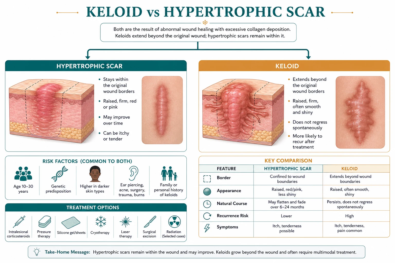
By morphology and clinical behaviour
Keloid
BEYOND wound margin — does NOT regress
- **Extends BEYOND** original wound into surrounding normal skin (claw-like / pseudopodial)
- **Does NOT regress** spontaneously; often continues to grow for months to years
- Recurrence after simple excision: **50-100%**
- High-tension sites: sternum, deltoid, upper back, earlobes, jawline, neck
- Strong racial predisposition (Fitzpatrick IV-VI, 15-fold higher)
- Histology: thick hyalinised, **keloidal collagen** bundles in whorls; reduced elastic fibres; random orientation
- Genetic predisposition common; HLA associations described
Hypertrophic scar
WITHIN wound margin — may regress
- **Confined WITHIN** the original wound margin
- Often **regresses** spontaneously over 12-24 months
- Recurrence after excision: **lower** than keloid
- Flexor surfaces, across joints, and any wound under tension
- Strongly associated with **burns** and **surgical wounds closed under tension**
- Histology: **parallel myofibroblast/collagen nodules**, oriented along tension lines
- Often improves with pressure, silicone, and intralesional steroid
Normal scar / atrophic scar
Flat — physiologic healing
- **Flat, pale, soft**, within wound margins
- Atrophic scar: depressed below the surface (e.g. acne scars, striae)
- Mature scar: collagen remodelling complete, type I collagen predominates
- No active fibroblast proliferation; no further change after 12-18 months
- No treatment required; can be revised for cosmesis if desired
By site and trigger (clinically useful subtyping)
Epidemiology & Risk Factors
Keloids are a global problem with a striking racial and age distribution. They are uncommon in fair-skinned populations and markedly more common in skin of colour.[2][4]
Risk factors and precipitants — the high-yield list: [1]
- Skin of colour — Fitzpatrick IV-VI; the single biggest risk factor. Reported in 4.5-16% of Black, Hispanic, and Asian populations, compared with under 1% of Caucasians. The fibroblasts in darker skin are intrinsically more active and produce more collagen per cell in response to growth factors.
- Genetic predisposition — autosomal dominant with incomplete penetrance in some families; several loci (e.g. chromosomes 2q23, 7p11) and HLA alleles (HLA-DRB1*, HLA-DQA1*0501, HLA-DQB1*0501) have been associated; IL-6 promoter polymorphisms, p53 codon 72 polymorphism, and connective-tissue growth factor variants are also implicated. Syndromic associations include Rubinstein-Taybi (keloids + broad thumbs + short stature) and Dubowitz (keloids + microcephaly + intellectual disability + eczema).[12]
- Age — peak incidence 10-30 years; rare in the elderly and in pre-pubertal children. The peak follows the rise in dermal fibroblast activity at puberty and may reflect androgen/oestrogen sensitivity.
- Site of injury — high-tension and highly mobile areas: chest/sternum, deltoid/shoulder, upper back, jawline, neck, ear (especially after piercing), and the abdomen. Keloids spare the palms, soles, and genitalia (no pilosebaceous unit, lower fibroblast density).
- Type of injury — minor skin trauma: ear piercing, acne, vaccination, insect bite, burn, surgical incision, laceration, tattoo, injection site. Even a clean surgical incision can produce a keloid in a susceptible individual. Delayed epithelialisation, foreign body, infection, and wound tension all increase risk.
- Pregnancy and oestrogen exposure — keloids may enlarge during pregnancy due to oestrogen-driven fibroblast proliferation; they may also regress partially post-partum. Keloids typically regress after the menopause.
- Wound-healing factors — wound infection, foreign-body reaction, delayed epithelialisation, increased wound tension perpendicular to the skin tension lines (Langer's lines), and repeated trauma (e.g. friction from clothing on a sternal wound) all perpetuate fibroblast activation.
Why high-tension sites? The chest, shoulder, and suprapubic regions lie perpendicular to the lines of skin tension, so even a small wound is pulled apart at the edges during normal movement. This mechanical force activates mechanotransduction pathways in fibroblasts (including the focal-adhesion kinase / ERK / MRTF-SRF axis) that drive collagen synthesis. The clinical correlate is that incisions placed parallel to the skin tension lines heal with less scarring than those placed perpendicular to them. [1]
Pathophysiology
The pathophysiology of keloid and hypertrophic scar formation is a disordered remodelling phase of wound healing, with the underlying wound having progressed normally through haemostasis, inflammation, and proliferation.[1][4]
The normal wound-healing cascade (a brief recap, since the scar disorder is a deviation from this):[1]
- Haemostasis (minutes-hours) — platelet plug, fibrin clot, release of PDGF, TGF-beta, EGF from platelet alpha-granules.
- Inflammation (days 1-3) — neutrophils clear bacteria, macrophages (M1 → M2) clear debris and release cytokines (TGF-beta, PDGF, FGF, VEGF) that recruit fibroblasts and angiogenic cells.
- Proliferation (days 3-21) — fibroblasts synthesise type III collagen, glycosaminoglycans, and elastin; granulation tissue forms; angiogenesis is active; keratinocytes migrate to re-epithelialise; myofibroblasts (TGF-beta-driven) contract the wound.
- Remodelling (weeks-months, up to 1-2 years) — type III collagen is replaced by type I collagen; matrix metalloproteinases (MMPs) remodel the ECM; fibroblasts and myofibroblasts undergo apoptosis; the wound matures and gains tensile strength (never reaching more than ~80% of normal skin). [1]
In a keloid or hypertrophic scar, the remodelling phase fails to switch off — fibroblasts persist, continue to deposit collagen, and the scar grows beyond the original wound margin (keloid) or thickens within it (hypertrophic scar).[2][4]
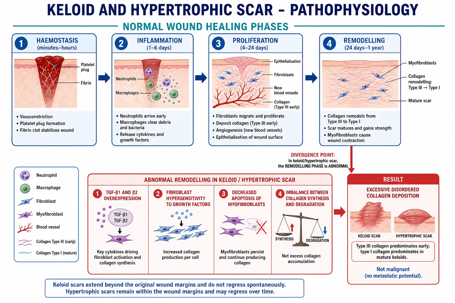
Molecular mechanisms — the high-yield detail
TGF-beta / SMAD axis
Master regulator of fibrosis
- **TGF-beta1 and TGF-beta2** are upregulated in keloid fibroblasts; both drive fibroblast proliferation and collagen synthesis
- **TGF-beta3** is anti-fibrotic and is *reduced* in keloids (a relative deficiency that promotes scarring)
- TGF-beta signals through **SMAD2/3** (profibrotic) versus SMAD1/5/8 (antifibrotic); keloid fibroblasts shift the balance toward SMAD2/3
- Downstream effect: ↑ collagen I, III, fibronectin, α-SMA; ↓ collagenase (MMP-1)
Fibroblast biology
More cells, more collagen, less death
- Increased fibroblast **proliferation** in response to PDGF, EGF, IGF-1
- Keloid fibroblasts are **hyper-responsive** — they produce 3-4× more collagen per cell than normal fibroblasts
- **Decreased apoptosis** of keloid fibroblasts and myofibroblasts (Bcl-2 ↑, Bax ↓; resistant to FasL-mediated death)
- Abnormal collagen **lattice contraction** in keloid-derived fibroblasts in vitro
Collagen remodelling
Synthesis wins, degradation loses
- **Increased collagen synthesis** — particularly type III collagen early; mature keloids shift toward type I
- Altered **type I:III ratio** — normal scar ~4:1, keloid ~3:1 (favours type III)
- **Decreased MMP-1, MMP-3, MMP-9** (collagenases) in keloid tissue
- **Increased TIMP-1, TIMP-2** (tissue inhibitors of metalloproteinases) — net reduction in collagen degradation
- Result: **net collagen accumulation**
Other contributors
Genetic, vascular, mechanical
- **Genetic**: HLA-DRB1*, IL-6 promoter polymorphism, p53 codon 72 polymorphism; rarely somatic p53 mutations (mostly wild-type with abnormal protein distribution)
- **Vascular**: increased microvessel density, ↑VEGF, ↑HIF-1α under chronic hypoxia
- **Mechanical**: wound tension activates focal-adhesion-kinase/ERK/MRTF-SRF → ↑α-SMA and collagen
- **Immunological**: T-helper-2 cytokine profile (↑IL-4, IL-13) promotes fibrosis; mast cells and macrophages are abundant
- **Hypoxia**: relative hypoxia in the early wound drives VEGF and angiopoietin, supporting the dense microvasculature
Mechanical tension — a unifying concept
Mechanotransduction is increasingly recognised as a central driver of keloid biology. The high-tension sites (chest, deltoid, sternum) experience continuous stretch; fibroblasts sense this via integrins, focal adhesions, and the actin cytoskeleton, transducing it into a profibrotic gene-expression programme. The therapeutic corollary is that off-loading tension is itself a treatment — pressure garments, silicone, and tension-releasing surgical techniques (e.g. subcutaneous advancement flaps, z-plasties) all reduce scar formation. Botulinum toxin injection reduces dynamic wound tension by paralysing underlying musculature and is a logical adjunct in high-tension sites.[2][3]
Clinical Presentation
Keloid
A keloid presents as a firm, rubbery, nodular or lobulated mass that has grown beyond the boundaries of the original wound into the surrounding normal-appearing skin. The classic descriptor is "claw-like" or "pseudopodial" extensions (the crab claw of its Greek etymology). The surface is usually smooth and shiny, often with telangiectasia, and the colour evolves from erythematous or pink in the early phase to purple and ultimately hyperpigmented or hypopigmented in mature lesions.[2][4]
Common clinical features: [1]
- Morphology — firm, rubbery, nodular or lobulated, with claw-like extensions beyond the wound margin.
- Surface — smooth, shiny, sometimes with telangiectasia; occasionally bosselated or pedunculated (especially earlobe keloids).
- Colour — pink/red in early lesions, purple in intermediate, brown/hyperpigmented in mature lesions in skin of colour.
- Symptoms — pruritus (itch) is the most common and often most distressing; pain, tenderness, burning, and hyperaesthesia are frequent. Spontaneous discharge is rare.
- Site — chest/sternum, shoulders/deltoid, upper back, jawline, earlobes, neck, abdomen. Palms, soles, and genitalia are spared (low fibroblast density in volar skin).
- Onset — usually weeks to months after injury; can grow for months to years and may continue to enlarge indefinitely.
- Triggers — minor skin trauma: ear piercing, acne, vaccination, insect bite, surgical incision, burn, tattoo, injection site. Spontaneous keloids (no trauma history) occur in ~3% of cases, usually on the chest, and should raise the question of an underlying genetic syndrome.
- Number — usually solitary; multiple keloids in distinct sites suggest a strong genetic predisposition.
- Course — does not regress; continues to grow slowly over months to years; recurrence after treatment is the rule rather than the exception. [1]
Hypertrophic scar
A hypertrophic scar is a raised, erythematous, firm scar that remains WITHIN the boundaries of the original wound. It typically develops earlier (within 4-8 weeks of injury) than a keloid, may be intensely red and pruritic in the early months, and tends to regress spontaneously over 12-24 months, although it can persist and become a significant functional or cosmetic problem.[1][4]
Common clinical features: [1]
- Morphology — raised, firm, often linear or following the wound shape; surface may be smooth or nodular.
- Site — flexor surfaces, across joints, and any wound closed under tension. Burns (especially deep partial-thickness) are a classic cause of extensive hypertrophic scarring.
- Course — develops 4-8 weeks after injury; mature scar forms by 6-12 months; may regress (flatten, soften, fade) over the following 12-24 months.
- Contracture — burns-related hypertrophic scars may contract across joints, restricting range of motion (a functional emergency if a finger, neck, or limb is involved).
- Symptoms — pruritus, erythema, tightness; pain is less common than in keloid. [1]
Atypical presentations and exam-relevant variants
- Paediatric keloids — rare under 10 years, but keloids in this group tend to be more aggressive, larger, and more resistant to treatment. Avoid radiation because of lifetime cancer risk. Silicone + intralesional steroid + cryotherapy are preferred; lower steroid doses are used.
- Pregnancy-associated keloids — can enlarge during pregnancy (oestrogen-driven fibroblast proliferation) and partially regress post-partum.
- Spontaneous keloids — chest, no trauma history; consider genetic syndromes (Rubinstein-Taybi, Dubowitz, Goeminne syndrome) and refer to clinical genetics if there are other features.
- Eyelid keloid — rare; can obstruct the visual axis; functional priority.
- Periorificial keloid (mouth, nostril, ear canal) — can restrict opening; functional priority.
- Keloid on a functional joint — can cause contracture and restrict movement; aggressive treatment warranted. [1]
Differential Diagnosis
The diagnosis of a typical keloid is clinical — a mass of fibrocollagenous tissue extending beyond the original wound margin in a susceptible individual. However, several conditions can mimic or coexist with a keloid, and the examiner may present them as differentials: [1]
Hypertrophic scar
WITHIN wound — may regress
- **Stays WITHIN** the wound margin; develops earlier (4-8 weeks)
- **May regress** spontaneously over 12-24 months
- Common on flexor surfaces, across joints, and in burns
- Histology: parallel-arranged myofibroblasts and collagen in nodular pattern
- Pressure garments and silicone often effective; generally better prognosis
Dermatofibroma
Benign — within skin
- **Firm nodule within the skin** (not arising from a wound); legs are commonest site
- **Dimple sign** (Fitzpatrick sign) on lateral compression — retracts downward
- Histology: spindle-cell fibrohistiocytic proliferation with overlying epidermal hyperplasia
- Usually solitary and stable; no relation to skin tension
Dermatofibrosarcoma protuberans (DFSP)
Low-grade sarcoma — BIOPSY
- **Slowly growing plaque or nodule**; can resemble a keloid or hypertrophic scar
- **CD34+** on immunohistochemistry; storiform spindle cells on histology
- **Local invasion** with finger-like projections — needs wide local excision (2-3 cm margins) or Mohs
- **Imatinib** for unresectable or metastatic disease (PDGFR inhibitor)
- Distinguishing features: very slow growth, indurated plaque, atypical location, fixation to deeper tissue
Scar sarcoidosis
Granulomatous — biopsy
- Sarcoid granulomas developing within a **pre-existing scar**
- Lesion may suddenly enlarge, become purple, or ulcerate
- **Biopsy** shows non-caseating granulomas; serum ACE and chest imaging for systemic sarcoid
- Treatment: intralesional or oral corticosteroid; treat underlying sarcoidosis
Lobomycosis (keloidal blastomycosis)
Tropical — Lacazia loboi
- Chronic fungal infection of skin and subcutaneous tissue, **endemic in the Amazon basin**
- Produces **keloid-like nodules** — hence the name
- Caused by *Lacazia loboi* (unculturable on routine media); biopsy with PAS or Grocott stain
- Affecting dolphins and humans; treatment: surgical excision, itraconazole (long course), cryotherapy
- Differential for any keloid in a returning traveller or from an endemic area
Desmoid tumour (aggressive fibromatosis)
Deep — locally aggressive
- **Deep, locally aggressive** fibroblastic proliferation
- Intra-abdominal (mesentery, retroperitoneum) or in the abdominal wall / shoulder girdle
- Firm, non-tender, fixed mass; may grow rapidly
- Histology: long sweeping fascicles of bland myofibroblasts; β-catenin positive (CTNNB1 mutation)
- Treatment: surgery (high recurrence), NSAIDs, anti-oestrogens, tyrosine-kinase inhibitors
Keloidal basal cell carcinoma / keloid-like SCC
Mimics — BIOPSY
- Histological mimics of a keloid in biopsy specimens
- Keloidal BCC: thick hyalinised collagen bundles resembling keloidal collagen, with basaloid nests at the periphery
- Keloid-like SCC: well-differentiated SCC with desmoplastic stroma
- **Suspect in any non-healing, ulcerated, or atypical "keloid"** — always biopsy before treating as benign
Metastatic cutaneous carcinoma
Rapid growth — biopsy
- **Rapidly growing nodule(s)** in a patient with known internal malignancy
- Common primaries: breast, lung, melanoma, GI, kidney
- Often firm, may be skin-coloured, pink, or pigmented; can ulcerate
- **Biopsy**: histology + immunohistochemistry for primary site
- Distinguishing: rapid growth, atypical location, systemic features
The "think before you diagnose a keloid" rule: [1]
- No history of trauma — think scar sarcoidosis, spontaneous keloid, dermatofibroma, malignancy.
- Atypical location — palms, soles, genitalia — not keloid.
- Rapid growth, ulceration, or fixation — biopsy to exclude sarcoma or carcinoma.
- Persistent growth despite adequate treatment — biopsy to exclude DFSP or scar sarcoidosis.
- Recurrence after excision with appropriate adjuvant — re-evaluate the diagnosis and the adjuvant timing. [1]
Clinical & Bedside Assessment
The diagnosis of a typical keloid or hypertrophic scar is clinical, and the assessment is directed at confirming the diagnosis, planning treatment, and providing a baseline for monitoring. The detailed clinical and bedside assessment includes:[2][4][9]
Focused history: [1]
- Trigger and timing — what was the inciting event (surgery, piercing, acne, burn, trauma, spontaneous)? When did the scar start to develop, and what has its growth pattern been?
- Symptoms — pruritus, pain, tenderness, burning, restricted movement. Impact on daily activities, sleep, and psychological well-being.
- Prior treatment and response — what has been tried (intralesional steroid, surgery, silicone, laser), for how long, and with what response and side effects?
- Personal and family history — personal keloid history, family history of keloids, dark skin type, syndromic features (e.g. Rubinstein-Taybi), pregnancy, hormonal exposure.
- Patient goals and expectations — what does the patient want? Symptom relief, flattening, colour change, or functional improvement? Realistic counselling is essential. [1]
Focused examination: [1]
- Scar morphology — measure length, width, height, and the extent of extension beyond the original wound margin (a tape measure is helpful for documentation and comparison over time).
- Surface — smooth, shiny, bosselated, pedunculated, ulcerated, crusted.
- Colour — pink, red, purple, brown, hyperpigmented, hypopigmented.
- Consistency — firm, rubbery, hard, soft.
- Tenderness — palpate for tenderness, allodynia.
- Range of motion — for scars over joints, document active and passive range.
- Number and distribution — multiple scars suggest strong genetic predisposition.
- Standardised photography — same lighting, distance, and angle; useful for monitoring treatment response. [1]
Scoring instruments — reproduced verbatim: [1]
- Vancouver Scar Scale (VSS) — a 0-13 (sometimes 0-14) scale that scores four parameters:[9]
- Pigmentation (0-2): 0 = normal; 1 = hypopigmented; 2 = hyperpigmented.
- Vascularity (0-3): 0 = normal; 1 = pink; 2 = red; 3 = purple.
- Pliability (0-5): 0 = normal; 1 = supple (flexible with minimal resistance); 2 = yielding (gives way to pressure); 3 = firm (inflexible, not easily moved, resistant to manual pressure); 4 = banding (rope-like tissue that blanches with extension); 5 = contracture (permanent shortening causing deformity).
- Height (0-3): 0 = flat; 1 = under 2 mm; 2 = 2-5 mm; 3 = over 5 mm.
- Maximum: 13. Higher scores = worse scar. Validated for burn scars; widely used for any scar.
- Patient and Observer Scar Assessment Scale (POSAS) — a more recent, patient-centred scale. Patient scale (symptoms: pain, itching, colour, stiffness, thickness, irregularity) and Observer scale (vascularity, pigmentation, thickness, relief, pliability) — each scored 1-10. Total ranges 12-120; higher = worse. More responsive to change than VSS and incorporates the patient's voice.
Imaging: [1]
- High-frequency ultrasound (20-50 MHz) — measures scar thickness objectively (in mm); useful for monitoring response to treatment and for distinguishing an elevated scar from an underlying mass.
- Standardised clinical photography — serial photos at the same magnification and lighting allow objective comparison of scar size, colour, and contour over time. Photography is the cheapest and most useful monitoring tool.
- MRI — reserved for lesions suspected of deep extension (DFSP, desmoid). [1]
Investigations
The diagnosis of a keloid is clinical, and most cases require no investigation beyond a careful history and examination. Investigations are reserved for atypical cases, suspected malignancy, or for monitoring treatment response.[2][4]
Indications for biopsy (the "not a typical keloid" rule): [1]
- Rapid growth, ulceration, or fixation — exclude dermatofibrosarcoma protuberans, squamous cell carcinoma, or metastatic carcinoma.
- No history of trauma in a non-syndromic patient — consider scar sarcoidosis, malignancy, or an alternative diagnosis.
- Atypical location (palms, soles, genitalia) — keloid is rare; consider alternative.
- Recurrence despite adequate combined treatment — reconsider the diagnosis.
- Atypical morphology — pigmentation, ulceration, asymmetry, or features that do not fit the textbook keloid. [1]
Histology (when performed): [1]
- Keloid — the dermal collagen is composed of thick, hyalinised, eosinophilic ("keloidal") collagen bundles arranged in whorls or nodules that extend beyond the original wound margin. The collagen bundles are randomly oriented (not parallel to the skin surface) and are accompanied by reduced elastic fibres, increased microvessel density, and a perivascular lymphoplasmacytic infiltrate. The overlying epidermis is usually normal. There is no atypia.
- Hypertrophic scar — parallel-arranged myofibroblasts and collagen in nodular patterns, oriented along the lines of skin tension. The collagen is thinner and less hyalinised than in keloid, and α-smooth muscle actin-positive myofibroblasts are abundant (driving the contracture). [1]
Keloid histology
Whorls of thick hyalinised collagen
- **Thick, hyalinised, eosinophilic collagen bundles** (the "keloidal collagen" sign)
- Bundles arranged in **whorls/nodules** that extend beyond the original wound
- **Random orientation** of collagen (not parallel to surface)
- **Reduced elastic fibres**
- Increased **microvessel density**
- No cytological atypia
- Sparse perivascular lymphoplasmacytic infiltrate
Hypertrophic scar histology
Parallel myofibroblasts and collagen
- **Parallel-arranged myofibroblasts** and collagen, oriented along tension lines
- **Nodular pattern** with thinner collagen bundles
- **Less hyalinisation** than keloid
- **α-smooth muscle actin** positive myofibroblasts (drive contracture)
- Increased microvessel density (less than keloid)
- No atypia
- Generally more cellular than mature keloid
Other investigations (rarely required): [1]
- Blood tests — no diagnostic value. Check baseline glucose or HbA1c in patients receiving repeated intralesional corticosteroid; FBC if methotrexate or 5-FU is considered.
- Imaging — high-frequency ultrasound for thickness monitoring; MRI only if deep invasion is suspected. [1]
- Photography — standardised, serial, in the medical record. [1]
Management — Resuscitation
Keloids and hypertrophic scars are not life-threatening emergencies and rarely require acute resuscitation. There are, however, three scenarios that may present urgently: [1]
- Airway or visual compromise from periorificial or eyelid keloid — urgent assessment; may need surgical release and adjuvant therapy.
- Joint contracture from hypertrophic burn scar — early surgical release, intensive physiotherapy, and adjuvant therapy; prevention is the goal.
- Severe pain, infection, or ulceration of a keloid — analgesia, treat secondary infection (rare), and address the underlying cause. [1]
In all cases, psychosocial support is important — keloids and hypertrophic scars are a significant source of distress, anxiety, depression, and body-image disturbance, particularly in cosmetically sensitive sites (face, earlobes, chest). Address these from the first consultation. [1]
Management — Definitive & Stepwise
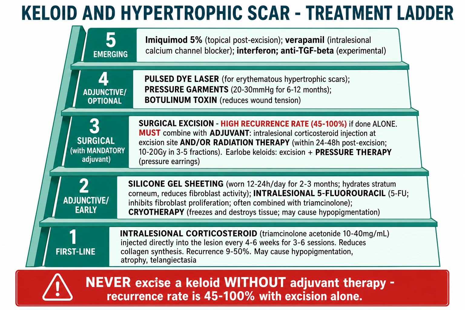
Management of keloids is chronic, multimodal, and prone to recurrence; no single treatment is curative. The cornerstone is realistic counselling, combination therapy, and adjuvant treatment after any surgical intervention.[2][3][4][6][9]
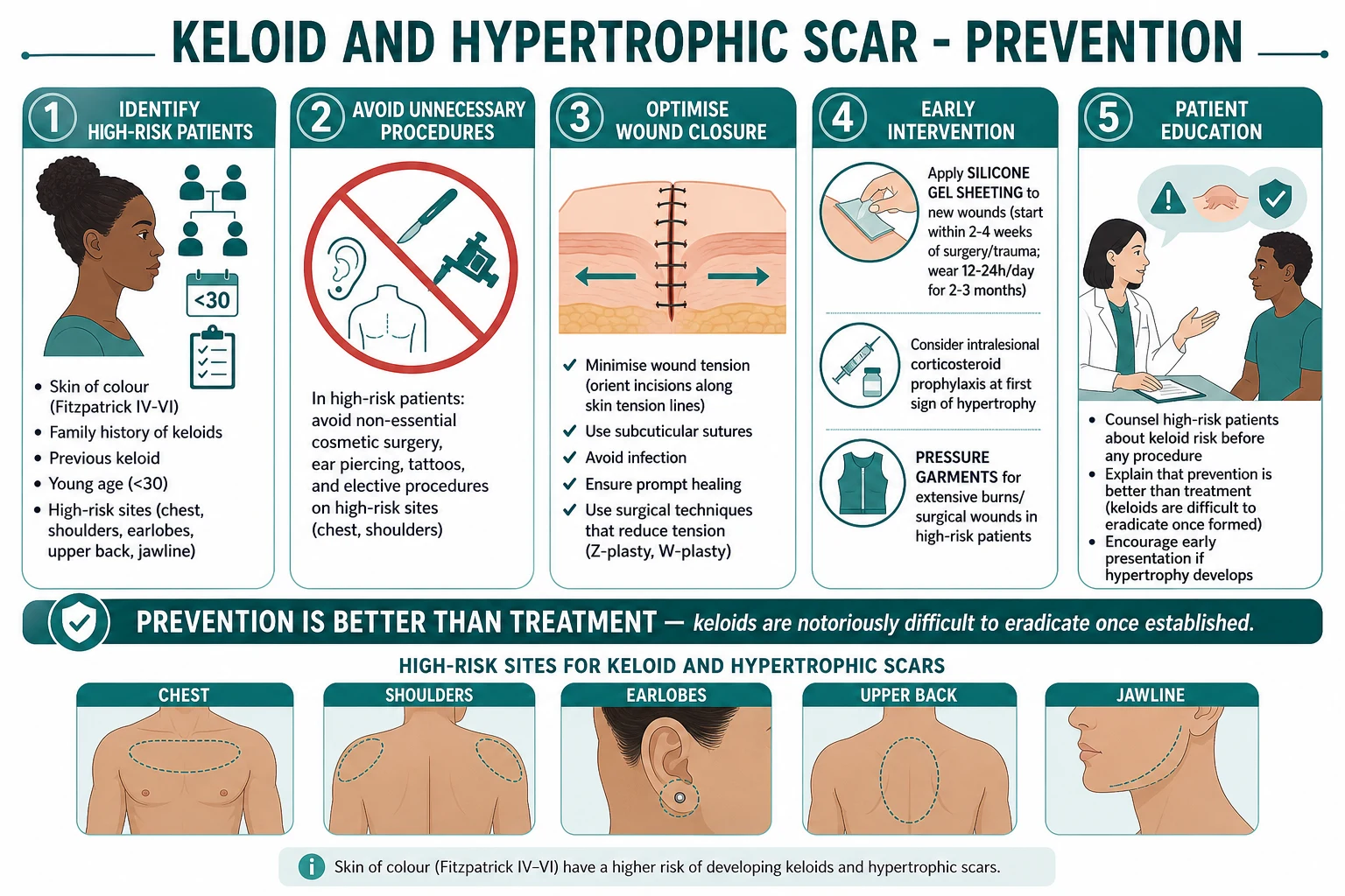
Prevention — the most effective strategy [1]
The prevention ladder
Identify high-risk patients
Skin of colour (Fitzpatrick IV-VI); personal or family history of keloids; age 10-30; planned procedure on a high-tension site (chest, sternum, deltoid, earlobe)
Avoid unnecessary procedures
No elective cosmetic surgery, ear piercing, tattoos, or other skin trauma in high-risk patients on high-tension sites. Counsel extensively and document.
Optimise surgical technique
Incisions parallel to skin tension lines; meticulous haemostasis; layered closure; minimise tension; avoid foreign material and infection; use fine non-absorbable sutures removed early (5-7 days for face, 7-14 for body)
Start silicone gel sheeting early
Apply within 1-2 weeks of epithelialisation; wear 12-24 h/day for at least 2-3 months (and up to 6 months for high-risk sites)
Consider prophylactic intralesional steroid
Single intra-operative triamcinolone 10-40 mg/mL injection at the closure site ± 2-4 weekly for 1-2 months in highest-risk patients
Pressure therapy for burns and earlobe keloids
Pressure garments (15-40 mmHg) 12-23 h/day for 6-12 months from epithelialisation; pressure earrings for earlobe keloids 24 h/day for up to 12 weeks post-excision
First-line: Intralesional corticosteroid
Triamcinolone acetonide 10-40 mg/mL injected directly into the lesion every 4-6 weeks for 3-6 sessions is the first-line treatment for both keloid and symptomatic hypertrophic scar.[2][3][4][6][8]
- Mechanism — anti-inflammatory; reduces fibroblast proliferation and collagen synthesis; increases collagen degradation; vasoconstrictive.
- Dose and dilution — earlobe and small facial lesions: 10 mg/mL; small body lesions: 20 mg/mL; chest, back, and large keloids: 40 mg/mL. Maximum single-session dose is generally capped at 40-80 mg to avoid systemic effects.
- Volume — inject 0.1-0.5 mL per cm² of scar; do not exceed 2-3 mL per session; blanching of the scar signals adequate intralesional deposition.
- Frequency — every 4-6 weeks; typical course 3-6 sessions; some lesions need 6-12 sessions.
- Response rate — 50-70% of keloids show flattening and symptomatic improvement; recurrence 9-50%.
- Combination — triamcinolone + 5-FU (often 0.1 mL of 50 mg/mL 5-FU per 0.9 mL of 10-40 mg/mL triamcinolone) is more effective than triamcinolone alone for refractory keloids.
- Side effects — skin atrophy, telangiectasia, hypopigmentation (particularly in skin of colour), local pain, Cushingoid features with very high cumulative doses.
- Cautions — avoid intralesional injection into a previously irradiated area; use lower doses in children and in dark-skinned patients (hypopigmentation risk). [1]
Adjunctive non-invasive treatments
Silicone gel sheeting / gel
Universal — prevention & early treatment
- **12-24 h/day** for **2-3 months** (or longer in high-risk cases)
- **Mechanism**: hydration + occlusion of the stratum corneum → reduced fibroblast activity, reduced cytokine release
- Available as sheets (reusable, washed daily) or self-drying silicone gel (cosmetically preferable for face/scalp)
- Effective for **prevention** (start 1-2 weeks post-epithelialisation) and **treatment** of early scars
- Adverse effects: minor skin irritation, sweating, poor adhesion in hot/humid conditions; rare contact dermatitis
- Evidence base: multiple RCTs and meta-analyses support efficacy, although trial quality is variable
Pressure therapy / pressure garments
15-40 mmHg for 6-12 months
- **15-40 mmHg** sustained pressure for **12-23 h/day** for **6-12 months**
- Standard of care for **burn-related hypertrophic scars**; reduces thickness, pliability, and erythema
- **Earlobe keloids**: **pressure earrings** worn 24 h/day for up to **12 weeks** after excision
- Mechanism: local hypoxia → fibroblast apoptosis, decreased collagen synthesis
- Adverse effects: skin breakdown, blistering, poor compliance (bulky, hot, uncomfortable)
Cryotherapy
Intralesional > contact
- **Intralesional cryotherapy** (a needle probe inserted into the scar) is more effective than contact/spray cryotherapy
- **Contact/spray cryotherapy**: liquid nitrogen, freeze-thaw cycles; flattening 50-75% in small keloids
- Combined with intralesional steroid: more effective than either alone
- **Adverse effects**: hypopigmentation (significant in skin of colour — counsel), blistering, pain, skin atrophy
Surgical excision (always with adjuvant)
Surgical excision of a keloid is contraindicated as a stand-alone procedure because the recurrence rate is 50-100%, and the recurrent scar is often larger and more symptomatic than the original. Excision is reserved for large, symptomatic, refractory lesions and must always be combined with adjuvant therapy.[2][3][4][8][9]
- Post-excision adjuvant options (any of, ideally combined):
- Intralesional corticosteroid — triamcinolone 10-40 mg/mL injected into the wound edges at the time of closure, then every 2-4 weeks for 1-2 months; reduces recurrence to ~30-50%.
- Post-excision radiation therapy (RT) — the most effective single adjuvant, with recurrence reduced to 10-15%. Best given within 24 hours of excision (most centres ≤24 h; some up to 48 h). Modalities: electron beam (10-20 Gy in 3-5 fractions) or high-dose-rate brachytherapy (iridium-192; 12-20 Gy). Reserved for adults, large/refractory keloids, and ear/chest keloids; avoided in children and pregnancy.[3][8]
- Pressure therapy — pressure earrings 24 h/day for up to 12 weeks for earlobe keloids; pressure garments for burn-related scars.
- Silicone gel sheeting — start at epithelialisation.
- 5-FU / imiquimod / verapamil — second-line adjuvants.
- Combined modality (excision + radiation + intralesional steroid) is standard for high-risk lesions and reduces recurrence to single digits.
- Earlobe keloids — excision + pressure earring (or magnetic compression disc) for 6-12 weeks is a particularly effective and well-tolerated combination.[10]
Intralesional non-steroid agents
5-Fluorouracil (5-FU)
Antimetabolite — combined with steroid
- **50 mg/mL** intralesional; **weekly** for 4-8 weeks; often combined with triamcinolone
- **Mechanism**: pyrimidine antimetabolite → inhibits fibroblast proliferation and collagen synthesis
- Effective as monotherapy and as adjuvant post-excision; reduced recurrence vs excision alone
- **Adverse effects**: local pain, hyperpigmentation, ulceration (rare), **bone-marrow suppression** with very high cumulative doses
- **Avoid in pregnancy** (teratogenic)
Verapamil
Calcium-channel blocker — intralesional
- **2.5 mg/mL** intralesional, 0.5-2 mL per session, every 2-4 weeks
- **Mechanism**: calcium-channel blockade → inhibition of fibroblast proliferation, ↑ collagenase, ↓ TGF-beta
- Evidence: small RCTs suggest benefit; not as well-established as steroid
- **Adverse effects**: rare; transient local pain
- May be useful as a steroid-sparing agent
Bleomycin
Antitumor antibiotic — refractory keloids
- **Intralesional bleomycin** (0.1-1 mL of 1.5 IU/mL) by **multiple puncture / tattoo technique**, every 4-6 weeks, 2-4 sessions
- **Mechanism**: induces fibroblast apoptosis; inhibits collagen synthesis
- Effective for **refractory** keloids; response rate 60-90% in small series
- **Adverse effects**: local hyperpigmentation, skin atrophy, rare pulmonary/systemic toxicity with high cumulative dose
- **Avoid in pregnancy**; caution in renal impairment
Interferon alpha-2b
Cytokine — anti-fibrotic
- **Intralesional IFN-α-2b** 1.5-3 MIU twice weekly for 4 weeks, or post-excision
- **Mechanism**: anti-fibrotic cytokine; ↓ collagen I/III synthesis, ↑ collagenase
- **Adverse effects**: flu-like syndrome (fever, myalgia), fatigue, depression; expensive
- Reserved for refractory keloids
Laser and light therapy
- Pulsed dye laser (PDL, 585-595 nm) — first-line laser for erythematous hypertrophic scars and as an adjunct in keloid management; reduces erythema, flattens, and improves pliability by selective photothermolysis of microvessels. Combination with intralesional steroid is more effective than either alone. Multiple sessions every 4-8 weeks.
- Fractional CO₂ laser (10,600 nm) — for texture and contour improvement of mature keloids and hypertrophic scars; stimulates collagen remodelling. Adjunct to intralesional steroid and 5-FU.
- Fractional non-ablative laser (1540 nm) — alternative with less downtime; effective for texture and dyschromia.
- Intense pulsed light (IPL) — for the erythema and telangiectasia of scars; less effective than PDL for the scar itself.
- Combination laser + intralesional steroid + 5-FU is the standard multimodal laser approach in many scar centres.[3][9]
Topical and emerging therapies
- Imiquimod 5% cream — applied post-excision for 6-8 weeks; immunomodulator (TLR-7 agonist) that induces interferon and anti-fibrotic cytokines. Evidence is mixed; best evidence is in earlobe keloids. Side effects: local irritation, crusting, hypopigmentation.
- Tacrolimus 0.1% ointment — calcineurin inhibitor; reported to reduce pruritus and erythema; limited evidence in keloid.
- Methotrexate — low-dose systemic (5-15 mg weekly); reported in small case series for refractory keloid; not first-line.
- Onion extract (e.g. mederma) — popular but no convincing evidence of efficacy over placebo; should not be relied upon.
- Botulinum toxin A — intralesional, 5-10 IU per cm² every 2-3 months; reduces dynamic wound tension by paralysing underlying muscle; meta-analyses suggest benefit as an adjunct, particularly in high-tension sites. Mechanistically appealing (mechanotransduction).
- Stem cell–derived exosomes — emerging research suggests that mesenchymal-stem-cell-derived exosomes can reduce keloid fibrosis in vitro and in animal models; clinical trials are ongoing; not yet standard of care.[5]
Comparison of treatment options for keloid
First-line — non-invasive
Universal
- **Intralesional triamcinolone** 10-40 mg/mL every 4-6 weeks (3-6 sessions)
- **Silicone gel sheeting** 12-24 h/day for 2-3 months
- **Pressure** 15-40 mmHg 12-23 h/day for 6-12 months (burns, earlobe)
- **Cryotherapy** (intralesional preferred) for small lesions
Second-line — combination
Refractory or larger lesions
- **Triamcinolone + 5-FU** (combined intralesional)
- **Pulsed dye laser** (585-595 nm) — every 4-8 weeks; combination with steroid
- **Fractional CO₂ laser** — texture/contour
- **Imiquimod** post-excision (earlobe keloids especially)
- **Botulinum toxin** (intralesional) — high-tension sites
Third-line — surgery + adjuvant
Large, refractory lesions
- **Surgical excision + post-excision radiation** (within 24 h) — gold standard for high-risk keloids
- **Excision + intralesional steroid** + **silicon/pressure**
- **Earlobe: excision + pressure earring 24 h/day × 12 wk**
- **Combined excision + RT + steroid** for sternal/deltoid keloids
- **Verapamil, bleomycin, interferon, methotrexate** for refractory lesions
Specific Subtypes & Scenarios
Earlobe keloids
The commonest keloid in many series, often bilateral after ear piercing, particularly in adolescents and young adults of African or Asian descent. Typically pedunculated, firm, and may be very large. First-line: surgical excision with pressure earring 24 h/day for up to 12 weeks, or magnetic compression disc. Combination with intralesional triamcinolone at the time of closure and 2-4 weekly for 1-2 months post-operatively. Imiquimod is an option for earlobe keloids post-excision. Recurrence is lower at this site than for sternal or deltoid keloids, especially with combined therapy.[2][10]
Sternal / chest / deltoid keloids
The highest-tension and most resistant sites. Multimodal therapy is the rule: excision + post-excision radiation (within 24 h) + intralesional steroid + silicone sheeting + pressure garment. Even with combined therapy, recurrence is in the 10-30% range. Excision alone is contraindicated. The patient must be counselled about the high risk of recurrence and the realistic goals of treatment (symptom relief, partial flattening). [1]
Burn-related hypertrophic scars
A distinct clinical entity. Extensive deep partial-thickness and full-thickness burns frequently produce hypertrophic scarring with contracture across joints. The cornerstone of management is early, sustained pressure garment therapy (15-40 mmHg, 12-23 h/day for 6-12 months, sometimes up to 18 months) from the time of epithelialisation, plus silicone gel sheeting, intralesional steroid, and intensive physiotherapy to prevent contracture. Functional contractures (across a finger, neck, axilla, or joint) may require surgical release and skin grafting with subsequent compression and steroid. [1]
Acne keloids (chest and back)
A form of scarring in which acne lesions on the chest and back heal as keloidal or hypertrophic papules and plaques, often with surrounding comedones and inflammatory acne. The first step is to control the active acne (topical retinoids, benzoyl peroxide, oral isotretinoin if severe). Intralesional triamcinolone and silicone gel sheeting are then added; laser (PDL, fractional CO₂) for established lesions. [1]
Spontaneous keloids
Keloids without a clear inciting trauma account for ~3% of cases, usually on the chest. The differential is wide (scar sarcoidosis, malignancy). Biopsy is reasonable if the clinical picture is atypical. If confirmed, treat as a standard keloid. Multiple spontaneous keloids in a young patient should prompt consideration of a genetic syndrome (Rubinstein-Taybi, Dubowitz) and referral to clinical genetics. [1]
Paediatric keloids
Keloids in children are uncommon before puberty and are generally more aggressive. Avoid radiation because of the lifetime cancer risk. First-line: silicone gel sheeting + intralesional triamcinolone (lower concentrations: 5-10 mg/mL) + cryotherapy. Surgical excision with adjuvant (but not radiation) is reserved for refractory lesions. [1]
Pregnancy and keloids
Keloids may enlarge during pregnancy due to oestrogen-driven fibroblast proliferation. Avoid radiation and 5-FU / bleomycin / methotrexate (all teratogenic). Safe options: silicone gel sheeting, pressure therapy, intralesional triamcinolone (in low doses, with informed consent), and topical emollients. Reassess post-partum; some pregnancy-associated keloids regress partially. [1]
Complications & Pitfalls
- Recurrence — the cardinal complication. 50-100% after excision alone; 9-50% after excision with adjuvant; lowest (single digits) with combined excision + radiation + steroid. Recurrence is often worse than the original lesion.
- Cosmetic deformity — particularly in cosmetically sensitive sites (face, earlobes, chest). The psychosocial impact is significant and often underestimated by clinicians.
- Psychosocial distress — depression, anxiety, social avoidance, body-image disturbance, sleep disturbance from pruritus. Screen formally in any keloid patient.
- Functional limitation — contracture across joints, restriction of mouth/nostril/eye opening, restriction of limb movement. May require surgical release and grafting.
- Treatment-related complications:
- Intralesional corticosteroid — skin atrophy, telangiectasia, hypopigmentation (significant in skin of colour), local pain, Cushingoid features with high cumulative dose.
- Cryotherapy — hypopigmentation (especially in skin of colour), blistering, pain, atrophy.
- Radiation — late carcinogenesis (skin, thyroid, breast in chest keloids), hypopigmentation, fibrosis, telangiectasia. Absolute contraindication in children and pregnancy.
- 5-FU — local ulceration, hyperpigmentation, rare bone-marrow suppression.
- Bleomycin — local hyperpigmentation, rare pulmonary toxicity with high cumulative dose.
- Laser — post-inflammatory hyperpigmentation, transient erythema, oedema, rare scarring. [1]
Classic pitfalls (the ones examiners test): [1]
- Excision of a keloid without adjuvant — guaranteed recurrence, often worse than the original.
- Biopsy not done in atypical "keloids" — missing a dermatofibrosarcoma protuberans or scar sarcoidosis.
- Radiation in a child — lifetime cancer risk; absolute contraindication.
- 5-FU or bleomycin in pregnancy — teratogenic.
- Cryotherapy as monotherapy in a skin-of-colour patient — hypopigmentation may be more distressing than the keloid.
- Underestimating the psychosocial impact — failing to ask about sleep, mood, social functioning.
- Recommending elective cosmetic surgery or piercing to a keloid-prone patient — the single most preventable keloid is the one that never forms.
- Forgetting the contralateral side — bilateral earlobe keloids after piercing are common; treat both or counsel about the high risk of recurrence on the other side. [1]
Prognosis & Disposition
Keloids are a chronic condition with no definitive cure. The course is characterised by persistent or slowly progressive growth, frequent recurrence after treatment, and significant psychosocial impact. Hypertrophic scars, by contrast, generally improve spontaneously over 12-24 months, although they may persist or contract across joints.[2][4]
Prognostic factors: [1]
- Site — earlobe keloids have the best prognosis (lowest recurrence after combined therapy); chest/sternal/deltoid keloids have the worst.
- Size — small (under 1 cm) lesions respond better than large (over 5 cm) lesions.
- Duration — early treatment (within 6-12 months of onset) is more effective than late treatment of mature lesions.
- Genetic predisposition — strong family history predicts more aggressive disease and higher recurrence.
- Treatment adherence — pressure garments and silicone sheeting require sustained use; non-adherence predicts recurrence.
- Combination therapy — combined modality (excision + radiation + steroid) outperforms monotherapy. [1]
Disposition: [1]
- Most keloids and hypertrophic scars are managed in the outpatient dermatology or plastic-surgery scar service.
- Refer to a multidisciplinary keloid service for: refractory disease, recurrence after combined therapy, large lesions, paediatric or pregnant patients, and consideration of radiation therapy.
- Refer to clinical genetics for: multiple spontaneous keloids, syndromic features (Rubinstein-Taybi, Dubowitz), strong family history of keloids.
- Biopsy and onward referral for atypical features suggesting malignancy (DFSP, SCC, scar sarcoidosis). [1]
Follow-up: [1]
- 3-6 monthly review for active treatment; annual review for established disease.
- Standardised photography at each visit for objective comparison.
- Monitor for recurrence for at least 12-24 months after apparent cure.
- Re-counsel about prevention in any new planned procedure. [1]
Special Populations
- Children — keloids are uncommon before puberty but more aggressive when they occur. Avoid radiation (lifetime cancer risk). First-line: silicone gel sheeting, intralesional triamcinolone (lower doses 5-10 mg/mL), cryotherapy. Surgical excision is reserved for refractory lesions, with adjuvant therapy other than radiation (e.g. compression, intralesional steroid, 5-FU). Pressure therapy is well tolerated in children and parents should be counselled on adherence. Psychosocial support is critical in adolescents.
- Pregnancy — keloids may enlarge during pregnancy due to oestrogen-driven fibroblast proliferation. Avoid radiation and 5-FU / bleomycin / methotrexate (teratogenic). Safe options: silicone gel sheeting, pressure therapy, intralesional triamcinolone in low doses (counsel about the small systemic-absorption risk). Post-partum reassessment; some keloids regress partially.
- Darker-skinned patients (Fitzpatrick IV-VI) — highest risk group; larger, more aggressive, and more numerous keloids; higher rate of post-inflammatory hypopigmentation from cryotherapy and intralesional steroid — counsel and use lower doses; silicone and pressure are well tolerated. Empathetic, culturally sensitive care is essential.
- Elderly — keloids are uncommon in the elderly and may regress after the menopause. The main consideration in older patients is the differential (DFSP, malignancy) and the safety of any surgical or radiation-based intervention in the context of comorbidity.
- Immunocompromised — no direct effect on keloid biology, but the differential in an immunocompromised patient with a new scar-like lesion is broader (atypical mycobacteria, deep fungal infection, Kaposi sarcoma). Biopsy if in doubt.
- Burn survivors — a unique population with extensive hypertrophic scarring, contracture, and pruritus. Pressure garments (15-40 mmHg 12-23 h/day for 6-12 months from epithelialisation), intensive physiotherapy to prevent and treat contracture, silicone, and intralesional steroid for symptomatic lesions. Functional contractures may need surgical release and grafting. Long-term psychological support is essential.
- Keloid-prone patient requesting elective surgery or piercing — counsel extensively, document the discussion, offer alternatives, and prophylactic silicone sheeting + intralesional steroid at closure if the procedure is unavoidable. [1]
Evidence, Guidelines & Regional Differences
The management of keloids and hypertrophic scars is supported by a broad but heterogeneous evidence base, dominated by small RCTs, retrospective case series, and expert consensus rather than large multicentre trials. The most influential international documents are:[6][7][8][9]
- Mustoe 2002 (International Clinical Recommendations on Scar Management) — the foundational document that established silicone and intralesional steroid as first-line therapies.[7]
- Gold 2014 (Updated International Clinical Recommendations, Part 2) — algorithms for prevention and treatment.[8]
- Monstrey 2014 (Updated Scar Management Practical Guidelines) — the European (JPRAS) consensus, with practical algorithms for non-invasive and invasive measures.[9]
- Ogawa 2021 (Nippon Medical School Protocol) — the Japanese consensus that emphasises a structured, multimodal approach.[6]
- Multiple systematic reviews (Walsh 2023) of intralesional steroid, 5-FU, bleomycin, laser, and radiation; in general, combination therapy outperforms monotherapy, and the highest-quality evidence supports post-excision radiation and combined intralesional therapy for refractory disease.[3]
Regional differences: [1]
- US (AAD) — first-line intralesional triamcinolone; combination with 5-FU; pulsed dye laser; post-excision radiation widely available.
- UK (BAD) — first-line silicone and intralesional steroid; pressure garments for burns; NICE recommends onward referral for refractory disease.
- Europe (Monstrey / EBA) — emphasis on structured prevention (silicone, tension-free closure) and multimodal therapy.
- Japan (Ogawa / NMS Protocol) — long experience with post-excision radiation and brachytherapy; silicone and steroid as universal first-line; emphasis on early intervention.
- India (IADVL / ICMR) — high prevalence in skin-of-colour population; intralesional triamcinolone as first-line; pressure earrings for earlobe keloids; radiation is less commonly used (access and acceptance). [1]
Areas of controversy: [1]
- Radiation after keloid excision — highly effective (recurrence 10-15%) but controversial in younger patients and in women with chest keloids because of the small lifetime risk of secondary malignancy. The risk-benefit analysis favours radiation in adults with refractory or recurrent keloids; absolute contraindication in children and pregnancy.
- Imiquimod post-excision — evidence is mixed; some RCTs show benefit, others do not.
- Interferon alpha-2b — efficacious but expensive and flu-like side effects limit use.
- Botulinum toxin — biologically appealing (mechanotransduction rationale) but evidence is heterogeneous; useful as adjunct, not as monotherapy.
- Verapamil — small RCTs suggest benefit; not as well-established as steroid.
- Stem-cell exosomes — promising preclinical data; not yet standard of care.[5]
Exam Pearls
KELOIDS — features to remember
K-E-L-O-I-D-S
The pivotal distinction: keloid extends BEYOND the wound margin; hypertrophic scar stays WITHIN
Histology: thick hyalinised keloidal collagen in whorls, reduced elastic fibres
High-tension sites (chest, deltoid, sternum, earlobe, jawline); incisions parallel to skin tension lines reduce scarring
Pathophysiology: TGF-beta1/beta2 overexpression, fibroblast hypersensitivity, reduced apoptosis
First-line treatment: 10-40 mg/mL every 4-6 weeks for 3-6 sessions
Skin of colour (Fitzpatrick IV-VI) is the single biggest risk factor
Excision MUST be combined with adjuvant (radiation within 24 h, intralesional steroid, pressure)
High-yield pearls for the exam: [1]
- Keloid extends BEYOND the original wound margin; hypertrophic scar stays WITHIN — the single most important distinction.
- Keloids do NOT regress spontaneously; hypertrophic scars may regress over 12-24 months.
- First-line treatment of both: intralesional triamcinolone acetonide 10-40 mg/mL every 4-6 weeks.
- Silicone gel sheeting 12-24 h/day for 2-3 months is universal first-line adjunct — both prevention and treatment.
- Surgical excision of a keloid is contraindicated as monotherapy — recurrence is 50-100%; must be combined with adjuvant (intralesional steroid, post-excision radiation within 24 h, pressure earrings for earlobe).
- Skin of colour (Fitzpatrick IV-VI) has a 15-fold higher risk of keloid formation.
- High-tension sites: chest, sternum, deltoid, upper back, jawline, earlobes, neck; keloids spare palms, soles, and genitalia.
- TGF-beta1 and TGF-beta2 overexpression is the central cytokine abnormality; TGF-beta3 is anti-fibrotic.
- Pressure therapy 15-40 mmHg for 12-23 h/day for 6-12 months is standard for burn-related hypertrophic scars; pressure earrings 24 h/day for up to 12 weeks for earlobe keloids post-excision.
- Earlobe keloids are the commonest and most amenable to treatment; chest/sternal keloids are the most resistant.
- Avoid radiation in children and pregnancy (lifetime cancer risk, teratogenicity).
- Avoid 5-FU, bleomycin, and methotrexate in pregnancy (teratogenic).
- Histology of keloid: thick hyalinised, eosinophilic, "keloidal" collagen bundles in whorls; histology of hypertrophic scar: parallel myofibroblast/collagen in nodular pattern.
- Biopsy a "keloid" that is rapidly growing, ulcerated, or atypical — exclude DFSP, SCC, scar sarcoidosis, or metastatic carcinoma.
- Vancouver Scar Scale scores pigmentation, vascularity, pliability, and height (0-13); POSAS is the more responsive, patient-centred scale.
- Combination therapy outperforms monotherapy for established keloids; multimodal approach is the standard.
- Prevention is better than treatment — identify high-risk patients, avoid unnecessary procedures, optimise wound closure, start silicone early.
- Mechanical tension drives keloid biology — pressure garments, silicone, off-loading tension, and botulinum toxin all reduce scar formation.
- Botulinum toxin reduces dynamic wound tension by paralysing underlying musculature; useful adjunct in high-tension sites.
- Pregnancy can enlarge keloids (oestrogen-driven fibroblast proliferation); reassess post-partum.
- Spontaneous keloids (~3%) should raise suspicion of genetic syndromes (Rubinstein-Taybi, Dubowitz); refer to clinical genetics.
- Lobomycosis (keloidal blastomycosis) is a tropical differential — endemic in the Amazon basin; caused by Lacazia loboi.
- CD34-positive DFSP can mimic a keloid — biopsy any atypical or recurrent lesion.
- Standardised serial photography is the cheapest and most useful monitoring tool. [1]
Exam application bank (NEET-PG / INICET)
One-line answer
Keloids and hypertrophic scars are benign fibroproliferative disorders of the remodelling phase of wound healing with excessive collagen deposition. The pivotal distinction: a keloid extends BEYOND the original wound margins and does not regress; a hypertrophic scar stays WITHIN the wound and may regress over 12-18 months. Skin of colour (Fitzpatrick IV-VI; African, Asian, Hispanic) carries a 15-fold higher risk. Pathophysiology centres on TGF-beta1/beta2 overexpression, fibroblast hypersensitivity, reduced apoptosis of myofibroblasts, and a collagen-synthesis/degradation imbalance. Intralesional triamcinolone 10-40 mg/mL every 4-6 weeks is first-line; silicone gel sheeting, cryotherapy, 5-FU, verapamil, bleomycin, interferon, imiquimod, and PDL laser are adjuncts; surgical excision MUST be combined with adjuvant therapy (post-excision radiation within 24 h, intralesional steroid, pressu [1]
Worked stems (answer without another resource)
Stem 1 — Classic presentation. Map symptoms to mechanism; name the first investigation and first treatment step with dose/route if drug therapy is standard. [1]
Stem 2 — Unstable / complicated. List red flags that force immediate resuscitation, theatre, ICU, antidote, or reperfusion — and what you do in the first 15 minutes. [1]
Stem 3 — Atypical group. Elderly, pregnancy, child, or immunocompromised: how presentation and thresholds change. [1]
Stem 4 — Differential trap. Name the three closest mimics and one discriminator for each. [1]
Stem 5 — Disposition. Who goes home with safety-netting, who is admitted, who needs HDU/ICU/theatre, and what follow-up is mandatory. [1]
Rapid viva checklist
- Definition + classification
- Pathophysiology chain
- Bedside signs / criteria
- Score with exact components (if any)
- Emergency bundle
- Definitive therapy with doses
- Complications of disease and of treatment
- Special populations
- Guideline/trial name if classic
- Three exam traps
Coverage self-check
If you cannot answer any stem above from this page alone, re-read the matching section — the page is intended to be self-sufficient for final-prof and NEET-PG/INICET questions on Keloid and Hypertrophic Scar.
References
- [1]Wang PH, Huang BS, Horng HC, et al. Wound healing J Chin Med Assoc, 2018.PMID 29169897
- [2]Ekstein SF, Wyles SP, Moran SL, et al. Keloids: a review of therapeutic management Int J Dermatol, 2021.PMID 32905614
- [3]Walsh LA, Wu E, Pontes D, et al. Keloid treatments: an evidence-based systematic review of recent advances Syst Rev, 2023.PMID 36918908
- [4]Jeschke MG, Wood FM, Middelkoop E, et al. Scars Nat Rev Dis Primers, 2023.PMID 37973792
- [5]Zhou C, Zhang B, Yang Y, et al. Stem cell-derived exosomes: emerging therapeutic opportunities for wound healing Stem Cell Res Ther, 2023.PMID 37101197
- [6]Ogawa R, Dohi T, Tosa M, et al. The Latest Strategy for Keloid and Hypertrophic Scar Prevention and Treatment: The Nippon Medical School (NMS) Protocol J Nippon Med Sch, 2021.PMID 32741903
- [7]Mustoe TA, Cooter RD, Gold MH, et al. International clinical recommendations on scar management Plast Reconstr Surg, 2002.PMID 12142678
- [8]Gold MH, McGuire M, Mustoe TA, et al. Updated international clinical recommendations on scar management: part 2--algorithms for scar prevention and treatment Dermatol Surg, 2014.PMID 25068544
- [9]Monstrey S, Middelkoop E, Vranckx JJ, et al. Updated scar management practical guidelines: non-invasive and invasive measures J Plast Reconstr Aesthet Surg, 2014.PMID 24888226
- [10]Khalid FA, Farooq UK, Saleem M, et al. The efficacy of excision followed by intralesional 5-fluorouracil and triamcinolone acetonide versus excision followed by radiotherapy in the treatment of ear keloids: A randomized control trial Burns, 2018.PMID 29534885
- [11]De Decker I, Hoeksema H, Verbelen J, et al. The use of fluid silicone gels in the prevention and treatment of hypertrophic scars: a systematic review and meta-analysis Burns, 2022.PMID 35367089
- [12]Heitzer E, Seidl H, Bambach I, et al. Infrequent p53 gene mutation but UV gradient-like p53 protein positivity in keloids Exp Dermatol, 2012.PMID 22417303