Dermatology · General Medicine
Keloids & Hypertrophic Scars
Also known as Keloid · Hypertrophic scar · Abnormal scarring · Fibroproliferative scar
Keloids and hypertrophic scars are fibroproliferative disorders of wound healing characterised by excessive collagen deposition in response to skin injury. The pivotal distinction: hypertrophic scars stay within the original wound boundaries and often regress spontaneously, whereas keloids extend beyond the wound margins into surrounding normal skin, do not regress, and recur after excision. Risk factors include darker skin (Fitzpatrick IV-VI, up to 15-fold higher risk), age 10 to 30, genetic predisposition, wound tension (chest, shoulders, upper back, jawline, earlobes), infection, burns, foreign body, and piercings. Diagnosis is clinical. First-line treatment is intralesional corticosteroid (triamcinolone acetonide 10-40 mg/mL every 4-6 weeks), with silicone gel sheeting for prevention and early lesions. Surgical excision of keloids must be combined with adjuvant therapy (intralesional steroid or radiation within 24-48 hours), because excision alone has a 50-100 percent recurrence rate.
On this page & tools
Your progress
Saved locally on this device.
Exam tags
Red flags
Overview & Definition
Scarring is the body's default repair mechanism when the dermis is breached. In most people this process self-limits, producing a flat, pliable, asymptomatic line. In a genetically susceptible minority, however, the proliferative phase of wound healing refuses to switch off: fibroblasts keep depositing collagen, the scar thickens, and the boundary between repair and neoplasia blurs. The two ends of this abnormal scarring spectrum are the hypertrophic scar and the keloid.[1][4]
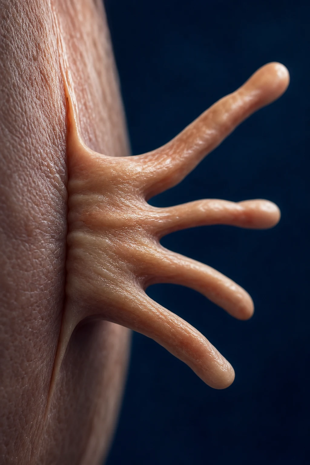
A keloid (from the Greek chele, meaning crab claw) is a firm, rubbery, fibroproliferative dermal lesion that extends beyond the boundaries of the original wound into surrounding normal skin. Keloids do not regress spontaneously, continue to grow for months to years, and recur aggressively after surgical excision. A hypertrophic scar, by contrast, is a raised scar that remains confined within the original wound margins and frequently flattens and softens over 12 to 24 months without intervention.[1][6]
This distinction is not semantic trivia. It determines prognosis, treatment intensity, and the patient's expectations. Hypertrophic scars are often self-limiting and respond well to conservative measures; keloids are chronic, recurrent, and require multimodal treatment with adjuvant therapy whenever surgery is attempted. Keloids are also a source of significant cosmetic and psychological distress, particularly in darker-skinned populations where they are commonest, larger, and more aggressive.[4][6]
Classification
Abnormal scars are classified by their relationship to the original wound margins and, secondarily, by their clinical behaviour (onset, regression, symptoms). The classification is fundamental because it dictates the entire management plan.[1][4]
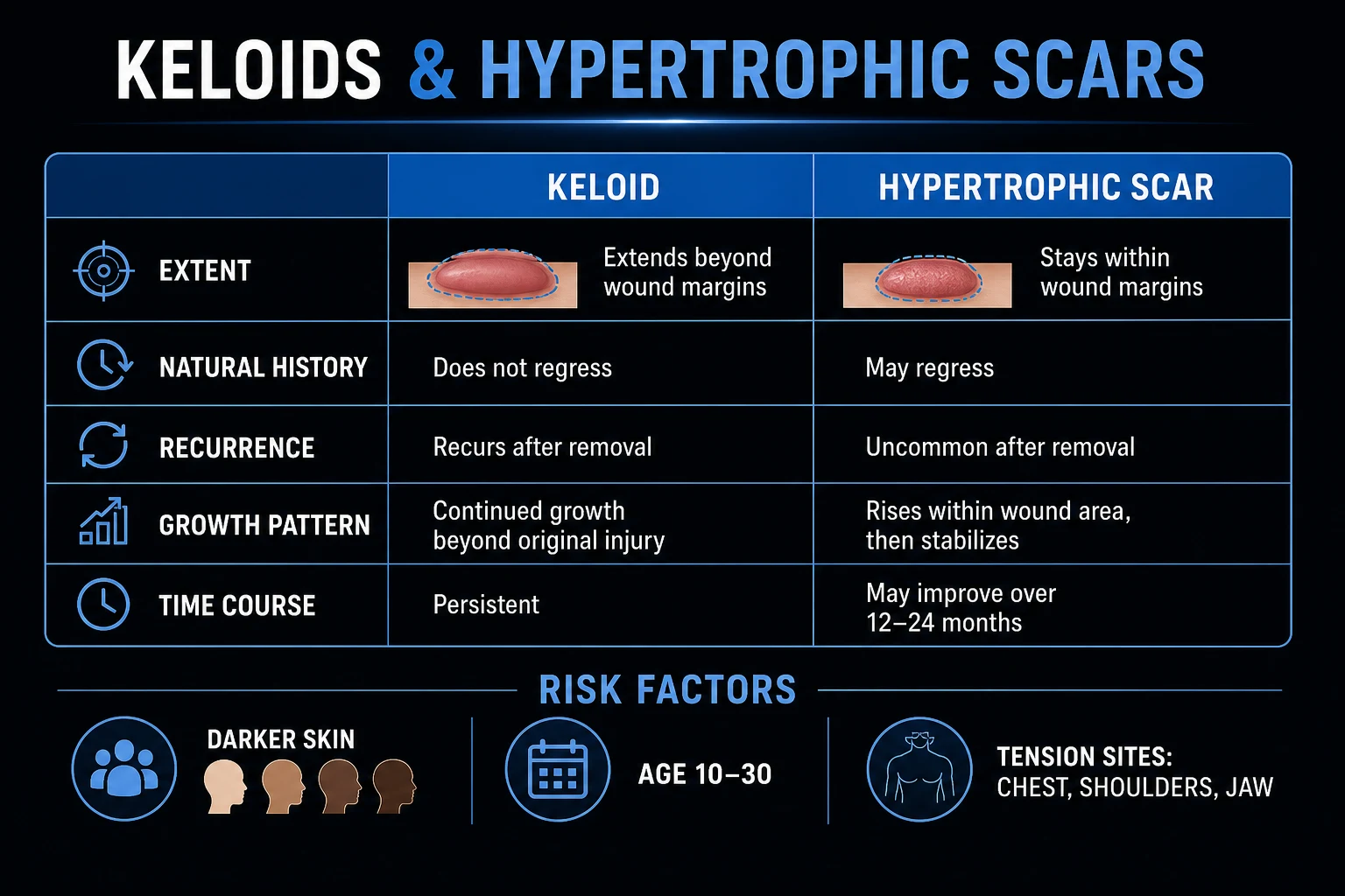
Hypertrophic scar
- Raised but **WITHIN** wound margins
- Onset 4-8 weeks after injury
- **Often regresses** over 12-24 months
- Less pruritic, less painful
- Responds well to treatment
- Any site, but common on flexor surfaces and burns
- Recurrence after excision: low if combined with adjuvant
Keloid
- Extends **BEYOND** wound margins (crab claw)
- Onset weeks to months; may grow for years
- **Does NOT regress** — permanent
- Often pruritic, painful, tender, hypersensitive
- Difficult to treat; high recurrence
- Tension sites: sternum, shoulders, upper back, jawline, earlobes, neck; **spares palms, soles, genitalia**
- Recurrence after excision alone: 50-100 percent
A practical clinical sub-classification also recognises spontaneous keloids (no history of trauma, usually on the chest, around 3 percent of cases), earlobe keloids (pedunculated, post-piercing), and massive keloids (large plaque-like lesions that may ulcerate or restrict movement). These scenarios carry different treatment implications, discussed under Specific Subtypes below.[4]
Epidemiology & Risk Factors
Keloids and hypertrophic scars are global disorders, but they disproportionately affect specific populations. The single strongest epidemiological signal is skin pigmentation: individuals with Fitzpatrick skin types IV to VI — predominantly people of African, Asian, and Hispanic ancestry — have an estimated 15-fold higher risk of keloid formation than lighter-skinned individuals.[4][6]
Population prevalence in darker-skinned groups ranges from approximately 4.5 to 16 percent, and keloid disorder is estimated to affect roughly 100 million people worldwide who acquire significant scarring from elective surgery each year. Sex distribution is roughly equal (F=M), though some series report a slight female predominance — likely an artefact of greater earlobe piercing and cosmetic-presentation rates rather than true biological skew.[1][3]
The peak age of onset is 10 to 30 years, reflecting the high fibroblast proliferative activity of youth. Keloids are uncommon before age 10 and rare in the elderly, in whom fibroblast activity wanes. Pregnancy and puberty may accelerate keloid growth, implicating oestrogen and growth-hormone signalling.[4]
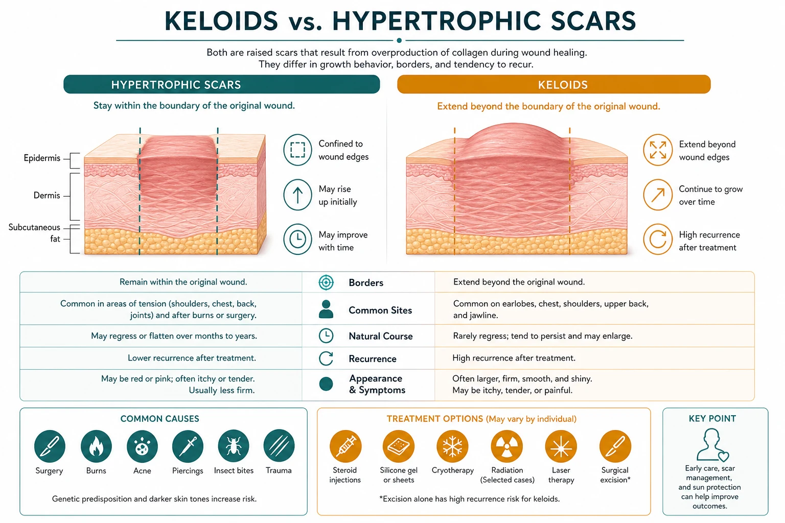
The risk factors can be grouped into patient factors and wound factors.[4][6]
Patient (intrinsic) risk factors:[4]
- Dark skin (Fitzpatrick IV-VI) — the dominant risk factor, 15-fold increased risk
- Age under 30 — peak fibroblast activity
- Family history / genetic predisposition — autosomal dominant with incomplete penetrance in some kindreds; HLA-DRB1, IL-6, and p53 polymorphisms implicated
- Pregnancy and puberty — hormonal influence on growth
Wound (extrinsic) risk factors:
- High skin tension sites — sternum/chest (highest), shoulders/deltoid, upper back, jawline, neck (these correlate with Langer's lines perpendicularity and muscle pull)
- Wound infection — prolongs inflammation, delays epithelialisation
- Burns — especially deep dermal and full-thickness; high incidence of hypertrophic scarring
- Foreign body — suture reaction, embedded material, talc
- Excessive wound tension at closure — poor surgical technique
- Delayed epithelialisation — wounds taking longer than 2-3 weeks to re-epithelialise are far more likely to scar abnormally
- Type of injury — surgery, piercings, acne, vaccinations (BCG), insect bites, traumatic lacerations[1][2][7]
A clinically useful rule: any wound that takes longer than three weeks to re-epithelialise is at high risk of hypertrophic scarring in a susceptible patient. This underpins the aggressive early use of silicone and pressure garments in burn centres.[7] Geographically, keloid disorder is most prevalent in sub-Saharan Africa, South and East Asia, the Indian subcontinent, and Latin America, and among their diaspora populations. Within any population, a subset of patients are unmistakably "keloid-prone": they keloid after every minor injury, often develop multiple and bilateral lesions, and frequently have a first-degree relative similarly affected. Recognising this phenotype is essential because it reframes every future medical and surgical decision for that patient — from BCG vaccination site selection to the wisdom of an elective cosmetic procedure. A simple screening question for any darker-skinned young patient undergoing surgery is whether they or a close family member have ever formed a raised, spreading scar.[4][6]
Pathophysiology
Normal wound healing unfolds in three overlapping phases: inflammation (days 1-6), proliferation (days 4-24, with fibroblast invasion, granulation tissue, and type III collagen deposition), and remodelling (3 weeks to 18 months, when type III collagen is replaced by stronger type I collagen and the scar matures). Keloids and hypertrophic scars represent a failure of the remodelling phase to terminate — the fibroblast keeps producing collagen well beyond physiological needs.[4][9]
Cellular and molecular drivers
At the cellular level, keloid fibroblasts behave abnormally in four ways. First, they proliferate excessively and show reduced sensitivity to signals that normally switch them off. Second, they resist apoptosis (programmed cell death), so they persist in the wound far longer than normal fibroblasts. Third, they overproduce collagen — particularly type III — and fourth, they show increased sensitivity to growth factors such as TGF-beta, platelet-derived growth factor (PDGF), epidermal growth factor (EGF), and insulin-like growth factor 1 (IGF-1).[9][10]
The collagen ratio shifts: normal skin contains roughly a 4:1 ratio of type I to type III collagen, while in keloids this falls to around 3:1 as type III collagen accumulates. The collagen bundles in keloids also become thick, hyalinised, and eosinophilic (so-called keloidal collagen), arranged in disorganised whorls rather than the parallel arrays of normal scars.[4]
The central molecular culprit is transforming growth factor beta (TGF-beta). The isoforms TGF-beta1 and TGF-beta2 are profibrotic and upregulated in keloids, driving fibroblast proliferation and collagen synthesis. TGF-beta3, by contrast, is anti-fibrotic — and recombinant TGF-beta3 (avotermin) was investigated as an anti-scarring therapy. The balance between these isoforms partly explains why some wounds over-heal.[5][9]
A second key imbalance is in the matrix metalloproteinase (MMP) system. MMPs (especially MMP-1 and MMP-3) degrade collagen, while tissue inhibitors of metalloproteinases (TIMPs) block them. In keloids, MMP activity is reduced and TIMP expression is increased — so collagen is produced faster than it can be broken down, and the scar inexorably grows.[4][5] A third driver is the immune-inflammatory milieu. Keloids are now understood not merely as a fibroblast problem but as a fibro-inflammatory disorder: mast cells, macrophages (especially the profibrotic M2 phenotype), and T lymphocytes are enriched in keloid tissue. Mast-cell degranulation releases histamine and tryptase, which stimulate fibroblast collagen synthesis and explain the pruritus that dominates the symptom profile. Inflammatory cytokines such as interleukin-6 (IL-6), tumour necrosis factor alpha (TNF-alpha), and IL-1 are elevated, with IL-6 in particular driving the acute-phase response and collagen gene transcription. This immune activation is why keloids are red, tender, and itchy, and why they respond to anti-inflammatory measures such as corticosteroid.[9][5]
Vascular, genetic, and mechanical contributions
Keloids are richly vascular. Microvessel density is increased, and local hypoxia drives vascular endothelial growth factor (VEGF) production, which in turn stimulates fibroblast activity — a self-perpetuating fibrovascular loop that single-cell transcriptomic studies have recently confirmed at the gene-expression level.[10]
Genetics load the gun. Keloid disorder clusters in families, with autosomal dominant inheritance and incomplete penetrance described in some lineages. Susceptibility loci include HLA-DRB1, interleukin-6 (IL-6) polymorphisms, and mutations affecting the p53 tumour-suppressor pathway. The shared HLA-DRB1 associations and the fact that keloids virtually never occur on the palms, soles, or genitalia (skin with different embryological origin and tension properties) point to a gene-environment interaction between immune-genetic background and local mechanical forces.[4][9]
Mechanical tension is the dominant local driver. Keloids arise almost exclusively at sites of high static or dynamic skin tension — the central chest (over the sternum), shoulders, upper back, and jawline. Wounds oriented perpendicular to Langer's lines, or closed under tension, are far more likely to scar abnormally. Tension is also why keloids recur after excision: cutting them out and re-closing under the same tension simply restarts the pathological process. This is the biological rationale for botulinum toxin (which paralyses local musculature and reduces tension) and for pressure therapy.[7]
Clinical Presentation
The patient typically presents weeks to months after a documented skin injury — a surgical incision, a burn, a laceration, an ear piercing, an acne lesion, or a vaccination — with a progressively enlarging, raised, firm mass at the site. A smaller fraction (around 3 percent) report no antecedent trauma (spontaneous keloids), almost always on the chest.[4][6]
Morphology
The lesion is firm and rubbery on palpation, with a smooth, shiny surface that may show overlying telangiectasia. Colour ranges from pink to erythematous in early lesions, through to purple, deep brown, or hyperpigmented in established keloids, particularly in darker skin. The margins are the diagnostic feature: claw-like or lobulated extensions project into the surrounding normal skin beyond the original wound, unlike a hypertrophic scar whose raised portion exactly mirrors the incision line.[1][6]
Clinical course: normal scar versus hypertrophic scar versus keloid
Symptoms
Although many keloids are asymptomatic, a substantial proportion are symptomatic and the symptoms can be the patient's chief complaint:[1]
- Pruritus — the commonest symptom, often intense
- Pain and tenderness — variable but can be severe
- Burning sensation and hyperaesthesia over the lesion
- Cosmetic concern — often the presenting complaint, especially on the face and earlobes
- Psychological distress — anxiety, depression, social avoidance, impaired quality of life
Site distribution
Keloids show a striking site predilection that mirrors skin tension and embryological skin type:[1][4]
High-risk sites
- Sternum / central chest (highest tension, most resistant)
- Shoulders and deltoid
- Upper back
- Jawline and chin
- Neck
- Earlobes (post-piercing, often bilateral)
- Cheek and pre-auricular skin
Low-risk / spared sites
- Palms and soles (rarely affected)
- Genitalia (very rare)
- Eyelids
- Scalp
The classic earlobe keloid after piercing — bilateral, pedunculated, lobulated masses — is a frequently tested presentation. Bilateral earlobe keloids in a young woman of African or Asian ancestry following piercing is a near-pathognomonic scenario.[6]
Atypical presentations
- Children and adolescents — more aggressive growth, larger lesions, greater lifetime recurrence burden; radiation must be avoided
- Pregnancy — pre-existing keloids may enlarge rapidly, implicating oestrogen and growth hormone
- Massive keloids — large plaque-like lesions on the chest or back that may ulcerate, restrict shoulder movement, or cause chronic pain
- Refractory keloids — recurrent despite multiple treatments, often at central chest sites; suggest the need for combination therapy[4]
Differential Diagnosis
The differential divides into the single most important mimic (hypertrophic scar) and a list of other dermal nodular or plaque-like lesions that can resemble a keloid, some of which are dangerous and require biopsy.[4][6]
Hypertrophic scar
- Raised but WITHIN wound margins
- Develops within 4-8 weeks
- Often regresses over 1-2 years
- Distinguished by margin confinement
- Less aggressive treatment needed
Dermatofibroma
- Firm dermal nodule, usually on legs
- Dimple sign (retracts on lateral compression)
- Not trauma-related
- No claw-like extensions
- Dimble sign and central pigmentation
Desmoid tumour (aggressive fibromatosis)
- Deep, rapidly growing mass
- Chest wall or abdominal wall common
- No antecedent wound
- Biopsy essential — spindle cells, beta-catenin nuclear staining
- Locally invasive, high recurrence
Scar sarcoidosis
- Sarcoid granulomas within a pre-existing scar
- Scar becomes raised, red, tender
- May be presentation of systemic sarcoidosis
- Biopsy: non-caseating granulomas
- Check chest radiograph and serum ACE
Lobomycosis (keloidal blastomycosis)
- Endemic in Amazon basin
- Caused by Lacazia loboi
- Keloid-like nodules, slowly progressive
- Biopsy: yeast-like cells in chains (Grocott stain)
- Geographic/exposure history
Dermatofibrosarcoma protuberans (DFSP)
- Slowly enlarging plaque or nodule
- Often on trunk, can mimic keloid
- CD34-positive on histology
- t(17;22) COL1A1-PDGFB translocation
- Biopsy mandatory for atypical/recurrent 'scar'
The clinical reasoning in differentiating these hinges on three questions. First, is there a wound history? A lesion arising at the exact site of a documented injury and extending beyond it is a keloid until proven otherwise; a nodule without trauma context shifts the differential toward dermatofibroma or DFSP. Second, does it stay within or extend beyond the wound? Margin confinement favours hypertrophic scar, which itself is distinguished from a normal immature scar by its persistence and height beyond six months. Third, is it behaving atypically? Rapid growth, ulceration, fixation to deep structures, or recurrence after a well-executed combined regimen should prompt biopsy — because dermatofibrosarcoma protuberans in particular is notorious for masquerading as a keloid on the trunk for years before diagnosis. Scar sarcoidosis deserves special mention: a pre-existing scar that suddenly becomes raised, red, and tender may signal systemic sarcoidosis, and biopsy reveals non-caseating granulomas rather than keloidal collagen.[4][6]
Clinical & Bedside Assessment
Assessment is clinical and focuses on confirming the keloid versus hypertrophic distinction, quantifying severity, and establishing a baseline for treatment response.[1][8]
History should establish:[1][8]
- The inciting injury and its date (or spontaneous onset)
- Growth trajectory — stable, slowly enlarging, or rapidly progressive
- Symptoms — itch, pain, tenderness, cosmetic and psychological impact
- Prior treatments and their responses (intralesional steroid, surgery, laser, cryotherapy, silicone, pressure)
- Family history of keloids or abnormal scarring
- Skin type (Fitzpatrick), ethnicity
- Patient expectations — realistic counselling is essential
Examination should document:[1][8]
- Location, size (in mm), and number of lesions
- Morphology — surface, colour, border definition, claw-like extensions
- Palpation — firmness, pliability, tenderness, fixation to deeper structures
- Functional impact — range of motion if over a joint
Two validated scar assessment scales are widely used. The Vancouver Scar Scale (VSS) scores scars from 0 to 14 across four parameters — vascularity (0-3), height/thickness (0-3), pliability (0-5), and pigmentation (0-2) — with higher scores indicating worse scarring. The Patient and Observer Scar Assessment Scale (POSAS) incorporates both patient-reported symptoms (pain, itch, colour, pliability, thickness) and observer-rated features, and is increasingly preferred because it captures the patient experience that pure observer scales miss.[8]
Standardised serial photography is essential for monitoring treatment response over time, as is high-frequency (20-50 MHz) cutaneous ultrasound, which objectively measures scar thickness and can distinguish a superficial keloid from an underlying deeper mass when clinical doubt exists.[8]
Investigations
Diagnosis is clinical — no investigation is required for a typical keloid in a characteristic site with a clear history of injury.[1][6]
Biopsy (4 mm punch) is reserved for atypical features: rapid growth, ulceration, fixation to deep structures, recurrence despite adequate first-line therapy, or a lesion behaving unlike a keloid. Histology of a keloid shows thickened, hyalinised, eosinophilic collagen bundles (keloidal collagen) in the dermis arranged in disorganised whorls, with a relative reduction in elastic fibres and increased fibroblast density. Hypertrophic scars, in contrast, show a nodular arrangement of myofibroblasts and collagen in parallel arrays without the hyalinised keloidal collagen. This histological distinction is less reliable than the clinical one and is rarely the deciding factor.[4]
High-frequency cutaneous ultrasound provides objective, reproducible thickness measurements useful for monitoring treatment response. Optical coherence tomography and cutaneous reflectance spectroscopy are research tools. There are no diagnostic blood tests for keloid disorder.[8]
Management — Resuscitation
Keloid and hypertrophic scar management is not an emergency. The immediate priorities at first presentation are:[6]
- Confirm the diagnosis clinically and distinguish keloid from hypertrophic scar — this alone redirects the entire plan.[6]
- Address the symptom burden. Intensely pruritic or painful keloids can be debilitating; intralesional triamcinolone often relieves both itch and pain within days of the first injection.
- Assess psychosocial impact. Depression, anxiety, body-image disturbance, and social avoidance are common, especially with facial and chest keloids. Acknowledge the distress, set realistic expectations, and involve psychology where appropriate.
- Treat any secondary infection of an ulcerated lesion (rare) with appropriate antibiotics and wound care.
- Counsel on realistic outcomes. Keloids are chronic and recurrent; no single treatment guarantees cure; combination therapy and long-term follow-up are the norm. Over-promising sets up dissatisfaction and loss of trust.
Management — Definitive & Stepwise
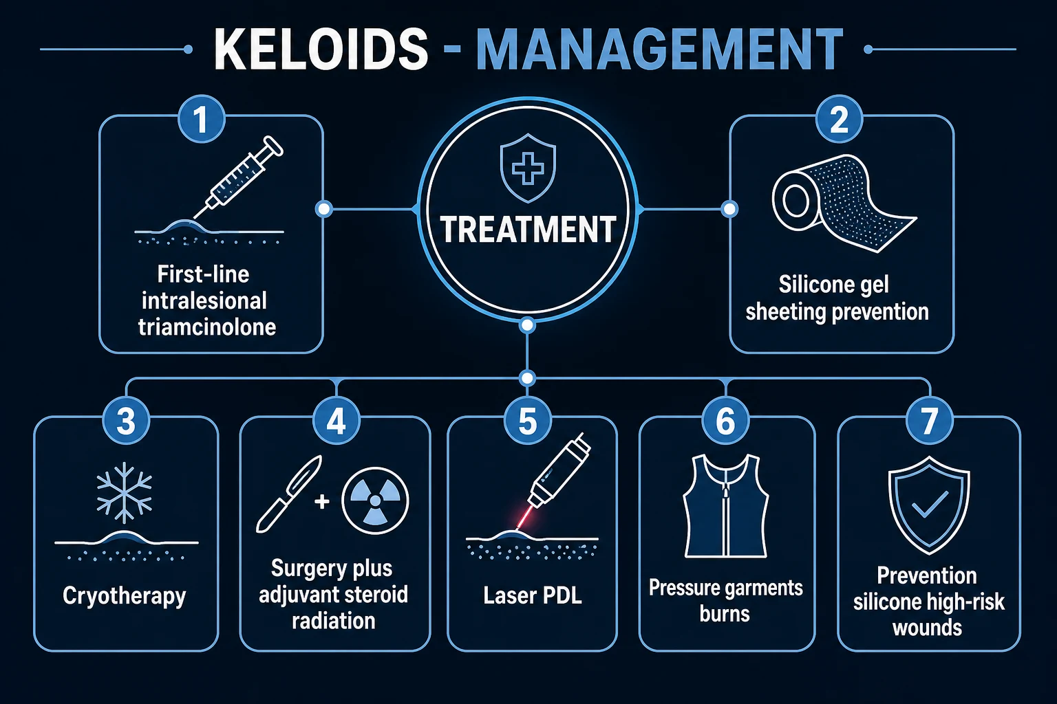
Treatment is site-specific, lesion-specific, and patient-specific. The ladder below reflects the consensus of the Japan Scar Workshop, the European updated scar management guidelines, and recent systematic reviews. A central principle: monotherapy is inadequate for established keloids — combination therapy is standard.[1][2][8]
[1] [2]Stepwise treatment algorithm for keloid/hypertrophic scar
Confirm diagnosis clinically
History of injury + lesion extending BEYOND wound margins = keloid; WITHIN margins = hypertrophic. Photograph and document with Vancouver Scar Scale.
Prevention and early lesions (immature scar, under 6 months)
Silicone gel sheeting 12-24 h/day for 2-3 months +/- pressure therapy. Avoid elective procedures in keloid-prone patients.
First-line established lesion
Intralesional triamcinolone acetonide 10-40 mg/mL every 4-6 weeks, 4-6 sessions. Continue silicone concurrently.
Second-line / partial response
Add cryotherapy (small lesions), pressure therapy (burns/earlobes), 5-FU intralesional (refractory), or pulsed dye laser (erythematous lesions).
Surgical excision (if lesion refractory or functionally limiting)
ONLY with adjuvant: intralesional steroid at closure AND/OR radiation (electron beam/brachytherapy) within 24-48 h. Never excise a keloid alone (50-100 percent recurrence).
Refractory / recurrent
Combination: bleomycin, interferon, imiquimod, verapamil, botulinum toxin; re-excision with adjuvant if appropriate.
Long-term surveillance
Review at 3, 6, 12 months; serial photography; early intervention at first sign of recurrence.
First-line therapies
Intralesional corticosteroid — triamcinolone acetonide 10-40 mg/mL injected directly into the lesion every 4-6 weeks for 4-6 sessions — is the first-line treatment for established keloids and symptomatic hypertrophic scars. It suppresses fibroblast proliferation and collagen synthesis and accelerates collagen degradation. Systematic review evidence reports a 50 to 70 percent response rate with flattening and symptom relief, but recurrence rates of 9 to 50 percent. Lower concentrations (10 mg/mL) suit softer, early lesions and the face; 40 mg/mL is used for dense, established keloids and the trunk. Side effects include skin atrophy, telangiectasia, hypopigmentation (a particular concern in darker skin), and rarely systemic absorption. The needle is passed into the body of the lesion (not beneath it) until the keloid blanches; total dose per session should respect the safe triamcinolone ceiling (typically 40 mg per session in adults).[1][11]
Silicone gel sheeting — applied for 12 to 24 hours per day for 2 to 3 months — is the cornerstone of both prevention (on high-risk closed wounds from the day of epithelialisation) and treatment of early, immature scars. It is non-invasive, well tolerated, and supported by multiple randomised trials and consensus guidelines, though its mechanism (occlusion, hydration, electrostatic effects, modulation of keratinocyte signalling) remains incompletely understood. It is the first choice for the patient who wishes to avoid injections.[2][8]
Second-line and adjunctive therapies
Cryotherapy — liquid nitrogen delivered as freeze-thaw cycles, typically 2-3 per session every 4 weeks — flattens small to medium keloids in 50 to 75 percent of cases. It works by inducing cellular damage and microvascular injury that reduces fibroblast activity. Its principal drawback is permanent hypopigmentation, which is cosmetically significant in darker skin, and pain. Intralesional cryotherapy probes (a needle delivering cold from within) reduce surface pigment injury but require specialised equipment.[19][6]
Pressure therapy — garments or clips delivering 15 to 40 mmHg of pressure for 12 to 23 hours per day for 6 to 12 months — is especially valuable for burn hypertrophic scars and earlobe keloids (using clip-on pressure earrings). It works by inducing tissue ischaemia and reducing collagen synthesis. Compliance is the main limiting factor, as garments are hot, uncomfortable, and conspicuous; burn centres achieve better adherence through dedicated therapists.[7][8]
Intralesional 5-fluorouracil (5-FU) — 50 mg/mL injected weekly, often combined with triamcinolone — inhibits fibroblast proliferation. Systematic reviews support its efficacy both alone and in combination, and the 5-FU/steroid combination is widely used for refractory keloids and after excision. Reported response rates are 70 to 85 percent.[12][20]
Pulsed dye laser (585 nm) — reduces erythema and flattens immature, vascular keloids and hypertrophic scars by targeting the microvasculature. A Cochrane review concluded the evidence is of low certainty but consistent with benefit, particularly when combined with intralesional steroid. It is most useful for the red, early, symptomatic lesion.[14]
Surgery — and the non-negotiable adjuvant
Surgical excision alone for a keloid is treatment failure waiting to happen: recurrence is 50 to 100 percent, and the recurrent keloid is frequently larger than the original (the "keloid reflex"). Excision is therefore only performed with an adjuvant: intralesional corticosteroid at the time of excision and/or post-excision radiation delivered within 24 to 48 hours.[18]
Radiation — superficial electron beam or brachytherapy — given as a single fraction or a short fractionated course immediately after excision reduces recurrence to approximately 10 to 15 percent. The efficacy is well established; the concern is long-term malignancy risk, which modelling studies suggest is very low for the doses used but mandates avoidance in children, pregnant women, and over radiosensitive organs such as the breast and thyroid. Consent must include a discussion of this theoretical risk.[18][6] A practical note on injection technique: the triamcinolone is deposited into the substance of the keloid (intralesional), not subcutaneously, using a 27-30 gauge needle, with the endpoint of uniform blanching indicating adequate infiltration. Subcutaneous injection wastes the drug and causes atrophy of surrounding fat. For very firm, established keloids, pre-treatment with cryotherapy one session earlier can soften the lesion and ease injection. The patient should be warned that response is gradual over weeks, that 4 to 6 sessions are typically required, and that 9 to 50 percent will recur — so realistic counselling at the outset prevents disappointment.[11][6]
For radiation, the typical regimen is superficial kilovoltage or electron beam at 6-10 Gy per fraction for 3-5 fractions, or a single 15-20 Gy fraction, delivered within 24 to 48 hours of excision. Brachytherapy catheters placed at the wound bed at the time of excision offer precise dose delivery to the scar bed while sparing surrounding normal tissue. The total biologically effective dose correlates with recurrence reduction. Radiation is reserved for adults with recurrent or refractory keloids at sites where the long-term theoretical malignancy risk is acceptable, and is never used in children, pregnant women, or over the breast or thyroid without explicit risk-benefit discussion and documented consent.[18]
Third-line and emerging therapies
Bleomycin (intralesional)
- Low-dose intralesional injection
- Meta-analysis: effective for refractory keloids and hypertrophic scars
- Mechanism: inhibits collagen synthesis, induces apoptosis
- Risk: skin necrosis, pigmentation change
Interferon alpha-2b (intralesional)
- Anti-fibrotic cytokine
- Injected post-excision to reduce recurrence
- Reduces type I/III collagen synthesis
- Limited by cost, flu-like side effects
Imiquimod 5% (topical)
- Immune response modifier (TLR-7 agonist)
- Applied to scar bed post-excision, especially earlobe keloids
- Meta-analysis: mixed evidence; may reduce earlobe recurrence
- Local erythema/irritation common
Verapamil (intralesional)
- Calcium-channel blocker reduces collagen synthesis
- Meta-analysis: comparable to triamcinolone
- Often combined with other agents
- Multiple injections required
Botulinum toxin A (intralesional)
- Reduces wound tension by paralysing local muscle
- Meta-analysis: benefit as adjunct to steroid
- Mechanism addresses the tension driver
- Best for facial and jawline keloids
A 2024 systematic review and several meta-analyses confirm that combination therapy outperforms monotherapy, and that intralesional corticosteroid remains the backbone around which most regimens are built.[3][5]
Prevention
Because keloids are so difficult to eradicate once established, prevention in the susceptible patient is disproportionately valuable. The prevention bundle for any keloid-prone patient undergoing surgery or sustaining a wound includes:[2][7]
SCAR-FREE
Specific Subtypes & Scenarios
Earlobe keloids — typically pedunculated, bilateral, post-piercing. They respond well to excision followed by pressure earrings (clip-on delivering sustained pressure) or topical imiquimod applied to the wound bed for 6-8 weeks post-excision. Recurrence after excision alone is high; adjuvant is essential. Combining excision with intralesional steroid at closure and pressure earrings yields the lowest recurrence.[15][6]
Chest and sternal keloids — the highest-tension, most resistant site. Multimodal treatment is standard: intralesional triamcinolone and 5-FU, pulsed dye laser for erythema, and — if excision is contemplated — post-excision radiation. Patients should be counselled that complete resolution is unlikely and that the goal is flattening and symptom control.[1][4]
Burn hypertrophic scars — develop weeks after deep dermal or full-thickness burns, often widespread. The foundation is pressure garment therapy begun as soon as epithelialisation is complete and continued for 6-12 months, combined with silicone, physiotherapy to prevent contractures, and splinting. Intralesional steroid targets particularly thick or symptomatic areas. Early and aggressive rehabilitation is the single biggest determinant of functional outcome.[7][8]
Acne keloids — small, multiple keloids on the chest, shoulders, and back driven by acne inflammation. Treat the acne aggressively (oral retinoid if severe) and inject individual lesions with low-dose intralesional triamcinolone. Pulsed dye laser can address erythema.[6]
Spontaneous keloids — no history of trauma, usually central chest. Treat as standard keloids. Biopsy if atypical.[4]
Paediatric keloids — more aggressive growth, decades of potential recurrence ahead. Avoid radiation (lifetime malignancy risk). Prefer silicone, intralesional triamcinolone at lower doses, cryotherapy, and pressure. Delay elective surgical excision where possible.[6]
Complications & Pitfalls
The cardinal complication of keloid disorder is recurrence after treatment, particularly after surgical excision alone (50-100 percent). The recurrent keloid is often larger than the original, eroding patient confidence and complicating future management. This is why excision without adjuvant is a pitfall, not a treatment.[18]
Beyond recurrence, complications fall into disease-related and treatment-related groups:[4][6]
Disease-related:
- Cosmetic deformity — disfigurement, particularly at cosmetically sensitive sites (face, earlobes, chest)
- Psychological distress — depression, anxiety, social avoidance, impaired self-image and quality of life
- Functional limitation — joint contractures (especially burns), restricted shoulder or neck movement
- Chronic symptoms — intractable itch, pain, and hyperaesthesia
- Ulceration of massive keloids, rarely with secondary infection The psychological dimension deserves emphasis because it is frequently under-recognised. Keloids on cosmetically sensitive sites — the face, earlobes, and central chest — can dominate a patient's self-image, driving social withdrawal, intimate-relationship avoidance, and clinical depression. Several quality-of-life studies document Dermatology Life Quality Index (DLQI) scores in keloid patients comparable to those of psoriasis or atopic dermatitis. Asking about impact on mood, relationships, and occupational functioning, and offering psychology referral or patient-support resources alongside physical treatment, is part of comprehensive care — and a question examiners increasingly probe in communication and ethics stations.[6][3]
Treatment-related pitfalls:[6]
- Skin atrophy, telangiectasia, and hypopigmentation from intralesional corticosteroid — the hypopigmentation is particularly distressing in darker skin and may be permanent
- Permanent hypopigmentation from cryotherapy — a major drawback in Fitzpatrick IV-VI skin
- Radiation risk — theoretical long-term malignancy, mandating avoidance in children and pregnancy
- Teratogenicity — 5-FU and bleomycin must be avoided in pregnancy
- Recurrence after excision alone — the classic error; never excise a keloid without an adjuvant plan
- Failure to biopsy an atypical lesion — missing dermatofibrosarcoma protuberans or metastatic carcinoma masquerading as a keloid
Prognosis & Disposition
Keloid disorder is chronic and recurrent; there is no cure. Realistic goals are flattening, symptom control, and prevention of further growth, not complete resolution. Hypertrophic scars, by contrast, generally improve over 12-24 months and may flatten spontaneously.[1][6]
Prognosis depends on site, size, patient factors, and treatment. Earlobe keloids have the best outlook (excision + adjuvant achieves durable control). Chest, shoulder, and sternal keloids have the worst prognosis — highest recurrence, most resistant to all modalities. Early treatment (within months of onset, while the scar is immature and vascular) is more effective than late treatment of established, hyalinised keloids. Combination therapy consistently outperforms monotherapy.[2][3]
Patients are managed as outpatients in dermatology or plastic surgery, typically with a structured course of intralesional injections and adjunctive measures, followed by long-term surveillance. Indications for specialist referral include large or multiple lesions, cosmetically sensitive sites, treatment failure, and diagnostic uncertainty. Burn scars are best managed in a multidisciplinary burn rehabilitation service.[8]
Special Populations
Darker skin (Fitzpatrick IV-VI) — highest incidence, largest and most aggressive lesions, and greatest risk of treatment-induced hypopigmentation from cryotherapy and intralesional steroid. Silicone gel sheeting (no pigment risk) is preferred for prevention; when steroid is used, lower concentrations and careful technique minimise atrophy and pigment change. Cultural significance of earlobe piercing demands sensitive counselling.[4][6]
Children and adolescents — more aggressive keloids and a lifetime of recurrence risk. Radiation is contraindicated (cumulative malignancy risk). Prefer silicone, pressure, cryotherapy, and intralesional triamcinolone at lower doses. Defer elective excision where possible.[6]
Pregnancy — avoid radiation, 5-FU, and bleomycin (teratogenic or abortifacient). Silicone and pressure are safe. Pre-existing keloids may enlarge under hormonal influence; reassure that growth often stabilises postpartum.[5]
Burn survivors — hypertrophic scarring is common after deep burns. The bedrock is pressure garment therapy from the moment of epithelialisation, maintained for 6-12 months, combined with silicone, physiotherapy, and splinting to prevent contractures. Multidisciplinary burn rehabilitation is essential.[7][8]
Keloid-prone patient requesting elective surgery or piercing — counsel explicitly on the high recurrence and growth risk. Where possible, recommend alternatives. If the procedure is unavoidable, plan prophylactic silicone from day one, minimise tension, and schedule early review.[2] A word on cost and access. Keloid treatment is frequently prolonged, multi-session, and often not reimbursed by public or insurance systems, which classify it as cosmetic despite its genuine symptom and quality-of-life burden. This reality shapes real-world management: many patients receive fewer sessions of intralesional steroid than ideal, and costly modalities (laser, brachytherapy) are concentrated in tertiary centres. Practising within resource constraints — prioritising the universally accessible and effective first-line (intralesional steroid, silicone) and reserving expensive technology for refractory cases — is part of pragmatic, equitable care.[3][6]
Evidence, Guidelines & Regional Differences
The evidence base for keloid treatment is moderate-to-low certainty overall, dominated by small randomised trials, systematic reviews, and consensus guidelines rather than large pragmatic trials. Headline sources:[1][3]
- Japan Scar Workshop Consensus 2018 (Ogawa) — the clinical practice framework widely cited globally, codifying the prevention-treatment ladder and the principle of tension-reduction surgery.[1]
- Ogawa 2020 algorithms update — a 10-year revision of the widely used treatment and prevention algorithms, endorsing intralesional corticosteroid as first-line, silicone for prevention, and the mandatory adjuvant principle for excision.[2]
- Monstrey 2014 updated scar management practical guidelines (JPRAS) — European consensus covering non-invasive and invasive measures, the foundation of European practice.[8]
- Khansa 2016 (Plast Reconstr Surg) — evidence-based scar management review integrating technique and technology.[7]
- Cochrane review on laser (Leszczynski 2022) — found low-certainty evidence that pulsed dye laser may improve scar erythema and thickness, but robust trials are lacking.[14]
- Meta-analyses support intralesional triamcinolone, 5-FU, bleomycin, verapamil, and botulinum toxin as effective monotherapies and combinations.[11][12][13][16][17]
- Radiation after excision (Gold 2020) — confirms reduction of recurrence to approximately 10-15 percent, with attention to selection of patients.[18]
Silicone gel sheeting and intralesional corticosteroid are universal first-line across Japan, Europe, North America, and India. Radiation availability and acceptance vary: brachytherapy requires specialised equipment and is concentrated in tertiary centres; cultural acceptance of radiation for a benign condition differs, with greater hesitation in younger patients and in regions with limited radiotherapy access. Pressure garment therapy is well established in burn services globally but compliance varies with climate (hot, humid regions reduce adherence). Traditional and herbal remedies are used in some cultures and should be asked about, as they may delay evidence-based treatment or cause contact dermatitis.
[1][8]Controversies include the role of intralesional cryotherapy versus surface cryotherapy, the optimal radiation regimen (single fraction versus fractionated, electron beam versus brachytherapy), the place of botulinum toxin (mechanism-based but pragmatic trial data limited), and the true long-term malignancy risk of low-dose radiation in children and young adults. There is broad consensus, however, that combination therapy and early intervention outperform monotherapy and late referral.[3][5]
A persistent gap is the absence of large, multicentre pragmatic trials comparing head-to-head the many intralesional agents (steroid, 5-FU, bleomycin, verapamil) and combination regimens, so most current recommendations rest on small randomised studies and meta-analysis. Translational research into TGF-beta3 analogues (avotermin), interferon formulations, gene therapy targeting keloid fibroblasts, and stem-cell or adipose-derived therapies offers future directions, but none is yet in routine practice. For now, the clinician's task is to combine proven modalities thoughtfully, treat early, counsel honestly, and follow patients long-term — the measures that most reliably improve both the physical scar and the patient's quality of life.[3][5]
Exam Pearls
Quick self-test: A 22-year-old woman of African ancestry presents with bilateral, firm, rubbery masses on both earlobes that have enlarged progressively since piercing 8 months ago. They are itchy and tender. What is the diagnosis, first-line treatment, and the principle if surgery is contemplated?
Diagnosis: bilateral earlobe keloids. First-line: intralesional triamcinolone acetonide 10-40 mg/mL every 4-6 weeks plus silicone gel sheeting. If excised, it must be combined with an adjuvant — pressure earrings, imiquimod 5 percent to the wound bed, or intralesional steroid at closure — because excision alone recurs 50-100 percent. Counsel against re-piercing.[6][15]
Exam application bank (NEET-PG / INICET)
One-line answer
Keloids and hypertrophic scars are fibroproliferative disorders of wound healing characterised by excessive collagen deposition in response to skin injury. The pivotal distinction: hypertrophic scars stay within the original wound boundaries and often regress spontaneously, whereas keloids extend beyond the wound margins into surrounding normal skin, do not regress, and recur after excision. Risk factors include darker skin (Fitzpatrick IV-VI, up to 15-fold higher risk), age 10 to 30, genetic predisposition, wound tension (chest, shoulders, upper back, jawline, earlobes), infection, burns, foreign body, and piercings. Diagnosis is clinical. First-line treatment is intralesional corticosteroid (triamcinolone acetonide 10-40 mg/mL every 4-6 weeks), with silicone gel sheeting for prevention and early lesions. Surgical excision of keloids must be combined with adjuvant therapy (intralesional [1]
Worked stems (answer without another resource)
Stem 1 — Classic presentation. Map symptoms to mechanism; name the first investigation and first treatment step with dose/route if drug therapy is standard. [1]
Stem 2 — Unstable / complicated. List red flags that force immediate resuscitation, theatre, ICU, antidote, or reperfusion — and what you do in the first 15 minutes. [1]
Stem 3 — Atypical group. Elderly, pregnancy, child, or immunocompromised: how presentation and thresholds change. [1]
Stem 4 — Differential trap. Name the three closest mimics and one discriminator for each. [1]
Stem 5 — Disposition. Who goes home with safety-netting, who is admitted, who needs HDU/ICU/theatre, and what follow-up is mandatory. [1]
Rapid viva checklist
- Definition + classification
- Pathophysiology chain
- Bedside signs / criteria
- Score with exact components (if any)
- Emergency bundle
- Definitive therapy with doses
- Complications of disease and of treatment
- Special populations
- Guideline/trial name if classic
- Three exam traps
Coverage self-check
If you cannot answer any stem above from this page alone, re-read the matching section — the page is intended to be self-sufficient for final-prof and NEET-PG/INICET questions on Keloids & Hypertrophic Scars.
References
- [1]Ogawa R, Akita S, Akaishi S, et al. Diagnosis and Treatment of Keloids and Hypertrophic Scars-Japan Scar Workshop Consensus Document 2018 Burns Trauma, 2019.PMID 31890718
- [2]Ogawa R. The Most Current Algorithms for the Treatment and Prevention of Hypertrophic Scars and Keloids: A 2020 Update of the Algorithms Published 10 Years Ago Plast Reconstr Surg, 2022.PMID 34813576
- [3]Walsh LA, Wu E, Pontes D, et al. Keloid treatments: an evidence-based systematic review of recent advances Syst Rev, 2023.PMID 36918908
- [4]Berman B, Maderal A, Raphael B. Keloids and Hypertrophic Scars: Pathophysiology, Classification, and Treatment Dermatol Surg, 2017.PMID 27347634
- [5]Murakami T, Shigeki S. Pharmacotherapy for Keloids and Hypertrophic Scars Int J Mol Sci, 2024.PMID 38731893
- [6]Bailey J, Schwehr M, Beattie A. Management of Keloids and Hypertrophic Scars Am Fam Physician, 2024.PMID 39700364
- [7]Khansa I, Harrison B, Janis JE. Evidence-Based Scar Management: How to Improve Results with Technique and Technology Plast Reconstr Surg, 2016.PMID 27556757
- [8]Monstrey S, Middelkoop E, Vranckx JJ, Bassetto F. Updated scar management practical guidelines: non-invasive and invasive measures J Plast Reconstr Aesthet Surg, 2014.PMID 24888226
- [9]Nangole FW, Agak GW. Keloid pathophysiology: fibroblast or inflammatory disorders? JPRAS Open, 2019.PMID 32051841
- [10]Shim J, Oh SJ, Yeo E, Park JH. Integrated Analysis of Single-Cell and Spatial Transcriptomics in Keloids: Highlights on Fibrovascular Interactions in Keloid Pathogenesis J Invest Dermatol, 2022.PMID 35123990
- [11]Hochman B, Locali RF, Matsuoka PK, Ferreira LM. Intralesional triamcinolone acetonide for keloid treatment: a systematic review Aesthetic Plast Surg, 2008.PMID 18418647
- [12]Bijlard E, Steltenpool S, Niessen FB. Intralesional 5-fluorouracil in keloid treatment: a systematic review Acta Derm Venereol, 2015.PMID 25805099
- [13]Kim WI, Kim S, Cho SW, Cho MK. The efficacy of bleomycin for treating keloid and hypertrophic scar: A systematic review and meta-analysis J Cosmet Dermatol, 2020.PMID 32243056
- [14]Leszczynski R, da Silva CA, Pinto ACPN, et al. Laser therapy for treating hypertrophic and keloid scars Cochrane Database Syst Rev, 2022.PMID 36161591
- [15]Klotz T, Munn Z, Aromataris EC, Greenwood JE. Imiquimod to prevent keloid recurrence postexcision: A systematic review and meta-analysis Wound Repair Regen, 2020.PMID 31587416
- [16]Bi M, Sun P, Li D, Dong Z. Intralesional Injection of Botulinum Toxin Type A Compared with Intralesional Injection of Corticosteroid for the Treatment of Hypertrophic Scar and Keloid: A Systematic Review and Meta-Analysis Med Sci Monit, 2019.PMID 31006769
- [17]Kuang J, An P, Li W. Comparative efficacy and safety of verapamil and triamcinolone in keloid and hypertrophic scar treatment: a meta-analysis J Cosmet Laser Ther, 2021.PMID 34278918
- [18]Gold MH, Nestor MS, Berman B, Goldberg D. Assessing keloid recurrence following surgical excision and radiation Burns Trauma, 2020.PMID 33225004
- [19]van Leeuwen MCE, van der Wal MBA, Bulstra AJ, et al. Intralesional cryotherapy for treatment of keloid scars: a prospective study Plast Reconstr Surg, 2015.PMID 25626801
- [20]Shin JY, Kim JS. Could 5-Fluorouracil or Triamcinolone Be an Effective Treatment Option for Keloid After Surgical Excision? A Meta-Analysis J Oral Maxillofac Surg, 2016.PMID 26529198