Dermatology · Medicine
Keratinocytic pathology (BCC, SCC, actinic keratosis)
Also known as Keratinocyte carcinoma pathology · Non-melanoma skin cancer histology · BCC SCC actinic keratosis histopathology · Cutaneous squamous carcinoma pathology · Basal cell carcinoma pathology
Board-level dermatopathology leaf on keratinocytic neoplasia: actinic keratosis and field cancerization, SCC in situ (Bowen), invasive cutaneous SCC with high-risk features, keratoacanthoma spectrum, and basal cell carcinoma subtypes. Emphasises adequate sampling, report elements that change surgery (depth, PNI, subtype, margins), Ber-EP4 pitfalls, and management mapping without replacing dedicated clinical BCC/SCC topics.
On this page & tools
Your progress
Saved locally on this device.
Exam tags
Red flags
Definition & Scope
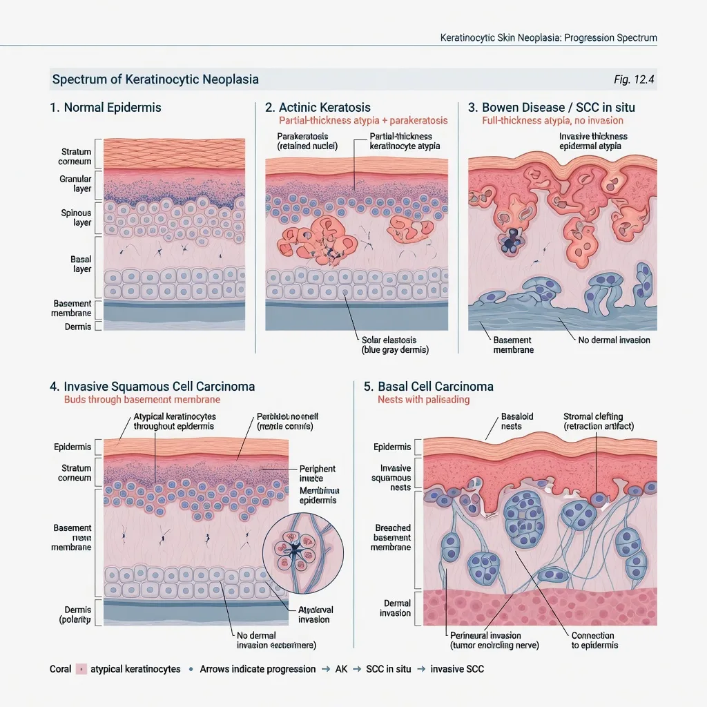
Keratinocytic pathology interprets neoplasms of epidermal keratinocytes — the bulk of “non-melanoma” / keratinocyte skin cancer practice — spanning precursors (actinic keratosis, AK), SCC in situ, invasive cutaneous SCC (cSCC), the keratoacanthoma (KA) spectrum, and basal cell carcinoma (BCC).[2][3][4] This leaf is the histology and reporting companion to clinical topics on AK, BCC, SCC, Mohs, and margins.
| Teaching label | Core histologic idea | Why it changes care |
|---|---|---|
| AK | Partial-thickness atypia ± parakeratosis, solar elastosis | Field vs lesion-directed therapy |
| SCCIS / Bowen | Full-thickness atypia, intact BM | Complete treatment; not “just eczema” |
| Invasive SCC | Tumour through BM into dermis/deeper | Risk features → surgery/MDT |
| KA spectrum | Crateriform well-differentiated proliferation | Complete excision often preferred |
| BCC | Basaloid nests, palisading, clefting | Subtype drives Mohs vs excision |
Field Cancerization
Chronic UV exposure produces clones of genetically altered keratinocytes across a field, not only the scaly papule you freeze.[7] Clinically this explains multiple AKs, subclinical dysplasia, and recurrent “new” lesions adjacent to treated spots. AAD guidelines frame AK management as both lesion-directed and field-directed care when burden is high.[8][9]
Sampling That Makes Pathology Useful
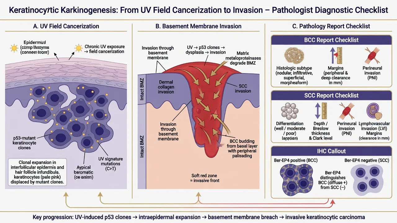
Technique decides whether subtype and invasion can be assessed.[3][5]
| Clinical question | Prefer | Hazard of wrong sample |
|---|---|---|
| Suspected invasive SCC | Full-thickness punch/incision to deep dermis/subcutis as appropriate | Superficial shave understages depth |
| Ill-defined infiltrative BCC | Punch or incision sampling representative edge | Tiny shave misses infiltrative strands |
| Classic nodular BCC for confirmation | Adequate scoop/shave may suffice if deep dermis included | Base transected → “at least” language |
| Field mapping | Multiple mapped samples | Random one AK does not map the field |
Always state on the form: site, size, clinical diagnosis/differential, prior treatment (cryo, 5-FU, imiquimod, PDT), immunosuppression, and whether margins are intended as diagnostic only. [1]
Exam application bank (NEET-PG / INICET)
One-line answer
Board-level dermatopathology leaf on keratinocytic neoplasia: actinic keratosis and field cancerization, SCC in situ (Bowen), invasive cutaneous SCC with high-risk features, keratoacanthoma spectrum, and basal cell carcinoma subtypes. Emphasises adequate sampling, report elements that change surgery (depth, PNI, subtype, margins), Ber-EP4 pitfalls, and management mapping without replacing dedicated clinical BCC/SCC topics.
Worked stems (answer without another resource)
Stem 1 — Classic presentation. Map symptoms to mechanism; name the first investigation and first treatment step with dose/route if drug therapy is standard. [1]
Stem 2 — Unstable / complicated. List red flags that force immediate resuscitation, theatre, ICU, antidote, or reperfusion — and what you do in the first 15 minutes. [1]
Stem 3 — Atypical group. Elderly, pregnancy, child, or immunocompromised: how presentation and thresholds change. [1]
Stem 4 — Differential trap. Name the three closest mimics and one discriminator for each. [1]
Stem 5 — Disposition. Who goes home with safety-netting, who is admitted, who needs HDU/ICU/theatre, and what follow-up is mandatory. [1]
Rapid viva checklist
- Definition + classification
- Pathophysiology chain
- Bedside signs / criteria
- Score with exact components (if any)
- Emergency bundle
- Definitive therapy with doses
- Complications of disease and of treatment
- Special populations
- Guideline/trial name if classic
- Three exam traps
Coverage self-check
If you cannot answer any stem above from this page alone, re-read the matching section — the page is intended to be self-sufficient for final-prof and NEET-PG/INICET questions on Keratinocytic pathology (BCC, SCC, actinic keratosis).
Expanded exam teaching (depth pass)
Clinical reasoning
For Keratinocytic pathology (BCC, SCC, actinic keratosis), examiners test whether you can prioritise life threats, choose the right first test, and give specific therapy (agent, dose, route, timing). Generic phrases without numbers score poorly.
Mechanism → feature map
Build a short chain: cause → pathophysiologic intermediate → clinical feature → complication. Every major symptom in the classic vignette should sit on that chain.
Investigation strategy
- Bedside/first-line tests that change immediate management
- Confirmatory or staging tests
- What a normal result does not exclude
- When not to delay treatment for imaging (unstable patient)
Management ladder
- Resuscitation / ABC / sepsis or haemorrhage bundle as relevant
- Specific antidote / procedure / antimicrobial / reperfusion / surgery
- Supportive care and monitoring targets
- Definitive long-term therapy and secondary prevention
- Disposition and safety-net advice
Special populations
Always prepare one line each for children, pregnancy, elderly, renal/hepatic impairment, and immunocompromised patients when the topic allows.
Pitfalls that fail candidates
- Treating the number not the patient
- Missing pregnancy status when relevant
- Imaging before stabilisation
- Wrong empiric cover or wrong antidote timing
- Incomplete counselling on recurrence, adherence, or red-flag return
Board-level dermatopathology leaf on keratinocytic neoplasia: actinic keratosis and field cancerization, SCC in situ (Bowen), invasive cutaneous SCC with high-risk features, keratoacanthoma spectrum, and basal cell carcinoma subtypes. Emphasises adequate sampling, report elements that change surgery (depth, PNI, subtype, margins), Ber-EP4 pitfalls, and management mapping without replacing dedicated clinical BCC/SCC topics. [1]
Structured revision sheet
Must-know numbers and names
List every score, size threshold, dose, and time window from this topic on a blank page from memory, then check against the sections above.
Three classic MCQ angles
- Most likely diagnosis given a vignette
- Next best step in management
- Most appropriate investigation
Three classic SAQ angles
- Pathophysiology in five steps
- Management algorithm with doses
- Complications and prevention
Clinical station flow
Greet → focused history → targeted exam → investigations → explain diagnosis → emergency care → definitive plan → safety-net / follow-up → answer examiner questions on mechanism and pitfalls.
[1]Actinic Keratosis — Histology Essentials
- Partial-thickness keratinocyte atypia (basal and lower spinous layers more than full epidermis)
- Alternating ortho- and parakeratosis (“flag sign” teaching image)
- Solar elastosis in the dermis
- Sparing of adnexal epithelium in some patterns (helpful teaching pearl, not absolute) [1]
When atypia becomes full-thickness, language shifts toward SCC in situ. Borders are continuous in real life — clinicopathologic correlation prevents false comfort. [1]
SCC in Situ (Bowen Disease) & Invasive SCC
SCC in situ
Full-thickness epidermal keratinocyte atypia without invasion through the basement membrane. May be UV-related on sun-exposed skin or HPV-related in anogenital sites — site history matters for work-up framing.[4][6]
Invasive cutaneous SCC
Invasion into dermis (or deeper) defines invasive disease. Contemporary reviews emphasise incidence, risk factors, diagnosis, and staging elements that pathologists and clinicians share.[3][4][5][6]
High-yield report elements (exam list): [1]
| Element | Why it matters |
|---|---|
| Diagnosis + differentiation | Poor differentiation raises risk |
| Depth / thickness / anatomic level | Primary local risk driver |
| Perineural invasion (PNI) | Recurrence / aggressive pathway |
| Lymphovascular invasion | Metastatic risk context |
| Margins (peripheral & deep) | Immediate re-excision decisions |
| Host factors on request | Transplant, CLL, drugs |
Lower risk teaching profile
- Well differentiated
- Superficial invasion
- No PNI/LVI
- Clear margins
- Immunocompetent host
Higher risk teaching profile
- Poorly differentiated
- Deep invasion
- PNI present
- Positive/close deep margin
- Transplant / heavy field damage
Keratoacanthoma Spectrum
KA presents as a rapidly growing crateriform nodule; histology shows a well-differentiated crateriform keratinocytic proliferation that can closely mimic well-differentiated SCC.[12] Board stance: many centres manage KA-like lesions with complete excision and path review rather than assuming spontaneous resolution — especially in adults and immunosuppressed patients.[12][4]
Basal Cell Carcinoma — Subtype Is Destiny
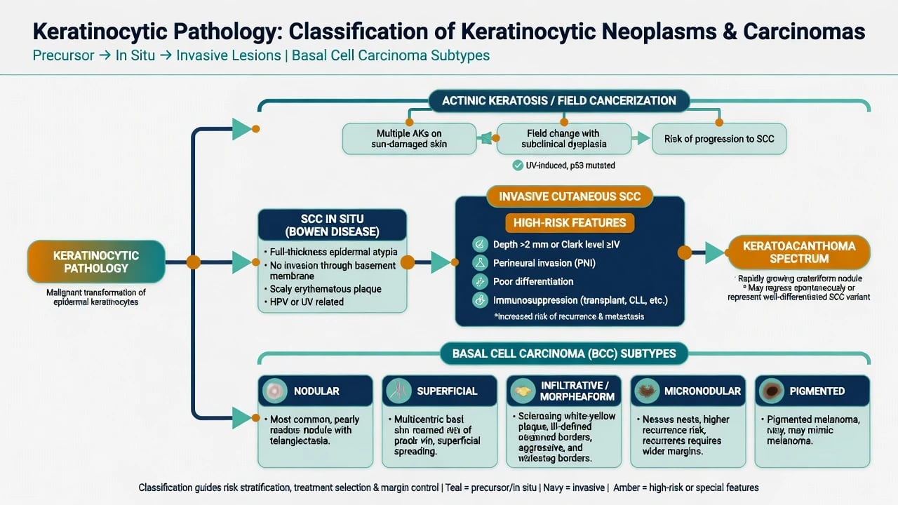
BCC is the commonest keratinocyte carcinoma clinically; European interdisciplinary guidance and clinical reviews stress diagnosis, subtype, and treatment selection.[1][2]
Classic nodular BCC histology
- Basaloid nests of varying size
- Peripheral palisading
- Retraction artefact (stromal clefting)
- Mucinous stroma in many cases [1]
Why subtype must be reported
| Subtype | Path teaching point | Surgical implication |
|---|---|---|
| Nodular | Well-circumscribed nests | Often standard excision if borders clear clinically |
| Superficial | Multifocal buds from epidermis | Selected topical/PDT options in guidelines for appropriate cases; still needs correct diagnosis |
| Infiltrative / morpheaform | Thin strands in sclerotic stroma | Ill-defined; Mohs / wide careful margins |
| Micronodular | Small nests, skip areas | Higher recurrence if under-treated |
| Pigmented | Melanin in tumour/stroma | Clinical melanoma mimic — path settles |
Immunohistochemistry — Support, Not Magic
Ber-EP4 classically labels BCC and helps separate BCC from SCC in ambiguous nests.[10] Limitations matter on boards: Bowen disease can show Ber-EP4 reactivity, so morphology and the full panel context still rule.[11] EMA, high-molecular-weight keratins, and p63/p40 enter selected adnexal and spindle differentials — never as a lone “SCC stain” without architecture.
Differentials That Catch Candidates
| Trap | Mimic of | Mitigation |
|---|---|---|
| Pseudoepitheliomatous hyperplasia | SCC | Look for regular reactive pattern + cause (ulcer, infection) |
| Inflamed SK / wart | SCC | Architecture of SK/HPV changes; clinical photo |
| Trichoepithelioma / other adnexal | BCC | Papillary mesenchymal bodies, IHC panels, expert review |
| Pagetoid Bowen | Melanoma in situ / EMPD | Cytokeratins vs melanocytic markers |
| Desmoplastic SCC | Scar / morpheaform BCC | Cytokeratin IHC, clinical thickness |
From Report to Management Algorithm
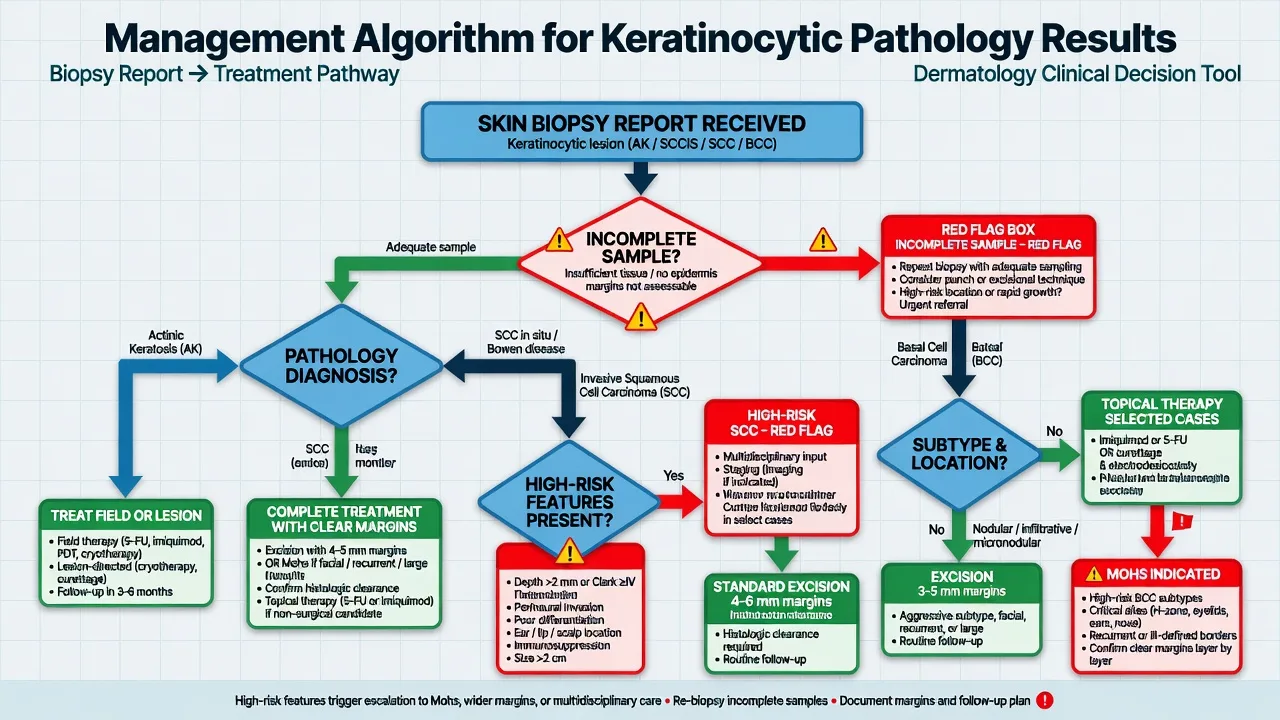
Reading order (board habit)
- Is there keratinocyte atypia? Partial vs full thickness vs invasive.
- Is the basement membrane breached? If yes, measure and describe invasion.
- BCC or SCC lineage? Architecture first; Ber-EP4 only if needed.
- Subtype / differentiation / PNI / margins.
- Host context — transplant, prior incomplete treatment.
- Write action language — clear, close, or positive margins; suggest re-excision/Mohs when indicated by subtype/risk.
Mapping (principles, not a dose table)
- AK: lesion- and field-directed options per AAD guidance; photoprotection always.[8][9]
- SCCIS: complete destruction/excision with clinical margin discipline — do not leave untreated full-thickness atypia on critical sites.
- Invasive SCC: surgery with risk-adapted margins; high-risk features → specialist/MDT.[3][4][5]
- BCC: low-risk nodular/superficial pathways vs high-risk infiltrative/micronodular → Mohs or careful margin control per European consensus framing.[1][2]
Regional & Exam Notes
MBBS/NEET-PG expect the spectrum table and red flags. MRCP/IADVL add transplant SCC risk and field cancerization. FACD/FRCDerm/ABD push subtype language, PNI reporting, Ber-EP4 limits, and Mohs indications. Skin of colour: pigmented BCC and under-recognised AK on non-classic sites need deliberate exam. [1]
Bottom Line
Report keratinocytic lesions so a surgeon can act: how thick is the atypia, has it invaded, what is the subtype, are nerves involved, and are margins free? Architecture first; stains second; clinical field and host always in the room.[1][4][7][10]
References
- [1]Peris K, Fargnoli MC, Kaufmann R, et al. European consensus-based interdisciplinary guideline for diagnosis and treatment of basal cell carcinoma-update 2023 Eur J Cancer, 2023.PMID 37604067
- [2]Heath MS, Bar A. Basal Cell Carcinoma Dermatol Clin, 2023.PMID 36410973
- [3]Waldman A, Schmults C. Cutaneous Squamous Cell Carcinoma Hematol Oncol Clin North Am, 2019.PMID 30497667
- [4]Wysong A. Squamous-Cell Carcinoma of the Skin N Engl J Med, 2023.PMID 37314707
- [5]Que SKT, Zwald FO, Schmults CD. Cutaneous squamous cell carcinoma: Incidence, risk factors, diagnosis, and staging J Am Acad Dermatol, 2018.PMID 29332704
- [6]Jiang R, Fritz M, Que SKT. Cutaneous Squamous Cell Carcinoma: An Updated Review Cancers (Basel), 2024.PMID 38791879
- [7]Willenbrink TJ, Ruiz ES, Cornejo CM, et al. Field cancerization: Definition, epidemiology, risk factors, and outcomes J Am Acad Dermatol, 2020.PMID 32387665
- [8]Eisen DB, Asgari MM, Bennett DD, et al. Guidelines of care for the management of actinic keratosis: Executive summary J Am Acad Dermatol, 2021.PMID 34111497
- [9]Eisen DB, Asgari MM, Bennett DD, et al. Guidelines of care for the management of actinic keratosis J Am Acad Dermatol, 2021.PMID 33820677
- [10]Tellechea O, Reis JP, Domingues JC, et al. Monoclonal antibody Ber EP4 distinguishes basal-cell carcinoma from squamous-cell carcinoma of the skin Am J Dermatopathol, 1993.PMID 8238781
- [11]Kogut M, Toberer F, Enk AH, et al. Limitations of Ber-EP4 for distinction of Bowen disease from basal cell carcinoma J Cutan Pathol, 2016.PMID 26765054
- [12]Bahmad HF, Stoyanov K, Mendez T, et al. Keratoacanthoma versus Squamous-Cell Carcinoma: Histopathological Features and Molecular Markers Dermatopathology (Basel), 2024.PMID 39449378