Dermatology · Medicine
Langerhans cell histiocytosis
Also known as Langerhans cell histiocytosis (LCH) · Histiocytosis X · Eosinophilic granuloma · Hand-Schüller-Christian disease · Letterer-Siwe disease
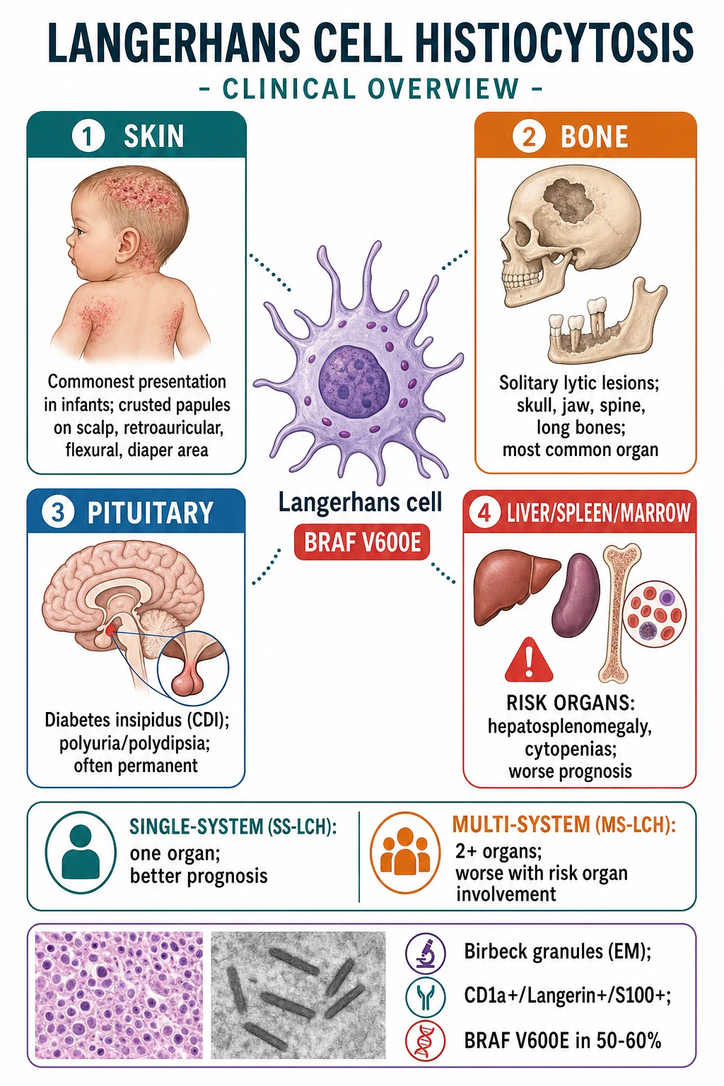
Langerhans cell histiocytosis (LCH) is a clonal neoplastic proliferation of abnormal Langerhans cells (myeloid dendritic cells). Classified as single-system (SS) or multi-system (MS), with risk organs (liver, spleen, bone marrow) conferring worse prognosis. Cutaneous presentation: seborrhoeic dermatitis-like crusted papules on scalp/flexures/diaper area (commonest in infants). Histopathology: Langerhans cells with grooved/coffee-bean nuclei + Birbeck granules on EM (tennis-racket-shaped, pathognomonic) + CD1a+, CD207/Langerin+, S100+ immunohistochemistry. BRAF V600E mutation in ~50-60%. Organ involvement: bone (solitary lytic — skull 'geographic skull', jaw 'floating teeth'), pituitary (diabetes insipidus), skin, lymph nodes, liver/spleen/marrow (risk organs), lung (smoking). Historical syndromes: eosinophilic granuloma (solitary bone), Hand-Schüller-Christian triad (skull lesions + DI + exophthalmos), Letterer-Siwe (acute multisystem, infants). Treatment: skin — topical steroids/nitrogen mustard; bone — curettage/intralesional steroids; multisystem — cytarabine or cladribine; refractory — BRAF inhibitors (vemurafenib, dabrafenib).
On this page & tools
Your progress
Saved locally on this device.
Exam tags
Red flags
Definition and Classification
Langerhans cell histiocytosis (LCH) is a clonal neoplastic proliferation of abnormal Langerhans cells — myeloid-derived dendritic cells that normally reside in the epidermis and mucosa. It is now classified as a myeloid neoplasm (not a reactive/inflammatory disorder).[1][3]
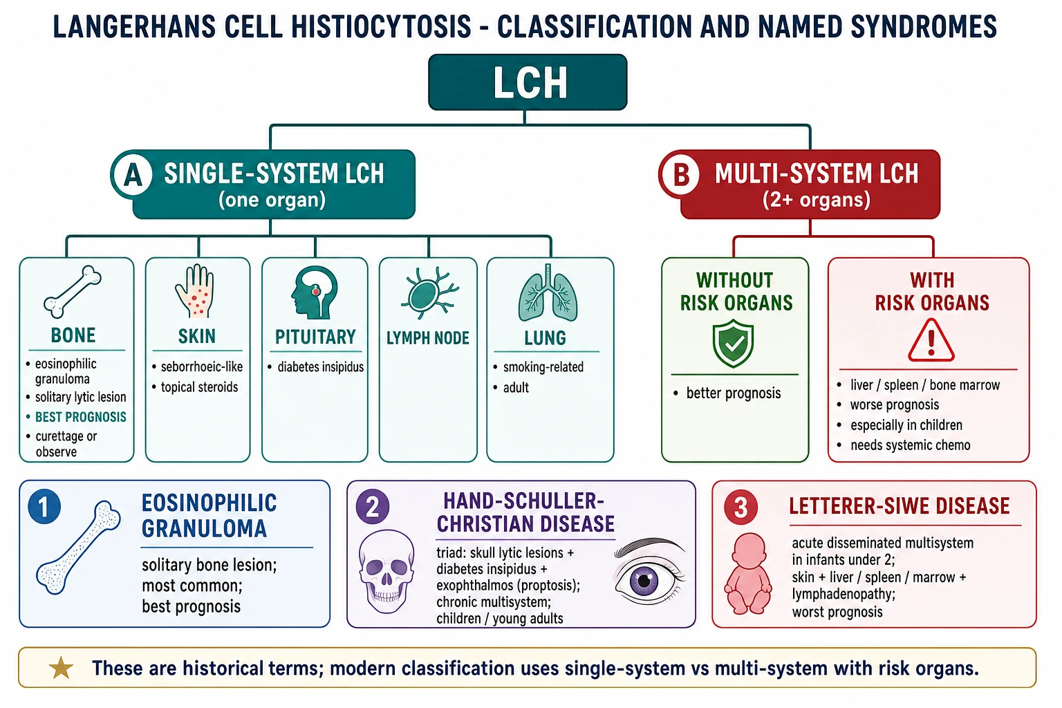
LCH is classified by the number of organ systems involved and the presence of risk organs:[1][5]
| Category | Definition | Prognosis |
|---|---|---|
| Single-system (SS-LCH) | One organ system involved | Better |
| Multi-system (MS-LCH) without risk organs | ≥2 organ systems; no liver/spleen/marrow involvement | Intermediate |
| Multi-system (MS-LCH) with risk organs | ≥2 systems including liver, spleen, or bone marrow | Worse (especially children; mortality up to 20%) |
Pathophysiology and Molecular Genetics
- LCH arises from clonal proliferation of pathological Langerhans cells that are trapped in an immature myeloid state (myeloid dendritic cell precursors) rather than mature epidermal Langerhans cells.[3]
- BRAF V600E mutation in ~50-60% of LCH — constitutive activation of the MAPK signalling pathway (RAS-RAF-MEK-ERK) drives proliferation and survival.[1][3]
- MAP2K1 (MEK1) mutations in ~10-15% of BRAF-negative cases.
- Other mutations: ERBB3, ARAF, KRAS, NRAS, PIK3CA — collectively, the MAPK pathway is activated in nearly all LCH cases.[3]
- The mutation burden varies by disease extent — multisystem disease with risk-organ involvement has a higher mutant allele frequency (more myeloid precursors carry the mutation) than single-system disease.[3]
Cutaneous Presentation
Skin involvement is the commonest presenting feature in infants and children, and is often the first sign leading to diagnosis:[1][4]
- Seborrhoeic dermatitis-like eruption: crusted, scaly, yellow-brown papules and plaques on the scalp, retroauricular area, flexures, diaper/inframammary/perineal regions. Often misdiagnosed as seborrhoeic dermatitis, atopic dermatitis, or irritant diaper dermatitis.
- Petechiae and purpura within the crusted papules (especially in flexural/inframammary/perineal areas) — a characteristic clue.
- Vesiculopustular variant: may mimic varicella or impetigo in neonates.
- Nodular variant: reddish-brown nodules, sometimes ulcerated.
- Oral mucosa: gingival hypertrophy, mucosal ulceration, jaw involvement ("floating teeth").
- Congenital self-healing reticulohistiocytosis (Hashimoto-Pritzker disease): present at birth with multiple reddish-brown nodules/papules that self-resolve over weeks to months — a self-limited form of cutaneous LCH (though systemic evaluation is still required to exclude occult disease).[3]
Pathophysiology & Molecular Biology
LCH is a clonal neoplastic proliferation of bone-marrow-derived dendritic cell precursors (Langerhans cells) with activating mutations in the MAPK pathway in nearly all cases. The cell of origin is a CD34+ myeloid progenitor that differentiates toward a Langerhans-cell phenotype but is arrested at an immature stage. Three molecular subgroups exist: [1]
- BRAF V600E mutation (≈ 50-60% of cases) — the commonest. Drives constitutive RAS-RAF-MEK-ERK activation. BRAF V600E-positive disease is associated with higher relapse risk and increased chronic disease burden but does not preclude cure.
- MAP2K1 (MEK1) mutation (≈ 25%) — another MAPK driver; mutually exclusive with BRAF.
- Other MAPK alterations (≈ 15%) — ARAF, MAP2K2, NRAS, KRAS, BRAF fusions. [1]
The same BRAF V600E mutation is found in hairy cell leukaemia, melanoma, papillary thyroid carcinoma, and Erdheim-Chester disease, supporting a true neoplastic basis. However, the indolent course in some patients and spontaneous regression of congenital forms suggests a complex biology where additional hits (immune microenvironment, second mutations) determine behaviour. [1]
[1]Quick numbers for the examiner
BONES-F — historical LCH syndromes
Eosinophilic granuloma — most common presentation, often solitary, best prognosis
Mandible/maxilla lytic lesions; premature tooth loss
Seborrhoeic-like rash with petechiae in flexures — biopsy for confirmation
Hand-Schuller-Christian triad: skull lesions + DI + exophthalmos
Crusted papules on scalp, flexures; petechiae clue
Multisystem disease relapses in approximately 30% even after first-line chemotherapy
Systemic Features and Organ Involvement
Bone (most common organ involved)
- Solitary lytic lesion (eosinophilic granuloma) — the most common LCH manifestation.
- Skull: punched-out lytic lesion ("geographic skull" or "map-like" appearance on X-ray).
- Jaw: alveolar bone loss → "floating teeth" on X-ray (teeth appear to float without bony support).
- Spine: vertebral body collapse → vertebra plana (flat vertebra).
- Long bones, pelvis, ribs: painful lytic lesions; may cause pathological fractures.
- Symptoms: localised pain, swelling, tenderness.[1][4]
Pituitary / Endocrine
- Diabetes insipidus (central): infiltration of the posterior pituitary or pituitary stalk → polyuria and polydipsia. The commonest endocrine manifestation; often permanent even after treatment. Screen with water deprivation test and MRI pituitary.[1][5]
- Growth hormone deficiency, anterior pituitary dysfunction (less common).
Other organs
- Lymph nodes: cervical most common; may be the only site (single-system lymph node LCH).
- Liver/spleen: hepatosplenomegaly, cholestasis, transaminitis, synthetic dysfunction (risk organ — worse prognosis).
- Bone marrow: cytopenias — anaemia, thrombocytopenia, neutropenia (risk organ; poor prognosis, especially children).
- Lung (pulmonary LCH): smoking-related; cystic and nodular interstitial pattern on HRCT (upper lobe predominant); may progress to pneumothorax, pulmonary fibrosis. Usually single-system in adults.[5]
- CNS neurodegeneration: cerebellar ataxia, cognitive/behavioural decline, pyramidal/extrapyramidal signs (neurodegenerative LCH); may be BRAF-related; poor prognosis; no effective treatment.[1]
Historical named syndromes (now unified under LCH)
- Eosinophilic granuloma: solitary bone lesion (most common; best prognosis).[1]
- Hand-Schüller-Christian disease: the classic triad of skull lytic lesions + diabetes insipidus + exophthalmos (proptosis) from orbital involvement. Chronic multisystem; typically children/young adults.[1]
- Letterer-Siwe disease: acute disseminated multisystem disease in infants less than 2 years — skin + liver/spleen/bone marrow + lymphadenopathy + fever. Worst prognosis.[1]
Histopathology
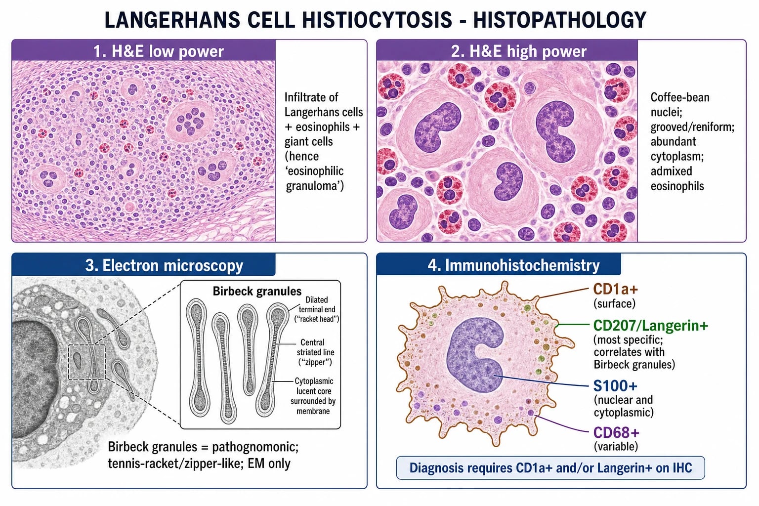
The histological hallmark is the Langerhans cell:[1][4]
- H&E: sheets and infiltrates of large cells with characteristic grooved, reniform ("coffee-bean") nuclei and abundant eosinophilic cytoplasm. Admixed eosinophils (may be very numerous — "eosinophilic granuloma"), lymphocytes, neutrophils, and multinucleated giant cells.
- Electron microscopy: Birbeck granules — pathognomonic cytoplasmic organelles with a rod-shaped/zipper-like body and a dilated terminal end (resembling a tennis racket). Composed of Langerin.[1]
- Immunohistochemistry (IHC):
- CD1a+ (surface glycoprotein) — diagnostic.
- CD207 / Langerin+ — the most specific marker; a C-type lectin that forms Birbeck granules; correlates with their presence.
- S100+ (nuclear and cytoplasmic) — sensitive but not specific.
- CD68+ (variable).
- Diagnosis requires CD1a+ and/or Langerin+ on IHC (Birbeck granules on EM are confirmatory but rarely needed now that IHC is available).[4]
CLS — the three IHC anchors for LCH
Surface glycoprotein; diagnostic anchor; reliably positive on formalin-fixed paraffin-embedded tissue.
Most specific marker; C-type lectin that polymerises into Birbeck granules; replaces EM in routine practice.
Sensitive but not specific (also melanocytes, nerves, some carcinomas); nuclear and cytoplasmic staining.
Differential Diagnosis
The clinical and histological mimics of LCH are wide and depend on the organ involved. Cutaneous LCH in infants is frequently misdiagnosed as common inflammatory dermatoses; bone LCH mimics infection and malignancy; pulmonary LCH in adults can be mistaken for cystic lung disease. A biopsy with CD1a and Langerin immunohistochemistry is the deciding test in equivocal cases.[1][3][4]
[1]Investigations and Diagnosis
- Tissue biopsy (skin, bone, lymph node) → histology + IHC (CD1a, Langerin, S100).[5]
- Staging (to determine single vs multi-system and plan treatment):[1][5]
- Skeletal survey or whole-body MRI / PET-CT — detect bone lesions.
- FBC + differential — screen for bone marrow involvement (cytopenias).
- LFTs — screen for liver involvement (cholestasis, transaminitis).
- Water deprivation test ± MRI pituitary — for diabetes insipidus.
- Chest HRCT — for pulmonary LCH (cysts, nodules).
- Urine osmolality — for DI.
- Endocrine assessment — growth hormone, anterior pituitary.
- BRAF V600E mutation testing — on tissue; guides targeted therapy for refractory disease.[1][3]
Management
Single-system skin LCH
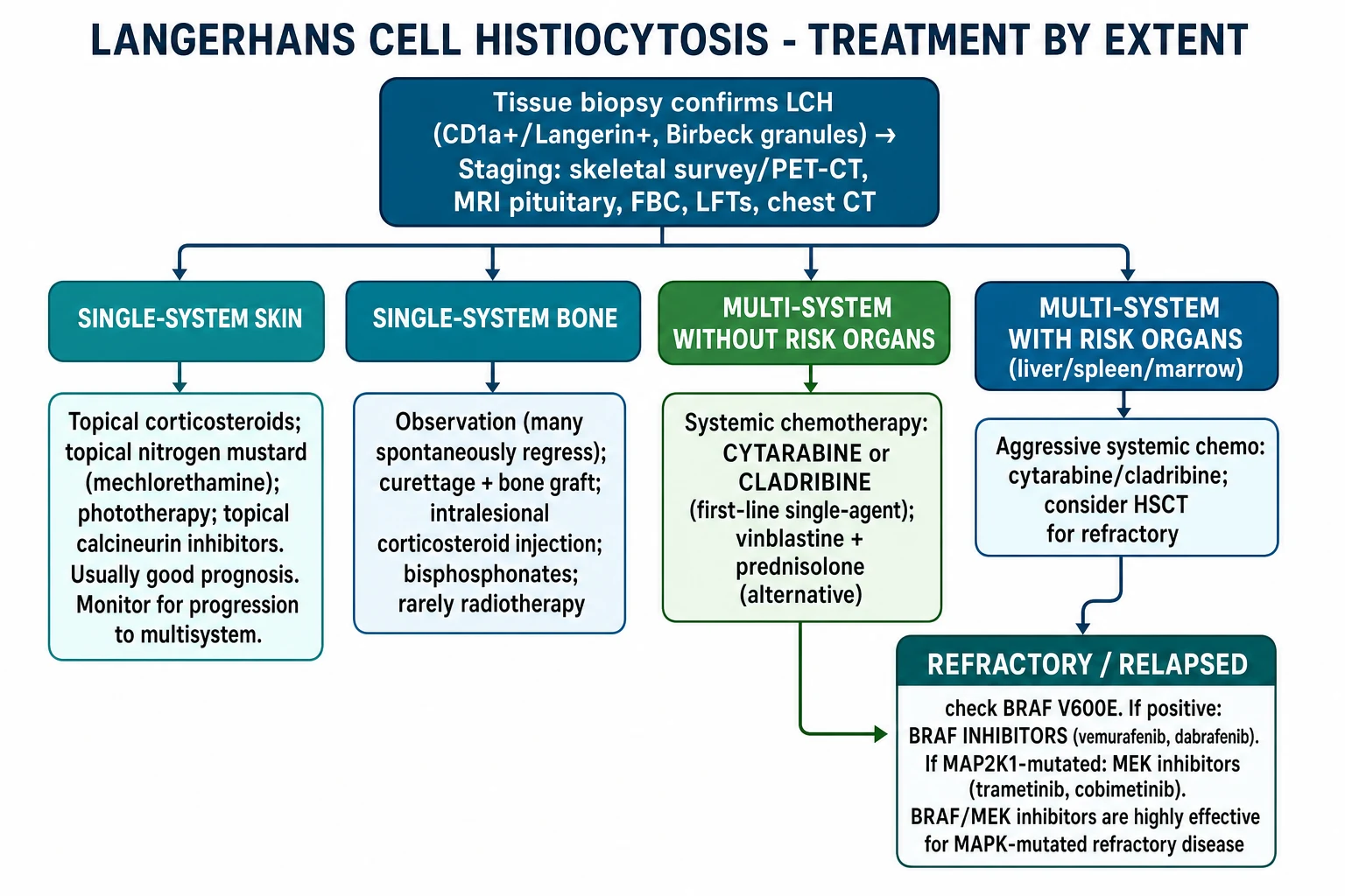
- Topical corticosteroids (potent), topical nitrogen mustard (mechlorethamine), topical calcineurin inhibitors, phototherapy (nbUVB, PUVA).
- Good prognosis; monitor for progression to multisystem (regular follow-up).[1]
Single-system bone LCH (eosinophilic granuloma)
- Observation — many solitary lesions spontaneously regress.
- Curettage + bone grafting (for symptomatic or structurally important lesions).
- Intralesional corticosteroid injection (triamcinolone).
- Bisphosphonates (for painful lesions).
- Localised radiotherapy (rarely; reserved for inaccessible or refractory lesions).[5]
Multi-system LCH
- Systemic chemotherapy:
- Haematopoietic stem cell transplant (HSCT) — for refractory multisystem disease with risk-organ involvement.[1]
Refractory / relapsed disease
- BRAF V600E testing on tissue — guides targeted therapy.[1][3]
- BRAF inhibitors — vemurafenib, dabrafenib — highly effective for BRAF V600E-mutated refractory/relapsed LCH.
- MEK inhibitors — trametinib, cobimetinib — for MAP2K1-mutated or BRAF-inhibitor-refractory disease.[3]
- Targeted therapy has transformed the outlook for refractory disease.[5]
Landmark clinical trials — LCH-III and LCH-IV
Multisystem-LCH management rests on two large international Histiocyte Society studies that define the modern standard of care: [1]
-
LCH-III (NCT00472035; 2001–2008): the Gadner et al. Blood 2013 publication.[6] Landmark trial randomised multisystem-LCH patients into a low-risk arm (RO−, six vs. twelve months of continuation therapy) and a high-risk arm (RO+, vinblastine + prednisone ± methotrexate). Key outcomes: RO− 5-year reactivation reduced from 54% (6-month arm) to 37% (12-month arm), p = 0.03 — twelve months of continuation therapy became the international standard for non-risk MS-LCH. RO+ 5-year survival improved to 84% (compared with approximately 62% in LCH-I and 69% in LCH-II); adding methotrexate conferred no survival benefit but added toxicity, so the vinblastine + prednisone backbone alone remained the standard. The first 6–12 weeks of induction are the most predictive period for ultimate outcome; patients who do not achieve an active-disease response by week 12 are candidates for salvage. Permanent long-term consequences (DI, hearing loss, sclerosing cholangitis, neurodegeneration) occur in 30–50% of MS survivors — the price of cure.
-
LCH-IV (NCT02205762; 2012–2025, recruitment closed): the successor study, an ambitious risk-adapted protocol with seven strata covering first-line MS-LCH, second-line non-risk salvage, second-line RO+ salvage, stem-cell transplantation, isolated tumourous/neurodegenerative CNS disease, observation of localised SS-LCH, and long-term follow-up. Randomisations within Stratum I compare 12 versus 24 months of continuation therapy with or without 6-mercaptopurine maintenance. The trial formally integrates BRAF V600E testing at diagnosis and embeds targeted-therapy questions for RO+ refractory disease. Results will refine the duration of maintenance, the role of oral mercaptopurine, and the placement of BRAF/MEK inhibitors within the first-line pathway. [1]
Second-line therapy — cladribine, cytarabine, clofarabine
When frontline vinblastine/prednisone fails (no disease resolution by week 12 of induction, or reactivation within 6 months of stopping therapy), or when initial disease is RO+ and refractory, purine analogues are the established second-line agents:[1][5][7]
- Cladribine (2-chlorodeoxyadenosine, 2-CdA, Litak/Leustatin): a purine analogue resistant to adenosine deaminase; concentrates in lymphocytes and histiocytes. Standard dosing 5–6.5 mg/m²/day IV over 5 days per cycle, repeated every 3–4 weeks for up to 6 cycles, often capped at 3 cycles in RO+ salvage to limit cumulative toxicity. In adult LCH, a low-dose oral or subcutaneous regimen (0.14 mg/kg/day × 5 days per cycle) is used. The pivotal Donadieu et al. Blood 2015 international phase 2 study ([7]) evaluated cladribine + cytarabine in 34 children with refractory RO+ MS-LCH: overall response rate 92%, 5-year survival 85% — but at the cost of substantial haematologic toxicity (pancytopenia in 100%, severe infection in approximately 50%, prolonged cytopenias). The authors recommended capping cumulative cladribine at 200 mg/m² and limiting to ≤3 cycles, with administration only at centres experienced in AML-grade chemotherapy or HSCT.
- Cytarabine (Ara-C): a pyrimidine analogue with strong single-agent activity in LCH. Dosing typically 100–200 mg/m²/day continuous IV infusion over 5 days (range 100–1,000 mg/m²; high-dose Ara-C 1–3 g/m² reserved for refractory cases and intracranial ND-LCH). The 2015 Donadieu regimen combined cytarabine 200 mg/m²/day × 5 days with cladribine 6 mg/m²/day × 5 days, repeated every 4 weeks. Synergy arises because cladribine inhibits ribonucleotide reductase and depletes dATP pools, while cytarabine competes for DNA polymerase — combined effect exceeds either agent alone against proliferating histiocytes.
- Clofarabine (Evoltra/Clolar): a second-generation purine nucleoside analogue with improved intracellular retention and broader cytotoxic profile than cladribine; approved for paediatric relapsed ALL and studied in RO+ LCH. Typical dosing 40 mg/m²/day × 5 days IV over 2 hours. Case-series data (children and adults with cladribine-refractory disease) show partial and complete responses, but myelosuppression, liver toxicity, and capillary leak syndrome limit wider use; reserved for salvage after cladribine + cytarabine failure or to bridge patients to HSCT.
Second-line agents at a glance for the examiner
BRAF/MEK inhibitor dosing — reference protocols for refractory LCH
Where BRAF/MEK inhibitors are indicated for refractory LCH, dosing follows the adult melanoma protocols (extrapolated to paediatrics on body-surface area):[3][5][8][9][10]
-
Vemurafenib (Zelboraf) — BRAF V600E inhibitor.[8][9][11] Standard adult dose 960 mg orally twice daily (i.e. four 240-mg tablets morning and evening, total 1,920 mg/day), taken on an empty stomach (≥1 hour before or ≥2 hours after a meal). The pivotal VE-BASKET phase 2 study (Diamond et al. JAMA Oncol 2018, [8]) treated adults with BRAF V600-mutant ECD and LCH; response rate in LCH exceeded 90% in cross-disease analysis. Common toxicities: arthralgia, photosensitivity, keratoacanthoma and cutaneous squamous cell carcinoma (~10–25% in adults), QT prolongation, transaminitis, pyrexia, fatigue. Paediatric liquid formulation has been used at 45–55 mg/kg/day (maximum 960 mg BD); weight-based dosing is critical because clearance is proportional to body weight. Continuous therapy is mandatory because drug holiday leads to relapse within weeks to months in most patients.
-
Dabrafenib (Tafinlar) — BRAF V600E inhibitor. Standard adult dose 150 mg orally twice daily as monotherapy (i.e. one 75-mg capsule twice daily, total 300 mg/day), or 150 mg BD when combined with trametinib. Paediatric dosing is weight-banded and uses an oral suspension: 5.25 mg/kg/day (younger children divided BD; older children at 150 mg BD if ≥46 kg). Pre-medications: avoid concurrent potent CYP3A4 inhibitors and inducers. Common toxicities: pyrexia (often >38.5°C with rigors — a clinical signature compared with vemurafenib), chills, headache, arthralgia, hyperkeratosis and cutaneous SCC (lower than vemurafenib), uveitis, hyperglycaemia, ventricular ejection fraction (LVEF) decline, QTc prolongation. The Whitlock et al. Blood Adv 2023 paediatric trial ([10]) reported an overall response rate of 76.9% with dabrafenib monotherapy and 58.3% with dabrafenib + trametinib in BRAF V600-mutated recurrent/refractory paediatric LCH; both arms superior to historical chemotherapy.
-
Trametinib (Mekinist) — MEK1/2 inhibitor, downstream of BRAF. Monotherapy dose in histiocytosis is 2 mg orally once daily (adults and paediatrics ≥1 year at approximately 0.04–0.06 mg/kg/day adjusted to BSA). For BRAF-mutant histiocytosis, trametinib is the rational alternative when BRAF inhibitors are contraindicated (e.g. cutaneous SCC risk in adults) and is the partner drug for BRAF/MEK combination therapy. When combined with dabrafenib: trametinib remains 2 mg daily and dabrafenib 150 mg BD. Common toxicities: acneiform rash (more than BRAF inhibitors), diarrhoea, stomatitis, LVEF decline (~10%), ocular toxicity (serous retinopathy / retinal vein occlusion), elevated CK, pneumonitis, hepatitis. Trametinib has the advantage of working regardless of BRAF status — useful in BRAF wild-type, MAP2K1-mutant, ARAF-mutant, KRAS/NRAS-mutant histiocytoses, and post-BRAF-inhibitor resistance. [1]
BRAF/MEK inhibitor dosing for refractory LCH
Diabetes insipidus
- Desmopressin (DDAVP) — for polyuria/polydipsia. Often permanent (pituitary damage is irreversible).[1]
Special Populations and Mixed Histiocytosis
LCH exists within a wider family of histiocytic disorders that share MAPK-pathway-driven myeloid neoplasia but have distinct clinical phenotypes and histology. Recognising the boundaries and overlaps matters for prognosis and treatment.[3][5]
[1]Histiocytosis family at a glance
Prognosis
- Single-system LCH: excellent (>90% survival); many bone lesions spontaneously regress.
- Multi-system without risk organs: good (>80% survival).
- Multi-system with risk organs (liver/spleen/marrow): worse (mortality ~10-20% in children; age less than 2 years is an additional risk factor).[1]
- Neurodegenerative LCH: progressive; no effective treatment; poor quality-of-life outcome.[1]
- Long-term sequelae: diabetes insipidus (often permanent), growth hormone deficiency, skeletal deformity, hearing loss, neurodegeneration, secondary malignancies (especially after chemotherapy/radiotherapy).[5]
Exam Pearls
[1]Red Flags
Exam application bank (NEET-PG / INICET)
One-line answer
Langerhans cell histiocytosis (LCH) is a clonal neoplastic proliferation of abnormal Langerhans cells (myeloid dendritic cells). Classified as single-system (SS) or multi-system (MS), with risk organs (liver, spleen, bone marrow) conferring worse prognosis. Cutaneous presentation: seborrhoeic dermatitis-like crusted papules on scalp/flexures/diaper area (commonest in infants). Histopathology: Langerhans cells with grooved/coffee-bean nuclei + Birbeck granules on EM (tennis-racket-shaped, pathognomonic) + CD1a+, CD207/Langerin+, S100+ immunohistochemistry. BRAF V600E mutation in ~50-60%. Organ involvement: bone (solitary lytic — skull 'geographic skull', jaw 'floating teeth'), pituitary (diabetes insipidus), skin, lymph nodes, liver/spleen/marrow (risk organs), lung (smoking). Historical syndromes: eosinophilic granuloma (solitary bone), Hand-Schüller-Christian triad (skull lesions + DI +
Worked stems (answer without another resource)
Stem 1 — Classic presentation. Map symptoms to mechanism; name the first investigation and first treatment step with dose/route if drug therapy is standard. [1]
Stem 2 — Unstable / complicated. List red flags that force immediate resuscitation, theatre, ICU, antidote, or reperfusion — and what you do in the first 15 minutes. [1]
Stem 3 — Atypical group. Elderly, pregnancy, child, or immunocompromised: how presentation and thresholds change. [1]
Stem 4 — Differential trap. Name the three closest mimics and one discriminator for each. [1]
Stem 5 — Disposition. Who goes home with safety-netting, who is admitted, who needs HDU/ICU/theatre, and what follow-up is mandatory. [1]
Rapid viva checklist
- Definition + classification
- Pathophysiology chain
- Bedside signs / criteria
- Score with exact components (if any)
- Emergency bundle
- Definitive therapy with doses
- Complications of disease and of treatment
- Special populations
- Guideline/trial name if classic
- Three exam traps
Coverage self-check
If you cannot answer any stem above from this page alone, re-read the matching section — the page is intended to be self-sufficient for final-prof and NEET-PG/INICET questions on Langerhans cell histiocytosis.
[1]References
- [1]Rodriguez-Galindo C, Allen CE. Langerhans cell histiocytosis Blood, 2020.PMID 32106306
- [2]Moore PF. Histiocytic Diseases Vet Clin North Am Small Anim Pract, 2023.PMID 36270835
- [3]McClain KL, Bigenwald C, Collin M, et al. Histiocytic disorders Nat Rev Dis Primers, 2021.PMID 34620874
- [4]McKinney RA, Wang G. Langerhans Cell Histiocytosis and Other Histiocytic Lesions Head Neck Pathol, 2025.PMID 39998733
- [5]Goyal G, Tazi A, Go RS, et al. International expert consensus recommendations for the diagnosis and treatment of Langerhans cell histiocytosis in adults Blood, 2022.PMID 35271698
- [6]Gadner H, Minkov M, Grois N, et al. Therapy prolongation improves outcome in multisystem Langerhans cell histiocytosis Blood, 2013.PMID 23589673
- [7]Donadieu J, Bernard F, van Noesel M, et al. Cladribine and cytarabine in refractory multisystem Langerhans cell histiocytosis: results of an international phase 2 study Blood, 2015.PMID 26194764
- [8]Diamond EL, Subbiah V, Lockhart AC, et al. Vemurafenib for BRAF V600-Mutant Erdheim-Chester Disease and Langerhans Cell Histiocytosis: Analysis of Data From the Histology-Independent, Phase 2, Open-label VE-BASKET Study JAMA Oncol, 2018.PMID 29188284
- [9]Haroche J, Cohen-Aubart F, Emile JF, et al. Dramatic efficacy of vemurafenib in both multisystemic and refractory Erdheim-Chester disease and Langerhans cell histiocytosis harboring the BRAF V600E mutation Blood, 2013.PMID 23258922
- [10]Whitlock JA, Geoerger B, Dunkel IJ, et al. Dabrafenib, alone or in combination with trametinib, in BRAF V600-mutated pediatric Langerhans cell histiocytosis Blood Adv, 2023.PMID 36884302
- [11]Haroche J, Cohen-Aubart F, Bugnet E, et al. Reproducible and sustained efficacy of targeted therapy with vemurafenib in patients with BRAF(V600E)-mutated Erdheim-Chester disease J Clin Oncol, 2015.PMID 25422482